BEHAVIORAL AND BRAIN SCIENCES (1990) 13, 289-331 Printed in the United States of America
B-Afferents: A fundamental
division of the nervous system
mediating hoxneostasis?
James C. Prechtl*
Terry L* Powley
Laboratory of Regulatory Psychobiology, Department of Psychological Sciences, Purdue University, West Lafayette, IN 47907
Electronic mail: [email protected]@brazil.psych.edu *Reprint requests should be addressed to: James C. Prechtl, Department of Neuroscience, A-001, University of California, San Diego, La Jolla, CA
92093
Abstract? The peripheral nervous system (PNS) has classically been separated into a somatic division composed of both afferent and efferent pathways and an autonomic division containing only efferents. J. N. Langley, who codified this asymmetrical plan at the beginning of the twentieth century, considered different afferents, including visceral ones, as candidates for inclusion in his concept of the "autonomic nervous system" (ANS), but he finally excluded all candidates for lack of any distinguishing histological markers. Langley's classification has been enormously influential in shaping modern ideas about both the structure and the function of the PNS. We survey recent information about the PNS and argue that many of the sensory neurons designated as "visceral" and "somatic" are in fact part of a histologically distinct group of afferents concerned primarily autonomic function. These afferents have traditionally been known as "small dark" neurons or B-neurons. In this target article we outline an association between autonomic and B-neurons based on ontogeny, cell phenotype, and functional relations, grouping them together as part of a common reflex system involved in homeostasis. This more parsimonious classification of the PNS, made possible by the identification of a group of afferents associated primarily with the ANS, avoids a number of confusions produced by the classical orientation. It may also have practical implications for an understanding of nociception, homeostatic reflexes, and the evolution of the nervous system. Keywords; autonomic; capsaicin; dorsal root ganglion; nerve growth actor; neuroimmunology; nociception; sensory neurons; substance P; sympathetics; tachykinins; visceral afferents
1. introduction
All of neuroscience uses the fundamental classification that distinguishes the central and peripheral nervous systems - the CNS and PNS, respectively - and classifies the elements of the PNS into subdivisions on the basis of three dichotomies: afferent and efferent, skeletal and autonomic, and somatic and visceral. Given the paradig-matic importance of such a taxonomy, it is noteworthy that neither this accepted systematics nor the resulting assignments have been rigorously reconsidered since the beginning of the twentieth century, when they were considered tentative. In this target article, we suggest that the categorization of PNS afferents, including a description of their relation to the autonomic nervous system (ANS), should be revised to reflect new informa-tion about morphology, histochemistry, ontogeny, and function.
As J. N. Langley developed his autonomic nervous system concept, he carefully considered the possibility that there was a specific class of afferents that also be-longed to this division of the nervous system. In 1903, after having reviewed a number of possible criteria for classifying afferents, Langley (1903, p. 25) tentatively concluded that autonomic afferents were those fibers "which give rise to reflexes in autonomic tissues, and
which are incapable of directly giving rise to sensation." He was reluctant to classify afferents according to func-tional criteria, however, and hoped to rely eventually on morphological criteria. At the end of his 1903 paper he wrote: "Further progress awaits the discovery of some distinguishing histological character [i.e., marker of auto-nomic afferents]."
Eighteen years later, in his book, The Autonomic
Nervous System, Part I (1921), Langley excluded once
and for all the possibility of autonomic afferents (see Figure 1). Although he planned (Langley 1921, p. 9) to reexamine this conclusion in a later publication, the sequel to his 1921 work never appeared, and his "effer-ents only" version of the ANS has been a source of continuing controversy.
A number of neuroscientists, including authors of ma-jor treatises on the ANS (Gabella 1976; Mitchell 1953; Pick 1970), have expressed dissatisfaction with the "efferents only" concept. Nevertheless, it is commonly accepted in textbooks and in the general literature. Even grant proposals have been criticized for suggesting that specific afferents are associated with the ANS (see Nor-gren 1985). Recently, some investigators have broken with tradition and used terms such as "sympathetic af-ferents" (Foreman et al. 1986; Malliani 1982; Malliani et al. 1973; Morgan et al. 1986) and "parasympathetic
af-Prechtl & Powley: B-Afferents
Langley's PNS Concept (1921)
Nerves
(Dromic and A n t i
-dromic action) Sympathetic Parasympathetic
(Plexuses of Auerbach and Meissner)
Bulbo-sacral (oro •*>-anaI)
r
Figure 1. Langley's (1921, p. 10) classification of the pe-ripheral nervous system. Note that all afferents are classified as "somatic," including those with antidromic actions.
ferents" (Kawatani et al. 1985; Mei 1985). Such desig-nators have important uses and in some cases have satis-fied specific needs of specialists. It is uncertain whether they are sufficiently cogent to gain general acceptance, however (see section 2.3). To date at least, none of these terms has stimulated a reformulation in the general literature.
A common justification for such designators has been that the afferents in question are found in the nerves, which are labeled "sympathetic" or "parasympathetic." Langley was aware of the existence of afferents in auto-nomic nerves but he did not regard this as a sufficient reason for invoking another autonomic neuron category. He did not consider the ANS concept an invention for heuristic convenience, but rather a discovery that could be objectively repeated. Earlier, Gaskell (1886, p. 2) had asserted that the test of a "real" fundamental division of the nervous system was whether the proposed functional differences were "bound up with morphological dif-ferences." This approach had enabled Gaskell (1886) to delineate the bulbar, thoracolumbar, and sacral visceral nervous outflows and to achieve a revolutionary sim-plification of the nineteenth-century neuroanatomy. The morphological difference that Gaskell used was the con-spicuously small size of myelinated preganglionic auto-nomic fibers. In a similar fashion, Langley identified "somatic" efferents by their association with multinuclear striated muscle; later he turned to pharmacological crite-ria in order to distinguish sympathetic and parasympa-thetic divisions. Such distinctive neuronal traits or mark-ers were considered to reflect distinct ontogenetic histories. In effect, Langley's attempt at systemization was really intended to be not just a special-purpose classification, but a "natural" grouping, analogous to the groupings of some taxonomists (see Mayr 1982). It is in the same spirit of "natural" taxonomy that we readdress the issue of PNS classification.
For more than 100 years, morphologists have con-trasted two cell types in the dorsal root ganglia (see Scharf 1958): A-neurons and B-neurons (section 3). In the past decade this A-B neuron typology has been reintroduced, particularly by histochemists and pharmacologists, in a series of papers that has radically revised our thinking about sensory neurons (see Hokfelt et al. 1976; Jancso et al. 1977). Furthermore, many of the groups of dorsal root ganglion (DRG) neurons that have been distinguished
(histochemically or pharmacologically) have charac-teristics that seriously challenge the validity of current classification systems. For example, substance P-contain-ing afferents, a subpopulation of B-neurons (section 3), not only mediate autonomic functions but have key on-togenetic and phenotypic traits similar to those of auto-nomic motor neurons. Nevertheless, according to Lan-gley's classification (Figure 1), these neurons would be, and indeed traditionally have been, lumped together with all other spinal afferents. Alternatively, they are fragmented into "general somatic" and "general visceral" categories by the doctrine of nerve components (Herrick 1903; 1927).
In this target article we analyze the inadequacies of the current classification systems and propose an alternative, more parsimonious ordering. Our two main hypotheses are: (1) B-neurons represent a major division of the PNS, more fundamental than the traditional categories of "vis-ceral" and "somatic." (2) The ANS is the motor system most closely related to the B-neuron division. In one sense, the B-neurons characterized here represent the afferent division that Langley had searched for, but provi-sionally ruled out.
2. Classification of nerwes and neurons
The current terminology and concepts can be put in perspective by identifying their roots in some of the more influential contributions to PNS classification and to the idea of homeostasis.
The distinctiveness of visceral function must first have been recognized when these inner parts were found to move of their own accord, usually without the knowledge of their owner. In the seventeenth century, such observa-tions led to the idea of involuntary nervous function, introduced to the study of functional anatomy by Thomas Willis (Sheehan 1936). According to Willis (1664, 1965), the cerebrum mediated volition via the flow of animal spirits through the spinal cord and the cerebellum medi-ated involuntary functions through the vagal and inter-costal (sympathetic chain) nerves. The cerebrospinal
sys-tem came to be viewed as the main substrate of volition
and sensation. Willis concluded that only the very strong volitions could pass through the cerebellum to the invol-untary nerves and thereby modify semivolinvol-untary func-tions such as breathing. Similarly, he considered mild visceral reactions to be insensible because they were blocked from the cerebrum by the cerebellum. Though few neuroscientists today would consider using volition as a criterion for classifying nerves, most of the autonomic or
vegetative nervous system concepts, from Willis' time to
the present, have been based partly on notions that can be traced to presuppositions about consciousness or voli-tion. Gaskell (1916) went so far as to call this division the "involuntary nervous system."
The forerunner of modern autonomic-somatic concepts was suggested by Xavier Bichat (1827/1977). Bichat di-vided the life of an organism into an organic mode, controlled by the ganglionic system (autonomic ganglia and plexuses), and an animal mode, mediated by the cerebrospinal system. The function of organic life was nutrition; that of animal life, coordination of relations with the external environment. Whereas the organic
Prechtl & Powley: B-Afferents mode was passive and stationary, consisting of a
continu-ous rhythm of assimilation and excretion, the animal mode consisted of variable and episodic behaviors such as locomotion, manipulation, and communication.
BIchat's functional neuroanatomy also had a mor-phological basis:
What anatomist, In fact, has not been struck with the differences that exist between the nerves of these two systems? Those of the brain [cerebrospinal or somatic] are larger, less numerous, whiter, more compact in texture and exhibit less variety. On the contrary, the extreme tenuity, great number, especially towards the plexuses, greyish colour, remarkable softness and vari-eties extremely common are characters of the nerves coming from the ganglions [autonomic] if we except those of communication with the cerebral nerves [communicating rami] and some of those which unite together these small nervous centers. (Bichat 1977, p. 72)
Unfortunately, Bichat underestimated the significance of the white communicating rami and promoted the idea of a decentralized, completely independent, ganglionic (autonomic) nervous system.
Shortly after Bichat's career was ended by his death at the age of 31, Charles Bell (1811, 1966) introduced the Idea of specific nerve components. According to Bell, nerves were capable of different functions because they had distinct components with distinct central connec-tions. Previous anatomists had divided the nervous sys-tem according to the presumed specializations of entire nerves. Bell implied that classification should not be based on nerves, which have a mixture of functional components, but on the parcels of central organization (e.g., nuclei, laminae), "That the nerves of sense, the nerves of motion, and the vital [autonomic] nerves, are distinct through their whole course, though they seem sometimes united In one bundle; and that they depend for their attributes on the organs of the brain to which they are severally attached" (Bell 1966, p. 274). In Bell's time, the same neural element was thought to be able to transmit both different sensations (modalities) and the power of motion.
Three-quarters of a century later, the osmium nerve fiber stain was Introduced by Schultz (see Sheehan, 1936) and the power of Bell's idea was realized In the mor-phological analyses of Gaskell (1886). Gaskell discovered that "real" (1886, p. 2) functional components of the nervous system were correlated not only with distinctive features of central organization but also with mor-phological traits such as specific fiber size. Gaskell ac-cordingly discriminated the bulbar, thoracolumbar, and sacral autonomic outflows from the somatic fiber compo-nents and thereby laid the foundation for the PNS classifi-cation systems that prevail today: the doctrine of nerve components and the autonomic nervous system. 2.1. Doctrine of nerwe components. According to Herrick (1903), the purpose of the doctrine of nerve components was to divide the PNS into units that had both functional and structural significance. The main idea of the doctrine was that different functional nerve components could be defined by the similarities of their central and peripheral terminal projections. In the case of sensory components, however, It was seldom possible to analyze peripheral
terminal/receptor specializations, so peripheral path-ways were used as the discriminating criteria instead. The most fundamental division of functions and nerve compo-nents was thought to be reflected in the distinction between the visceral responses to internal stimuli and the bodily or somatic responses to external stimuli (Herrick 1903). The inward distribution of innervation was accord-ingly assumed to correspond to a "visceral" type of function; the outward distribution innervation corre-sponded to a "somatic" type.
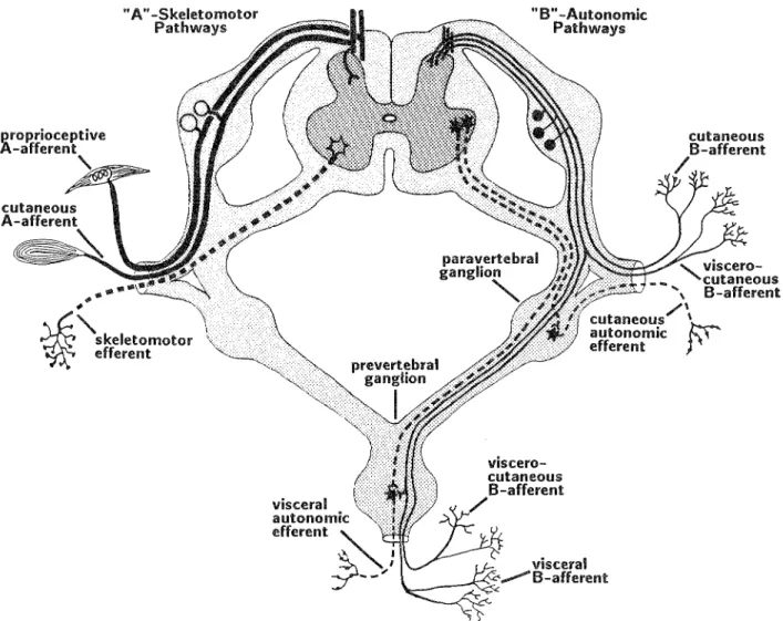
At the turn of the century, these notions of inner/outer functional divisions were validated by their correspon-dence with discrete features of CNS organization (see Figure 2a). In later years, however, the doctrine was applied according to the pathway of the peripheral pro-cesses: Fibers which coursed in visceral nerves, and thus extended inward, were designated "visceral"; fibers of the cerebrospinal nerves were classified as "somatic." In the case of afferents, recent results do not bear out the doctrine's purported correlation between central or-ganization and peripheral nerve pathway. Although the central terminals of spinal cord afferents do indicate a dichotomous organization, this seems to be related more to fiber type than to inner or outer functions or nerve pathway. In general, the large myelinated affe rents ter-minate in the deep layers of the dorsal'horn (especially laminae III & IV); the smaller, unmyelinated, and A-delta fibers terminate predominantly in the superficial layers (laminae I and II; see Figure 2b). Although the fibers of the large myelinated afferents are found mostly, if not exclusively, in somatic nerves that innervate the outer tissues (e.g., skin, muscles, joints), the distal processes of the smaller and unmyelinated afferents are found in all nerve types, passing both to inner and outer tissues. Moreover, some of these unmyelinated afferents have dichotomizing collaterals that pass through both visceral and somatic nerves (Bahr et al. 1981; see neuron labeled "viscero-cutaneous" in Figure 4). In conclusion, there seem to be no compelling reasons for classifying afferents on the basis of gross innervation territories (inner/outer) or the pathways they take.
2.2. Langiey's search for autonomic afferents. As dis-cussed in the Introduction, Langley searched for mor-phological distinctions that would define the afferents falling under his ANS concept, and when he could find none, he declared the ANS to be a purely motor system. This conclusion resulted partly because the fiber stain available to Langley (osmium) would not reveal the ma-jority of visceral afferents, which were unmyelinated (see Langley 1922). When he compared visceral and somatic afferents, he compared the exceptional splanchnic A-beta fibers (myelinated), which Innervate mesenteric Pacinian corpuscles (cat), with the more common myelinated fibers of the somatic nerves; of course, they were alike. Apart from the technical limitations of his day, how-ever, Langley's search was also hampered by the prevail-ing preconceptions. From the beginnprevail-ing, Langley con-sidered the spinal ganglia and nerves as the anatomical territory of the "somatic" nervous system. Only after he failed to find sensory neurons in the sympathetic ganglia (Langley 1903) did he look to the dorsal root ganglia. Langley would accordingly consider an afferent as auto-nomic only if at some point it had passed through
path-Prechtl & Powley: B-Afferents
•(outer)
(inner)
Somatic sensory column Visceral sensory column Visceral motor column Somatic motor column
Medial group (A-afferents)
Lateral group (B-afferents)
Figure 2. Early and modern views of spinal cord organization. Top (a): Early conception of a cross section of the human spinal cord showing the four columns that were hypothesized to be basic to the doctrine of nerve components. The somatic and visceral sensory columns were considered the projection zones of afferents from outer and inner tissues, respectively. Based on Figure 67 of Herrick's (1927) An Introduction to Neurology. Bottom (b): Modern view of spinal afferent organization. In some species, the cutaneous and proprioceptive afferents (the ontogenetically earliest afferents) form a fascicle that enters the spinal cord medially; they have large fibers and collaterals that terminate in the ventral layers of the dorsal horn. (Propriocep-tive collaterals are not shown.) The visceral and somatic (cut-aneous, articular, muscular), or late-arriving, afferents form the lateral group (lateral group); have small fibers and terminate chiefly in the superficial layers of the dorsal horn (darkly shaded area). Adapted from Maxwell & Rethelyi (1987).
ways that Langley had considered a priori to be ex-clusively "autonomic." Thus, because none of the cutaneous or other nonvisceral afferents took a circuitous path through the sympathetic ganglia, none of them could be considered autonomic. As a result of this approach, the dermis has become a reflexological oddity; its only efferent innervation is autonomic (e.g., vasomotor, pil-oraotor, sudomotor), and its only afferent innervation is called "somatic." (As will be discussed later, many of these so-called somatic afferents elicit autonomic reflexes when stimulated.)
This dependence on nerve pathway as a classification
criterion represents a return to the cerebrospinal system concept of Willis and its functional associations. Langley (1921) objected to using volition as a classification criteri-on because of its subjectivity, but he persisted with Willis' idea that the cerebrospinal nerves were dis-tinguished by their ability to produce conscious sensa-tions. The observation that sympathetic nerve stimula-tion produced pain reinforced Langley's view that sympathetic nerve afferents did not differ from other spinal cord afferents arid thus should also be considered somatic (Langley 1903, pp. 21, 26).
2 3 . Contemporary terms and usages. Currently, differ-ent PNS classifications are used for differdiffer-ent purposes. Herrick's terminology is used for "visceral" afferents and Langley's term "autonomic" is indispensable for discuss-ing the innervation of tissues that are difficult to describe as visceral (e.g., bone marrow, sweat glands, brown adipose tissue).1 The term "somatic" is applicable to both systems but its different connotations cause confusion. Some scientists use the term simply to designate those nerves that do not innervate the viscera; others attach functional connotations in the tradition of Langley (e.g., conscious sensation). For example, in one prominent physiology textbook published in the United States (Guyton 1986), visceral pain is listed under the rubric "Somatic Sensations II."
Other classifications of peripheral neurons in addition to the systems of Herrick and Langley have contributed to the current terminology and blend of meanings. Table 1 is a list of these classifications and the criteria used to establish them. The more recent groupings of afferents under the labels "sympathetic" and "parasympathetic" (see Introduction) do not conflict with the B-neuron grouping proposed here; they could include separate subpopulations of neurons. The observation that B-neurons are involved in the functioning of autonomic reflexes is a strong argument that they are associated with the ANS. The fact that they are found in sympathetic and parasympathetic pathways is less compelling. Again, as Bell (1811) concluded, nerves do not correspond to spe-cific neuron classes. Many of the accepted nerve labels are either arbitrary or do not accurately indicate their fiber compositions. For example the abdominal vagus, although thought of as a parasympathetic nerve, contains sympathetic (Ahlman et al. 1979) and other yet uniden-tified fibers (Prechtl & Powley 1987; 1990).
3. A- and B-afferents: Alternatiwes to somatic and visceral classes
We now know that there are two major populations of afferents in the dorsal root ganglia: the A- and the B-neurons. These two populations are produced by suc-cessive waves of cell proliferation (Carr & Simpson 1978, chick; Hamburger & Levi-Montalcini 1949, chick; Law-son & Biscoe 1979, mouse; LawLaw-son et al. 1984, rat). The A-neuron class is known to consist predominantly of proprioceptive and mechanoreceptive afferents; the B-class consists mostly of thermoreceptive and nociceptive afferents. Thus far, a nervous system classification has not been proposed that is based on these two afferent popula-tions; rather, the A- and B- designators have been applied
Prechtl & Powley: B-Afferents
Table 1. Peripheral sensory neuron classifications
Work Classification Criteria
Head et al. (1905) Sherrington (1906) Gasser and Erlanger
(1929); Heinbecker et al. (1934) Lloyd (1943)
Contemporary authors
Deep sensibility; protopathic sensi-bility; epicritic sensibility
Proprioceptive; interoceptive; extero-ceptive
A-, B-, and C-fibers«
Afferent groups I, II, III, Wh
Sympathetic, parasympathetic af-ferents
Function, morphology, receptive field size, regeneration profile
Function, responses to adequate stimuli Conduction velocity, electrophysiological traits, fiber size
Reflex type, fiber size, peripheral termi-nation
Ability to elicit ANS reflexes, found in sympathetic or parasympathetic nerves
a
Fiber classification includes efferents.
b
Lloyd's classification was originally formulated for analyzing hind limb reflexes in the cat.
secondarily to the traditional designators, "somatic" and "visceral," and have usually been used only for describing ganglion cell morphology. In this target article we argue that the ontogeny of afferent populations is far more significant and more fundamental than other classification criteria. As will be shown, the A-B distinction seems to be correlated not only with many other distinguishing cell traits and organizational features, but also with a particu-lar class of functions. Put another way, we conjecture that Langley, if he had had access to today's data, would have assigned B-neurons to the autonomic afferent category that he left empty.
3.1. "Large light" and "small dark" cells* In light-micro-scopic studies conducted since the nineteenth century, the A- and B-neuron populations have been referred to as "large light" and "small dark" cells (for reviews see Andres 1961; Lieberman 1976; Scharf 1958). The desig-nators "A" and "B" have been applied only recently by electron microscopists. Although some investigators had initially argued that, the difference in light-microscopic density of the A- and B-neurons was artifactual, ultra-structural studies have since indicated that it is related to the distribution of Nissl substance and neurofilaments. The A-neurons appear lighter because their Nissl sub-stance is gathered in clumps within a lattice of relatively translucent neurofilament bundles, whereas the somata of the small dark neurons interfere more thoroughly with light because their granular endoplasmic reticulum and free ribosomes are more concentrated and more evenly distributed (see Figure 3). Other ultrastructural features distinctive of the small dark cells (B-neurons) include more highly developed Golgi apparatus, lysosomal bodies, and • Golgi-associated smooth endoplasmic re-ticulum with associated lysosomes (Lieberman 1976), and a concentric zonal distribution of membrane-bound organelles (Rambourg et al. 1983, rat; Sommer et al. 1985, mouse). With the use of prolonged nerve stimula-tion and electron microscopy, Duce and Keen (1977, rat) demonstrated that the differences in infrastructure of A-and B-neurons could not be explained on the basis of different activity states.
Although the B-neurons are small, the size distribu-tions of A- and B-neuron populadistribu-tions overlap
consider-ably (Lawson 1979, mouse; Lawson et al. 1984, rat; J. Price 1985, rat). B-neuron cell diameters range from 15 fxm to 35 |xm; those of A-neurons range from |xm 15 to 70 fxm, but most are greater than 40 |xm. Using comput-erized statistical analysis, Lawson (1979, mouse) demon-strated that the distribution of cell sizes in the DUG can be described by no fewer than two Gaussian distribu-tions.
Other morphological differences are also found at the light-microscopic level. The A-neurons have prominent spiraling glomeruli and have mostly large myelinated fibers. Their unipolar cell stems are also myelinated, and in the central zone of the ganglion, at a node of Ranvier, each stem divides into central and peripheral myelinated processes of roughly equal diameter (Ha 1970, cat). The B-neurons do not have complicated glomeruli and most of their fibers are unmyelinated, although some are thinly myelinated (Andres 1961, rat). The unipolar cell stem of the unmyelinated B-neurons, and possibly that of the thinly myelinated ones, forms a central process that is several times thinner than either the stem or the pe-ripheral process (Ha 1970, cat).
Recently, the presumed A- and B-neuron populations of the rat have been discriminated histochemically. The light-microscopically identifiable A-neurons were found to bind the neurofilament protein antibody RT97 (Kai-Kai etal. 1986, rat; Lawsonetal. 1984, rat;}. Price 1985, rat; Sharp et al. 1982, rat), and the size distribution of RT97-labeled neurons correlated well with the theoretical sta-tistical distributions generated to describe the cell size distribution of the A-neuron population (Lawson et al. 1984, rat). The B-neurons were found to contain arginine vasopressin (Kai-Kai et al. 1986, rat). When RT97 and arginine vasopressin probes were applied to adjacent sections of the same DRG, they stained complementary populations.
Although arginine vasopressin may be a marker of all B-neurons in the rat, it has long been known that both overlapping and nonoverlapping subpopulations of B-neurons can be identified by various histpchemical mark-ers,2 which include bombesin/gastrin-releasing peptide (Panulaetal. 1983, rat), the corticotropin-releasing factor (Skofitsch et al. 1985, rat), fluoride-resistant acid phos-phatase (Knyihar-Csillik & Csillik 1981, rat; J. Price
Prechtl & Powley: B-Afferents El-Neurons ONTOGENY Histochemical Marker Fiber Types s Groups Receptor Endings Modalities lODD fpion, ccepttlo
Itch, crude touch
Figure 3. A- and B-neuron characteristics. The essential criterion for grouping B-neurons is a common ontogenetic history. Thus far, B-neurons are known to have been born late and to have a characteristic sensitivity to nerve growth factor during development. Most of the proposed divisions are tentative extrapolations that have not yet been directly tested using the ontogenetic criteria. Although most neurons with fibers that conduct impulses in the A-delta velocity range (central panel) belong to the B class, D hair receptor afferents (which also conduct in the A-delta range) may be an exception; their central morphology (see Brown 1981) and electrophysiological properties (see Koerber et al. 1988) differ from those of other B-neurons (A-delta and C-fibers). Neuron illustrations adapted from Rambourg et al. (1983).
1985, rat), oxytocin (Kai-Kai et al. 1986, rat), somatostat-in, substance P (Hokfelt et al. 1976, rat; Tuchscherer & Seybold 1985, rat), and vasoactive intestinal polypeptide (Lundberg et al. 1978, guinea pig)- At least one'sensory neuropeptide, the calcitonin gene-related peptide, does not seem to be a discriminator of B-neurons because it is found in DRG neurons of all sizes (Ju et al. 1987, rat). The histochemistry and fiber type data suggest that the A- and B-neuron populations are the basis of the prin-cipally dichotomous organization of DRG afferents within the dorsal horn that has been summarized or implied in recent reviews (Brown 1981; Cervero 1986; Hunt 1983; Maxwell & Rethelyi 1987; Perl 1984; D. D. Price 1986; Ruda et al. 1986; Willis 1985): Peripheral to or within the dorsal root entry zone, DRG afferent processes are segre-gated into medial and lateral groups (Figure 2b). The lateral group distinctively contains small, unmyelinated, and thinly myelinated fibers; the medial group contains mostly large, thickly myelinated fibers. Whereas the fibers of the lateral group terminate predominantly in the superficial layers of the dorsal horn (Rexed's laminae I, II,
and V), the collateral terminals of the medial group terminate most densely in the more ventral layers (es-pecially laminae "III and IV). The termination pattern of the lateral group matches closely the combined termina-tion patterns of the various histochemically identified B-neuron processes (see de Groat 1986; Hunt 1983; Kai-Kai et al. 1986; Ruda et al. 1986). Also indicative of the lateral group's origin is the observation that, like B-neurons, the lateral group appears late in ontogeny, as do the neurons of the superficial layers of the dorsal horn (Altman & Bayer 1984). Moreover, some anatomical features of the two groups may be explained by their different on-togenies. For example, the hair receptor afferents of the medial group characteristically descend medially in the dorsal horn, curve laterally and ascend dorsolaterally, and terminate predominantly in laminae III and IV (see Figure 2b). The developmental explanation for such cir-cuitous paths is that although these early-arriving axons initially form a straight path to rely neurons, they are later dragged ventromedially as the dorsal portion of the devel-oping spinal cord rotates (Altman & Bayer 1984). The
lateral group (B-neuron) processes arrive after the rota-tion phase and thus do not become distorted,,
3o2. B-neurons of different species and deweiopmentai stages. Comparative analyses of D.RG cytology are not available, but the light-microscopic literature includes reports of similar cell populations in a variety of verte-brates, including the cat, cow, fox, horse, human, lamprey, lemur, mouse, pig, rabbit, and rat (see Scharf 1958). They have also been identified in electron micro-scopic studies of the rabbit (Dawson et al. 1955; Tennyson 1965), guinea pig (Hess 1955), rat (Andres 1961), frog (Berthold 1966), and mouse (Sommer et al. 1985).
Most studies have reported on the cell structure of the adult ganglia; however, large- and small-celled popula-tions have also been reported in the embryonic ganglia of the chicken (Hamburger & Levi-Montalcini 1949), sheep, rabbit (Tennyson 1965), and mouse (Lawson 1979; Lawson & Biscoe 1979). Furthermore, there is evidence that the large- and small-celled embyronic populations represent immature versions of the A- and B-neuron phenotypes found in the adult. With the use of thymidine labeling, Lawson and Biscoe (1979; also Lawson 1979) demonstrated that large and early-differentiating neu-roblasts of the murine DRG mature into A-neurons and that the smaller, late-differentiating neuroblasts mature Into B-neurons. A striking feature of the chick embryo DRG is that for a period of development during and after ganglion cell differentiation the two cell types are clearly segregated in different zones of the ganglion (Hamburger & Levi-Montalcini 1949). The large neurons occupy the lateroventral zone (and are thus called LV cells); the small ones occupy the mediodorsal zone (and are called MD cells).
For decades, the two neuron populations of the chick embryo DRG were designated LV and MD cells, and the issue of homology with other adult vertebrate DRG cells did not arise. Recently, however, (Barakat et al. 1985; Philippe et al. 1986) have applied the A- and B-neuron terminology to the chick embryo cells, presumably be-cause myelin-associated glycoprotein immunoreactivity was localized specifically in chick embryo MD cells (Omlin et al. 1984), and this immunocytochemical stain-Ing In hatching-age chick ganglion cells was correlated with B-neuron morphology (Philippe et al. 1986). Other grounds for postulating an equivalence between B-neu-rons and MD cells are: (1) Both MD neuB-neu-rons (Hamburger & Levi-Montalcini 1949) and B-neurons (Lawson 1979,
mouse; Lawson & Biscoe 1979, mouse) are born in the
second of the two overlapping waves of cell proliferation. (2) Root ganglion substance P immunoreactivity, which is restricted to B-neurons in the chick embryo DRG, is found only in the MD population (Fontaine-Penis et al. 1985; New & Mudge 1986). (3) These substance P-con-taining MD neurons project primarily to the avian dorsal horn homologues of Rexed's laminae I and II (La Valley & Ho 1983; New & Mudge 1986), the same laminae to which B-neurons project. (4) The peripheral processes of MD neurons seem to show the same broad distribution as B-neurons (Honig 1982).
3.3. neuron mefameres in the cranial root ganglia. B-neurons are not restricted to spinal root ganglia but are
Prechtl & Powley: B-Afferents found also in the metameric (segmentally homologous) ganglia of the cranial nerve roots.
Like the spinal nerves, some cranial nerves (5th, 7th, 9th, and 10th) have dorsally positioned roots and have been called dorsal root nerves (Romer 1970). Most such nerves have two ganglia; the more proximal of the two is known as the root ganglion and is considered a metamere of the spinal root ganglia. The cranial root ganglia, like their spinal counterparts, are embryonic derivatives of the neural crest and include the jugular ganglion of the 10th nerve, the superior ganglion of the 9th nerve, and the cells that occupy the most proximal portion of the trigeminal ganglion in early development. In contrast, the more distal ganglia, such as the nodose and petrosal, have a placodal embryonic origin (Narayanan & Narayanan 1980, chicken). The trigeminal nerve ganglion also has placode-derived neurons that aggregate distally in the nerve root (Hamburger 1961, chicken), but they do not form an anatomically distinct distal ganglion.
Cranial B-neurons accordingly, have many of the same characteristics as spinal B-neurons, such as late matura-tion, small cell size, and the presence of substance P (Fontaine-Perus et al. 1985, chicken). The presumed trigeminal B-neurons of adult specimens have compara-ble ultrastructures (Jacobs et al. 1975, rat; Peach 1972,
rat) and have the same neuropeptides: arginine
vas-opressin, oxytocin (Kai-Kai et al. 1985, rat), substance P, somatostatin, and vasoactive intestinal polypeptide (Kum-mer & Heym 1986, guinea pig; see also Ruda et al. 1986). They also show similarly heavy staining for acid phos-phatases and acetylcholine esterases, although dif-ferences have been found in monoamine oxidase staining (Kalina & Wolman 1970, rat). Finally, the afferents of cranial nerves 5, 7, 9, and 10, which would be presumed to be B-neurons because of their fiber type or function (thermoceptive or nociceptive), or the presence of sub-stance P, terminate densely in the spinal trigeminal subnucleus caudalis (Cuello et al. 1978, rat [trigeminal substance P]), the medullary continuation of the dorsal horn.
3.4. Lumping and splitting. Thus far, few subpopulations of the A and B classes have been studied in detail, and the existence of additional small but distinct populations cannot be ruled out. Electron microscopists who have systematized the classification of ganglion cells have typ-ically included a transitional group that has the ultrastruc-tural features of both A and B classes, such as the A3 cell
groups of Rambourg et al. (1983, rat) and Andres (1961, 7% of rat DRG cells). Also, some very small neurons have been distinguished from the B-neurons and are consid-ered to constitute a C class (Rambourg et al. 1983, rat; Sommer et al. 1985, 1% of mouse DRG cells). These additional groups may account for the 5% of DRG neu-rons that Lawson et al. (1984, rat) were unable to classify with the use of densitometry, or for the RT97 antibody. Although the histochemical markers tested thus far with rat tissue suggest a sharp division among most ganglion cells, future histochemical identifications will have to be correlated with ontogenetic and ultrastructural traits, both within and between species.
As shown in Figure 3, B-neurons have a number of traits that distinguish them from other DRG neurons. Traits such as presence of substance P are not listed
Prechtl & Powley: B-Afferents
because, unlike the presence of arginine vasopressin, they distinguish only a subpopulation of B-neurons. The demarcation of A- and B-neuron groups proposed in Figure 3 is based primarily on the hypothesis and im-plicitly accepted principle that ontogenetic history pro-vides the most crucial data for cell classification. Just as phylogenetic history (when it can be deduced) is predic-tive of the characters and behaviors of related organisms, ontogeny should best predict cell phenotype because it is through modifications of the ontogenetic sequence that cellular specializations emerge. Cell lineage, in combina-tion with epigenetic factors (e.g., timing, posicombina-tion, micro-environment) ultimately determines the differentiated state of the cell; in most instances neurons do not become similar as a result of responding to dissimilar ontogenetic variables. Nevertheless, we know little about the rules of ontogeny, or about how to interpret and weight different ontogenetic data. In this target article, the late birth of the B-neuron population is regarded as an indicator of specific developmental potentials that are not possessed by the A-neurons.
The test of this ontogenetically based classification will be whether it can parsimoniously describe the functional organization of the PNS. That is, the information that a DUG afferent belongs to the ontogenetically defined A or B class should tell us more about its structure, connective relations, and function than the information that its pe-ripheral fiber passed through a somatic or visceral nerve.
4. Close relationship of B-afferents and autonomic neurosis
An association between B-neurons and the ANS is sug-gested by the peripheral distribution of B-afferents, by their role in homeostatic reflex functions (section 5), and by the fact that virtually all root ganglion visceral afferents belong to this population. Often cell populations with collaborative or closely related functions have similar phenotypic and ontogenetic traits (e.g., T- and B-lym-phocytes), and this seems to be true of B-neurons and ANS neurons.
4.H. Fhenotypic similarities. At the most superficial level, B-neurons, with their less differentiated morphologies, tend to resemble principal (postganglionic) autonomic neurons. The small dark cell types in some preparations have been mistaken for genuine sympathetic neurons. For instance, having used a prolonged osmium stain, Kiss (1932) maintained that the large groups of DRG cells (B-neurons) found in numerous vertebrates were actually ectopic sympathetic neurons. Moreover, in mammals, most sympathetic and B-neuron fibers are histologically indistinguishable (Gasser 1955) and conduct impulses in nearly identical velocity ranges - from 0.7 to 2.3 m/sec for superior cervical ganglion sympathetics; from 0.6 to 2.0 m/sec for dorsal root C-fibers (Patton 1960, p. 77). In contrast, most somatic (skeletomotor) efferents and af-ferent A-neurons conduct impulses at velocities several times greater.
A number of histochemical similarities have also been found between sympathetic3 neurons and certain sub-populations4 of neurons. For example, like many B-neurons, sympathetic neurons contain substance P
(Kes-sler et al. 1981, rat). Substance P has also been found in a variety of preganglionic ANS neurons, including sym-pathetic preganglionic neurons (Krukoff 1987, cat), preganglionic neurons extending to the ciliary ganglion (Erichsen et al. 1982, pigeon), and vagal preganglionic neurons extending to the cardiac ganglion (Bowers et al. 1986, bullfrog; Gibbons et al. 1987, toad). Other histo-chemical markers found both in the sympathetic ganglia and in subpopulations of B-neurons are tyrosine hydroxy-lase (j. Price 1985, rat), somatostatin, and vasoactive intestinal polypeptide (Fontaine-Perus et al. 1985, chick
embryo; Lundberg et al. 1982, guinea pig). Finally,
myelin-associated glycoprotein immunoreactivity, which is a marker of B-neurons in the chick embryo (see section 3.2), has also been found in cells of the sympathetic ganglia and adrenal medulla of the same preparation (Omlin et al. 1984).
4.2. Ontogenetic parallels* Perhaps the most significant ties between sympathetic and B-neurons are their late ontogeny and exceptional sensitivity to nerve growth factor. In a now historic experiment, Levi-Montalcini and Hamburger (1951) observed the morphological effects on a chick embryo of a factor released by grafted mouse sarcoma tumor fragments. The tumor fragments were grafted unilaterally to the base of a limb bud in 1.5- to 3-day-old chick embryos. When embryos were histo-logically examined on embryonic days 5 and 6, the un-treated limb showed the normal combined ingrowth of the LV neurons (embryonic A-afferents; see section 3) and skeletomotor efferents; in the tumor-bearing limb the LV fibers either bypassed the tumor or were blocked by it. In contrast, the embryos that were examined at later dates - once the MD cells (chick embryo B-neurons) and sympathetic neurons had differentiated and ex-tended neurites - exhibited a striking effect: the sym-pathetic ganglia and MD (B-neuron) zones of the dorsal root ganglia were hyperplastic, and these two neuronal groups had caused a "hyperinnervation" of the tumor. In the fifties, the substance responsible for these trophic and morphogenetic responses was isolated and named nerve growth factor (NGF; for review see Levi-Montalcini 1987).
More recently, NGF has been found to have a variety of trophic effects on a number of cell populations; sym-pathetic neurons (except "short-type" symsym-pathetics; see Harper & Thoenen 1981) and B-neurons, however, pro-vide the broadest demonstration of NGF's activities, including neurite extension and guidance (Levi-Mon-talcini 1987). These late-developing efferents and af-ferents may depend partly on NGF as a chemotactic or haptotactic factor for approximating their target tissues, even though much of their anatomical course has already been paved by the earlier-differentiating skeletomotor and A-neurons. Such a morphogenetic action could help explain the comparably broad innervation patterns of sympathetic and B-neurons and the observation that they jointly innervate some tissues to the exclusion of all other neuron types (e.g., cornea, Tervo et al. 1979; bone marrow, Bulloch 1985; sweat glands, Hokfeltetal. 1975). By citing as an example the responsiveness of these neuron groups to NGF, Black (1986) has suggested that conjoint responsiveness to trophic factors represents an evolutionary mechanism for regulating coinnervation
(e.g., adjustment of relative pathway size). Whether NGF played a role in evolution or even affects normal morphogenesis is still unclear; it is of interest here, however, that sympathetic and B-neurons show the same exceptional responsiveness to NGF both in vitro and in vivo.
Levi-Montalcini & Hamburger's (1951) study of the chick embryo serves to contrast the developmental tim-ing of A- and skeletomotor neurons with that of B- and sympathetic neurons. This differential timing is also re-flected in the ontogeny of reflex functions in mammals. Proprioceptive and exteroceptive reflexes appear early, in utero (see Windle 1944), whereas sympathetic (Smith et al. 1982, rat) and other C-fiber reflexes (Fitzgerald & Gibson 1984, rat) do not mature until well after birth. Although postnatal B-neurons no longer respond to NGF with neurite extension, most B-neurons, like sympathetic neurons, continue to show biochemical responses to NGF. Sympathetic neurons respond to NGF by increas-ing their levels of catecholamines (Harper & Thoenen 1981); the somatostatin- and substance P-containing B-neurons respond by increasing their respective neu-ropeptide contents (Kessler & Black 1981, rat).
A final ontogenetic argument for the relatedness of B-neuron and sympathetic cell types comes from results suggesting that they have similar developmental poten-tials. Until embryonic day 10, the cells of quail dorsal root ganglia back-transplanted into young (day-2) chick em-bryos migrate to the sympathetic ganglia and adrenal glands and differentiate into adrenergic phenotypes (i.e., sympathetic and chromaffin cells; for review, see Le Douarin 1982). Also, under specific conditions, cultured DRG cells are able to differentiate into catecholaminergic (sympathetic) neurons (Newgreen & Jones 1975, chicken; Xue et al. 1985, quail). The undifferentiated DRG cells that are capable of becoming sympathetic neurons may perhaps represent the same (or similar) precursors that have been found to differentiate under other culture conditions into small neurons that are immunoreactive for substance P and for myelin-associated glycoprotein (i.e., B-neurons; Barakat & Droz 1987).
5e Functional ties between B=neurons and autonomic neurons
The similarity between sympathetic and B-neuron phe-notypes is also reflected in their functional relations. By extrapolation from both fiber size and the conclusion that B-neurons are the afferents of layers I & II of the dorsal horn, we can infer that the "modalities"5 mediated by B-neurons include thermoception, nociception, tickle, itch, and crude (C-fiber) mechanoreception. Some B-af-ferents, therefore, must constitute the afferent limb of thermoregulatory and nociceptive sympathetic reflexes (see Janig 1985). Moreover, it is becoming increasingly clear that sympathetic and peptidergic B-neurons jointly mediate the neural control of the immune system (Felten et al. 1985; Payan et al. 1986).
Although thermoregulation and immunomodulation are generally considered autonomic functions, nocicep-tive reflexes associated with pain perception are often regarded functionally as "somatic" (see section 2.3). However, recent evidence from substance P-containing
Prechtl & Powley: B-Afferents B-neurons (SP-B neurons), the most thoroughly charac-terized nociceptive afferents, affirms that nociceptive reflexes invariably and most directly involve the ANS or autonomic effectors.
5.1. SB neurons and rejectiwe reflexes. Substance P-containing afferents represent a major subpopulation of B-neurons, by far the most studied ones. In rats SP-B neurons amount to from 10% to 30% of DRG neurons (Hokfelt et al. 1976; Kai-Kai et al. 1986), or as much as half of all B-neurons, depending on the segmental level sampled. The peripheral terminals of SP-B neurons are highly collateralized and broadly distributed via visceral and somatic nerves to a variety of tissues, including autonomic effectors. Some SP-B neurons have collaterals that also terminate in autonomic ganglia (Hokfelt et al. 1977; cat, guinea pig, rat; see Figure 4).
For the most part, SP-B neurons mediate reflexes that are elicited by injurious or potentially injurious stimuli (noxae), some of which contribute to pain perception (Henry 1980). In 1937, these and other B-afferents that mediate protective reflexes in the skin were described as constituting a "nocifensor" system by T. Lewis (see Lem-beck 1985; 1987). It now seems clear that the afferents that mediate analogous reflexes in the eyes and mucous membranes and have been known as the "common chem-ical sense" (see Moncrieff 1967) belong to the same system.
SP-B neurons mediate protective reflexes in three different ways. First, they mediate them in the least direct way by releasing substance P (and perhaps other neuroactive substances) to interneurons and to the in-terstitial space of the spinal cord. Physiological studies have shown that intrathecal substance P facilitates the flexor withdrawal reflex in response to noxae (Wiesen-feld-Hallin 1986, rat). Second, they mediate such re-flexes more directly (monosynaptically), by releasing substance P antidromically from axon collaterals that terminate on principal autonomic ganglion neurons. SP-B neurons have been found to form axodendritic synapses with prevertebral sympathetic neurons (Matthews et al. 1987, guinea pig); when stimulated, they produce slow, excitatory, postsynaptic potentials (Otsuka & Konishi 1983, rat). Finally, SP-B neurons mediate protective reflexes most directly by releasing locally in the effector tissue transmitter from stimulated nerve terminals or from the antidromically excited terminals of neighboring collaterals. These local terminal and axonal reflexes pro-vide the least ambiguous definition of SP-B neuron func-tion because they are fixed and independent of synaptic or integrative factors.
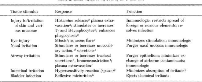
Local SP-B neuron reflexes vary from tissue to tissue, but in general they are rejective or immunologic (see Table 2) - that is, they purge, neutralize, or bar poten-tially harmful elements from epithelia or interstitial spaces. Since the writings of Bernard (1878, 1973), the interstitial fluid of tissues has come to be regarded as a carefully regulated milieu of electrolytes, gases, nu-trients, thermal energy, and metabolic by-products. The external environment is recognized as the source of nurture, but also as a threat. Homeostasis consists of the carefully monitored exchange between the two environ-ments, the assimilation of nutrients and warmth, and the rejection of noxae and foreign elements. Rejective
reflex-Freehtl & Powley: B-Afferents proprloceptiwe A-afferent \ cutaneous A-affereot cutaneous B-afferent wiseero-cutaneoys -afferent wiscero-cutaneous B-afferent visceral -afferent
Figure 4. Afferent and efferent components of the spinal nerves and ganglia. The left-hand side shows the A-afferent and skeletomotor (somatic) efferent (dashed line) divisions. The right-hand side shows the B-afferent and autonomic efferent (dashed lines) divisions. The viscero-cutaneous (or possibly viscera-muscular) neuron has collaterals that dichotomize into visceral and somatic pathways. (Drawing based on the electrophysiological data of Bahr et al. 1981). The collateral innervation of pre vertebral ganglia is consistent with findings that have been reported on substance P-containing B-neurons. Adapted in part from Carpenter (1976, p. 232).
es are therefore particularly important in tissues that are at risk, such as those which are specially adapted for nutrient assimilation, blood-gas exchange, thermal ex-change, waste concentration, and certain sensory func-tions (e.g., nasal mucosa).
Although many SP-B neuron reflexes could be de-scribed as rejective because they function to minimize the immediate effects of eliciting stimuli, it is doubtful that a single term can describe all of their actions. Fur-thermore, the visceral and somatic varieties of SP-B neurons are not likely to be functionally identical, be-cause their projection patterns within the superficial dorsal horn have been found to differ slightly (Cervero & Connell 1984, cat). Nevertheless, comparisons using a number of criteria, including the role in the defense of homeostasis, indicate that inner and outer SP-B neurons are far more alike than their "visceral" and "somatic" labels would suggest.
The role in the defense of homeostasis is not restricted to B-neurons that contain substance P. Eecently, Lem-beck (1987) summarized a number of findings on cap-saicin-sensitive B-afferents (SP-B neurons included) and
hypothesized that such afferents constitute a "neurogenic alarm system" or a "network of defense." The defensive reflexes he cites include neurogenic inflammation, heat loss thermoregulation, micturition, sneezing, lacrima-tion, salivalacrima-tion, splanchnic depressor responses, protec-tive skeletomotor reactions, and a number of endocrine responses such as catecholamine release in response to hypoglycemia (see also Lembeck 1985). In general, Lem-beck's (1987) insightful interpretation of capsaicin-sen-sitive B-afferents is compatible with the hypothesis of this target article. In support of the argument developed here about ANS relations, however, we would emphasize that the role of B-neurons in skeletomotor and behavioral defensive reflexes is functionally and synaptically less direct than in autonomic reflexes. Only autonomic neu-rons and autonomic effector tissues have been shown to be directly excited by B-neurons.
The boundaries outlined in Figure 3 suggest that B-neurons also include mechanoreceptive C-fiber afferents that are neither nociceptive nor capsaicin-sensitive (Fitzgerald 1983). If by ontogenetic definition these af-ferents are truly B-neurons, then a test of the present
Prechtl & Powley: B-Afferents
Table 2. Local rejective reflexes of SP-B neurons
Tissue stimulus Response Function
Injury to/irritation of skin and vari-ous mucosae Eye injury Nasal irritation Airway irritation Intestinal irritation Bladder infection
Histamine release;a plasma extra-vasation0; stimulates or increases T- and B-lymphocytesb; enhances
phagocytosis b
Miosisc; aqueous flarec
Stimulates or increases mucocili-ary action,^ secretions6
Stimulates or increases tracheal secretionsfc; broncoconstriction-^; plasma extravasation-^
Hypersensitivity reaction (spasm) § Reflexive micturitionh
Immunologic: restricts spread of foreign or noxious elements; re-solves infection
Minimizes stimulation; immunologic Purges nasal mucosa; immunologic Purges epithelium; minimizes ex-change of airborne contaminants; immunologic
Minimizes absorption of irritants? Ejects chemical irritants
Note: The most recent reference or best source of references is cited.
"Lembeck 1985. ^Payan et al. 1986.
c
Hakanson et al. 1.985. ^Lindberg & Mercke 1985.
e
Lundblad et al. 1983. /Lundberg et al. 1985. ^Goetzl et al. 1985. >*Maggi & Meli 1986.
hypothesis would be to examine whether their synaptic and functional relations are most direct with the ANS, or with skeletomotor neurons. Similarly the present hy-pothesis would predict that in the cat most visceral afferents have more in common with somatic B-afferents than with the exceptional A-beta visceral afferents (see section 2.2) because the A-beta visceral afferents, as judged by their fiber types, are probably not B-neurons, whereas most visceral afferents are.
§B implications
One implication of our second hypothesis is that nocicep-tive reflexes are simply one of a variety of autonomic reflex types. The autonomic correlates of nociception have traditionally been thought of as epiphenomena, that is, nonessential symptoms or indexes of nociception, and as examples of "soraatic-autonomic" integration. Accord-ing to the present model, nociception and ANS function are simply the afferent and efferent aspects of the same reflex arc. The validity of this perspective is borne out by the observation that PNS disorders that involve nocicep-tive B-afferents - such as cluster headache (Hardebo 1984), familial dysautonomia (see Pearson et al. 1982), and reflex dystrophies (see Procacci & Maresca 1987) -invariably involve abnormalities in autonomic function. Dyck et al. (.1983) have coined the term "hereditary sensory autonomic neuropathy" to refer to a number of disorders characterized by insensitivity to noxious stimuli and autonomic symptoms, and by the involvement of both small afferent and autonomic fibers. Also, because of the traditional PNS classification systems, clinical termi-nology has varied and has often encouraged different pathophysiological hypotheses (Procacci & Maresca 1987). For instance, some schools of neurology refer to
causalgia and similar disorders as algodys trophies, whereas others call them sympathetic dystrophies. The difference in terminology and concept can be traced to Langley's "autonomic nervous system" concept and its continual rejection by some of the European schools (see Procacci & Maresca 1987).
The most fundamental issue raised by the present hypothesis concerns the meaning of cell phenotypes with respect to the evolution of the nervous system. Some boundaries between the A- and B-neuron populations depicted in Figure 3 may be inaccurate, but if the proposition is generally correct, a question about the significance of this cytological discontinuity arises. Why is it that thermoreceptive and nociceptive afferents are born late, and discriminative mechanoreceptive and pro-prioceptive afferents are born early?
One possibility is that the discontinuity reflects an evolutionary shift from homeostatic nervous function alone to a function that includes behavior or skel-etomotor maneuvers. According to Romer (1970), the ancestor of the vertebrates was a passive, sea-dwelling filter-feeder with a life-style much like that of an intes-tine. Somatic function evolved with the addition of loco-motor devices such as tails and fins. Romer hypothesized that the original visceral and somatic components were so distinct anatomically and functionally that they could be thought of as two different animals that had been welded together. Although the two components have become progressively more integrated, in Romer's words (1970, p. 29), the "weld" is still an imperfect one.
Many scientists would judge the A-afferents to be more phylogenetically derived in terms of fiber type (Bishop 1959) and receptor ending specializations (Pieron 1952); moreover, these neurons give rise to the lemniscal sys-tem (see Mountcastle 1961). The evolutionary innovation represented by the appearance of A-afferents could be
Commentary/Prechtl & Powley: B-Afferents
the ability to handle highly discriminative and rapidly changing information about spatial relationships between the animal and the external environment. The primitive B-neurons, on the other hand, seem to be well suited for handling the kind of control system information necessary for the simple increases or decreases characteristic of homeostatic adjustments.
In summary, the variation on Homer's (1970) "imper-fect weld" interpretation suggested here is simply that the B- and A-neurons represent the descendant afferents of old and new nervous systems, the old one being originally charged with homeostatic functions and the new one charged with the reconnaissance and behavioral operations necessary for active animal-environment relations.
NOTES
1. There appears to be no consensus as to what a viscus is. Kuntz (1953) extended the definition to include glands and blood vessels - presumably grouping together all of the effectors innervated according to Langley's ANS concept. For a lively exchange on this unresolved issue, see Dart (1922) and Herrick (1922).
2. Words that refer to histochemical markings, such as "im-munoreactive," "content," and "labeling," should be taken to mean like immunoreactivity.
3» Currently, too little is known about the autonornic ganglia in general to pursue the idea that B-afferents are specifically related to sympathetics - or, for that matter, that sympathetics and parasympathetics constitute valid afferent cell classes. Simi-larly, the hypotheses developed here are restricted to B-af-ferents because relatively little is known about other afB-af-ferents that are also involved in autonomic reflexes, including the so-called special visceral afferents. The same reservations prevent-ing their incorporation into a more global classification scheme with B-afferents pertain to the enteric plexuses, the intrinsic sensory neurons that are thought to constitute the third subdivi-sion of the ANS (see Figure 1). To paraphrase Langley, further progress awaits the discovery of distinguishing histological characters.
4o Many traits are shared only by subsets of either B-neurons or ANS neurons; nevertheless, they may be regarded as evi-dence of a relationship. Such traits are called polythetic, as are the taxa they indicate (see Mayr 1982, p. 189; Sneath 1962). For membership in a polythetic taxon, no single trait is either necessary or sufficient.
5. In this article the word "modality" is used only in a loose sense in order to help identify the afferents in question; its use does not indicate an endorsement of any rigorous form of specificity theory.
^Correspondence should be directed to James C. Prechtl, Department of Neurosciences, A-001, University of California at San Diego, La Jolla, CA 92093.
Commentaries submitted by the qualified professional readership of this journal will be considered for publication in a later issue as Continuing Commentary on this article. Integrative overviews and syntheses are especially encouraged.
afferents by input
P. L. R. Andrews51 and I. N. C. Lawesb
^Department of Physiology, St. Georges Hospital Medical School, Cranmer Terrace, London, SW17 ORE, England and department of Biomedicai Sciences, University of Sheffield, Western Bank, Sheffield, S10 2TN, England
It is sometimes said in jest that research in anatomy progresses not through the discovery of new structures but through the renaming of old ones. On initial cursory reading of the target article, it appeared to be a good example of the genre; closer inspection, however, revealed that the article not only critically reviews the debate about the naming of the afferent nerves associated with the autonomic nervous system (ANS) but it also exploits this debate as a vehicle for suggesting a novel division of the peripheral nervous system on complementary functional and structural grounds. In introducing the term "autonomic nervous system," Langley (1921) included an explanatory state-ment: "It is more important that the new word should be used for new ideas than that the words should be accurately descrip-tive. " Prechtl & Powley's (P & P's) review is written in the spirit of this statement and our criticisms essentially revolve around the "idea" rather than the nomenclature.
The presence of afferent axons in nerves that Langley re-garded as part of the ANS has been demonstrated by histological and neurophysiological studies and is not a matter of dispute; but their designation as autonomic afferents is contested. On several grounds, P & P propose that the well-described B-afferents are in effect the anatomically distinct group of B-afferents sought by Langley as the afferent arm of the ANS. It appears to us that at best the B-afferents can be only a component of the autonomic afferent system (if, for the moment, one excludes the B-afferents supplying the skin; see below).
Vagal afferents supplying the cardiovascular, respiratory, and digestive systems probably outnumber afferents traveling with sympathetic nerves such as the splanchnic, and yet the scheme proposed excludes them from being autonomic on the grounds that they do not fulfill the criteria for being B-afferents (e.g., they are of placodal rather than neural crest origin). If we exclude vagal afferents from P & P's classification, where do we put them? They clearly convey information to the central ner-vous system (CNS), give rise to sensations by pathways (as indirect as, for example, splanchnic afferents), influence auto-nomic efferents in response to both noxious and innocuous stimuli (Andrews 1986) and, as with the B-afferents, are in-volved in rejective reflexes. P & P claim that their classification is more parsimonious; we agree, in the literal sense of the word, which means "stingy" (Oxford English Dictionary); and P & P have indeed been stingy in including only a minor population of the afferents associated with the ANS.
The role of a classification is to reduce complexity, but this new one will, we fear, increase it, not least because of its omission of several cranial afferents, particularly the vagus. Langley's classification shaped the concept of the ANS, particu-larly in the time before unmyelinated fibres could be easily studied. There is now sufficient evidence, however, to question the continuing value of his scheme and to assess whether we should bolster it further by fitting new data into it rather than by replacing it totally.
From an examination of the basis for ascribing an autonomic afferent role to the B-neurones, P & P extend the observations to propose the hypothesis that the B-afferent system (cutaneous and visceral components) is the substrate of a more fundamental division of the nervous system mediating homeostasis. This is a very interesting idea and meshes with a novel view of home-ostatic mechanisms published by one of us (Lawes 1989). We believe that a consideration of homeostatic mechanisms allows a true reconciliation of the functional and anatomical classification
Commentary/Frechtl & Powley: B-Afferents of B-afferents and non-B visceral afFerents; we therefore propose
the following scheme for discussion:
The distinction between A- and B-afFerents is not their con-nection to skeletomotor and autonomic efFerents, respectively. Neither does the autonomic regulation of homeostasis neces-sarily precede the skeletomotor equivalent. The original contri-bution of the nervous system to homeostasis was predominantly skeletomotor: AfFerents detected suboptimal conditions and skeletomotor efFerents withdrew the organism to a more con-ducive environment. Only later in phylogeny did the central nervous system acquire greater control of smooth muscle and glands and allow the autonomic mediation of homeostatic mech-anisms so prevalent in mammals.
Thus, skeletomotor escape/avoidance behaviour preceded autonomic regulation, at least as far as thermoregulation and osmoregulation are concerned. AfFerents such as thermorecep-tors (including cutaneous ones), presumably of the B-group, were therefore originally linked to protective behaviour ex-pressed via skeletomotor efFerents. Nociception, a prominent function of B-neurones, fits readily into this scheme. The func-tion of B-afFerents is not autonomic per se, but the detecfunc-tion of any threat, whether autonomic and requiring a homeostatic response or somatic and requiring a skeletomotor response. Conversely, A-afFerents detect and discriminate stimuli that are in themselves not threatening and do not require es-cape/avoidance or autonomic arousal. In this scheme, the role of the non-B afFerents (e.g., nodose components of the vagus) becomes clearer. They are a further component of the division of the nervous system mediating homeostasis and protection from harmful stimuli; emesis, in particular, illustrates how wrong it is to try to separate skeletomotor and autonomic function as there is a considerable involvement of both (Andrews & Hawthorn 1987). It is the purpose of the response and not its mechanism of mediation that is the crucial criterion.
In conclusion, the concept of A- and B-neurones is a good one, but it should be extended to include other afFerents that have similar properties, for example, those of the vagal nodose ganglion. Now we can abandon fruitless semantic arguments over whether there are skeletomotor and not autonomic af-ferents, or some somatic afFerents that more closely resemble visceral afFerents. Once attention is turned to what the afFerents do for the animal, these difficulties are resolved, and the biolog-ical significance of striking anatombiolog-ical and biochembiolog-ical dif-ferences is revealed.
To classify or not to classify:
That is the question
F. Cervero
Visceral Sensation Research Group, Department of Physiology, University of Bristol Medical School, University Walk, Bristol BS8 1TD, England
I have enjoyed reading Prechtl & Powley's (P & P's) target article as it is not usual to find scientific articles addressing general questions far beyond the minute details of fragmented data. It is good to know there are neuroscientists who can think of the nervous system as something other than a useful collec-tion of ion channels!
I suspect, however, that there are at least two main reasons why such articles as this one are uncommon nowadays. First, very few journals - though Behavioural and Brain Sciences is one of them - publish papers that address general issues rather than specific items of data. Second, and in this case more Important, we have acquired so much detailed information on the organization of the nervous system that it is virtually impos-sible to generalize without the exemptions outweighing the rules.
The kind of grand classification of the different elements of the
nervous system prominent at the turn of the century and so well reviewed by P & P (Langley, Gaskell, and the rest) was largely because little detailed information existed on those individual elements, such as their anatomical, electrophysiological, and neurochemical properties. Speculation has always been In-spired when not too much is known about an issue. My main objection to another grand classification of the peripheral ner-vous system along the lines of P & P is, therefore, that these generalisations are no longer helpful because we are now forced to twist many experimental observations to make everything fit into a grand scheme. It is true that the current classifications of, the peripheral nervous system are a bit of a mess (and P & P bring this point home very well), but does the new proposal of two distinct categories of primary afferent neurone agree with all the available data? Let us consider a few items:
(i) One of the most powerful spinal actions of afferent C-fibres is the activation of somatic motoneurones. In fact, not only can afferent C-fibres excite flexor motoneurones but they can also increase the excitability of the flexion reflex for prolonged periods of time. Yet P & P play down these affects as "less direct" than the activation of autonomic systems.
(ii) The neurotoxin capsaicin affects afferent C- and A-delta fibres but the functional properties of capsaicin-insensitive af-ferents connected to nociceptors are all similar to those of capsaicin-sensitive ones. Hence, it is not right to imply that capsaicin is a neurotoxin specific to B-afFerents.
(iii) It is possible to dissociate (anatomically, functionally, and behaviourally) the somatic components of nociceptive reflexes from the autonomic responses to a noxious stimulus. The central organization of these two reflex actions is quite different even though they may share a few common elements. I do not think that somatic nociceptive reflexes are simply a variety of a general autonomic reaction.
(iv) Although anatomical evidence exists for dichotomizing afferent fibres with branches in somatic and visceral nerves, no one has yet shown that these branches are connected to func-tionally active sensory receptors (as implied in P & P's Figure 4). It is important to point out that several investigators have tried (and failed) to find such dual receptive sites, as this is essential for a dichotomizing afferent fibre to have any real functional significance.
(v) One of the most powerful inputs to preganglionic sudomotor neurones is mediated by cutaneous Pacinian corpus-cles that are connected to the largest and fastest myelinated somatic afFerents. This is an example of an autonomic reflex input entirely supported by A-afFerents and hence a major exemption to P & P's classification.
So, the problem is not whether or not there are B-afferents but whether or not it is possible to classify primary afferent neurones into only two major categories and expect that all known properties of these neurones will fit neatly into this pair of very distinct bins. I was not persuaded by P & P's arguments; rather, I still think there are many different classes of primary afferent neurone. Under certain functional or behavioural cir-cumstances, some of these distinct primary afFerents can appear to have a common function, but I doubt that this major classifica-tion of the peripheral nervous system is useful or justifiable.
3-afferent classification apply
to wagai afferent neurons?
J. S. Davison and K. A. Sharkey
Department of Medical Physiology, Faculty of Medicine, University of Calgary, 3330 Hospital Drive N.W., Calgary, Alberta, Canada T2N 4N1 Electronic mail: 28552@ucdasum1 .hitnet
In their thought-provoking target article on B-afferents, Prechtl & Powley (P & P) raise a number of interesting hypotheses