Journal of Analytical Toxicology, Vol. 25, April 2001
Semiautomated High-Performance Liquid
Chromatographic Method for the Determination of
Benzodiazepines in Whole Blood
Anissa El Mahjoub and Christian Staub*
Institut Universitaire de M6decine L~gale, Geneva, Switzerland
Abstract [
A semiautomated method for the determination of five frequently prescribed benzodiazepines (BZD) (clonazepam, diazepam, flunitrazepam, midazolam, and oxazepam) in whole blood samples by reversed-phase high-performance liquid chromatography following simple online enrichment and clean-up on a short precolumn is described. After precipitation of protein and red cells with a mixture of organic solvents (methanol/acetonitrile, 50:50), the aliquot is centrifuged and the organic upper phase evaporated under a gentle stream of nitrogen. The residue is reconstituted by adding 500 pL of a mixture of phosphate buffer (20raM, pH 2.2) and acetonitrile (70:30, v/v). The sample is then directly introduced into the column-switching column. The precolumn is first washed with phosphate buffer at pH 7.2. Compounds retained on the precolumn are then eluted in the back-flush mode and separated on a C 8 semi-microcolumn (tichrospher select B, 125 x 3 mm). The BZD studied are determined by a diode-array detector at 254 nm. The method shows excellent linearity between 25 and 1000 ng/mL for clonazepam, flunitrazepam, and midazolam and between 25 and 5000 ng/mt for diazepam and oxazepam. The recoveries are around 80% for clonazepam and oxazepam and around 90% for the three others. Coefficients of variation for between-day and within-day assays are < 15% for low concentrations close to the limit of quantitation and < 5% for high concentrations.
Introduction
have been frequently reported (5-9). Further studies have been
carried out (10-12), and comparisons have been made between
GC-MS techniques and various immunoassays (13,14).
High-performance liquid chromatography methods (HPLC)
are often applicable to only one drug and its metabolites
(15-17). Furthermore, papers reporting screening procedures
(18) do not specifically address the problem of clinical and tox-
icological screening, as very often two or more unrelated drugs
are co-administered with benzodiazepines.
The chromatographic theory and method development be-
hind column-switching or online preconcentration techniques
have been reviewed and described (19-21).
This paper describes a column-switching technique in which
whole blood samples are injected, after protein and red cell
precipitation with a mixture of organic solvent, into a BioTrap
500 MS column (extraction column) and are then washed with
a mixture of 30raM phosphate buffer (pH 7.2) and acetonitrile
mobile phase (94:6, v/v). The drugs retained are then eluted
onto a C8 reversed-phase analytical column for determination.
The method presented is rapid and easily automated and al-
lows to determine BZD regularly encountered in clinical and
toxicological cases. The column-switching technique offers
several advantages: direct injection of biological fluids, non-
manual or robotic clean-up, semiautomated and minimized
contact with the infectious biological fluids, on-column en-
richment of analytes, and low cost per sample.
Benzodiazepines (BZD) are often used as antianxiety agents
in the treatment of psychiatric disorders (1--4). Various re-
searchers have reported total plasma BZD concentration related
to both clinical effect and toxicity. A variety of methods for the
detection and determination of BZD in biological matrices are
described in the literature.
Gas chromatography-mass spectrometry (GC-MS) methods
* Author to whom correspondence should be addressed: Christian Staub, Institut Universitairede M~decine L~gale, 9, avenue de Champel, 1211 Geneva 4, Switzerland. E-mail christian.staub@medecine.unige.ch.
Experimental
Chemicals
Clonazepam, diazepam, flunitrazepam, midazolam, and ox-
azepam were purchased from Promochem (Molsheim, France).
Human plasma was obtained from the University Hospital of
Geneva (Switzerland).
Monobasic and dibasic potassium phosphate and phosphoric
acid were purchased from Merck (Darmstadt, Germany), and
HPLC-grade acetonitrile was obtained from Romil (Cambridge,
England).
Stationary and mobile phases
The column-switching procedure was achieved by using a
BioTrap 500 MS (Chromtec, Hiigersten, Sweden), a new bio-
compatible extraction column offering repeated direct injection
of serum, plasma, supernatant of cell culture, or other complex
matrices, into the HPLC system without any clean-up proce-
dure (except a simple centrifugation). This biocompatible ex-
traction column is pH stable (between pH 2 and pH 11) with a
biocompatible external surface (cd-acid glycoprotein) and a
hydrophobic internal surface (hydrophobic polymer).
The surface within the pores is also a hydrophobic polymer,
and the matrix pores are small enough to exclude plasma pro-
Table I. Optimized Conditions
Component
Description
Extraction column Analytical column Loading
Transfer and analytical separation Detection Volume of plasma injected Biotrap MS 500 20 x 4.0-mm i.d Thermostated at 35~
Lichrospher select-B 125 x 3-mm i.d, 5pm Thermostated at 35~ K2HPO4, (30mM, pH 7.2) 5 min at 0.6 mL/min A = Acetonitrile (ACN), B = KH2PO 4, (20raM, ph 2.1) Linear gradient
0 rain: A/B 30:70 at 0.5 mL/min 30 min: A/B 35:65 at 0.3 mL/min 254 nm
50pL
Journal of Analytical Toxicology, Vol. 25, April 2001
tein and other macromolecular compounds.
The separation was performed using a C8 reversed-phase
semi-microcolumn (LiChrospher Select B, 125 x 3-mm i.d.,
5-1am particle size) and a guard column (Nucleosil NH2 8 x 4-
mm i.d., 5-1am particle size, Macherey-Nagel, Oensingen,
Switzerland). This method was recently validated (22).
The column switching mobile phase consisted of 30mM phos-
phate buffer (pH 7.2) and acetonitrile (94:6, v/v). The analytical
mobile phase comprised a mixture of 20raM phosphate buffer
(pH 2.1) and acetonitrile. The optimized conditions of this
technique are detailed in Table I.
Column-switching system
A representative column-switching technique set-up is given
in Figure 1. The online system consisted of two quaternary
pumps (model HP 1100) and two columns connected by an HP
1100 high-pressure six-port valve in back-flush configuration.
The chromatographic system was equipped with a diode-array
detector, an automatic injector, and an autosampler.
In extraction position, pump A pumped the extraction mobile
phase through the autosampler, into which the sample was in-
jected. A filterholder was inserted after the autosampler with a
2-1am biocompatibility filter. After passing through the filter, the
sample was transported to the extraction column via the six-
port valve. During this time, analytical pump B pumped the an-
alytical mobile phase through the column via the six-port valve.
In elution position, the mobile phase from pump A (the ex-
traction mobile phase) went to waste. The mobile phase from
pump B (analytical mobile phase) back-flushed from the ex-
traction column to the analytical column.
The strong elution power of the second mobile phase deliv-
ered by pump B caused analyte desorption from the extraction
column. The change in flow direction (back-flush) caused ad-
ditional analyte concentration.
Extraction position and separation position (original position)
Pump I
Pump 2
" ' ~ " ~ ~ ^~ Rotation 60"
Figure
1. Schematic representation of the column-switching set-up. Adapted from reference 23.Transfer position Pump 1
Pump 2
Journal of Analytical Toxicology, Vol. 25, April 2001 5 10 15 2D 25 30 Time ( m i n ) E E E ~ m ,= 0 N ~ ~I~ N ~K "O 0 Q. e~ 0 Time ( m i n )
Figure 2. Chromatogram of drug-free blood sample (top). Chromatogram of spiked blood with clonazepam (300 ng/mL), flunitrazepam (300 ng/mL), midazolam (300 ng/ml_), diazepam (I 000 ng/mL), and oxazepam (1000 ng/mL)(bottom), and methyIclonazepam (2000 ng/ml I.S.). Ex- traction conditions: injection, 50 ml. of blood sample (after precipitation) on BioTrap 500 MS extraction (20 x 4-mm i.d); mobile phase, acetoni- trile/phosphate buffer (pH 7.2, 0.3M). Analytical conditions: C8 reversed- phase column, Lichrospher Select B (I 25 x 3-mm i.d); mobile phase, acetonitrile/phosphate buffer (pH 2.1,0.2M) (35:65, v/v) at a flow rate of 0.3 mL/min UV detection at 254 nm.
Table II. Calibration Data for the Five Benzodiazepines (n = 3)
Range Coefficient of Line (pg/mL) correlation r Y = ax + b Clonazepam 0.025-1.0 0.995 0.0013x- 0.074 Diazepam 0.025-5.0 0.999 0.002x + 0.048 Flunitrazepam 0.025-1.0 0.996 0.0028x- 0.14 Midazolam 0.025-I .0 0.997 0.0017x + 0.18 Oxazepam 0.025-5.0 0.995 0.0015x- 0.34
In separation position, the valve was switched back to its original position (extraction position), the analyte fraction caused transfer into the analytical column, and analyte sepa- ration then took place in a conventional manner.
Eluent absorbance was monitored at 254 nm. HP Chem- Station (Hewlett-Packard Software no. G2170AA) was used for instrument control, data acquisition, and data handling.
Standard solutions
Stock standard solutions of clonazepam, diazepam, fluni- trazepam, midazolam, and oxazepam were prepared by dis- solution of each compound in methanol to obtain a concen- tration of I mg/mL. Stock solutions were stored at -20~ and remained stable for at least 24 months.
Biological standards were prepared at the required concen- trations by diluting the appropriate aliquots of the stock solu- tions with clean drug-free blood between 25 and 5000 ng/mL for both oxazepam and diazepam and between 25 and 1000 ng/mL for clonazepam, flunitrazepam, and midazolam (Figure 2).
Phosphate buffer
The extraction phosphate buffer (pH 7.5, 30mM) was pre- pared by transferring 2.7 mL of 1M KH2PO4 and 9.9 mL of 1M K2HPO4 into a 1000-mL volumetric flask and making up to volume with distilled water. The analytical phosphate buffer (pH 2.1, 20mM) was prepared by transferring 12.7 mL of 1M KH2PO4 and 22.3 mL of 1M H3PO 4 into a 1000-mL volumetric flask and making up to volume with distilled water. Buffer so- lutions were always freshly prepared and filtered through a 0.45-1Jm filter (Supelco, Bellefonte, PA) immediately before use.
Sample preparation
Whole blood samples (1 mL) were spiked with the different mentioned benzodiazepines at the desired concentrations and with 20 IJL of the appropriate internal standard (methylclon- azepam, 100 pg/mL). Spiked samples were mixed with 2 mL of a mixture of organic solvent (methanol/acetonitrile, 1:1), vortex mixed, and centrifuged for 10 min at 5000 rpm. The super- natant was evaporated to dryness and dissolved in 500 IJL of analytical mobile phase, then placed into glass vials. Fifty mi- croliters was processed online as described.
Table III. Repeatability and Reproducibility (n = 6)
Clonazepam Flunitrazepam Midazolam Concentration repeat.* repro, repeat, repro, repeat, repro.
(ng/mL) CV% CV% CV% CV% CV% CV%
Oxazepam Diazepam repeat, repro, repeat, repro.
CV% CV% CV% CV% 50 15.2 14.6 12.8 13.5 13.9 9.9 250 - - - 300 5.2 6.7 2.4 3.5 4.9 3.4 500 4.2 3.6 2.0 2.3 4.5 3.0 3000 - - 5000 - - - * A b b r e v i a t i o n s : repeat., r e p e a t a b i l i t y a n d repro., r e p r o d u c i b i l i t y . ~" N o t a n a l y z e d at this c o n c e n t r a t i o n . t _ _ _ 4.8 4.7 4.4 4.1 3.8 3.1 4.0 3.7 2.1 3.0 2.9 2.0
Journal of Analytical Toxicology, Vol. 25, April 2001
Results
Chromatography
The five benzodiazepines were well separated, and the whole procedure allowed an extremely clean chromatographic trace (Figure 2).
Linearity
Detector response linearity was evaluated by preparing five triplicate calibrations covering the following concentration ranges: 25 to 1000 ng/mL for clonazepam, flunitrazepam, and midazolam and 25 to 5000 ng/mL for diazepam and oxazepam. Linear regression lines were obtained by plotting peak-area ratios (the compound peak area divided by one of the internal standards, see Table II).
Mldazolam ~o CIonazepam
5 n ~ m L
IS
+il
9 ; : :,+
oo So 1oo 15o ~ o ~ 0 ~ o o 50 100 ISO ~ 2SO 3OO
Concentration (ng/mL) Concentration (ng/mL) Flunltrazepam 2O l \ o ... Is o . . . _ _ " : o I o I ~ 15o Z e ~ ~ o Concentration (ng/mL) Oxazepam Diazepam I$ 28.7 n g / m L 15 3 0 n g / m L =< _< 8 0 I I ! I O , - - , i i i
o 5o too ISO 2oo ~ o 3Oo o 5 0 100 1 5 0 2 0 0 2 5 o
Concentration (ng/mL) Concentration (ng/mL)
I
3 0 0
Figure 3. Curve determination of LOQ values.
Table IV. Recoveries Obtained with Spiked Plasma Samples (n = 6) Precision
Within-day reproducibility or repeatability (see Table III) was evaluated by replicate analysis (n = 6) of pooled blood at three different concentrations: 250, 3000, and 5000 ng/mL for di- azepam and oxazepam and 50, 300, and 500 ng/mL for the three others on the same day. The coefficients of variation (CV) obtained are shown in Table III.
Between-day reproducibility or reproducibility (Table III) was determined by replicate analysis (n = 6), over three days, at the same concentrations as for repeatability.
Assay detection limits
Limit of detection (LOD).
The LaD, defined as the lowest an-alyte concentration that can be clearly detected above the base- line signal, is estimated as three times the signal-to-noise ratio. The LaD was determined (n = 6) by injection of spiked blood with BZD in decreasing concentrations. LaD was determined to be around 15 ng/mL for clonazepam, flunitrazepam, and midazolam and 10 ng/mL for diazepam and oxazepam.
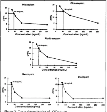
Limit ofquantitation (LOQ).
The LOQ (n = 6) is the lowestconcentration that can be measured on the standard curves with acceptable reproducibility (CV < 15%) (Figure 3). The lower pratical LOQ was 50 ng/mL for clonazepam, below 40 ng/mL for flunitrazepam and midazolam, and 30 ng/mL for diazepam and oxazepam.
Recovery
Recoveries of the five benzodiazepines studied were deter- mined by comparing the peak areas of the spiked blood samples and of the reference samples at six different concentrations. The reference samples were injected directly into the analytical column and the spiked blood samples were injected, after precipitation with a mixture of organic solvent, into the ex- traction column coupled to the analytical column through the switching valve.
As showed in Table IV, the method gives
acceptable
values for recoveries. The observed recovery is higher than 90% for allBZD,
except
clonazepam and oxazepam (around 80%).Clonazepam Flunitrazepam Midazolam Oxazepam Diazepam
Amount added mean mean mean mean mean
(ng/mL) recovery(%) recovery(%) recovery(%) recovery(%) recovery(%)
25 70.0 83.1 95.0 67.1 84.2 50 67.0 88.3 101.7 -* 100 76.7 87.0 113.0 - 250 - 72.3 88.3 300 76.7 84.0 100.3 - 500 82.8 105.7 101.3 76.3 88.3 800 81.0 97.7 107.7 - 1000 80.0 96.0 113.3 74.3 91.0 2000 - 74.3 104.0 3000 - 79.3 103.0 5000 - - 89.3 102.0
* Not analyzed at this concentration.
Application to Postmortem Blood Samples
Generally, postmortem whole blood is not as fluid as antemortem serum or plasma and is not easy to work with. Therefore, several sam- ples were chosen to demonstrate the potential of this method.
The first blood sample obtained from a de- ceased subject showed four benzodiazepines (desalkylflurazepam, flurazepam, desmethyl- diazepam, and oxazepam) and one antidepres- sant (venlafaxine) (Figure 4). The measured concentrations were as follows: desalkylflu- razepam, 42 ng/mL; flurazepam, 52 ng/mL; desmethyldiazepam, 310 ng/mL; and ox- azepam, 1150 ng/mL.
Journal of Analytical Toxicology, Vol. 25, April 2001
In the second blood sample, also obtained from a deceased
person, the following substances were clearly identified: ben-
zodiazepines (dernnoxeparn and desrnethyldiazeparn), one anti-
depressant (amitriyptiline), propranolol, and tramadol (Figure
5). The following concentrations were determined: demoxe-
pare, 995 ng/rnL; desmethyldiazeparn, 1095 ng/rnL; and ami-
tryptiline, 605 ng/mL.
In the third blood sample obtained from a deceased subject,
two benzodiazepines (lorazeparn and oxazepam) were detected
(Figure 6), and the following concentrations were measured:
lorazepam, 905 ng/mL and oxazeparn, 2075 ng/mL.
Conclusions
The HPLC procedure described for the simultaneous deter-
mination of benzodiazepines appears rapid and suitable for
routine analysis.
Satisfactory validation data were achieved for linearity, pre-
cision, and recovery. The LOQ allows measurement of thera-
peutic concentrations for most benzodiazepines and toxic
concentrations for flunitrazeparn. For the therapeutic concen-
tration of flunitrazepam, the supernatant is evaporated to dry-
*
ij !
-,,,i
JEt ...
1o
o,
nm
Figure 4. Chromatograms and UV spectra of blood sample obtained from a deceased subject. Extraction conditions: injection, 50 pL of blood sample (after precipitation), on BioTrap 500 MS extraction (20 x 4-ram i.d); mobile phase, acetonitrile-phosphate buffer (pH 7.2, 0.3M).
Analytical conditions: C8 reversed-phase column, Lichrospher Select B
(125 x 3-ram i.d); mobile phase, acetonitrile/phosphate buffer (pH 2.1,
0.2M) UV detection at 254 nm 2dO~ 5 0 , " " ~ . . . . , ~ , " ~ , " ~ , " ~ ' " a k ' " " ~ o ~ " ~ , " ~ -.~- . . . . n m ~ ~ ,,,,,v, ~,~ ,.~.,,..~ ~ ,,.,~ ~, ~.,~.o dk, z b ~ . ~ ~ o , ~ - - ~ . - :do " , J . - - ; , , ' n m
Figure 5. Chromatograms and UV spectra of blood sample obtained from
a deceased subject. Extraction conditions: injection, 50 pl. of blood
sample (after precipitation), on BioTrap 500 MS extraction (20 x 4-ram i.d); mobile
phase,
acetonitrile/phosphate buffer (pH 7.2, 0.3M). Ana-Igical conditions: C8 reversed-phase column, Lichrospher Select B
(125 x 3-mm i.d); mobile phase, acetooitrile/phosphate buffer (pH 2.1,
0.2M); UV
detection
at 254 nm 3 5 4 3 O 4 2 5 4 10 4 5 4t
t
T i m e ( r a i n )c~ o~al, l U ~ ~ I ~u,/r ~ lUlO & i ~ ~ m4aCClJD
s ' ,
. . . . ~ . . . . ,
Figure 6. Chmmatograms
and UV spectra of blood sample obtained
Nm a deceasedsubject.
Extraction conditions: injection, 50 pk of bloodsample (after
precipitation), on BioTrap 500 MS extraction (20 x 4-ram i.d); mobilephase,
acetonitrile/phosphate buffer (pH 7.2, 0.3M). Ana- lytical conditions: cg reversed-phase column, Lichrospher Select a (125 x 3-ram i.d); mobile phase, acetonitrile/phosphate buffer (pH 2.1,0.2M); UV detection at 254 nmJournal of Analytical Toxicology, Vol. 25, April 2001
ness and dissolved in 100--150 IJL of mobile phase.
The use of a column-switching technique allows to mini-
mization of contact with infectious biological fluids like blood
and plasma.
Finally, the proposed method is not only suitable for deter-
mining benzodiazepine in whole blood samples, but can also be
applied to other drugs such as antidepressants.
References
1. R.I. Shader and D.J. Greenblatt. Clinical implication of benzodi- azepines in pharmacokinetics. Am. J. Psych. 134:652-656 (1977).
2. K. Jinno, M. Taniguchi, and M. Hayashido. Solid phase micro ex- traction coupled with semi-microcolumn high-performance liquid chromatography for the analysis of benzodiazepines in human urine. J. Pharm. Biomed. Anal 17:1081-1091 (1998).
3. E. Tanaka, M. Terada, S. Misawa, and C. Wakasugi. Simultaneous determination of twelve benzodiazepines in human serum using a new reversed-phase chromatographic column on a 2-microns porous microspherical silica gel. J. Chromatogr. B 682:173-178
(1996).
4. J.B. Roberts and J.A. Tafuri. Clinical Management of Poisoning and Drug Overdose, L.M. Haddad and J.F. Winchester, Eds. W.B.
Saunder, Philadelphia, PA, 1990, pp 800-820.
5. K. Kudo, T. Nagata, K. Kimura, T. lmamura, and M. Noda. Sensi- tive determination of diazepam and N-desmethyldiazepam in human material using capillary gas chromatography-mass spec- trometry. J. Chromatogr. 431: 351-359 (1988).
6. N. De Giovanni and M. Chiarotti. Analysis of benzodiazepines. II. High-performance liquid chromatography-fluorescence detection after molecular rearrangement to acridanones. J. Chromatogr. 428:
321-329 (1988).
7. H. Maurer and K. Pfleger. Identification and differentiation of ben- zodiazepines and their metabolite in urine by computerized gas- chromatography-mass spectrometry. J. Chromatogr. 422:85-101
(1987).
8. M. Japp, K. Garthwaite, A.V. Geeson, and M.D. Osselton. Collec- tion of analytical data for benzodiazepines and benzophenones.
J. Chromatogr. 439:317-339 (1988).
9. A.J.H. Louter, E. Bosma, J.C.A. Schipperon, J.J. Vreuls, and U.A.Th. Brinkman. Automated on-line solid-phase extraction gas- chromatography with nitrogen phosphorus detection: determina- tion of benzodiazepines in human plasma. ]. Chromatogr. B 689: 35-43 (1997).
10. D.A. Black, G.D. Clark, V.M. Hayer, J.A. Garbin, and A.J. Saxon. Analysis of urinary benzodiazepines using solid-phase extraction and gas chromatography-mass spectrometry. J. Anal. Toxicol. 18:
185 (1994).
11. C. Moore, G. Long, and M. Marr. Confirmation of benzodiazepines in urine as trimethylsilyl-derivatives using gas-chromatog- raphy-mass spectrometry. J. Chromatogr. B 655:132-137 (1994).
12. K.M. Hold, D.J. Crouch, D.E. Rollins, D.G. Wilkins, D.V. Canfield, and R.A. Maes. Determination of alprazolam and 0~-hydroxyal- prazolam in human plasma by gas chromatography/negative-ion chemical ionization mass spectrometry. J. Mass Spectrom. 31:
1033-1038 (1996).
13. R.L. Fitzgerald, P.A. Rexin, and D.A. Herold. Detection benzodi- azepines: immunoassays compared with negative chemical ion- ization gas chromatography/mass spectrometry. Clin. Chem. 40:
373-380 (1994).
14. T. Nishikawa, H. Ohtani, D.A. Herold, and R.L. Fitzgerald. Com- parison of assay methods for benzodiazepines in urine, a receptor assay, two immunoassays and gas chromatography-mass spec- trometry. Am. J. Olin. Pathol. 107:345-352 (1997).
15. T.B. Vree, A.M. Boars, Y.A. Hekster, and E. Van der kleijn. Simul- taneous determination of chlordiazepoxid and its metabolites in human plasma and urine by means of reversed-phase high-per- formance liquid chromatography. J. Chromatogr. 224:519-525
(1981).
16. L. Wen-Nuei. Determination of clonazepam in serum by high pressure liquid chromatography. Ther. Drug Monit. 9:337-342
(1987).
17. M. Sajgo. Determination of tofisopam in serum by high-perfor- mance liquid chromatography. J. Chromatogr. ,117:303-307
(1981}.
18. R Mura, A. Piriou, P. Fraillon, Y. Paper, and D. Reiss. Screening pro- cedure for benzodiazepines in biological fluids by high-perfor- mance liquid chromatography using a rapid scanning multichannel detector. J. Chrornatogr. 416' 303-310 (1987).
19. D. Westerlund. Direct injection of plasma into column liquid chromatographic system (Review). Chromatographia 24" 155-164
(1987).
20. J.B. Lecaillon, N. Febvre, and C. Souppart. Influence of solute po- larity in column-switching chromatography for the assay of drug in plasma and urine (Review). J. Chromatogr. 317:493-506 (1984).
21. A. El Mahjoub and C. Staub. High-performance liquid chromato- graphic method for the determination of benzodiazepines in plasma or serum using the column-switching technique. J. Chro- matogr. 742:381-390 (2000).
22. A. El Mahjoub and C. Staub. Simultaneous determination of ben- zodiazepines in whole blood or serum by HPLC/DAD with a semi-micro column. J. Pharm. Biomed. Anal. 23:447-458 (2000).
23. J. Henderson and A. Grahn. Determination of drugs by direct in- jection of plasma into biocompatible extraction based on protein- entrapped hydropholic phases. J. Chromatogr. 660:119-129
(1994).
Manuscript received July 10, 2000; revision received October 2, 2000.