Simon Wildermuth Sebastian Leschka Hatem Alkadhi Borut Marincek Received: 6 September 2004 Revised: 26 November 2004 Accepted: 2 December 2004 Published online: 26 January 2005 # Springer-Verlag 2005
Multislice CT in the pre- and postinterventional
evaluation of mesenteric perfusion
Abstract Multislice computed tomography angiography (CTA) has been found feasible for the evaluation of visceral vasculature. The develop-ment of multislice technology has overcome past limitations. First, the faster scanning speed increases volume coverage during a single breath-hold and improves the exploi-tation of contrast medium. Second, the better spatial resolution results in nearly isotropic voxels allowing re-construction of high-resolution three-dimensional images with different algorithms. Volume rendering is ca-pable of displaying the visceral vas-culature from any external vantage point. Compared to conventional
angiography, CTA not only delineates vessels but also depicts the anatomi-cal relationship to adjacent structures and allows the evaluation of perfused organs. CTA also has become an emerging tool for the pre- and post-interventional assessment of vascular anatomy. The purpose of this pictorial essay is to present a spectrum of visceral vascular diseases and inter-ventional and surgical therapies, and to highlight the role of postprocessing for their evaluation.
Keywords Multislice computed tomography . Angiography .
Postprocessing . Mesenteric perfusion
Introduction
Computed tomography angiography (CTA) is a valuable minimally invasive tool for the visualization of visceral vasculature [1, 2]. When compared to conventional angi-ography, CTA is less expensive, saves examination time, and may save iodinated contrast dose and radiation expo-sure of the patient [3,4]. Moreover, CTA allows innumer-able arbitrary projections and is not limited to a single opacification. The introduction of 16-row multislice com-puted tomography (MSCT) has overcome most of the limitations of earlier scanner generations. With regard to faster scanning speed and improved spatial resolution, MSCT enables an increase in scan volume during a single breath-hold with a sub-millimeter resolution. Compared to 4-row or 8-row MSCT, the improved temporal resolution of 16-row MSCT ameliorates the contrast bolus exploitation and therefore decreases the opacification of overlaying
veins. The high z-plane resolution results in nearly isotropic voxels, allowing high-quality postprocessing algorithms. Multiplanar reformatted projections (MPR) are able to display the axial data sets in any desired oblique or curved plane. Another valuable algorithm is maximum intensity projection (MIP), which provides angiography-like dis-plays. The volume rendering (VR) technique makes use of the entire data set, thus giving an overview and may convey more information than MPR and MIP. It can show multiple internal and overlying features, and the flexibility of the VR algorithm allows the protocol to be tailored to the actual clinical problem, but when improperly used has the poten-tial hazard of loss of valuable information. Parameters such as brightness, opacity, window and level can be interac-tively modified, for example to better visualize small ves-sels at high opacity or to make organs more transparent at decreased opacity allowing the evaluation of intraparenchy-mal vasculature [5].
S. Wildermuth (*) . S. Leschka . H. Alkadhi . B. Marincek Department of Medical Radiology, Institute of Diagnostic Radiology, University Hospital Zurich, Rämistrasse 100,
8091 Zurich, Switzerland e-mail: [email protected] Tel.: +41-1-2553662
Imaging technique
CTA of the abdominal and pelvic vasculature was per-formed on a 16-row MSCT system (Sensation 16, Siemens Medical Solutions, Forchheim, Germany). After adminis-tration of 120 ml non-ionic intravenous contrast agent (Visipaque, Amersham Health, Little Chalfont, UK) at an injection rate of 3.0 ml/s and bolus tracking in the ab-dominal aorta, the CT scan was performed from the dia-phragm to the pubic symphysis. The scanning parameters were: collimation 16×0.75 mm, table feed 12 mm/rotation, tube potential 100 kV and tube current 225 mA. Oral contrast agent is not routinely administered because opaci-fied bowel would disturb postprocessing and segmentation of mesenteric vessels. We routinely perform dual phase imaging to allow both arterial and venous patency to be evaluated as well as the pattern of bowel wall and pa-renchyma enhancement to be defined. Axial images were reconstructed using a medium soft kernel (B30f). MPR, MIP, and VR images were routinely generated using a ra-diological workstation (Leonardo, Siemens Medical Solu-tions). For reconstruction of MPR and MIP a mean time of approximately 10 min was needed, whereas for recon-structing VR images between 15 and 30 min was required depending on the desired image quality and anatomical complexity of the individual case. Approximately 120 pa-tients were examined with this protocol between December
2003 and June 2004. The estimated mean radiation dose delivered to the patients was 10 mSv.
Discussion
Acute mesenteric ischemia
Acute mesenteric ischemia may be caused by occlusions of arteries or veins (Figs. 1, 2, 3), as well as by non-occlusive reduction of intestinal perfusion (Fig.4) [6, 7]. Rare causes of acute intestinal ischemia include sponta-neous dissection of mesenteric vessels or dissection of the abdominal aorta [8]. Previous studies have shown CTA to be highly sensitive and specific for the diagnosis of bowel ischemia by demonstrating arterial or venous thrombosis and alterations of bowel indicating ischemia [8–10]. Typ-ical changes include bowel dilatation (small bowel >3 cm, large bowel >6 cm) and wall thickening, abnormal or absent bowel wall enhancement, intramural hemorrhage, intestinal pneumatosis, and focal or diffuse intraperitoneal fluid collections [11]. Evaluation of the mesenteric vascu-lature by CTA may sometimes enable the underlying cause, such as atherosclerotic plaques, thrombus or occlu-sion, to be detected. Alterations of the bowel and the main vessels can usually be evaluated on axial images, while VR images offer the advantage of allowing the evaluation
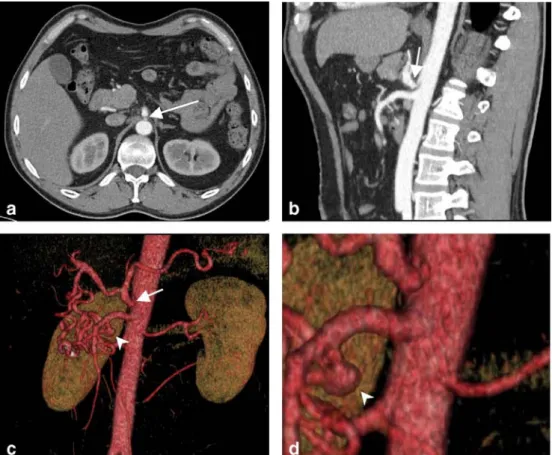
Fig. 1 A 47-year-old man with nonspecific abdominal pain. Axial image (a) and lateral MIP (b) reveal a filiform stenosis at the origin of the celiac artery (arrows) with poststenotic dila-tation. VR images (c and mag-nified view d) demonstrate the stenosis (arrows) as well as the collateralization through the pancreaticoduodenal arcade. In addition, an aneurysm of the inferior pancreaticoduodenal ar-tery (arrowheads) is depicted, most probably resulting from the high collateral flow through the superior mesenteric artery
of mesenteric vessels from the origin to the distal branches on a single projection.
Nonocclusive mesenteric ischemia (NOMI) is com-monly caused by decreased cardiac output resulting in splanchnic hypoperfusion. Typical angiographic signs for the diagnosis of NOMI include narrowing and vasocon-striction of multiple branches of the superior mesenteric artery, alternate dilatation and narrowing of intestinal branches, spasms of mesenteric arcades, impaired filling or occlusion of intramural vessels, reflux of contrast into the abdominal aorta, and spread out segmental arteries due to distention of bowel loops [7]. Some of these findings are difficult to depict on CTA. In addition, because the therapy of patients with NOMI consists of selective vaso-dilator infusion [12], some authors have proposed that a first-line conventional angiography be performed instead of CTA.
Segmental mediolytic arteriopathy
Segmental mediolytic arteriopathy (SMA) was first de-scribed in 1976 by Slavin and Gonzalez-Vitale [13]. It is characterized by a noninflammatory arteriopathy causing medial lysis of adult visceral arteries. Typical CT findings consist of thickening and dissection of the artery wall, multiple aneurysms of small vessels, and retroperitoneal bleeding (Fig. 5) [14]. Arterial wall thickening and dis-section can be clearly demonstrated on MPR or MIP, while the extent of saccular aneurysms is better delineated using the VR technique owing to the complex small vessel anat-omy of the typically affected arteries. Because of its rarity, no reliable information about the optimal therapeutic op-tion is available. Surgical or intervenop-tional repair of an-eurysms and endovascular stenting of arterial dissections may be necessary. Because of the similarity of SMA to polyarteritis nodosa, surgery with histopathological anal-ysis for better differentiation has been proposed [15].
Fig. 2 A 59-year-old man with known cardiac aneurysm after myocardial infarction 1 year previously and sudden abdomi-nal and left lower extremity pain. Axial image (a) displays a large mural thrombus in a left ventricular aneurysm (arrow) and multiple splenic infarctions (arrowheads). Coronal MPR (b) reveals segmental wall thicken-ing of the ileum (arrows) indi-cating bowel ischemia. The thickslab VR image (c) depicts the source of the bowel ischemia as an embolus in the ileocolic artery (arrow). Additional thickslab VR image (d) also demonstrates thromboembolic occlusion of the left common iliac artery (arrow)
Fig. 3 A 46-year-old woman admitted for evaluation of ve-nous bypasses. After occlusion of multiple visceral arteries 2 years previously, one bypass was connected from the inferior aorta to the celiac axis and superior mesenteric artery, and another from the right common iliac artery to branches of the superior mesenteric and common hepatic artery. Axial image (a) and axial MIP (b) show occlu-sion of the bypass
(arrows). Lateral MIP (c) de-monstrates the occluded bypass (arrow) and the unaltered occlusion of the celiac and su-perior mesenteric artery (arrow-head). VR image (d) depicts the occluded first bypass (arrow) and open second bypass (arrowhead). Another VR image (e) reveals the complex anatomy of the postoperative visceral vasculature and the advantage of reconstruction from 16-row CT data as compared to the 4-row CT examination 2 years pre-viously (f); the shorter scanning time results in an improved con-trast bolus exploitation and less venous overlay
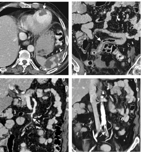
Fig. 4 A 61-year-old woman with elevated liver enzymes and abdominal pain after aortic and mitral valve replacement and known cardiac arrhythmia. Axial image (a) and coronal MIP (b) depict massive wall thickening and pneumatosis of the ascending and transverse colon indicating edema and ischemic necrosis (arrows). The coronal MIP (b) displays the normal opacification of the su-perior mesenteric vein, whereas the VR image (c) demonstrates the normal superior mesenteric artery branches without evidence of vessel occlusion (arrowhead). Considering the normally per-fused mesenteric vessels, hypo-perfusion due to low cardiac output is the most probable cause of the bowel ischemia in this case
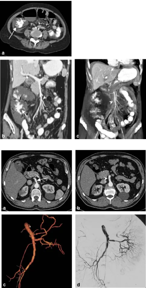
Fig. 5 A 38-year-old female with acute abdominal pain and vomiting. Axial image (a) shows caliber variability in the superior mesenteric artery (arrow). Another axial image (b) demonstrates an arterial gap reaching into the lumen (ar-rowhead). The VR image (c) clearly visualizes the caliber variability of the superior me-senteric artery. The diagnosis of segmental mediolytic arteriopa-thy was confirmed by selective conventional angiography (d)
Fig. 6 An 84-year-old woman with penetrating aortic aneu-rysm ulcer and emergency stent-grafting. Due to the extent of the aortic aneurysm, the stent graft needed to be inserted in the nondilated part of the aorta covering the celiac axis. To maintain visceral perfusion, a mesentericotruncal Shelhigh bypass had to be placed. Post-interventional axial image (a) depicts the excluded aneurysm sac (arrow) and the stent graft (arrowhead) in a normal posi-tion. Lateral MIP (b) and oblique VR image (c) delineate the anatomy after endovascular intervention and successful surgery showing the perfused Shelhigh bypass from the supe-rior mesenteric to the common hepatic artery
Fig. 7 A 40-year-old woman with known Takayasu arteritis. After complete occlusion of the aortic bifurcation, an extraana-tomical aortic bypass was in-stalled. To ensure visceral perfusion the celiac trunk and superior mesenteric artery had to be reinserted side-to-side to the bypass. Postoperative axial MIP (a) depicts the site of reinsertion to the celiac axis (arrow). The VR images (b and c) visualize the complex anatomy after sur-gery: The site of reinsertion of the celiac axis (thin arrow) and the side-to-side anastomosis of the superior mesenteric artery (black arrowhead) are excel-lently visualized. In addition, a stenosis with poststenotic dila-tation at the origin of the inferior mesenteric artery (thick arrow in b) is shown
Pre- and postinterventional and postsurgical imaging In emergency abdominal vascular disease, CTA provides the single most effective imaging tool [14]. In contrast to conventional angiography, CTA is not only able to detect the pathological vascular condition but also its effects on organs by delineating parenchymal perfusion [5]. In addi-tion, CTA can help the surgeon or interventional radiol-ogist plan treatment, for example by allowing the occluded vessel’s diameter to be measured or the communication points for stent graft placement to be defined [16]. Due to these advantages and the wide availability of CT, CTA with two- and three-dimensional reconstructions is in-creasingly becoming the initial and single imaging method for planning surgical or angiographic interventions [14].
Postsurgical evaluation of vascular anatomy is often complex and can only hardly be appreciated on axial
im-ages alone. Therefore postprocessing modalities, and in particular the VR technique, can be extremely helpful in understanding the extra-anatomical course of the postop-erative mesenteric vessels (Figs. 3, 6, 7,8).
Conclusion
Multislice CTA is an excellent tool to assess a wide variety of abdominal vascular diseases and offers a noninvasive imaging method for pre- and postinterventional evaluation of mesenteric perfusion.
The VR technique in particular provides images of high quality with excellent visualization of pathological condi-tions and their anatomical relacondi-tionship with adjacent struc-tures, that often cannot be confidently assessed by axial CT images alone.
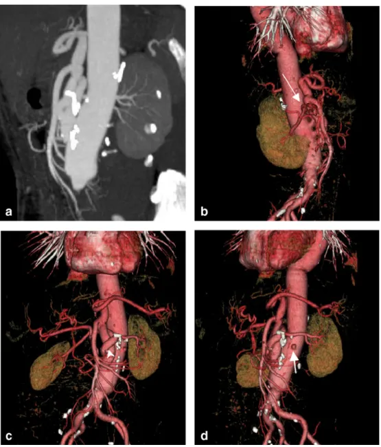
Fig. 8 A 65-year-old female with ruptured aortic abdominal aneurysm. The patient received an aortic graft with reinsertion of the visceral vasculature. Axial data sets, MPR and MIP (a) failed to clearly depict the new anatomy of the reinserted vessels. Owing to the three-dimensional effect and rotation capabilities, only VR images (b–d) are able to sufficiently demonstrate the reinserted renal and celiac arteries. Note the small aneurysm in the middle of the right renal artery (thin arrow in b), the tortuous course of the left renal artery around the celiac trunk (arrowhead in c), and the opacification of the renal parenchyma. The VR image (d) from a lateral oblique view shows the origin of the obstructed inferior mesenteric artery (thick arrow)
CTA allows the success or failure of vascular interven-tion to be determined and is consequently the preferred imaging modality for both pre- and postinterventional plan-ning and follow-up studies.
Acknowledgements This work was supported by the National Center of Competence in Research, Computer Aided and Image Guided Medical Interventions of the Swiss National Science Foundation.
References
1. Prokop M (1998) CT angiography of the abdominal arteries. Abdom Imag-ing 23:462–468
2. Schoepf UJ, Becker CR, Hofmann LK, Das M, Flohr T, Ohnesorge BM, Baumert B, Rolnick J, Allen JM, Raptopoulos V (2003) Multislice CT angiography. Eur Radiol 13:1946– 1961
3. Rubin GD, Shiau MC, Schmidt AJ, Fleischmann D, Logan L, Leung AN, Jeffrey RB, Napel S (1999) Computed tomographic angiography: historical perspective and new state-of-the-art using multi detector-row helical com-puted tomography. J Comput Assist Tomogr 23 [Suppl 1]:S83–S90 4. Kalra MK, Maher MM, Saini S (2003)
Multislice CT: update on radiation and screening. Eur Radiol 13:M129–M133
5. Johnson PT, Heath DG, Bliss DF, Cabral B, Fishman EK (1996) Three-dimensional CT: real-time interactive volume rendering. AJR Am J Roent-genol 167:581–583
6. McKinsey JF, Gewertz BL (1997) Acute mesenteric ischemia. Surg Clin North Am 77:307–318
7. Trompeter M, Brazda T, Remy CT, Vestring T, Reimer P (2002) Non-occlusive mesenteric ischemia: etiolo-gy, diagnosis, and interventional therapy. Eur Radiol 12:1179–1187 8. Fleischmann D (2003) MDCT of renal
and mesenteric vessels. Eur Radiol 13: M94–M101
9. Taourel PG, Deneuville M, Pradel JA, Regent D, Bruel JM (1996) Acute mesenteric ischemia: diagnosis with contrast-enhanced CT. Radiology 199:632–636
10. Kirkpatrick ID, Kroeker MA, Greenberg HM (2003) Biphasic CT with mesenteric CT angiography in the evaluation of acute mesenteric ische-mia: initial experience. Radiology 229:91–98
11. Lee R, Tung HK, Tung PH, Cheung SC, Chan FL (2003) CT in acute mesenteric ischaemia. Clin Radiol 58:279–287
12. Ernst S, Luther B, Zimmermann N, Bohner H, Wilke R, Feindt P, Furst G (2003) Current diagnosis and therapy of non-occlusive mesenteric ischemia (in German). Rofo 175:515–523 13. Slavin RE, Gonzalez-Vitale JC (1976)
Segmental mediolytic arteritis: a clin-ical pathologic study. Lab Invest 35:23–29
14. Frauenfelder T, Wildermuth S, Marincek B, Boehm T (2004) Non-traumatic emergent abdominal vascular conditions: advantages of multi-detec-tor row CT and three-dimensional imaging. Radiographics 24:481–496 15. Chan RJ, Goodman TA, Aretz TH, Lie
JT (1998) Segmental mediolytic arte-riopathy of the splenic and hepatic arteries mimicking systemic necrotiz-ing vasculitis. Arthritis Rheum 41:935–938
16. Piccoli G, Gasparini D, Smania S, Sponza M, Marzio A, Vit A, Bazzocchi M (2003) Multislice CT angiography in the assessment of peripheral aneu-rysms. Radiol Med (Torino) 106:504– 511