Single amino acid substitutions in the HsdR subunit of the type IB restriction enzyme EcoAI uncouple the DNA translocation and DNA cleavage activities of the enzyme
6
0
0
Texte intégral
(2) Nucleic Acids Research, 1999, Vol. 27, No. 13 2639. reminiscent of the P-D...(D/E)-X-K motif typical for the active site of many type II restriction enzymes (26). Here we describe properties of mutant forms of the type IB restriction endonuclease EcoAI containing alanine substitutions at the putative active site residues Asp-61, Glu-76 and Lys-78. We show that these residues are indeed important for restriction, but not for ATPase and DNA translocation activities of the enzyme. These data indicate that the N-terminal conserved region of HsdR contains active site residues for the DNA cleavage reaction.. addition of 1 vol of 0.1 M EDTA (pH 8). Plasmids pJP25 (one EcoAI site) and pDRM.1R (no EcoAI site) were used as DNA substrates (31,34). If required, plasmid pJP25 was cut with AlwNI to produce a linear substrate. DNA and enzyme concentrations used are indicated in the figure legends. Reaction products were analyzed on agarose gels run in 0.5× TBE buffer. DNA was visualised by ethidium bromide staining. If required, agarose gel images were digitised and quantified using NIH Image 1.61 software. ATPase assay. MATERIALS AND METHODS DNA manipulations The origin repair method (27) was employed for site-directed mutagenesis. The original procedure was modified by usage of a DH5α mutS strain to enhance efficiency of mutagenesis (28). The mutagenesis of the hsdR gene was performed using the plasmid pJP41, a derivative of pGLamB (28) in which the XbaI–BamHI fragment containing the lamB gene was replaced by the XbaI–BamHI fragment of pJP22 carrying the hsdR gene. The hsdR gene in pJP41 is under control of the T7 transcriptional and translational signals. For in vivo functional assays, individual mutations were transferred from pJP41 derivatives to pFFP30 harboring all three EcoAI genes in the natural transcriptional organisation (29). This was achieved by replacing the NcoI–DraIII region in pFFP30 by the NcoI–DraIII fragments from the mutant derivatives of pJP41, resulting in plasmids named pFFP30D61A, pFFP30G69D, pFFP30E76A and pFFP30K78A, respectively. Phage infection assay The cells were grown in LB medium containing, when required, ampicillin at a concentration of 200 µg/ml to select for appropriate plasmids expressing wild-type or mutant EcoAI genes. At an OD600 of ∼1, the cells were infected with serial dilutions of bacteriophage λvir and phage titre was determined as described previously (30). Protein preparations Wild-type and mutant HsdR subunits were over-produced in Escherichia coli BL21 from the appropriate pJP41 derivative (see above), and purified to apparent homogeneity as described previously (31). HsdM and HsdS subunits of EcoAI were also produced separately and purified as described (31). The wildtype and mutant EcoAI endonucleases were reconstituted by mixing HsdR, HsdM and HsdS subunits in a ratio of 6:2:1. At this subunit ratio, the wild-type endonuclease reached the maximal activity when incubated with equimolar amounts of a circular DNA substrate containing one EcoAI recognition site (31). The excess of HsdR was presumably required due to the weak association of the two HsdR subunits with the methylase observed during the endonuclease purification (32). EcoKI endonuclease was produced from pVMC3 and purified as described (33). DNA cleavage assay All DNA cleavage reactions were performed at 37°C in buffer C [50 mM Tris–HCl (pH 8.0), 10 mM MgCl2, 25 mM NaCl, 1 mM dithiothreitol, 0.2 mM AdoMet, 5 mM ATP]. Reactions were started by the addition of ATP and stopped by the. The ATPase activity of EcoAI mutants was measured by a colorimetric estimation of the inorganic phosphate released by ATP hydrolysis as described previously (34). Reaction conditions were the same as for the DNA cleavage assay. RESULTS The N-terminal conserved region of type I HsdR subunits contains an amino acid sequence motif reminiscent of the catalytic motif of type II restriction enzymes Several short conserved regions have been identified by alignments of HsdR polypeptides from different type I restriction enzyme families (14,23; Fig. 1). These include the seven DEAD-box motifs, clustered in the central part of each polypeptide, whose relevance for both restriction and ATPase activities was demonstrated by mutational analysis of EcoKI (24,25) and a region that precedes the DEAD-box motifs, which was named region X (14). After including a number of HsdR homologues from recently sequenced eubacterial and archaeal genomes for sequence alignment using the Clustal X program (35), we have noted that the resulting consensus sequence for the region X (Fig. 1B) is reminiscent of the PD...(D/E)-X-K catalytic motif of type II restriction enzymes such as EcoRI or EcoRV (26). The proline residue was not conserved in all HsdR subunits (Fig. 1B). However, proline is also absent in the catalytic motif of PvuII (36) and it is dispensable in the catalytic motifs of several other type II endonucleases (37,38), which indicates that this residue is not required for catalysis. In almost all HsdR sequences, the two acidic residues of the motif are spaced by 13 amino acids except for the HsdR subunits of EcoAI and EcoEI (IB family) in which the spacer contains 14 amino acids. Similarly, the distance between the catalytic acidic residues in the primary sequence of type II enzymes is (with a few exceptions) in the range from 9 to 19 amino acids (39). In all HsdR polypeptides, the spacer contains additional conserved residues such as Gly and is highly hydrophobic (Fig. 1B). In the following text the putative DNA cleavage motif of type I restriction enzymes will be indicated as D-X13-14-EX-K. Site-directed mutagenesis of EcoAI HsdR To investigate the functional importance of the D-X13-14-E-X-K sequence motif, we have produced mutants of the type IB restriction endonuclease EcoAI. The putative catalytic residues of EcoAI HsdR Asp-61, Glu-76 and Lys-78 (Fig. 1B) were individually substituted by an alanine. The conserved Gly-69 in the spacer between the acidic residues of the motif was also subjected to mutagenesis and it was replaced with an aspartic acid..
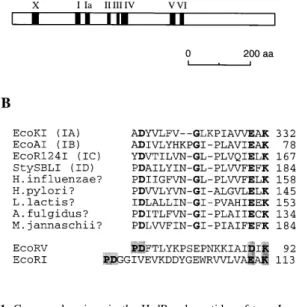
(3) 2640 Nucleic Acids Research, 1999, Vol. 27, No. 13. Table 1. In vivo restriction of λvir by EcoA mutants Plasmid. e.o.p.. (SD). pFFP30. 2.33 × 10⁻³. (± 0.45 × 10⁻³). pFFP30D61A. 0.47. (± 0.11). pFFP30G69D. 1.87 × 10⁻³. (± 0.18 × 10⁻³). pFFP30E76A. 0.49. (± 0.14). pFFP30K78A. 0.65. (± 0.11). The strain DH5α (restriction minus) was used as the host for the plasmids expressing the wild-type (pFFP30) or mutant EcoA genes. Single amino acid substitutions in the HsdR subunit are indicated in the names of pFFP30 derivatives. The values of e.o.p. are averages of four independent measurements. The standard deviations (SD) are shown in brackets.. Figure 1. Conserved regions in the HsdR polypeptides of type I restriction enzymes. (A) A diagram of the HsdR subunit of the type IB restriction enzyme EcoAI showing the location of the conserved amino acid sequence regions as defined previously (14,23). Based on mutational analysis data, the conserved sequence originally named region Y was reclassified to be the DEAD-box motif IV (25). The original motif IV is not shown. (B) An alignment of amino acid sequences of several known and putative HsdR polypeptides around the N-terminal conserved region X. The number at the end of each line indicates the position for the last amino acid in the corresponding sequences. Conserved amino acids are indicated in bold. The regions of the type II endonucleases EcoRI and EcoRV containing the P-D...(D/E)-X-K catalytic motif are also shown. The elements of the motif are in grey boxes. The protein identification numbers used by the National Center for Biotechnology Information are as follows: EcoKI, 730887; EcoAI, 304894; EcoR124I, 78918; StySBLI, 1679868; Haemophilus influenzae Rd, 1574743; Helicobacter pylori, 2314575; Lactococcus lactis, 3057061; Methanococcus jannaschii, 2129337; EcoRI, 135227; EcoRI, 135230.. Restriction phenotype of mutants The restriction phenotype of mutant endonucleases was determined by testing the ability of cells expressing these mutants to restrict the growth of unmodified bacteriophage λ. For this assay, individual mutations were transferred to the plasmid pFFP30 that contains all three EcoAI hsd genes in the natural transcriptional organisation (Materials and Methods). The resulting plasmids with mutant hsdR genes were transformed to DH5α and efficiency of plating (e.o.p.) of λ phage (e.o.p.; ratio of the phage titre on tested host to the titre on nonrestricting host) on these strains was determined (Table 1). The data from this experiment indicated that the D61A, E76A and K78A mutants had greatly impaired restriction activity in vivo. The G69D mutant had a wild-type restriction phenotype, indicating that the conserved Gly residue is not important for this function. In vitro DNA cleavage and ATPase activities of mutant enzymes For in vitro analysis, the mutant HsdR subunits, except for G69D, were separately overproduced and purified as described in the Materials and Methods and mixed in vitro with purified HsdM and HsdS subunits to reconstitute the endonuclease. As a substrate for DNA cleavage in vitro, the plasmid pJP25. Figure 2. DNA cleavage assay of EcoAI mutants. The supercoiled form of the plasmid pJP25 (a single EcoAI site) was used as DNA substrate. Plasmid pDRM.1R (no EcoAI site) served as non-specific DNA for control reactions. 20 nM DNA was incubated with 80 nM wild-type or mutant EcoAI enzymes in buffer C at 37°C for 10 min. Reactions were stopped by the addition of 1 vol of 0.1 M EDTA (pH 8) and analysed on a 0.9% agarose gel run in 0.5× TBE buffer at 50 V for 5 h. DNA was visualised by ethidium bromide staining. Lane 1, DNA size markers; lanes 2–6 contain pJP25 incubated with no enzyme, wild-type, D61A, E76A end K78A, respectively; lanes 7–11 contain pDRM.1R incubated with no enzyme, wild-type, D61A, E76A and K78A, respectively. Positions of supercoiled (SC) linear (L), nicked (NC) and supercoiled dimeric (SC dimer) forms of plasmid DNA are indicated on the right of the gel.. containing a single site for EcoAI was used. In agreement with the in vivo phenotypes, none of the mutant enzymes was able to cleave the plasmid DNA to its linear form even when present in a large excess over DNA (Fig. 2). Interestingly, the K78A mutant showed a significant nicking activity that was dependent on the presence of the EcoAI recognition site in the substrate (Fig. 2). The rate of the pJP25 DNA nicking by the K78A mutant was much lower than the rate of double-strand cleavage of this DNA by the wild-type enzyme (Fig. 3). The E76A mutant exhibited very little site-specific nicking activity and the D61A mutant did not show any nicking activity at all. None of the mutants exhibited any DNA cleavage activity on a linear DNA substrate containing two EcoAI recognition sites while the wild-type enzyme efficiently cleaved this substrate at random positions over the region between the recognition sites.
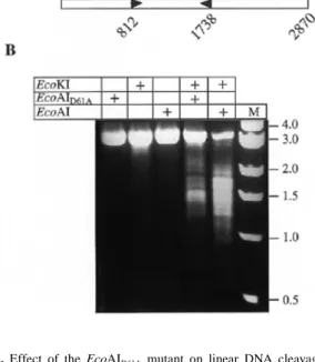
(4) Nucleic Acids Research, 1999, Vol. 27, No. 13 2641. Figure 3. Rate of plasmid DNA cleavage by wild-type EcoAI endonuclease compared to the rate of DNA nicking by EcoAIK78A mutant. Cleavage reactions were carried out in buffer C at 37°C and contained 20 nM pJP25 DNA and 80 nM enzyme. Aliquots removed during the reaction were analysed on a 0.9% agarose gel run in 0.5× TBE buffer at 3 V/cm for 5 h. DNA was visualised by ethidium bromide staining. The gel was quantified as described in Materials and Methods and relative intensity of the linear DNA bands for wild-type enzyme (square) and nicked circular DNA bands for EcoAIK78A mutant (circle) in each time point was calculated as a percentage of total DNA per lane.. Figure 4. Time course of ATP hydrolysis by EcoAI mutants. 20 nM pJP25 DNA (a single EcoAI site) was incubated with 80 nM enzyme in buffer C at 37°C. During the incubation, 10 µl aliquots were removed and the reaction was stopped by the addition of 1 vol of 0.1 mM EDTA (pH 8). The concentration of inorganic phosphate (Pi) released by ATP hydrolysis was measured as described in Materials and Methods.. (not shown). Despite the absence of double-strand DNA cleavage activity, all three mutants had a similar ATPase activity to the wild-type endonuclease, indicating that the mutations did not cause a gross change in enzyme structure and that DNA cleavage is not a prerequisite for ATP hydrolysis (Fig. 4). Thus, the in vitro analysis of the EcoAI mutants suggests that all three charged HsdR residues of the D-X13-14-E-X-K motif participate in the DNA cleavage reaction. DNA translocation by EcoAI mutants The ability of the EcoAI mutants to hydrolyse ATP suggested that these mutants could still translocate DNA. To investigate the DNA translocation activity of the mutant enzymes, we have examined their ability to promote linear DNA cleavage. Figure 5. Effect of the EcoAID61A mutant on linear DNA cleavage by the EcoKI endonuclease. (A) Diagram of DNA substrate (2870 bp) containing one EcoKI (K) site and one EcoAI (A) site that was prepared by AlwNI (unique site) cleavage of pJP25. DNA is represented as an open rectangle. The arrowheads indicate the orientation of the asymmetric recognition sites (tail-to-tail). The numbers show the position of the first base pair of the recognition sites. (B) Restriction assay. 13.25 nM DNA was incubated with 90 nM EcoKI and 80 nM EcoAID61A individually or in combination in buffer C at 37°C for 8 min. In a control experiment, wild-type EcoAI endonuclease was used instead of EcoAID61A. Reactions were stopped by addition of 1 vol of 0.1 M EDTA and analysed on a 1% agarose gel run in 0.5× TBE buffer at 80 V for 2 h. DNA was visualised by ethidium bromide staining. Presence of enzymes in individual reactions is indicated above each lane by (+). Lane M contains DNA size markers.. by the type IA restriction enzyme EcoKI within the region between EcoKI and EcoAI recognition sites, by serving as molecular blocks to EcoKI translocation. This assay is based on our observation that type I restriction enzymes have the potential to cleave DNA at physical translocation barriers such as Holliday junctions, suggesting that DNA translocation blockage is the only requirement for DNA cleavage to occur (7). For this experiment, the pJP25 plasmid DNA was linearised by AlwNI to produce a 2.9 kb linear substrate with one EcoAI site and one EcoKI site separated by 0.9 kb (Fig. 5A). Assuming that the collision of EcoKI with the EcoAI mutants occurs at random positions between the recognition sites, the cleavage of this substrate would generate pairs of fragments with sizes ranging from ∼0.8 to 2.0 kb. As expected, the incubation of the DNA substrate with a large excess of EcoKI for 8 min did not lead to significant DNA cleavage (Fig. 5B), consistent with the requirement for the presence of two enzyme recognition sites for efficient linear DNA cleavage (6,8,9). However, when the reconstituted EcoAID61A mutant was added together with EcoKI, extensive DNA cleavage occurred generating multiple products which appeared as a DNA smear on EtBr-stained gel, consistent with random cleavage (Fig. 5B). As judged from the electrophoretic mobilities of the cleavage products relative to DNA size markers, the cleavage events occurred predominately within the region between the EcoKI and EcoAI recognition sites, with some preference for a very short area located.
(5) 2642 Nucleic Acids Research, 1999, Vol. 27, No. 13. approximately in the middle of the DNA molecule (∼200 bp from the EcoKI site). Identical cleavage profiles were also obtained with the E76A and K78A mutants when combined with EcoKI (not shown). In a control experiment, EcoKI was combined with the wild-type EcoAI endonuclease. As observed previously (7), this enzyme combination led to efficient cleavage at random positions over the region between the EcoAI and EcoKI sites, with some preference for the area starting approximately half way between the sites and ending at the EcoKI site (Fig. 5). One can notice that DNA cleavage resulting from the cooperation between EcoKI and EcoAID61A mutant was less efficient as compared to the situation when the wildtype EcoAI endonuclease was present instead of the mutant (Fig. 5). This may reflect the fact that in the later case DNA cleavage can be catalysed by either enzyme while in the former case only EcoKI is active for cleavage. Thus, the fact that the EcoAI mutants could promote DNA cleavage by EcoKI far from the EcoAI recognition sequence clearly demonstrates that these mutants are capable of translocating DNA.. This may suggest a modular protein structure, with the latter domain involved in ATP-dependent DNA translocation. Some support for this assumption comes from our finding that alanine substitutions at the endonucleolytic active site of the HsdR subunit of EcoAI effectively uncoupled the DNA translocation and DNA cleavage activities of the enzyme. A protein structure composed of independent nuclease and helicase domains was described for the RecB subunit of the E.coli RecBCD enzyme (41). Mutations in the DEAD-box motifs of EcoKI impaired both ATPase and restriction activity (25). It is possible that interactions of the helicase-like domain with ATP and DNA are required for an activation of the cleavage domain, perhaps by positioning this domain in proximity to the DNA substrate. All DEAD-box mutants exhibited some sitespecific nicking activity (25), which may reflect a residual activity of the cleavage domain. The mutants produced in this work could be useful in studying the mechanism of DNA translocation by type I restriction enzymes, since the experiments with the wild-type enzyme are hampered by rapid cleavage of DNA substrates.. DISCUSSION. ACKNOWLEDGEMENTS. Conservation of an amino acid sequence motif reminiscent of the P-D…(D/E)-X-K catalytic motif of many type II restriction enzymes in the N-terminal region of all known and putative HsdR subunits (Fig. 1) suggested that this motif may be a candidate for the endonucleolytic active site of type I restriction enzymes. In the active centre of the type II endonucleases, the two acidic amino acid residues serve to coordinate a Mg2+ ion that is the cofactor for the phosphodiester bond hydrolysis. The lysine residue is thought to stabilise the doubly charged pentavalent transition state (26). We have made single alanine substitutions in the putative active site motif of the HsdR subunit of the type IB restriction enzyme EcoAI, formed by Asp-61, Glu-76 and Lys-78 residues. These amino acid substitutions resulted in enzymes that exhibited normal ATPase activity and the ability to translocate DNA, but they failed to cleave DNA. Hence, all the three mutations have the properties expected for the mutations at the active site residues for DNA cleavage; they uncouple ATP-dependent DNA translocation and restriction activities of the enzyme. It is quite likely that the homologous motifs in other type I restriction enzymes also form the endonucleolytic active site since they share common features with the active site residues of EcoAI: (i) they are located in the same part of the HsdR polypeptide and (ii) the sequences between the two acidic residues are homologous (Fig. 1). The K78A mutant of EcoAI exhibited a significant site-specific nicking activity (Figs 2 and 3). Since the D61A and E76A mutants had no or very little nicking activity (Fig. 2), the ability of the K78A mutant to nick plasmid DNA substrate may reflect residual activity of the mutated cleavage centre rather than the presence of an additional active site for DNA cleavage. Similarly, nicking activity was observed with EcoRV catalytic mutants (40). In addition, an alanine substitution at the lysine residue of EcoRV did not completely eliminate double strand cleavage activity while alanine mutants at either acidic residue were completely inactive in double-strand cleavage (40). In all HsdR polypeptides, the putative active site for DNA cleavage is located outside of the helicase-like domain (Fig. 1).. We are grateful to Daniel Panne for comments on the manuscript. This work was supported by grants from the Swiss National Science Foundation. REFERENCES 1. 2. 3. 4.. 5. 6. 7. 8. 9. 10.. 11.. 12. 13. 14. 15. 16. 17. 18. 19. 20.. Wilson,G.G. and Murray,N.E. (1991) Annu. Rev. Genet., 25, 585–627. Bickle,T.A. and Krüger,D.H. (1993) Microbiol. Rev., 57, 434–450. Yuan,R. (1981) Annu. Rev. Biochem., 50, 285–315. Bickle,T.A. (1993) In Linn,S.M., Lloyd,R.S. and Roberts,R.J. (eds), Nucleases. Cold Spring Harbor Laboratory Press, Cold Spring Harbor, NY, pp. 89–109. Studier,F.W. and Bandyopadhyay,P.K. (1988) Proc. Natl Acad. Sci. USA, 85, 4677–4681. Dreier,J., MacWilliams,M.P. and Bickle,T.A. (1996) J. Mol. Biol., 264, 722–733. Janscak,P., MacWilliams,M.P., Sandmeier,U., Nagaraja,V. and Bickle,T.A. (1999) EMBO J., 18, 2648–2658. Murray,N.E., Batten,P.L. and Murray,K. (1973) J. Mol. Biol., 81, 395–407. Szczelkun,M.D., Dillingham,M.S., Janscak,P., Firman,K. and Halford,S.E. (1996) EMBO J., 15, 6335–6347. Tomb,J.F., White,O., Kerlavage,A.R., Clayton,R.A., Sutton,G.G., Fleischmann,R.D., Ketchum,K.A., Klenk,H.P., Gill,S., Dougherty,B.A. et al. (1997) Nature, 388, 539–547. Bult,C.J., White,O., Olsen,G.J., Zhou,L., Fleischmann,R.D., Sutton,G.G., Blake,J.A., FitzGerald,L.M., Clayton,R.A., Gocayne,J.D. et al. (1996) Science, 273, 1058–1073. Murray,N.E., Gough,J.A., Suri,B. and Bickle,T.A. (1982) EMBO J., 1, 535–539. Price,C., Pripfl,T. and Bickle,T.A. (1987) Eur. J. Biochem., 167, 111–115. Titheradge,A.J.B., Ternent,D. and Murray,N.E. (1996) Mol. Microbiol., 22, 437–447. Dryden,D.T.F., Cooper,L.P., Thorpe,P.H. and Byron,O. (1997) Biochemistry, 36, 1065–1076. Janscak,P., Dryden,D.T.F. and Firman,K. (1998) Nucleic Acids Res., 26, 4439–4445. Taylor,I., Patel,J., Firman,K. and Kneale,G. (1992) Nucleic Acids Res., 20, 179–186. Dryden,D.T.F., Cooper,L.P. and Murray,N.E. (1993) J. Biol. Chem., 268, 13228–13236. Fuller-Pace,F.V., Bullas,L.R., Delius,H. and Murray,N.E. (1984) Proc. Natl Acad. Sci. USA, 81, 6095–6099. Meister,J., MacWilliams,M., Hubner,P., Jutte,H., Skrzypek,E., Piekarowicz,A. and Bickle,T.A. (1993) EMBO J., 12, 4585–4591..
(6) Nucleic Acids Research, 1999, Vol. 27, No. 13 2643. 21. Willcock,D.F., Dryden,D.T.F. and Murray,N.E. (1994) EMBO J., 13, 3902–3908. 22. Gorbalenya,A.E. and Koonin,E.V. (1991) FEBS Lett., 291, 277–281. 23. Murray,N.E., Daniel,A.S., Cowan,G.M. and Sharp,P.M. (1993) Mol. Microbiol., 9, 133–143. 24. Webb,J.L., King,G., Ternent,D., Titheradge,A.J. and Murray,N.E. (1996) EMBO J., 15, 2003–2009. 25. Davies,G.P., Powell,L.M., Webb,J.L., Cooper,L.P. and Murray,N.E. (1998) Nucleic Acids Res., 26, 4828–4836. 26. Pingoud,A. and Jeltsch,A. (1997) Eur. J. Biochem., 246, 1–22. 27. Ohmori,H. (1994) Nucleic Acids Res., 22, 884–885. 28. Prilipov,A., Phale,P.S., Van,G.P., Rosenbusch,J.P. and Koebnik,R. (1998) FEMS Microbiol. Lett., 163, 65–72. 29. Fuller-Pace,F.V., Cowan,G.M. and Murray,N.E. (1985) J. Mol. Biol., 186, 65–75. 30. Gubler,M. and Bickle,T.A. (1991) EMBO J., 10, 951–957. 31. Janscak,P. and Bickle,T.A. (1998) J. Mol. Biol., 284, 937–948. 32. Suri,B., Shepherd,J.C.W. and Bickle,T.A. (1984) EMBO J., 3, 575–579.. 33. Weiserova,M., Janscak,P., Benada,O., Hubacek,J., Zinkevich,V.E., Glover,S.W. and Firman,K. (1993) Nucleic Acids Res., 21, 373–379. 34. Janscak,P., Abadjieva,A. and Firman,K. (1996) J. Mol. Biol., 257, 977–991. 35. Thompson,J.D., Gibson,T.J., Plewniak,F., Jeanmougin,F. and Higgins,D.G. (1997) Nucleic Acids Res., 25, 4876–4882. 36. Cheng,X., Balendiran,K., Schildkraut,I. and Anderson,J.E. (1994) EMBO J., 13, 3927–3935. 37. Grabowski,G., Jeltsch,A., Wolfes,H., Maass,G. and Alves,J. (1995) Gene, 157, 113–118. 38. Lagunavicius,A. and Siksnys,V. (1997) Biochemistry, 36, 11086–11092. 39. Stahl,F., Wende,W., Jeltsch,A. and Pingoud,A. (1998) Biol. Chem., 379, 467–473. 40. Selent,U., Ruter,T., Kohler,E., Liedtke,M., Thielking,V., Alves,J., Oelgeschlager,T., Wolfes,H., Peters,F. and Pingoud,A. (1992) Biochemistry, 31, 4808–4815. 41. Yu,M., Souaya,J. and Julin,D.A. (1998) Proc. Natl Acad. Sci. USA, 95, 981–986..
(7)
Figure
Documents relatifs
tuberculosis form- ing part of the type II fatty acid synthase (FAS-II) system involved in mycolic acid biosynthesis: the malonyl-CoA::AcpM transacylase mtFabD, and the  -ketoacyl
If environmental and housing conditions are responsible for these differences, the effect on performance are partly associated to the stimulation of the immune system
NTrm4p Is Catalytically Active whereas CTrm4p Is Not—To test the tRNA MTase activity of the different proteins and verify that the product of methyl transfer is m 5 C, a mature
The dispersion observed for a given alloy regarding mass losses, average incubation times, pit densities and average lateral pit growth rate constants highlights the importance
In the present case, this parsimonious approach showed that-contrary to what one might think looking at Yalcin's and Ninan's simple shifty frameworks-there is a
GTPases. Anti-FLAG immunoprecipitates were prepared from wild-type or SZT2-null HeLa cells stably expressing the indicated cDNAs that had been starved of amino acids for
This study was aimed to determine amino acid composition of Infraspinatus, Triceps brachii, Longissimus thoraces, Biceps femoris, Semitendinosus, and Semimembranosus muscles from
Ebenso besteht die Gefahr einer Nervenastschädi- gung mit Neurombildung, wenn die Inzision nicht stumpf in der Tiefe ausgeweitet und die Schutzhülse beim Bohren nicht verwendet
