HAL Id: hal-02406334
https://hal.archives-ouvertes.fr/hal-02406334
Submitted on 19 May 2020
HAL is a multi-disciplinary open access
archive for the deposit and dissemination of
sci-entific research documents, whether they are
pub-lished or not. The documents may come from
teaching and research institutions in France or
abroad, or from public or private research centers.
L’archive ouverte pluridisciplinaire HAL, est
destinée au dépôt et à la diffusion de documents
scientifiques de niveau recherche, publiés ou non,
émanant des établissements d’enseignement et de
recherche français ou étrangers, des laboratoires
publics ou privés.
Host-derived viral transporter protein for nitrogen
uptake in infected marine phytoplankton
Adam Monier, Aurélie Chambouvet, David Milner, Victoria Attah, Ramón
Terrado, Connie Lovejoy, Hervé Moreau, Alyson Santoro, Evelyne Derelle,
Thomas Richards
To cite this version:
Adam Monier, Aurélie Chambouvet, David Milner, Victoria Attah, Ramón Terrado, et al..
Host-derived viral transporter protein for nitrogen uptake in infected marine phytoplankton. Proceedings
of the National Academy of Sciences of the United States of America , National Academy of Sciences,
2017, 114 (36), pp.E7489-E7498. �10.1073/pnas.1708097114�. �hal-02406334�
PNAS
ENVIRONMENTAL
SCIENCES
Host-derived viral transporter protein for nitrogen
uptake in infected marine phytoplankton
Adam Moniera,b,1, Aur ´elie Chambouveta,c,2, David S. Milnera,2, Victoria Attaha, Ram ´on Terradod, Connie Lovejoyb,e,
Herv ´e Moreauf, Alyson E. Santorog, ´Evelyne Derellef, and Thomas A. Richardsa,1
aLiving Systems Institute, School of Biosciences, College of Life and Environmental Sciences, University of Exeter, Exeter EX4 4QD, United Kingdom; bQu ´ebec Oc ´ean, Universit ´e Laval, Qu ´ebec, QC, Canada G1V 0A6;cLaboratoire des Sciences de l’Environnement Marin, CNRS UMR 6539, Institut Universitaire Europ ´een de la Mer, Universit ´e de Bretagne Occidentale, 29280 Plouzan ´e, France;dDepartment of Biological Sciences, Dornsife College of Letters, Arts, and Sciences, University of Southern California, Los Angeles, CA 90089-0371;eTakuvik International Laboratory (CNRS Unit ´e Mixte
Internationale 3376), D ´epartement de Biologie and Institut de Biologie Int ´egrative et des Syst `emes, Universit ´e Laval, Qu ´ebec, QC, Canada G1V 0A6;fCNRS UMR 7232, Sorbonne Universit ´es, Observatoire Oc ´eanologique de Banyuls, 66650 Banyuls-sur-mer, France; andgDepartment of Ecology, Evolution, and Marine Biology, University of California, Santa Barbara, CA 93106
Edited by W. Ford Doolittle, Dalhousie University, Halifax, NS, Canada, and approved July 10, 2017 (received for review May 18, 2017) Phytoplankton community structure is shaped by both bottom–
up factors, such as nutrient availability, and top–down processes, such as predation. Here we show that marine viruses can blur these distinctions, being able to amend how host cells acquire nutrients from their environment while also predating and lysing their algal hosts. Viral genomes often encode genes derived from their host. These genes may allow the virus to manipulate host metabolism to improve viral fitness. We identify in the genome of a phytoplankton virus, which infects the small green alga Ostre-ococcus tauri, a host-derived ammonium transporter. This gene is transcribed during infection and when expressed in yeast mutants the viral protein is located to the plasma membrane and res-cues growth when cultured with ammonium as the sole nitro-gen source. We also show that viral infection alters the nature of nitrogen compound uptake of host cells, by both increas-ing substrate affinity and allowincreas-ing the host to access diverse nitrogen sources. This is important because the availability of nitrogen often limits phytoplankton growth. Collectively, these data show that a virus can acquire genes encoding nutrient transporters from a host genome and that expression of the viral gene can alter the nutrient uptake behavior of host cells. These results have implications for understanding how viruses manipulate the physiology and ecology of phytoplankton, influ-ence marine nutrient cycles, and act as vectors for horizontal gene transfer.
Phycodnaviridae | NCLDV | prasinophytes | Mamiellophyceae | lateral gene transfer
P
hytoplankton underpin the biogeochemistry of the surface oceans and drive the marine carbon and nitrogen cycles (1). Nutrients fuel cyanobacteria and eukaryotic single-celled algae with the elements essential for organic matter biosynthe-sis, especially nitrogen (N) and phosphorus (P); hence nutrient availability exerts a bottom–up control on phytoplankton cell growth and oceanic productivity (2, 3). In particular, N and P are found in low concentrations over much of the open ocean, lim-iting phytoplankton growth rates. Nutrient limitation, including colimitation by several nutrients, results in competition among phytoplankton. In oligotrophic environments, and transiently nutrient-depleted environments, phytoplankton species have evolved a range of strategies to optimize nutrient acquisition (4). The genetic repertoire of N and P transporters shows evidence of gene duplication, differential loss, and horizontal gene trans-fer (HGT) in phytoplankton genomes as well as contrasting gene expression levels (5–9), suggesting that the evolution of these genes has been driven by adaptation to environmental limita-tion. Ammonium (NH+4) and nitrate (NO −
3) are commonly
avail-able N sources for marine phytoplankton (10, 11) and, as such, the gene repertoires of cyanobacterial and eukaryotic phyto-plankton are configured toward utilization of these two forms of inorganic N sources (12).
In addition to bottom–up nutrient limitation, phytoplankton community structures are influenced by top–down controls (13). For phytoplankton communities these include predation by graz-ers and viral infection (e.g., ref. 14). Marine viruses are the most abundant biological entities in the oceans; they are esti-mated to induce 1023 infections daily (15) and are thought to
control phytoplankton abundance, biomass, and species com-position through taxon-specific infections (16, 17). There is a growing appreciation of the role of viruses in oceanic nutrient cycles, based on lytic infections leading to the release of dissolved and particulate organic matter, which is then recycled by other microorganisms (15, 18). This virus-mediated process, referred to as the viral shunt, was suggested as a mechanism that main-tains availability of organic matter in the euphotic zone by lysing cells before they sink (15, 18). However, the influence of viruses on oceanic ecosystems extends beyond top–down host mortality. Genomic and metagenomic analyses show that marine viruses— either phages or eukaryotic viruses—harbor host-derived genes encoding a diverse range of putative functions (19–21), includ-ing whole biochemical pathways (22). These “auxiliary metabolic genes” (AMGs) may allow the virus to manipulate the host via metabolic reprogramming during infection and have been exper-imentally shown to alter the central carbon metabolism and pig-ment biosynthesis of cyanobacteria (23, 24) and to “reprogram”
Significance
Viruses often carry genes acquired from their host. In the present work, we show that a virus of a marine alga carries a gene encoding a transporter protein that mediates nutri-ent uptake. We confirm that the viral transporter protein is expressed during infection and show that the protein func-tions to take up sources of nitrogen. This is important because acquisition of nutrients often determines the ecological suc-cess of phytoplankton populations. This work demonstrates how a virus can amend host–viral dynamics by modulating acquisition of nutrients from the environment.
Author contributions: A.M., D.S.M., and T.A.R. designed research; A.M., A.C., D.S.M., V.A., R.T., and E.D. performed research; H.M. and E.D. contributed new reagents/analytic tools; A.M. analyzed data; and A.M., C.L., A.E.S., and T.A.R. wrote the paper.
The authors declare no conflict of interest. This article is a PNAS Direct Submission.
Freely available online through the PNAS open access option.
Data deposition: The sequence reported in this paper has been deposited in the GenBank database (accession no.KX254356). Phylogenetic and experimental data are available at the Zenodo data repository,zenodo.org/record/61901.
1To whom correspondence may be addressed. Email: a.monier@exeter.ac.uk or t.a.
richards@exeter.ac.uk.
2A.C. and D.S.M. contributed equally to this work.
This article contains supporting information online atwww.pnas.org/lookup/suppl/doi:10. 1073/pnas.1708097114/-/DCSupplemental.
lipid biosynthesis of eukaryotic algal cells (25–27). AMGs are thought to modulate host function to improve fitness of the virus and, in some cases, temporarily the host. For example, it was hypothesized that virally encoded putative phosphate trans-porters increase accumulation of P in host cells (28, 29), which may in turn increase virus fitness given that P-depleted phyto-plankton cells limit viral proliferation (e.g., refs. 30 and 31).
Given the evidence of HGT for genes involved in both N and P metabolisms (7, 9, 32), the presence of host-derived phosphate transporters in phytoplankton viral genomes (29), and the impor-tance of nutrient availability for phytoplankton and viral replica-tion (33), we hypothesize that funcreplica-tional N transporters would also be found in genomes of phytoplankton viruses. Indeed, a recent analysis of marine viral metagenomes extended the cat-alog of functions encoded by AMGs and reported the presence of genes putatively encoding NH+
4 transporters in metagenomic
assemblies which harbored phage genes (21). To confirm the existence of potential virus-mediated N-uptake processes, we searched available viral genomes for the presence of N trans-porters. Here we report the identification of a host-derived N transporter harbored by an algal virus, OtV6. This virus infects the green alga Ostreococcus tauri and we show that the viral transporter is transcribed during the infection cycle. Cloning and phenotype analysis in yeast demonstrate that the viral protein transports NH+4, methylammonium, and potentially a range of
alternative N sources and that the viral transporter mediates a higher rate of methylammonium uptake at low environmental concentrations compared with the O. tauri homolog. Algal cul-ture experiments show viral infection alters host nutrient uptake dynamics.
Results
OtV6 Genome Harbors a Putative NH+
4Transporter. To identify viral
transporter proteins putatively involved in N uptake, all avail-able viral amino acid sequences were screened using similarity searches based on hidden Markov models (HMM) encompassing the main N transporter protein families. These HMM searches discovered a single viral protein potentially involved in direct N uptake. This viral protein sequence [UniProtKB (34) identi-fier: H8ZJB2] generated a significant hit with the Amt/Mep/Rh superfamily HMM (SI Appendix, Fig. S1A). Members of this superfamily are integral membrane proteins involved in the elec-trogenic transport of ammonium ions, either the direct trans-port of NH+
4 or ammonia (NH3)/H+ cotransport (35). Several
proteins from this superfamily have been shown to mediate the uptake of methylammonium, which can be used as a radiolabeled tracer (14CH
3NH+3) to infer NH +
4 uptake rates (36). The viral
transporter identified is encoded in the genome of OtV6, a virus belonging to the Phycodnaviridae family of nucleocytoplasmic large dsDNA viruses [NCLDV (37)]. We name this viral puta-tive NH+
4 transporter vAmt (viral ammonium transporter).
Phycodnaviridae infect a broad range of eukaryotic algae (38). To date, 12 genome sequences of viruses infecting the prasinophyte alga Ostreococcus—a widely distributed marine Mamiellophyceae (39, 40) and the smallest known free-living eukaryotic cell—are available (41–46). These include viruses that infect one of three species: O. tauri, Ostreococcus lucimar-inus, and Ostreococcus mediterraneus. OtV6 infects a distinct population of O. tauri [shown to be resistant to another virus, OtV5 (44)], an alga originally isolated from a coastal northwest Mediterranean lagoon (47).
OtV6 Is Evolutionarily Distinct from Other Ostreococcus Viruses. To determine the phylogenetic position of OtV6 among the Phycod-naviridae that infect green algae, we used the OtV6 genomic data (44) for a maximum-likelihood (ML) phylogenetic analysis. The ML tree reconstruction was based on a concatenated alignment
of 22 conserved protein sequences (46) with a sampling of 7,668 sites. In the resulting ML phylogeny, OtV6 branched at the base of all other Ostreococcus viruses (Fig. 1); both the basal posi-tion of OtV6 and the clustering of all other Ostreococcus viruses in a single clade were strongly supported (100% bootstrap sup-port). This intermediate phylogenetic position was also found in a ML phylogenetic tree of the viral DNA polymerase B (915 sites;SI Appendix, Fig. S2) a gene commonly used as a marker for NCLDV phylogenetic and diversity analyses (48–50).
The phylogenies of both the viral core protein set and DNA polymerase B trees demonstrate that OtV6 is positioned almost equidistant between the sampled members of the Ostreococcus virus and Micromonas virus clades. By comparing the amino acid conservation levels of the 250 ORF sequences of OtV6 with those of the other 11 Ostreococcus viruses and 3 Micromonas viruses, we found that 20% of OtV6 ORFs were more similar to Micromonas virus homologs than to ones from Ostreococcus viruses (SI Appendix, Fig. S3). In addition, 13 OtV6 ORFs had homologs in the genomes of Micromonas viruses, which were absent from all other Ostreococcus viruses. Given current sam-pling of Phycodnaviridae genomes, these results suggest that OtV6 represents an intermediate prasinovirus lineage.
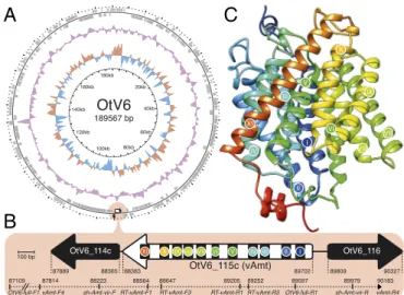
vAmt Is Virally Encoded and Expressed During Infection. Sequence searches and ML phylogenetic tree reconstructions confirmed the viral provenance of both vAmt flanking genes (OtV6 114c and OtV6 116;SI Appendix, Fig. S4). To rule out contamina-tion and genome assembly artifacts, we confirmed the presence of the vAmt-encoding gene (OtV6 115c) on the OtV6 genome by targeted PCR amplification (Fig. 2 A and B). Three sets of PCR primers were designed to amplify through the complete vAmt-encoding gene and its 50and 30flanking genes (Fig. 2B and
SI Appendix, Table S1; GenBank identifier: KX254356). These
results confirm that the transporter-encoding gene is linked to
Fig. 1. OtV6 branches basal to all other available Ostreococcus spp. viral genomes. ML phylogenetic tree of green algal viruses is inferred from a concatenated sequence alignment of 22 core proteins shared among these viruses (7,668 sites) under the LG+G+F model. The unrooted version of this tree is presented below the midpoint-rooted tree. A red circle indi-cates OtV6 branch; other colored circles represent the taxonomy of the viral hosts: green (Ostreococcus), purple (Bathycoccus), yellow (Micromonas), and white (chloroviruses, viruses of Chlorella, as outgroup clade). Node support was calculated from 1,000 nonparametric bootstrap replicates; only boot-strap values >90% are shown. (Scale bar represents the number of esti-mated substitutions per site.) The branch connecting the prasinoviruses to the chloroviruses was truncated for display.
PNAS ENVIRONMENTAL SCIENCES
A
B
C
Fig. 2. vAmt genomic context and putative protein structure. (A) Circu-lar depiction of the OtV6 genome. Oriented ORFs are mapped in gray on the outer ring. The light red circle indicates the vAmt locus along with neighboring ORFs. The colored plots drawn as intermediate rings display the biases in dinucleotide uses across the OtV6 genome: The red and blue plot shows the A+T skew, and the purple plot shows the G+C content. (B) vAmt locus and neighboring ORFs. A white arrow represents the vAmt ORF (OtV6 115c), and black arrows represent the 50 and 30 vAmt
flank-ing ORFs (OtV6 114c and OtV6 116, respectively;SI Appendix, Fig. S4 dis-plays the phylogenies of these flanking ORFs demonstrating viral prove-nance). The 11 predicted transmembrane domains along the vAmt ORF are indicated by black rectangles with colored circles and numbered with Roman numerals. ORF lengths were scaled according to the 100-bp scale bar. Genomic coordinates indicate the 50 and 30boundaries of each depicted
ORF. The PCR-amplified region is represented by a dashed line along with the corresponding amplicon genomic coordinates. (C) vAmt protein struc-ture prediction. The predicted protein strucstruc-ture is composed of 11 helices corresponding to the transmembrane domains indicated by colored circles and Roman numerals, as in B (seeSI Appendix, Fig. S6for sequence and structural comparisons of both host and viral copies).
the viral genes OtV6 114c and OtV6 116 and therefore resid-ing on the viral genome. In addition, no spliceosomal introns were identified in the vAmt-encoding gene, while all Ostreo-coccus spp. homologs have one intron with the exception of Ostreococcus sp. RCC809, which has no introns (SI Appendix, Fig. S1B).
Next we sought to confirm that the vAmt-encoding gene is expressed during infection. We isolated RNA from infected and uninfected O. tauri cultures in parallel. Two different sets of reverse transcription (RT)-PCR primers directed against the vAmt-encoding transcript (RT-vAmt primer sets; Fig. 2B and
SI Appendix, Table S1) amplified products in the infected
cul-tures; no amplicons were found in the uninfected cultures (SI
Appendix, Fig. S5). We sequenced both RT-PCR amplicons from
the infected cultures and confirmed that they corresponded to the vAmt-encoding gene and that this viral gene is expressed dur-ing infection.
vAmt Is an NH+
4 Transporter. The vAmt ORF is located on the
reverse strand of the OtV6 genome (Fig. 2A) and encodes a putative protein of 439 aa. Sequence similarity searches of UniProtKB demonstrate the Amt1.1 sequence from the OtV6 host O. tauri (UniProtKB identifier: A0A096PA30) has the highest similarity with 75.8% amino acid similarity and 62.3% nucleotide identity (SI Appendix, Fig. S6A). In the curated SwissProt database (34), the best match to the viral protein was Arabidopsis thaliana Amt1.3 sequence (Q9SQH9; 65.9% amino acid similarity with vAmt), which was shown to mediate NH+ 4
uptake in N-replete and N-deplete conditions (51).
The vAmt predicted protein secondary structure has 11 trans-membrane domains, a structural feature shared by other NH+ 4
transporter proteins including the O. tauri Amt1.1 (Fig. 2B and
SI Appendix, Figs. S6A and S7). These transmembrane domains
corresponded to 11 α-helices, which have been shown for several NH+
4 transporters to cross the membrane making up a conserved
hydrophobic pore and to contribute to the overall channel stabil-ity of the transporter, as revealed by crystal structures (52–54). In addition, the vAmt has a predicted extracellular N-terminal and cytosolic C-terminal topology, a topology also found for eukary-otic Amt proteins (35) (SI Appendix, Fig. S7; seeSI Appendix, Fig. S1Cfor comparison of Amt homolog C termini). Further-more, vAmt possesses another hallmark of NH+4 transporters:
two conserved histidine residues in the hydrophobic pore con-sistently found in helices V and X (SI Appendix, Fig. S6A) and shown to be essential for transport activity (55). In addition to the high level of sequence conservation between the vAmt and its O. tauri Amt1.1 homolog, protein structures inferred from viral (Fig. 2C) and host (SI Appendix, Fig. S6B) homo-logs showed a high level of structural similarity (SI Appendix, Fig. S6C).
To confirm the vAmt is a functional transporter, we cloned the vAmt-encoding gene (pAG416 GPD vAmt) and transformed it into the yeast Saccharomyces cerevisiae mutant 31019b (56), which has the three known native yeast NH+
4 Mep transporters
deleted (mep1∆mep2∆mep3∆). We compared growth of this mutant with parallel cultures of the same strain but contain-ing only an empty vector (pAG416 GPD) and under culture conditions where 100 µM or 500 µM NH+4 was available as
the sole N source. Complementation of the yeast mutant with vAmt increased growth rate and culture density, consistent with vAmt encoding a functional NH+
4 transporter (Fig. 3A andSI
Appendix, Fig. S8A). For comparison we showed that addition
of pAG416 GPD vAmt did not facilitate growth of the YNVW1 S. cerevisiae mutant (57), which has its urea transporter dur3 gene deleted (dur3∆), on medium containing 100 µM or 500 µM urea as sole N source (SI Appendix, Fig. S8), indicating that vAmt does not transport urea in addition to NH+4. GFP tagging of
either the vAmt C or N termini identifies a plasma membrane localization, with some additional patchy localization, in yeast (Fig. 3B). Similar patterns of localization have been reported for plant Amt transporters, when expressed and tagged in yeast (58).
Comparison of radiolabeled methylammonium uptake rates in yeast mutants (strain 31019b) complemented with either the vAmt-encoding or the O. tauri Amt1.1-encoding genes demon-strated that the viral variant encodes a protein mediating the uptake of methylammonium with a higher rate at lower concen-trations (e.g., 50–100 µM) compared with its O. tauri homolo-gous protein which showed an improved uptake rate at higher methylammonium concentrations (e.g., 500 µM; Fig. 3C). Strik-ingly, these analyses demonstrate that both transporter proteins have distinct substrate affinities and kinetics; specifically, O. tauri Amt1.1 transporter has a Km of 520 µM (±250 SE; Vmax=
3.38 ± 0.91 nmol·mg−1·min−1
) and the vAmt has a Kmof 30 µM
(±10; Vmax= 1.1 ± 0.1 nmol·mg−1·min−1). These distinct
prop-erties of host and viral transporters indicate that production of the vAmt protein during infection has the potential to alter the N-uptake dynamics of infected cells.
Using the OmniLog system (59), we investigated the range of N substrates that showed an increased respiration rate pheno-type in yeast. Comparisons of the NH+4-uptake–deficient 31019b
strain carrying pAG416 GPD vAmt with 31019b carrying only an empty vector (pAG416 GPD) were used to identify poten-tial alternative substrates of the vAmt transporter that resulted in increased respiratory rates. In addition to NH+4, this
pheno-type assay identified a significant increase in respiratory rate on
A
C
B
D
Fig. 3. vAmt is a functional NH+
4 transporter that takes up alternative
sub-strates, is localized to the cell membrane, and mediates an uptake at higher rate for low substrate concentrations than Ostreococcus Amt1.1 homolog when expressed in yeast. (A) Culture optical density (OD600nm) of an NH+4
uptake defective yeast mutant (strain 31019b) transformed either with an empty vector or with a vAmt-containing vector in two NH+
4 concentrations
(100 µM and 500 µM). Error bars represent SEs based on culture triplicates. The lines represent local polynomial regression fits (seeSI Appendix, Fig. S8 for the equivalent results of an additional experiment with O. tauri Amt1.1). Growth of the empty vector transformed mutant is facilitated by passive diffusion of NH+
4 across the yeast membrane and/or scavenging of
intracel-lular N stocks. (B) vAmt GFP fusion proteins expressed in yeast, cloned in frame in N terminus (Left) or in C terminus (Right). (Scale bars, 3-µm dis-tances.) (C) [14C]-Methylammonium uptake rates in mutant strain 31019b complemented with vAmt or the O. tauri Amt1.1 homolog. Lines show Michaelis–Menten curves; error bars represent SEs based on culture tripli-cates. (D) Omnilog comparison of mean point estimates and their 95% con-fidence intervals for N sources showing significant increase in respiration (area under the curve) of vAmt-transformed 31019b yeast cultures com-pared with non–vAmt-transformed 31019b cultures; data were normalized by subtracting the negative control. SeeSI Appendix, Fig. S8for complemen-tation experiments confirming culture growth phenotypes in all of these results apart from D,L-a-amino butyric acid where complementation is not confirmed; seeSI Appendix, Fig. S9for OmniLog Phenotype Microarray res-piration curves.
three alternative N sources: D,L-a-amino butyric acid, glucuron-amide, and D-mannosamine (Fig. 3D andSI Appendix, Fig. S9). Two of these three alternative vAmt substrates were further con-firmed by functional complementation experiments in yeast cul-tures (SI Appendix, Fig. S8). We note that this method likely underestimates the range of substrates transported by vAmt because alternative substrates may have no “metabolic” effect in yeast but could be used in other cellular backgrounds such as O. tauri.
Viral Infection Alters the Uptake Rate of Methylammonium by Ostre-ococcus Cells. Next we carried out infection experiments of O. tauri cultures with OtV6, to determine whether infection and expression of the vAmt-encoding gene altered the NH+ 4
uptake rates of the host alga (relative to noninfected, control cultures; n = 3). To this aim, we conducted an infection time-course experiment. For 16 h postinfection (hpi), we monitored
O. tauri and virus-like particle (VLP) abundances [enumerated by flow cytometry (FCM)] and methylammonium uptake rates (Fig. 4 andSI Appendix, Tables S2 and S3).
Based on FCM estimates of cell and VLP abundances, the ratio OtV6 particle to O. tauri cell [as a proxy for multiplicity of infection (60)] at the start of the experiment was 0.42 ± 0.06 SE; algal growth was not significantly affected by viral infection for the first 12 hpi (Fig. 4A). At 12 hpi, temporal change in cell abun-dance significantly differed between control and infected algal cultures (two-sample t test, t4= 7.03, P = 0.002; SI Appendix,
Table S2), with a sharp decrease of 11.8% on average in abun-dance for the infected cultures (corresponding to a loss of 0.59 × 10⁷ cells·ml−1 ± 0.12 SE); in contrast, the control O. tauri cul-tures were steadily growing, with an average gain of 33% between 8 hpi and 12 hpi (a gain of 1.44 × 10⁷ cells·ml−1± 0.53 SE).
This drop in O. tauri cell number in infected cultures indicates occurrences of cell burst and lysis caused by viral shedding. The OtV6 latent period is thus between 8 hpi and 12 hpi, a time frame comparable to that of other prasinoviruses of Ostreococ-cus and Micromonas spp. (31, 41). FCM enumeration of VLPs confirmed the reduction in VLPs before 12 hpi, consistent with virus adsorption followed by increase in VLPs after 12 hpi, which is in turn consistent with OtV6 particle release (Fig. 4A andSI
Appendix, Table S3).
During the infection experiment, we used two methods to track NH+4 flux in O. tauri cells. First, we determined [
14
C]-methylammonium uptake rates of standardized subsamples of O. tauri cells throughout the time-course experiments. This analysis demonstrated significant increases in methylammonium uptake rates for infected cells, consistent with viral infection altering the NH+
4 uptake phenotype of the phytoplankton host
(Fig. 4B). We also monitored by fluorometry NH+
4 concentration
in the cell-free culture media, which showed depletion at 8 hpi in
A
B
Fig. 4. Nitrogen source uptake during viral infection. Temporal dynamics of host cell and viral particle abundances (A) and [14C]-methylammonium
uptake rates (B) in OtV6-infected and noninfected O. tauri cultures. Three O. tauri cultures were exposed to OtV6 (time point 0 hpi) and three other noninfected cultures were used as controls. For each panel, (log2)-fold changes are based on time point 0 hpi (i.e., time of viral inoculation for infected O. tauri cultures); plots summarize the fold change distribu-tion of three cultures for a given time point during the experiment, and trend lines were estimated by local polynomial regressions (loess). Error bars represent SEs. Shaded overlays indicate incubator dark period (“night”). O. tauri cells and virus-like particles were enumerated using flow cytom-etry; uptake rates were estimated by tracing the isotope-labeled [14
C]-methylammonium in cultures taken as subsamples throughout the time-course experiment. SeeSI Appendix, Tables S2 and S3for two-sample t-test results.
PNAS
ENVIRONMENTAL
SCIENCES
NH+
4 concentration compared with 0 hpi in the infected cultures,
although not significant (SI Appendix, Fig. S10 and Table S2). In addition, we used quantitative PCR (qPCR) to quantify the transcript levels of several O. tauri and OtV6 genes (SI
Appendix, Table S4) during the latent period of infected
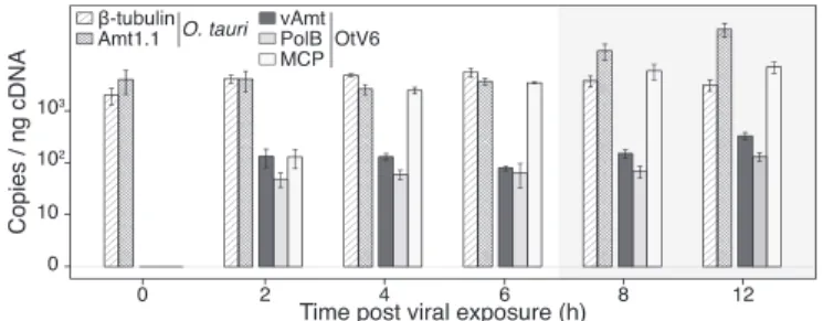
cul-tures (i.e., 0–12 hpi). The qPCR shows that transcription of the vAmt-encoding gene is maintained relatively constant dur-ing infection and at a similar level to that of the viral DNA polymerase B gene, while transcription of the gene encod-ing the major capsid protein increases until 4 hpi and is then expressed at a relatively high level throughout 4–12 hpi (Fig. 5). Taken together, VLP and host cell dynamics, along with gene transcription, all indicate a substantial viral infection of O. tauri cells resulting in an altered methylammonium uptake phenotype.
OtV6 Acquired vAmt from an Ostreococcus Host. We sought to explore the ancestry of the vAmt-encoding gene by conduct-ing phylogenetic tree reconstruction usconduct-ing a comprehensive sam-pling of protein sequences of the Amt/Mep/Rh superfamily (Fig. 6A). Proteins of this superfamily are found across all three domains of life and are involved in the transport (uptake and excretion) of ammonium ions through cell membranes (35). This superfamily of membrane proteins has a complex evolutionary history marked by gene duplications, losses, and HGT events, probably driven by environmental selection linked to N avail-ability (7, 32). In a phylogenetic study, McDonald et al. updated the Amt/Mep/Rh superfamily phylogeny (32), showing a parti-tioning into distinct clades: the monophyletic groups “Amt-Euk” (originally named Amt1) and “Mep,” as well as two distantly related clades, “Rh” (Rhesus) and a cluster of archaeal and bac-terial homologs grouped into “Mep-grade.” The Amt-Euk clade is composed of eukaryotic sequences, with a large representa-tion of phytoplankton and plant species. For the host of OtV6, O. tauri, four Amt/Mep/Rh transporters were identified in its genome, two Amt-Euk (eukaryotic origin; Amt1.1 and -1.2) and two Mepα[probably acquired via HGT; Amt2.1 and -2.2 (7)].
To determine which clade the vAmt groups within, a large-scale, approximate ML phylogenetic tree reconstruction was conducted based on an alignment of all available, nonredun-dant Amt/Mep/Rh protein sequences, including those from the marine microbial eukaryotic transcriptome sequencing project [MMETSP (61)] and which includes several Ostreococcus tran-scriptomes such as O. mediterraneus. The resulting Amt/Mep/Rh superfamily phylogeny (based on an alignment of 374 sites; Fig. 6A and SI Appendix, Fig. S11) demonstrates that the vAmt branched within the Amt-Euk clade. To determine the branching
Fig. 5. OtV6-infected O. tauri gene expression. Transcript levels were RT-qPCR monitored in infected O. tauri cultures (n = 3; same infected cultures as presented in Fig. 4) and expressed as copies per nanogram of cDNA library (displayed on a log10 scale, y axis). Hatched bars show transcript copy num-bers of two O. tauri genes (β-tubulin and the NH+
4 transporter Amt1.1);
solid color bars show copy numbers for three OtV6 genes (dark gray, viral NH+
4 transporter, vAmt; light gray, DNA polymerase B; white, major capsid
protein). Error bars represent SEs. Shaded overlays indicate incubator dark period (night).
position of the vAmt sequence more accurately, a ML phyloge-netic tree was reconstructed using a subset of Amt-Euk homologs (364 sequences, alignment composed of 429 sites; Fig. 6B and
SI Appendix, Fig. S12). This phylogeny shows the vAmt
branch-ing with the Ostreococcus Amt1.1 family radiation, demonstrat-ing that the OtV6 vAmt-encoddemonstrat-ing gene is derived from its host lineage, most likely via host-to-virus HGT. In addition to the recent identification of putative Amt transporters on phage con-tigs assembled from marine metagenomes (21), our phylogenetic analyses also demonstrated that two metagenomic sequences retrieved from the viral fraction of aquatic samples [Pacific solar salterns, San Diego (62)] branch with the OtV6 vAmt (Fig. 6B
andSI Appendix, Fig. S12), suggesting a wider geographical
dis-tribution of viral NH+
4 transporters.
Discussion
Here we demonstrate that a virus of the marine phytoplank-ton O. tauri, representing a distinct lineage of Ostreococcus virus (Fig. 1), harbors a gene encoding a transporter protein that is expressed during infection (Fig. 5 and SI Appendix, Fig. S5) and functions to take up NH+
4 (Fig. 3A andSI Appendix, Fig.
S8A) and a range of alternative fixed N sources (Fig. 3D andSI
Appendix, Figs. S8 and S9). Phylogenetic analyses show that the
viral transporter is the result of a HGT event and was acquired from the host Ostreococcus lineage (Fig. 6B).
Viral HGT and Functional Compatibility. Many host-derived AMGs, including the vAmt-encoding gene reported here, are highly similar at the sequence level to their host homologs. This has two implications: First, it complicates the analysis of viral metagenomes, as the process of host-to-virus HGT is likely to “contaminate” viral metagenomes with sequences that appear as though they should belong to larger sample filtration fractions (i.e., the fraction containing host genomes). This can be amelio-rated by the use of metagenome assembly methods that control for host-derived genes harbored by viral genomes (21). Second, it confirms that viruses are acquiring genes from their host lin-eage and therefore acting as vectors for gene transfers. HGT and viral integration of genes that function in how host cells interact with their environment demonstrate a selective scenario that can drive transfer of genes encoding functional traits. Indeed, bac-terial genomic islands often contain transporter-encoding genes and have been shown to undergo gene gain at high relative rates [for instance, in the pico-cyanobacterium Prochlorococcus (63)]. Genes that encode transporter proteins are relevant because they encode the proteins responsible for nutrient uptake and are “functionally compatible.” We define a functionally compat-ible gene as a single gene that encodes a complete trait and also requires little protein–protein interaction network complexity to result in a function (64, 65). As such, these gene classes are eas-ily lost, duplicated, or (re)acquired during the diversification of a lineage, leading to these genes having complex evolutionary his-tories often involving HGT events (7, 29, 32, 66).
The Role of vAmt During Infection. Both O. tauri and O.
lucimari-nus are able to grow with NH+
4 as the sole N source (67). Here
we demonstrate that the vAmt-encoding gene harbored by OtV6 is transcribed during viral infection (Fig. 5 andSI Appendix, Fig. S5), and NH+4 (methylammonium) uptake in infected
Ostreococ-cus cells is enhanced compared with that in uninfected cells (Fig. 4B). Interestingly, expression in a yeast mutant demonstrates that the viral vAmt transporter has a higher affinity (defined here as [14C]-methylammonium uptake rate) at low
environ-mental substrate concentrations compared with the algal trans-porter homolog, which has a higher relative NH+
4 uptake rate at
higher environmental concentrations. Competition for NH+4 is
fierce in many marine systems, given that it is often the preferred
Mep
βMep
α A. thaliana Amt2 S. cerevisae Rh H. sapiens RhA,B,C,D,CE Mep Amt-Euk E. coli AmtBMep
γ Mep-grade Eukaryote Bacteria Archaea SwissProt reviewed vAmt (OtV6)A
B
Fig. 6. vAmt phylogenetic ancestry shows it is derived by HGT from the host lineage. (A) Amt/Mep/Rh superfamily phylogenetic tree. This large-scale approximate ML tree was inferred under the WAG+G model. Putative homologs were recruited based on a similarity search using the Pfam HMM cor-responding to the Amt/Mep/Rh superfamily (PF00909) against UniRef100 (i.e., nonredundant version of UniProtKB), MMETSP protist transcriptomes, and predicted proteomes from various protist genome projects; the final curated alignment was composed of ∼20,000 protein sequences encompassing 374 sites. Curved black lines indicate the phylogenetic positions of the main clades (Amt-Euk, Mep, Mep-grade, and Rh) as well as Mep subclades (α, β, and γ). The red capsid graphics show the phylogenetic position of the vAmt within the superfamily tree, positioned within the Amt-Euk clade. For contextual reference, orange stars represent SwissProt reviewed protein entries; yellow, blue, and purple circles represent eukaryotic, bacterial, and archaeal proteins, respectively. SeeSI Appendix, Fig. S11for additional information, including local support values, MMETSP sequence positions, and scale bar. (B) vAmt evolu-tionary relationships with Amt-Euk (Amt1) homologs. This ML phylogenetic tree was inferred under the LG+I+G+F model, based on a multiple alignment of 364 proteins totaling 429 sites. These vAmt homologs were recruited and selected based on the Amt/Rh/Mep superfamily phylogenetic tree reconstruction. Green and red cell schematics represent green and red algal lineages. vAmt is highlighted in red with capsid graphics and branched within the prasino-phyte green algae, delimited by a green frame. Code numbers in front of species names represent sequence identifiers from the MMETSP transcriptomes (O. mediterraneus, Nephroselmis pyriformis, and Crustomastix stigmata), UniProtKB (O. lucimarinus and O. tauri), and the Ostreococcus sp. RCC809 genome project available at the DoE-Joint Genome Institute. Numbers in parentheses beside clade names are the number of sequences present in collapsed nodes. Branch node supports were computed from 1,000 nonparametric bootstrap replicates. Gray and black circles correspond to bootstrap values of ]80, 95%] and ]95, 100%], respectively. The dashed branches represent the phylogenetic placement of two short environmental sequences, with placement posterior probabilities indicated on their corresponding branches; both sequences originate from a saltern viral metagenome (NCBI BioProject: PRJNA28353), and their GenBank sequence identifiers are provided. SeeSI Appendix, Fig. S12for additional data and uncollapsed branch information.
PNAS
ENVIRONMENTAL
SCIENCES
source of inorganic N for phytoplankton cells (10). Expression of the vAmt-encoding gene during O. tauri infection may increase host NH+4 uptake rate and therefore may provide an
advan-tage to infected host cells over noninfected algae in terms of N uptake, allowing an infected population to temporarily out-compete other phytoplankton species, including uninfected sis-ter cells. As such, production of vAmt transporsis-ter proteins is likely to act to fulfill the extended N requirements of viral repli-cation within the infected host cell to sustain the physiological burden of viral replication through the uptake of various organic N sources.
Using comparative culture screening, we show that the vAmt protein potentially mediates the uptake of alternative organic N sources in addition to NH+
4. Amt transporter proteins are known
to mediate the uptake of at least one alternative to NH+4,
methy-lammonium (68). Most knowledge regarding substrate affinities of protein transporters derives from annotation resulting from sequence homology searches and few experimental studies; it is thus important to take into account, when assessing the sig-nificance of transporter proteins in an ecological context, that these proteins may display affinities to additional substrates not described via bioinformatic annotation (69).
Viral Manipulation of Host Elemental Composition.Harboring genes coding for functional nutrient transporters implies that phytoplankton viruses manipulate the elemental composition of their host cells during infection. This viral stoichiometric alter-ation would confer a strong advantage to viruses able to inter-fere with the hosts’ nutrient uptake capabilities by encoding their own transporter repertoires. Availability of nutrients to phyto-plankton hosts has been shown to be of critical importance for phytoplankton virus proliferation (33). Infection experiments on phytoplankton cultures depleted in P and N have shown that low concentrations of these nutrients can significantly hamper viral cycle dynamics in terms of latent period and burst size (i.e., the number of viral progeny per lysed cell) as well as decreasing the infectivity of viral progenies; such constraints on viral replica-tion have been shown for various clades of Phycodnaviridae (e.g., refs. 30 and 70), including for a Micromonas virus, a close relative of OtV6 (Fig. 1).
Although most studies have focused on the effect of low P availability on phytoplankton virus replication, recent studies have shown the importance of N availability for the quality of viral replication (31, 70). In particular, the burst size of PBCV1, a virus of the green alga Chlorella, was shown to decrease with higher C:N ratio of the algal host cell (70). Furthermore, the effect of N depletion was shown to be more detrimental to viral proliferation than low P availability for a Phycodnaviridae– phytoplankton system (31). These results are somewhat antici-pated because of the stoichiometric requirements of phytoplank-ton virus replication, which are exacerbated by the strong dis-crepancy in C:N:P ratios between the viral particle and the host phytoplankton cell (71). Viruses exhibit much lower C:P and C:N ratios than their hosts, as shown for PBCV1’s 17C:5N:1P compo-sition (72), compared with the Redfield ratio of 106C:16N:1P, the elemental composition of an average phytoplankton (71).
In the case of the Phycodnaviridae EhV, a virus of the coc-colithophore Emiliania, it was noted that viral genomes were being overproduced during the infection cycle, relative to the number of capsids being produced by the infected phytoplank-ton (73). Such a discrepancy between genome and capsid pro-ductions may be linked to a shortage in amino acids and/or lipids, a constituent of EhV lipid membrane, limiting viral production (73, 74). This is predictable for Phycodnaviridae given the N requirements of PBCV1 capsid, which is composed of ∼5,050 major capsid proteins (MCPs) (75). Based on the MCP protein sequence of PBCV1, the elemental composition of the complete
PBCV1 capsid would be C2161H3301N577O657S9, that is, a C:N
ratio of 4:1. Hence, the ability of OtV6 to express its gene encod-ing vAmt and enhance or maintain NH+4 uptake would allow the
host to fulfill the requirements in N imposed by the viral repli-cation, in turn providing a strong fitness advantage to OtV6 rel-ative to marine viruses without a N transporter gene repertoire. Further work will be required to understand the role of virally encoded nutrient transporters in the dynamics of viral replica-tion, especially in contrasting nutrient concentrations.
Blurring Top–Down and Bottom–Up Controls. Consistent with the idea that viral proteins act to amend host N metabolic function, a putative glutamine synthetase gene has also been identified in the genomes of NCLDVs such as Mimivirus and Mamavirus (76). These data suggest that viral reprogramming of host N metabolism may be a wider phenomenon (21, 77), as has been shown for photosynthetic function (78), sulfur oxidation (79), and lipid (25–27) and phosphate metabolism (80). Evidence that a viral lineage can acquire host genes to amend host nutrient uptake has implications for our understanding of phytoplankton ecology. Specifically, this phenomenon blurs the lines between bottom–up and top–down regulation of phytoplankton commu-nities, because here a top–down viral agent has acquired the host genes which allow it to amend phytoplankton nutrient acqui-sition from the environment, which is normally considered a bottom–up process.
Materials and Methods
vAmt Identification, Sequence, and Structure Analyses. To identify a viral transporter sequence putatively involved in NH+
4 uptake, all protein
sequences available in UniProtKB (34) and the NCBI nonredundant Refer-ence SequRefer-ence database (81) were searched using “hmmsearch,” part of the HMMer v3 software suite (82), with the Pfam (83) HMM corresponding to the NH+
4 transporter superfamily (Pfam release 3.0 identifier: PF00909). Hits
were filtered using E-value (1e−10) and gathering cutoffs. This large-scale
search identified only a single viral encoded putative transporter protein, OtV6 vAmt (HMMer search statistics with PF00909 model: score, 418.4; E value, 1e−129). To avoid missing additional putative viral NH+
4 transporters—
potentially due to inaccurate viral gene modeling—a HMM-based search was repeated on six-frame translations of all viral genomes available at the NCBI genomic sequence repository (ORF minimal size: 60-aa residues). In addition to the Pfam-based HMM searches, further searches using the TIGRfam (84) NH+4 transporter HMM (TIGR00836; release 15) were con-ducted using the aforementioned protein sequence datasets. These addi-tional searches did not recover any other viral sequences matching the NH+ 4
transporter HMMs.
Transmembrane domains of the vAmt and O. tauri Amt1.1 protein sequences were identified using TMHMM v2 (85). Protein structures were predicted using I-TASSER v4.2 (86); the C scores of the vAmt and O. tauri Amt1.1 3D structure models were 1.36 and 0.72, respectively. O. tauri Amt1.1 and vAmt predicted structures were aligned using MatchMaker [UCSF Chimera (87)].
Phylogenetic Tree Reconstructions. The Amt/Mep/Rh superfamily phylogeny was reconstructed using an approximate ML method as implemented in FastTree v2.1 (88) (compiled using the double-precision flag) and based on an alignment of 19,493 protein sequences sampled from UniProt100 [clustered at 100% sequence identity (34) to reduce redundancy]. HMM searches were also conducted against predicted protein sequences from the MMETSP transcriptomic data (61) and from protist genome projects avail-able at the DoE-Joint Genome Institute. All identified putative Amt/Mep/Rh sequences were then aligned using “hmmalign” (82) and sequences shorter than 50-aa residues were discarded. Alignment sites composed of more than 50% of gaps were discarded. Misaligned and/or false positive sequences were detected using preliminary phylogenetic reconstructions with FastTree (default parameters). Sequences that resulted in very long branches were then removed, allowing a final tree calculation using the “accurate” mode (-slownni) with the WAG+G substitution matrix.
For the ML phylogeny, different strategies were applied for putative homolog sampling. For the viral DNA polymerase B and the vAmt flank-ing ORFs, homologous protein sequences were identified with BLASTP (89)
similarity searches against UniProtKB. The homologous sequences were then aligned using the iterative refinement method E-INS-i as implemented in MAFFT v7.2 (90), edited with the trimAl v1.4 “strict” algorithm (91), and manually inspected and corrected. For the phylogeny based on the 22 core proteins shared among green algal viruses (46), the OtV6 predicted pro-teome was parsed using BLASTP and homologous sequences from other Ostreococcus spp. viruses were used as search queries. The resulting 22 OtV6 core proteins were then concatenated, aligned, and added to the original viral core protein alignment using MAFFT.
For the vAmt/Amt-Euk ML phylogenetic subtree, vAmt homologs were identified using the previous large-scale, superfamily phylogenetic recon-struction described above. Sequences branching with vAmt were identi-fied; corresponding full-length sequences were retrieved and aligned with MAFFT E-INS-i and manually inspected. After homolog sequence selection and alignment, the same methodology was applied for all ML phylogenetic tree reconstructions. The most likely tree was identified from 100 ML recon-structions, using RAxML v8.2 (92), and under the substitution model best fitting the data, as identified by ProtTest v3 (93) using the Akaike informa-tion criterion; branch support values were based on 1,000 nonparametric bootstrap replicates.
Metagenome Screening and Phylogenetic Mapping. To investigate the wider distribution of vAmt-like homologs, we screened metagenomes for simi-lar sequences in the Global Ocean Sampling and Tara Oceans giant virus datasets (50) as well as all aquatic metagenomes available at iMicrobe (94). Putative vAmt homologs were aligned to alignment corresponding to the vAmt subtree (Fig. 6B andSI Appendix, Fig. S12) using MAFFT (without alter-ing the original alignment) and then phylogenetically mapped onto the ML tree using pplacer v1.1 (95). Within this tree, two Amt-Euk sequences mapped to the vAmt branch.
vAmt Genomic Amplification. To confirm the vAmt-encoding gene was har-bored by OtV6, we designed specific primer pairs (listed inSI Appendix, Table S1) and the vAmt gene along with its 50and 30flanking regions were
amplified, represented by an ∼2,500-bp sequence. For each PCR, a negative control (distilled H2O) was included. PCRs (25 µL total volume) were
con-ducted using GoTaq Green Master Mix (Promega) with 1 µL of virus OtV6. Cycling reactions were as follows: 5 min at 95◦C; followed by 30 cycles of
30 s at 95◦C, 30 s at 54◦C, and 120 s at 72◦C; and with an additional 10-min extension at 72 ◦C. The PCRs were checked on a 1% agarose
gel stained with GelRed (Biotium). The positive PCRs were then directly cloned using the StrataClone PCR cloning kit (Agilent Technologies) accord-ing to the manufacturer’s instructions. One clone per library was selected and double-strand sequenced using universal M13 primers. All amplicon sequencing was performed externally by Eurofins Genomics.
Ostreococcus Culture Infection and vAmt RT-PCR. O. tauri cells from the RNP2 population [OtV5 resistant (44)] were grown in L1 medium exponen-tially under 12:12 light (32.3 µmol·m−2·s−1; 1,700 lux). Infection
experi-ments proceeded as follows: A total of 5 mL of O. tauri at exponential growth was infected using 250 µL of purified OtV6; as a negative con-trol, 5 mL of O. tauri culture was incubated with 250 µL of L1 medium. To test for the expression of the vAmt-encoding gene during the infection cycle, the O. tauri cultures were sampled 12 h after OtV6 inoculation; 1 mL of the infected and uninfected cultures was sampled and centrifuged at 6,200 × g during 10 min. After removal of the supernatant, the pellets were flash frozen and stored at−80◦C. RNA was extracted using the RNeasy
Plus Universal Kits (Qiagen). An extra step to remove all genomic DNA was added after the RNA extraction protocol, using the RTS DNase kit (MO BIO Laboratories, Qiagen).
Using three sets of primers that amplify an overlapping region of the vAmt-encoding gene (OtV6-full-F1/R1, vAmtF4/R4, and sh-Amt-vir-F/R; Fig. 2B andSI Appendix, Table S1), we first checked for the presence of DNA contamination in the RNA samples, using PCR amplification. For every PCR, we included a negative control (distilled H2O) and a positive control
(puri-fied OtV6). PCRs were performed in 25 µL total volume, using GoTaq Green Master Mix with 1 µL of RNA sample. Cycling reactions were as follows: 5 min at 95◦C; followed by 35 cycles of 30 s at 95◦C, 30 s at 50◦C,
and 45 s at 72◦C; with an additional 10-min extension at 72 ◦C. The
PCRs were checked on a 1% agarose gel stained with GelRed. The RT-PCRs were conducted using OneTaq One-Step RT-PCR (Qiagen). A total of 1 µL of RNA was mixed in 50 µL with 10 µL of Buffer 5 ×, 2 µL of 10 mM dNTP, 1 µL of each primer (0.2 mM final concentration), 2 µL enzyme mix, and finally 0.25 µL of RNaseOUT (10 units/µL; Invitrogen, Life Technolo-gies). Negative controls were made with 1 µL of water instead of the RNA
samples. Each PCR amplification was checked on a 1% agarose gel stained with GelRed. Positive RT-PCRs were cloned using a Strataclone PCR cloning kit; one clone per library was selected and double-strand sequenced using M13 primers.
Cloning and Functional Analysis of vAmt in Yeast. The vAmt ORF was synthe-sized de novo by Genscript, codon optimized for expression in S. cerevisiae, and fused to a C-terminal GFP tag in vector p426 GPD. For complementation assays, the vAmt ORF was amplified with primers vAmt-attF/R (SI Appendix, Table S1), using Phusion polymerase (New England Biolabs) to remove the GFP-coding region; cloned into pDONR221 using Gateway recombination (Life Technologies); and mobilized into pAG416 GPD (low-copy constitutive expression vector). For fluorescence microscopy, the ORF was mobilized into pAG426 GPD EGFP (N-terminal EGFP vector) or the original p426 GPD EGFP (C-terminal) construct was used.
For yeast transformations, competent cells were prepared as described in Thomson et al. (96), mixed with ∼500 ng of plasmid DNA, and pulsed at 1.5 kV in an Eppendorf electroporator. S. cerevisiae strain 31019b was trans-formed with pAG416 GPD vAmt or pAG416 GPD empty vector, as described above. Transformed yeast 31019b cultures were grown to stationary phase at 30◦C and centrifuged at 3,200 × g for 2 min, washed twice in water, and
diluted to OD600nm0.1 in 10 mL yeast nitrogen base (YNB) liquid medium
[0.19% YNB without amino acids and without (NH4)2SO4; Formedium]
containing a final concentration of 0.1 mM or 0.5 mM (NH4)2SO4,
D-mannosamine, D-glucuronamide, or D,L-a-amino butyric acid. Cells were then incubated at 30◦C and OD
600nmmeasurements were taken at 24-h
intervals. This method was also used to assess growth of S. cerevisiae YNVW1 (∆dur3) and Σ23346c (wild type) on urea, with YNB supplemented with urea (0.1–2 mM) instead of (NH4)2SO4. R v3 and ggplot2 (97) were used for
statistical analyses and plots.
Spinning-Disc Confocal Microscopy. GFP constructs were transformed into S. cerevisiae strain BY4742 as described above, grown to midlog phase, and suspended in PBS. Spinning-disc confocal microscopy of EGFP-labeled cells was performed using an Olympus IX81 inverted microscope and a CSU-X1 Spinning-Disc unit (Yokogawa). A × 60/1.35 oil or × 100/1.40 oil objective was used with a 488-nm solid-state laser to excite the EGFP fluorophore. A Photometrics CoolSNAP HQ2 camera (Roper Scientific) was used for imaging with the VisiView software (Visitron Systems).
Transporter Protein Phenotyping. To prepare cells for OmniLog Pheno-type Microarray (PM) plates (Biolog), each yeast strain (S. cerevisiae 31019b transformed with either pAG416 GPD or pAG416 GPD vAmt) was grown on YNB complemented with potassium nitrate and uracil at 30◦C for 48–72 h. Colonies were suspended in Yeast Nutrient Sup-plement solution (Biolog) and adjusted to 62% turbidity. A total of 250 µL of cell suspension was made up to a final volume of 12 mL of inoculating fluid, containing 1 × IFY-0, 1 × Dye Mix D, 50 mM D-glucose, 1 mM disodium pyrophosphate, and 2 mM sodium sulfate, and 100 µL was inoculated into each well of a PM3 MicroPlate (N sources). Assays were run in triplicate using independently obtained transformants. OmniLog Phe-notype Microarray outputs were analyzed by normalizing each individual plate against well A01 (negative control) to control for any background growth as a result of the inoculation solution and then analyzed using the R package opm (98). Data were aggregated using the “opm-fast” method, analyzed using the area under curve (AUC) parameter, and tested by t test to detect significant increase in respiration rates in the pAG416 GPD vAmt strain.
Yeast Methylammonium Uptake Assays. S. cerevisiae strains were grown in 25 mL minimal proline medium (99) for 16 h at 30◦C with shaking. Cells
were then diluted and grown until early log phase and harvested by cen-trifugation at 1,000 × g for 3 min. Cells were washed once and sus-pended in 2 mL medium lacking proline. The dry weight of each sample was noted, before 90-µL aliquots of cells were exposed to five concentrations (5–500 µM) of methylamine [14C] hydrochloride for 1 min at 24◦C. Reac-tions were stopped by the addition of 1 mL 120 mM methylamine hydrochlo-ride. Background adsorption was also calculated by exposing cells to 1 mL of 120 mM unlabeled methylamine hydrochloride before the addition of the radiolabeled substrate. Cells were collected by centrifugation at 14,000 × g for 3 min, washed, and suspended in 500 µL deionized water, and then radioactivity was determined by liquid scintillation counting in a liquid scintillation analyzer (LS 6500; Beckman Coulter) after addition of 2.5 mL Emulsifier-Safe scintillation mixture solution (Perkin-Elmer). The R package drc (100) was used to calculate Michaelis–Menten kinetics and curves.
PNAS
ENVIRONMENTAL
SCIENCES
Infection Time-Course Experiments. Two 15-mL cultures of O. tauri were inoc-ulated with OtV6 viruses and the infection was allowed to proceed until culture bleaching after 5 d. Cell debris were removed by centrifugation at 3,200 × g for 20 min followed by filtering through a 0.2-µm syringe fil-ter. Viruses were concentrated 10-fold using a 50-kDa Amicon Ultra-15 Cen-trifugal Filter (Merck Millipore) followed by centrifugation at 3,200 × g for 2.5 min. A total of 18 mL O. tauri cultures were cultured using 10 mL precul-ture and 8 mL media, 2 d before the experiment. To begin the infection time-course experiments, 750 µL OtV6 was added and at each time point, 1 mL sample was pelleted and frozen in liquid nitrogen for RNA preser-vation and the supernatant was stored at−80◦C for fluorometric NH+ 4
detection. Two 200-µL samples were also taken for flow cytometry: (i) 2 µL 25% glutaraldehyde was added and the sample incubated for 15 min before freezing in liquid nitrogen for O. tauri counts and (ii) 8 µL 25% glutaralde-hyde was added and the sample incubated for 30 min at 4◦C before
freez-ing for viral counts. O. tauri and VLP abundances were monitored by a FACSCanto flow cytometer (BD Biosciences) according to their right-angle scatter and the fluorescence emission due either to the chlorophyll a pig-ment for O. tauri (101) or to SYBR Green I (Roche Diagnostics) staining for VLPs (102). A 900-µL sample was also removed for assessing methylam-monium uptake at each time point. Cells were harvested by centrifuga-tion at 5,000 × g for 3 min and suspended in 450 µL media. For each sam-ple, the uptake rate for methylamine [14C] hydrochloride was determined
as previously described. The orthophtaldialdehyde (OPA) method (103) was used for fluorometric NH+
4 detection in cell-free medium (supernatant, see
above). A total of 20 µL of the working reagent (OPA, borate buffer, and sodium sulfite) was added to 80 µL of the culture sample (or NH+
4 standard);
samples and standards were processed on a 96-well plate, which was incu-bated in the dark at 25◦C with shaking for 2–3 h. Fluorescence was then read at 355 nm excitation and 420 nm emission on an Infinite M200 plate reader (Tecan).
Quantitative PCR. RNA was extracted using the RNeasy Plus Universal Mini kit (Qiagen) following the manufacturer’s instructions and incorporating a 5-min elution step using 30 µL RNase-free water. Residual DNA was removed using the Turbo DNA-free kit (Ambion, Life Technologies) and 6 µL RNA reverse transcribed using the Superscript III First Strand Synthesis SuperMix (Invitrogen) and oligo(dT)20primers, following the manufacturer’s
instruc-tions. cDNA was quantified using a Qubit ssDNA Assay Kit (Life Technolo-gies) and stored at−20◦C before performing qPCR. Plasmids for each gene
of interest were generated by Phusion polymerase (New England Biolabs) PCR using cDNA templates, followed by A-tailing using Taq polymerase and cloning using a Strataclone PCR cloning kit (Agilent Technologies). Each plasmid was confirmed by sequencing (Eurofins Genomics) and serial dilu-tions (108 to 10 copies) were used to generate standard curves for each primer pair and probe. Efficiencies ranged from 96% to 102% (SI Appendix, Table S4). qPCRs were performed in a StepOnePlus Real-Time PCR system (Thermo Fisher Scientific). Each 25-µL reaction contained 12.5 µL TaqMan Gene Expression Master Mix (Thermo Fisher Scientific), 900 nM each primer, 250 nM hydrolysis probe, and 1 µL cDNA per plasmid DNA and was per-formed in duplicate alongside no-template and minus-RT controls. Cycling conditions were as follows: UDG activation for 2 min at 50◦C and DNA
poly-merase activation for 10 min at 95◦C, followed by 40 cycles of 15 s at 95◦C
and 1 min at 60◦C. ROX was used as an internal reference dye for analysis
of CT values, which were determined using StepOne Software v2.3 (Thermo Fisher Scientific), and standard curves were used for quantification of each gene.
Data Access. Phylogenetic and experimental data are available at the Zenodo data repository:zenodo.org/record/61901. PCR-amplified sequence assem-bly of the vAmt-encoding locus was deposited in GenBank (KX254356). ACKNOWLEDGMENTS. We thank C. Salmeron and the cytometry platform of the Observatoire Oc ´eanologique de Banyuls-sur-Mer, as well as C. Lam-bert and the cytometry core facilities of the Laboratoire des Sciences de l’Environnement Marin (Institut Universitaire Europ ´een de la Mer) for assis-tance in FCM counts. We are grateful to Dr. A-M. Marini (Universit ´e Libre de Bruxelles) and to Dr. G. P. Bienert (Leibniz Institute of Plant Genetics and Crop Plant Research) for yeast mutant strains. We thank Prof. K. Haynes, Dr. F. Maguire, Dr. F. Savory, Prof. N. Smirnoff and Dr. J. G. Wideman (Univer-sity of Exeter) for helpful comments. A.M. and T.A.R. are funded by the Royal Society, through Newton and University Research fellowships, respectively. This work is supported in part by research grants from The Gordon and Betty Moore Foundation (GBMF5514), Leverhulme Trust (PLP-2014-147), and the University of Exeter. The University of Exeter OmniLog facility is supported by a Wellcome Trust Institutional Strategic Support Award WT105618MA. Phylogenetic reconstructions were computed on the Data Intensive Aca-demic Grid (National Science Foundation, MRI-R2 Project DBI-0959894).
1. Falkowski P (2012) Ocean science: The power of plankton. Nature 483:S17–S20. 2. Arrigo KR (2005) Marine microorganisms and global nutrient cycles. Nature 437:
349–355.
3. Moore C, et al. (2013) Processes and patterns of oceanic nutrient limitation. Nat
Geosci 6:701–710.
4. Litchman E, Klausmeier CA, Schofield OM, Falkowski PG (2007) The role of functional traits and trade-offs in structuring phytoplankton communities: Scaling from cellular to ecosystem level. Ecol Lett 10:1170–1181.
5. Hildebrand M (2005) Cloning and functional characterization of ammonium trans-porters from the marine diatom Cylindrotheca fusiformis (Bacillariophyceae). J Phycol 41:105–113.
6. Allen AE, Vardi A, Bowler C (2006) An ecological and evolutionary context for inte-grated nitrogen metabolism and related signaling pathways in marine diatoms. Curr
Opin Plant Biol 9:264–273.
7. McDonald SM, Plant JN, Worden AZ (2010) The mixed lineage nature of nitrogen transport and assimilation in marine eukaryotic phytoplankton: A case study of Micromonas Mol Biol Evol 27:2268–2283.
8. Dagenais-Bellefeuille S, Morse D (2013) Putting the N in dinoflagellates. Front
Micro-biol 4:369.
9. Terrado R, Monier A, Edgar R, Lovejoy C (2015) Diversity of nitrogen assimilation pathways among microbial photosynthetic eukaryotes. J Phycol 51:490–506. 10. Mulholland MR, Lomas MW (2008) Nitrogen uptake and assimilation. Nitrogen in the
Marine Environment, eds Capone D, Bronk D, Mulholland M, Carpenter E (Academic,
New York), pp 303–384.
11. Glibert PM, et al. (2016) Pluses and minuses of ammonium and nitrate uptake and assimilation by phytoplankton and implications for productivity and community composition, with emphasis on nitrogen-enriched conditions. Limnol Oceanogr 61: 165–197.
12. Gobler CJ, et al. (2011) Niche of harmful alga Aureococcus anophagefferens revealed through ecogenomics. Proc Natl Acad Sci USA 108:4352–4357.
13. Worm B, Lotze HK, Hillebrand H, Sommer U (2002) Consumer versus resource control of species diversity and ecosystem functioning. Nature 417:848–851.
14. Pasulka AL, Samo TJ, Landry MR (2015) Grazer and viral impacts on microbial growth and mortality in the Southern California current ecosystem. J Plankton Res 37: 320–336.
15. Suttle CA (2007) Marine viruses—Major players in the global ecosystem. Nat Rev
Microbiol 5:801–812.
16. Brussaard CP (2004) Viral control of phytoplankton populations—A review. J Eukaryot
Microbiol 51:125–138.
17. Rohwer F, Thurber RV (2009) Viruses manipulate the marine environment. Nature 459:207–212.
18. Wilhelm SW, Suttle CA (1999) Viruses and nutrient cycles in the sea. BioScience 49:781–788.
19. Breitbart M (2012) Marine viruses: Truth or dare. Annu Rev Mar Sci 4:425–448. 20. Hurwitz BL, Hallam SJ, Sullivan MB (2013) Metabolic reprogramming by viruses in the
sunlit and dark ocean. Genome Biol 14:R123.
21. Roux S, et al. (2016) Ecogenomics and potential biogeochemical impacts of globally abundant ocean viruses. Nature 537:689–693.
22. Monier A, et al. (2009) Horizontal gene transfer of an entire metabolic pathway between a eukaryotic alga and its DNA virus. Genome Res 19:1441–1449. 23. Thompson LR, et al. (2011) Phage auxiliary metabolic genes and the redirection of
cyanobacterial host carbon metabolism. Proc Natl Acad Sci USA 108:E757–E764. 24. Dammeyer T, Bagby SC, Sullivan MB, Chisholm SW, Frankenberg-Dinkel N (2008)
Efficient phage-mediated pigment biosynthesis in oceanic cyanobacteria. Curr Biol 18:442–448.
25. Rosenwasser S, et al. (2014) Rewiring host lipid metabolism by large viruses deter-mines the fate of Emiliania huxleyi, a bloom-forming alga in the ocean. Plant Cell 26:2689–2707.
26. Malitsky S, et al. (2016) Viral infection of the marine alga Emiliania huxleyi triggers lipidome remodeling and induces the production of highly saturated triacylglycerol.
New Phytol 210:88–96.
27. Ziv C, et al. (2016) Viral serine palmitoyltransferase induces metabolic switch in sphingolipid biosynthesis and is required for infection of a marine alga. Proc Natl
Acad Sci USA 113:E1907–E1916.
28. Wilson WH, Carr NG, Mann NH (1996) The effect of phosphate status on the kinetics of cyanophage infection in the oceanic cyanobacterium Synechococcus sp. wh78031.
J Phycol 32:506–516.
29. Monier A, et al. (2012) Phosphate transporters in marine phytoplankton and their viruses: Cross-domain commonalities in viral-host gene exchanges. Environ Microbiol 14:162–176.
30. Maat DS, Crawfurd KJ, Timmermans KR, Brussaard CP (2014) Elevated CO2 and phos-phate limitation favor Micromonas pusilla through stimulated growth and reduced viral impact. Appl Environ Microbiol 80:3119–3127.
31. Maat DS, Brussaard CP (2016) Both phosphorus-and nitrogen limitation constrain viral proliferation in marine phytoplankton. Aquat Microb Ecol 77:87–97.
32. McDonald TR, Dietrich FS, Lutzoni F (2012) Multiple horizontal gene transfers of ammonium transporters/ammonia permeases from prokaryotes to eukaryotes: Toward a new functional and evolutionary classification. Mol Biol Evol 29:51–60.

![Fig. 4. Nitrogen source uptake during viral infection. Temporal dynamics of host cell and viral particle abundances (A) and [ 14 C]-methylammonium uptake rates (B) in OtV6-infected and noninfected O](https://thumb-eu.123doks.com/thumbv2/123doknet/13736839.436852/5.877.453.818.606.875/nitrogen-infection-temporal-dynamics-particle-abundances-methylammonium-noninfected.webp)