HAL Id: hal-03176756
https://hal.archives-ouvertes.fr/hal-03176756
Submitted on 22 Mar 2021
HAL is a multi-disciplinary open access
archive for the deposit and dissemination of
sci-entific research documents, whether they are
pub-lished or not. The documents may come from
teaching and research institutions in France or
abroad, or from public or private research centers.
L’archive ouverte pluridisciplinaire HAL, est
destinée au dépôt et à la diffusion de documents
scientifiques de niveau recherche, publiés ou non,
émanant des établissements d’enseignement et de
recherche français ou étrangers, des laboratoires
publics ou privés.
Sortilin-derived peptides promote pancreatic beta-cell
survival through CREB signaling pathway
Guillaume Daziano, Nicolas Blondeau, Sophie Béraud-Dufour, Amar
Abderrahmani, Carole Rovère, Catherine Heurteaux, Jean Mazella, Patricia
Lebrun, Thierry Coppola
To cite this version:
Guillaume Daziano, Nicolas Blondeau, Sophie Béraud-Dufour, Amar Abderrahmani, Carole Rovère,
et al.. Sortilin-derived peptides promote pancreatic beta-cell survival through CREB signaling
path-way. Pharmacological Research, Elsevier, 2021, 167, pp.105539. �10.1016/j.phrs.2021.105539�.
�hal-03176756�
Pharmacological Research 167 (2021) 105539
Available online 15 March 2021
1043-6618/© 2021 The Author(s). Published by Elsevier Ltd. This is an open access article under the CC BY-NC-ND license (http://creativecommons.org/licenses/by-nc-nd/4.0/).
Sortilin-derived peptides promote pancreatic beta-cell survival through
CREB signaling pathway
Guillaume Daziano
a, Nicolas Blondeau
a, Sophie B´eraud-Dufour
a, Amar Abderrahmani
b,
Carole Rov`ere
a, Catherine Heurteaux
a, Jean Mazella
a, Patricia Lebrun
a,1,*,
Thierry Coppola
a,1,*aUniversit´e Cˆote d′Azur, CNRS, IPMC, Sophia Antipolis, F-06560, France
bUniversit´e Lille, CNRS, Centrale Lille, Universit´e Polytechnique Hauts-de-France, UMR 8520-IEMN, F-59000 Lille, France
A R T I C L E I N F O Keywords: Beta-cell protection Calcium influx CREB activation Neuropeptide Sortilin-released propeptide A B S T R A C T
Deterioration of insulin secretion and pancreatic beta-cell mass by inflammatory attacks is one of the main pathophysiological features of type 2 diabetes (T2D). Therefore, preserving beta-cell mass and stimulating in-sulin secretion only in response to glucose for avoiding the hypoglycemia risks, are the most state-of-the-art option for the treatment of T2D. In this study we tested two correlated hypothesis that 1/ the endogenous peptide released from sortilin, known as PE, that stimulates insulin secretion only in response to glucose, protects beta-cells against death induced by cytokines, and 2/ Spadin and Mini-Spadin, two synthetic peptides derived from PE, that mimic the effects of PE in insulin secretion, also provide beneficial effect on beta-cells survival. We show that PE and its derivatives by inducing a rise of intracellular calcium concentration by depolarizing the membrane protect beta-cells against death induced by Interleukin-1β. Using biochemical, confocal imaging and cell biology techniques, we reveal that the protective effects of PE and its derivatives rely on the activation of the CaM-Kinase pathway, and on the phosphorylation and activation of the transcription factor CREB. In addition, Mini-Spadin promotes beta-cell proliferation, suggesting its possible regenerative effect. This study highlights new possible roles of PE in pancreatic beta-cell survival and its derivatives as pharmacological tools against diabetes.
1. Introduction
Insulin is a hormone produced and released by pancreatic beta-cells to regulate glucose homeostasis. Imbalance of insulin secretion and the tissue needs caused by insulin resistance leads to diabetes, one of the deadliest diseases worldwide. In diabetes, glucose-induced insulin secretion (GSIS) of beta-cells is impaired. In addition, the loss of insulin secretion is exacerbated by the loss of beta-cell mass (BCM) that is estimated of 30–50% in long-term disease, resulting from increased
beta-cell apoptosis and possibly dedifferentiation.
Major mechanisms coupling diabetes development to impaired GSIS and the loss of BCM involve alteration of positive signaling pathways. Among them, CREB by its anti-apoptotic and trophic effects plays a central role in beta-cell survival and growth [1–4] and a decrease in its expression leads to beta-cell dysfunction and death [3]. Induction of CREB is a key mechanism from which glucagon-like peptide 1 (GLP-1) exerts its incretin physiological action and therefore its mimetics that are used as antidiabetic drugs, ameliorate beta-cell function and
Abbreviations: ADP, Adenosine diphosphate; ATP, Adenosine triphosphate; KATP, ATP-sensitive potassium channel; Bcl2, B-cell lymphoma 2; BCM, Beta cell mass;
BrdU, 5-Bromo-2′-deoxyuridine; CaMK, Ca2+/calmodulin kinases; PKA, cAMP dependent protein kinase; CREB, cAMP response element binding protein; CRE, CREB- responsive elements; cAMP, cyclic AMP; Ex-4, Exendin-4; ERK, Extracellular signal-regulated kinase; FCS, Fetal calf serum; IRS2, Insulin receptor substrate 2; GLP- 1RA, GLP-1 receptor agonist; GLP-1, Glucagon like peptide 1 analogs; GSIS, Glucose-stimulated insulin secretion; HRP, Horse Radish Peroxidase; NTR3, Neurotensin receptor-3; NF-κB, Nuclear factor-kappa B; PKA, Protein kinase A; PBS, Phosphate buffered saline; PI3K, Phosphatidylinositol 3-kinase; INS-1E, Rat Insulinoma-1E cells; Spa, Spadin; PE, Sortilin propeptide; TREK-1, TWIK1-related K+channel; K2P, Two-pore domain potassium channel; T2D, Type 2 diabetes; VDCC, Voltage dependent calcium channel.
* Corresponding authors.
E-mail addresses: Lebrun@ipmc.cnrs.fr (P. Lebrun), Coppola@ipmc.cnrs.fr (T. Coppola).
1 These co-authors share the position of last author.
Contents lists available at ScienceDirect
Pharmacological Research
journal homepage: www.elsevier.com/locate/yphrshttps://doi.org/10.1016/j.phrs.2021.105539
Pharmacological Research 167 (2021) 105539
2 preserves BCM [5,6]. This suggests that endogenous molecules that induce the expression and/or CREB activation support beta-cell function and preserve BCM, and are of potential therapeutic interest against diabetes.
In beta-cells, both insulin secretion and CREB activation are directly dependent on cytosolic calcium concentration and therefore linked to ion channels. The voltage-dependent calcium channels (VDCC), which enable calcium entry into the cell in response to glucose-induced de-polarization, stimulate CREB phosphorylation [7]. Beside of VDCC, beta-cells also express two-pore domain channel (K2P) TREK-1 [8,9].
TREK-1, a background K(+) channel that plays a major role in setting the resting membrane potential of many cell types, is inhibited by an endogenous peptide of 44 amino acids (Gln1-Arg44) [10]. This sortiline-released peptide (PE) also known as neurotensin receptor-3 (NTR3) propeptide results from the cleavage of the precursor form of NTR3/sortilin by furin [11] and is detectable in the bloodstream advo-cating for a potential physiological importance [10].
With respect to glucose homeostasis, in insulin-secreting beta cells INS-1E, PE induces a 20% increase of calcium response [12]. A synthetic analog, we have generated to investigate PE physiological, known as Spadin (Spa), that reproduces the inhibitory action of PE on TREK-1
[10], potentiates glucose-induced insulin secretion [8]. Therefore, beyond being a useful tool to investigate the physiological role of PE, the development and optimization of Spadin-based peptides may constitute a potential opportunity in search of new antidiabetics acting on the membrane potential of pancreatic beta-cell [13].
Targeting membrane potential of pancreatic beta-cell is already a well-accepted therapeutic strategy for type 2 diabetes, as exemplified by the use of glucagon like peptide 1 analogs (GLP-1) [14,15]. GLP-1 syn-ergizes insulin secretion only in a context of hyperglycemia [16] by modulation of the membrane potential and its correlated Ca2+ signaling pathways [17]. Indeed, GLP-1 exerts its effects by binding to a G-protein coupled receptor (GLP-1R) at the surface of the pancreatic beta-cells inducing downstream signaling pathways such as activation of cAMP dependent protein kinase (PKA) that are also known as potent triggers of growth factor cascades and cell survival signal pathways [6]. This dual effect on insulin secretion by both ameliorating beta-cell function and BCM preservation makes the GLP-1 mimetics the most advanced anti-diabetic drugs on the market.
Interestingly, Spa activates the CREB pathway galvanizing growth factor cascades in brain, thereby leading to increased synaptogenesis and neurogenesis and showing antidepressive effect [10,18,19]. As a similar activation of the CREB pathway should demonstrate beneficial effect on pancreatic beta-cells, we investigated the role of PE and the therapeutic potential of its derivatives Spa and Mini-Spa in the beta-cell protection against apoptotic cell death by Interleukin-1β (IL-1β) and staurosporine. Indeed to propose innovative option, beside Spa, we also tested its new synthetized derivative that has an increased affinity for TREK-1 receptor and a higher stability in vivo. This potent derivative is a shortened Spa analog, defined as Mini-Spa (PE-[22–28]), which has a 400-fold higher affinity for TREK-1 than Spa [20].
In this study, we provide the evidence that endogenous PE may play a major protective role on pancreatic beta-cells in triggering the phos-phorylation and activation of CREB. Moreover such beta-cell protection against apoptotic cell death, phosphorylation and activation of CREB were also reproduced by Spa and Mini-Spa. Beyond elucidated a phys-iological role of PE, our finding may pave the way for the identification of a new class of antidiabetics.
2. Material and methods
2.1. Cell culture, transfection and pharmacological treatments
The insulin-secreting cell line INS-1E [21] was cultured in RPMI 1640 medium supplemented with 10% fetal calf serum (FCS), 50 UI/ml penicillin, 50 mg/ml streptomycin, 0.1 mM sodium pyruvate and
0.001% β-mercaptoethanol. Transient transfection experiments were performed using the Lipofectamin 3000 transfection Kit (Invitrogen). Ex-4, Staurosporine, IL-1β, LY294002, H89 PKA inhibitor and CaM-Kinases inhibitor KN93 were purchased from Sigma Aldrich (St Quentin Fallavier, France).
INS-1E were exposed to different peptides at the following concen-trations: PE (100 nM), Spadin (Spa, 10 or 100 nM), Mini-Spadin (Mini- Spa, 10 nM), Ex-4 (100 nM) in the presence of IL-1β (10 ng/ml) during 48 h or Staurosporine (1 µM) during 2 h, PI3-kinase pathway inhibitor LY294002 (1 µM), H89 (1 µM) and KN93 (1 µM).
All experiments were performed in triplicate on at least three inde-pendent experiments (three different cell passages), in a minimum of 2 plates per plating period. In all experiments, 3–4 wells within each 12- well plate were devoted to vehicle control, in order to minimize inter- plate variability.
2.2. Caspase assay
Caspase 3/7 activity was measured using the Caspase-Glo 3/7 assay system (#G8090) (Promega France). In brief, INS-1E cells were seeded in 96-well plates and incubated with PE(100 nM), (Spa (100 nM), Mini- Spa (10 nM) or Ex-4 (100 nM), in the presence of apoptosis inducers IL- 1β (10 ng/ml, during 48 h) or Staurosporine (1 µM, during 2 h).
Activity of caspase 3/7 was measured in cell lysate by the production of aminoluciferin and its subsequent consumption by luciferase. This generates a luminescent signal proportional to caspase activity. Exper-imental procedure was performed following the manufacturer’s guide-lines. Results are expressed as arbitrary units (AU) of luminescent signal normalized by protein amount (μg) in each sample.
2.3. CREB transcriptional activity assays
CREB transcriptional activity was measured using the Granuphilin promoter Luciferase construct (Gra-Luc) described in [2]. In this construct, the human Granuphilin promoter containing CREB-Responsive Elements (CRE) has been subcloned upstream of the luciferase coding sequence. The plasmid was introduced into beta-cells by transient transfection using the Lipofectamin 3000 transfection Kit (Invitrogen). CREB activity was measured using the Luciferase Assay System (E1500) (Promega France), according to the manufacturer’s protocol. CREB activity is proportional to the luminescent signal and results are expressed as % or fold of control condition.
2.4. Measurement of cytosolic calcium
The Fura-2AM loading protocol was previously described [22]. Loaded cells were visualized under an inverted epi-fluorescence micro-scope (AxioObserver, Carl Zeiss, France) using a Fluar 40x/1.3 oil im-mersion objective. The intracellular Fura-2AM was sequentially excited at 340 and 380 nm with a Xenon arc-lamp through a high-Speed mul-ti-filter wheel. For each excitation wavelength, the fluorescence emis-sion was discriminated by a same 400 LP dichroic mirror and a 510/40 band-pass filter. Fluorescence images were acquired every 10 s on an EMCCD camera (Cascade 512, Roper Scientific, Evry, France). Calcium images were analyzed with MetaMorph, Metafluor software (Universal Imaging). Fura-2AM fluorescence intensity is expressed as change rela-tive to the initial fluorescence ratio (F340/380). A minimum of 5–7 cells per field has been analysed in a minimum of 3–5 experiments for each condition.
2.5. Immunocytochemistry
INS-1E cells seeded on glass coverslips coated with 2 mg/ml poly-L- Lysine were incubated for 24 h in RPMI 1640 medium supplemented with the indicated concentrations of peptides. The cells were fixed in 4% paraformaldehyde and incubated for 2 h with the first antibody diluted
in PBS (rabbit polyclonal Anti-Phospho (Ser133) CREB (1:500) or mouse monoclonal Anti-BrdU (1:7000), BD France), pH 7.5, 0.1% goat serum, 0.3% Triton-X-100 and 20 mg/ml bovine serum albumin (BSA). The coverslips were then incubated for 30 min with the secondary antibody and mounted for confocal microscopy (Zeiss LSM780). The images shown are representative of 3 independent experiments. Characteristics of antibodies used for immunocytochemistry are detailed in Suppl table 1 in Supplementary material section.
2.6. Western blot analysis
Solubilized proteins from beta-cells were loaded at 50 μg in Laemmli
sample buffer, separated on 10% SDS PAGE gels and then transferred to a nitrocellulose membrane. Membranes were blocked and incubated with antibodies directed against phosphorylated or total forms of Akt (1:1000) and ERK1/2 (1:1000) (Cell Signaling, Ozyme, France), over-night at 4 ◦C. Afterwards, membranes were incubated 45 min with
secondary antibody (adapted for species of first antibody) coupled to Horse Radish Peroxidase (HRP). Protein bands were revealed, and im-ages acquired with FX Fusion (Vilber) and analyzed with the ImageJ software (US NIH, Bethesda, MD, United States) (Schneider et al., 2012). Original Western Blots can be found as Supplementary material (Suppl S1: INS-1E treated with 100 nM PE; Suppl S2: INS-1E treated with 100 nM Spadin; Suppl S3: INS-1E treated with 10 nM Mini-Spadin). Char-acteristics of antibodies used for Western Blotting are detailed in Suppl table 1 in Supplementary material Section.
2.7. 5-Bromo-2′-deoxyuridine (BrdU) incorporation assay
Proliferation of INS-1E cells was assessed using a BrdU incorporation assay. INS-1E cells that were seeded in 12-wells plates at the density of 12 × 104 cells/well onto poly-L-Lysine coated coverslips were incubated
with or without the different peptides for 24 h (Spa 100 nM, Mini-Spa 10 nM or Ex-4 100 nM) and exposed to 10 μM BrdU for the last 2 h.
Next, they were fixed with 4% formaldehyde and permeabilized with 1% Triton X100. Proliferative BrdU positive cells were revealed by immunochemistry. The number of BrdU positive cells has been normalized per total cell number and results are expressed as fold over control.
3. Results
3.1. PE, Spa and Mini-Spa reduce apoptosis of beta cells submitted to cytotoxic conditions
To investigate the protective effect of PE and its synthetic analogs Spa and Mini-Spa, the peptides were co-cultured together with the cytotoxic and diabetogenic agent IL-1β and compared to that of Ex-4 (100 nM) known to protect INS-1E cells against apoptosis induced by IL-1β [23]. Measurement of caspase-3/7 activity indicated that exposure of cells to IL-1β for 48 h strongly enhanced caspase-3/7 activity (125.4 ± 1.1 AU/µg protein) as compared to control condition (64.4 ± 6.0 AU/µg protein) (Fig. 1A).
Application of PE (100 nM, which was the maximal physiological circulating concentration we have measured) drastically dampens caspase-3/7 activity of INS-1E cells (95.7 ± 5.3 AU/µg protein) in response to IL-1β (125.4 ± 1.1 AU/µg protein). This decrease in IL-1β- induced caspase-3/7 activity was also afforded by its synthetic de-rivatives Spa (100 nM) and Mini-Spa (10 nM) (respectively 113.7 ± 4.3 and 104.6 ± 4.6 AU/µg protein). Moreover the reduction of IL-1β- induced caspase-3/7 activity by PE, Spa and Mini-Spa was similar to the one observed in presence of Ex-4 (108.2 ± 6.3 AU/µg protein).
As this experimental setting suggested there could be a difference in the intensity of protection by PE and its analog Spa, we investigated the protective effects of PE, Spa and Mini-Spa in a harsher model of apoptosis caused by Staurosporine (Fig. 1B).
Incubation of INS-1E cells with Staurosporin for 2 h induced a 2-fold increase of caspase-3/7 activity (Ctl: 60.1 ± 8.3 AU/µg prot vs Stau: 137.0 ± 3.5 AU/µg prot) (Fig. 1B). We found that caspase-3/7 activa-tion caused by staurosporine was prevented by PE (115.9 ± 6.6 AU/µg protein) and by both derivatives Spa (117.7 ± 6.9 AU/µg protein) and Mini-Spa (108.4 ± 9.0 AU/µg protein). The reduction of caspase-3/7 activation by Ex-4 (101.3 ± 6.4 AU/µg protein) and the three peptides were similar (Fig. 1B). These data indicated that PE and its derivatives Spa and Mini-Spa protect beta cells from apoptosis induced by different cytotoxic challenges.
3.2. PE, and its derivatives, Spa and Mini-Spa, activate the Akt and ERK survival and proliferative pathways in INS-1E beta cells
Since ERK1/2 control the phosphorylation and protein level of CREB and play a key role in glucose-mediated pancreatic β-cell survival [24], we first investigated the influence of PE, Spa and Mini-Spa on Phos-phatidyl Inositol 3-kinase (PI3K) and MAP kinases pathways by measuring the phosphorylation level of Akt and ERK1/2, respectively. Concerning the PI3-kinase pathway, PE (100 nM), Spa (10 nM) and Mini-Spa (10 nM) exerted a strong activation on both kinases in INS-1E cells (Fig. 2A).
The effects of the native peptide PE and its two derivatives (Spa and MiniSpa) on Akt and ERK were different. The PE effect on the Fig. 1. PE, Spa and Mini-Spa prevent beta cells apoptosis induced by IL-1β and Staurosporine cytotoxic challenges. INS-1E cells were cultured under serum deprivation (Ctl) and treated or not with PE (100 nM), Spa (100 nM), Mini-Spa (10 nM) or Ex-4 (100 nM) in the presence of IL-1β (10 ng/ml) during 48 h (A) or Staurosporine (1 µM) during 2 h (B). The analysis of caspase-3/7 activity identifies a significant reduction of caspase-3/7 activity, normally induced by IL-1β (A) and staurosporine (B), in presence of PE, Spa or Mini-Spa. Values expressed in arbitrary units are the mean ± SEM of 3 independent experiments (*p < 0.05, **p < 0.01, ***p < 0.001 different from IL-1β- or staurosporine- stimulated conditions; #p < 0.01 different from PE condition). Results are expressed as Arbitrary Units (luminescent signal) normalized by the amount of protein (μg).
Pharmacological Research 167 (2021) 105539
4 phosphorylation of the two kinases was faster than Spa and Mini-Spa. PE induced phosphorylation as early as 5 min after stimulation, whereas PE derivatives showed a maximal Akt and ERK1/2 phosphorylation at 20 min that returned to basal level after 30 min (Fig. 2A). This result suggests that PE and its derivatives Spa and Mini-Spa protect cells via activation of the Akt and ERK pathways.
As PE, Spa- and Mini-Spa induce membrane depolarization and cal-cium influx [8,20], we then investigated whether Akt activation by Spa or Mini-Spa accounts for the rise of calcium influx. To do so, intracellular calcium of cells cultured with Spa or Mini-Spa was measured in the presence of the PI3K/Akt inhibitor (LY294002). LY294002 failed pre-venting an increase of the intracellular calcium concentration by Spa
(100 nM) (1.23 ± 0.01 for Spa+LY vs 1.02 ± 0.01 for LY294002 con-dition or 1.00 ± 0.01 for basal concon-dition) and Mini-Spa (10 nM) (1.09 ± 0.01 for Mini-Spa+LY vs 1.01 ± 0.01 for LY294002 condition and 1.00 ± 0.01 for basal condition) (Fig. 2B). This result demonstrates that the activation of the PI3K/Akt pathway in response to Spa or its derivative occurs downstream of the Ca2+influx and not the opposite.
3.3. PE and its derivatives prevent stress-induced decrease of phosphorylation level and transcriptional activity of CREB factor
As CREB is regulated by AKT [25] and ERK1/2 and Akt are directly involved in cell fate modulation [26], we investigated whether PE, Spa Fig. 2. PE, Spa and Mini-Spa activate ERK and Akt signaling pathways and induce calcium influx in INS-1E cells. (A) Representative Western blots of INS-1E cells stimulated by 100 nM of PE, 10 nM of Spa or Mini-Spa during 5–30 min. The Akt and ERK1/2 phosphorylation (pAkt and pERK1/2) was evaluated by immuno-blotting and normalized by labeling the same blot with antibodies directed against total Akt or total ERK1/2 (3 distinct experiments). (B) Cytosolic calcium variations were analyzed measuring the Fura-2AM absorbance ratio (340/380) of INS-1E cells in response to the PI3-kinase pathway inhibitor LY294002 (1 µM) and a sub-sequent exposition to Spa (100 nM) or Mini-Spa (10 nM). (***p < 0.001 compared to LY294002 condition alone.
or Mini-Spa application could induce activation of CREB, a well-known marker of cell survival [6,27]. To achieve this, we cultured INS-1E cells with Spa or Mini-Spa in a serum-deprived condition, a well-accepted model for inducing stress and subsequent death in INS-1E cells [22, 26]. CREB activity was monitored by measuring its transcriptional ac-tivity on SYTL4, also called Granuphilin. The SYTL4 gene contains the cAMP responsive elements that bind CREB [1,28]. We therefore inves-tigated whether PE, Spa and Mini-Spa could promote CREB transcrip-tional activation by following the expression level of luciferase as reporter under the control of the human Granuphilin promoter (Gra-Luc).
Fig. 3A shows that PE (85.00% ± 6.59) prevents the reduction in CREB activity normally induced by the serum deprivation (-FCS: 54.70% ± 1.86), and with a similar intensity than the protection ach-ieved by Ex-4 (-FCS+ Ex-4: 80.61% ± 3.50) used as positive control for its capacity to stimulate CREB activity [29]. This protection was also achieved by the PE-derivatives Spa (64.87% ± 1.95) or Mini-Spa (79.44% ± 3.57), suggesting that PE benefit may be reproduced by its synthetic derivatives.
Moreover the dose used and amplitude of the CREB activity protec-tion suggest that Mini-Spadin (10 nM) effects are improved in compar-ison with that of Spadin (100 nM), in agreement with its better specificity and affinity for TREK-1 channel [20].
As exemplified with Mini-Spa, immunostaining shows that the PE- derivatives induced protection of CREB activity revealed by Luciferase activity of Gra-Luc in correlation with an increased level of phosphor-ylation on CREB Ser133 [25] (Fig. 3B). Interestingly, an increased number of pCREB-positive cells was observed in presence of Mini-Spa (55%) as compared to the -FCS condition (32% of pCREB positive cells). The increase factors induced by Mini-Spa (1.72) and by Ex-4 (1.84) were comparable. It therefore suggested that PE-derivatives similarly to what has been already demonstrated for GLP-1 and ana-logs, may protect beta cells by promoting CREB phosphorylation. 3.4. PE, Spa and Mini-Spa induce transcriptional CREB activity through CaM-Kinases pathway
Our previous results suggested that PE, Spa and Mini-Spa triggered beneficial effects on beta cell survival and that CREB phosphorylation induced by Spa and Mini-Spa rely on a Ca2+-dependent signaling pathway. We then went further in the comprehension of the protective mechanism(s) induced by PE and derivatives, investigating their effect on two distinct pathways of CREB activation: the cAMP-dependent
Protein Kinase A (PKA) pathway and the well-known non-canonical pathway activated by calcium, Ca2+/calmodulin-dependent protein
ki-nases (CaM-Kiki-nases) [30–33]. INS-1E cells transfected with the Gra-Luc construct were incubated with PE (100 nM), Spa (100 nM), Mini-Spa (10 nM) or Ex-4 (100 nM), in presence of the PKA inhibitor H89 or the CaM-Kinases inhibitor KN93 to decipher the pathways triggered by PE and its synthetic derivatives. Fig. 4A shows that the inhibition of the PKA pathway failed to block the protective effect of PE, Spa and Mini-Spa on CREB activity (PE: 1.25 ± 0.08, Spa: 1.15 ± 0.03, Mini-Spa: 1.11 ± 0.02 vs H89: 1.00 ± 0.01, in fold of CREB activity). However, as expected, inhibition of PKA abolished the sole improvement of CREB activity by Ex-4 (Ex-4: 0.99 ± 0.03 vs H89: 1.00 ± 0.01, in fold of CREB activity).
In contrast, the inhibition of the cAMP-dependent Protein Kinases (CaM-Kinases) pathway by KN93 suppressed the protective stimulation of CREB activity induced by PE, Spa and Mini-Spa (PE: 0.96 ± 0.02; Spa: 0.97 ± 0.04 and Mini-Spa: 1.11 ± 0.06 vs KN93: 1.00 ± 0.02, in fold of CREB activity) without affecting the beneficial effect of Ex-4 (Ex-4: 1.28 ± 0.04 vs KN93: 1.00 ± 0.02, in fold of CREB activity, Fig. 4B).
To further get insight on the correlation between the protective ac-tion by Spa and Mini-Spa and the involvement of CaM-Kinases pathway, we investigated whether the protective action of Mini-Spa was the consequence and not the origin of the calcium influx. Fig. 4C shows that the presence of the CaMK inhibitor KN93 failed to inhibit the increase of the intracellular calcium concentration induced by Mini-Spa (Mini- Spa+KN93: 1.58 ± 0.02 vs KN93: 1.04 ± 0.01). These results strongly suggest that Mini-Spa protective effects rely on CaMK activation by a mechanism that does not involve PKA, thereby differing from GLP-1 receptor agonists action.
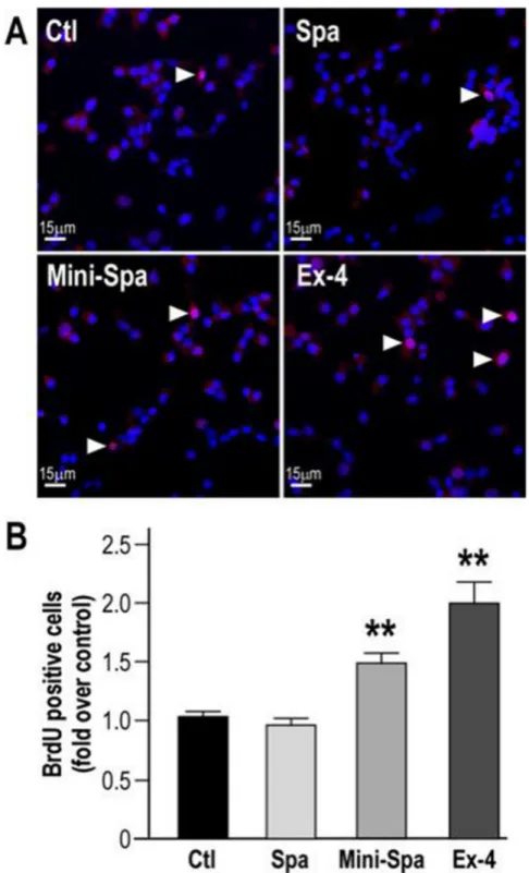
3.5. Mini-Spadin protects from inhibition of INS-1E cells proliferation by serum deprivation
Akt/CREB pathway also controls an essential feature of beta-cell proper physiology that is cell growth [6]. As individuals with type 2 diabetes could benefit from agents that preserve or expand beta cell mass [34], we investigated the potential of both PE synthetic derivatives to prevent inhibited proliferation of serum-starved beta-cells. Using BrdU incorporation experiments in serum starved cells incubated for 24 h with Spa and Mini-Spa, the results presented in Fig. 5A and B show that Spa failed to maintain INS-1E cell proliferation under serum deprivation (-FCS) (Spa: 0.26 ± 0.03 vs -FCS: 0.27 ± 0.03, in fold of +FCS condition). In contrast, Mini-Spa and as expected, Ex-4 [35], had
Fig. 3. PE, Spa and Mini-Spa maintain CREB activity in INS-1E challenged by serum-deprivation. (A) INS-1E cells were transiently transfected with the Gra-Luc plasmid, incubated 24 h in the presence or absence of serum and treated or not with PE (100 nM), Spa (100 nM), Mini-Spa (10 nM), or Ex-4 (100 nM). Results are mean ± SEM of (at least) 5 independent experiments (17 for +/- SVF and Spa, 11 for Mini-Spa, 13 for Ex-4 and 5 for PE) and values are expressed as % of CREB activity. (**p < 0.01, ***p < 0.001 compared to –FCS condition). (B) Immunofluorescent staining was used to evaluate the number of pCREB positive cells (phospho- CREB (Ser133, in red) and Hoescht 33342 (in blue)). The images shown are representative of 3 independent experiments. (For interpretation of the references to color in this figure legend, the reader is referred to the web version of this article.)
Pharmacological Research 167 (2021) 105539
6 similar protective effects on INS-1E cell proliferation in serum deprived condition (Mini-Spa: 0.46 ± 0.05 and Ex-4: 0.53 ± 0.05, in fold of +FCS condition).
4. Discussion
The PE derivative Spa is now emerging as a promising peptide for the treatment of depression [10]. The anti-depressive effect of Spa relies on the modification of the neuron morphology [36] and activation of the CREB pathway [10]. Beside of treating depression, Spa may also be insightful for moving forward about the understanding of mechanisms modulating insulin secretion. We previously showed that Spa potenti-ates glucose-induced insulin secretion in cell and animal models [8]. The level of potentiation of insulin secretion by Spa was seen as comparable with the one triggered by GLP-1 receptor agonists, including Exendin-4, which are currently used as antidiabetic drugs [37]. As a considerable effort is placed for making Spadin-analogs usable in clinics against depression, gaining evidence for their potential in another clinical application is of high interest. Another important point was to confirm that it was possible to work on the synthetic PE analog to ameliorate its efficiency. Indeed, the shorten formulation of Spadin that present an improved in vivo stability and bioavailability, and a reduced manufacturing cost as compared to the lead Spa, Mini-Spa rivals Exen-din at a concentration 10 times lower. However, future investigation is required for confirming this hypothesis, which would move forward about the clinical interest of these molecules for diabetes. Herein, we provide the evidence that Spa and its short derivative Mini-Spa protect beta cells against apoptotic death caused by IL-1β or staurosporine in a mechanism involving the PI3/Akt pathway. Akt and other MAPKs
including ERK, PKA and CaMK stimulate the phosphorylation and thereby, the activation of the transcription factor CREB [38]. Once activated, CREB is pivotal for regulating the expression of genes required for the secretory function [1]. Impairment of the CREB activity as observed in response to a diabetogenic environment such as chronic hyperglycemia, leads to a profound defect in beta-cell function [1,39]. Therefore using an in vitro model of chronic exposure of INS-1E cells or rat or human islets to high glucose that also result in a decrease CREB protein expression [3] would have probably been more representative of the physiological condition rather than using a serum deprivation model to reveal the beneficial effect of PE and derivatives on CREB. Nonethe-less, our findings indicate that the effect of the PE-derived peptides is a consequence of the increase in intracellular calcium influx able to generate a sufficiently robust intracellular signal for activating CREB via CaMK, but not via PKA. Because, CaMK-II and IV play a crucial role in the beta-cells and neurons function, it is possible that they account for the effects of Spa and Mini-Spa on CREB [40,41]. We also demonstrate that blocking PI3K/Akt or CaMK pathways does not prevent intracel-lular calcium increase, while blocking the protective effect of Spa and Mini-Spa. This suggests that the intracellular calcium influx could be the upstream key for inducing CaMK/CREB pathway and finally beta-cell survival induced by Spa and Mini-Spa. Such mechanism has also been raised in neurons [2,20], indicating a conserved function of the PE de-rivatives in neuroendocrine cells. As Spa causes beta-cell depolarization, which is a key event that promotes calcium influx in beta cells, it is tempting to speculate a role of the plasma membrane potential in the CREB activation. This idea may also be supported by the effect on the membrane potential of GLP-1 receptor agonist that potently activates CREB [17,38]. By linking small depolarization events induced by PE and Fig. 4. CaM-Kinases, but not cAMP-dependent PKA pathway mediates the effect of PE, Spa and Mini-Spa on CREB activity. (A) INS-1E cells were transiently transfected with the Gra-Luc plasmid, incubated 24 h under serum deprivation in presence of PE (100 nM), Spa (100 nM), Mini-Spa (10 nM) or Ex-4 (100 nM) and exposed to (A) H89 (1 µM) or (B) KN93 (1 µM). ***p < 0.001 compared to H89 or KN93 condition respectively. Values expressed in fold of basal CREB are mean ±SEM of all the experiments done (3–6 independent experiments) per condition. (C) Cytosolic calcium variations were analysed by measuring the Fura-2AM absorbance ratio (340/380) of INS-1E cells in response to the CaM-Kinases inhibitor KN93 and a subsequent exposure to Spa (100 nM) or Mini-Spa (10 nM). ***p < 0.001 compared to KN93 condition.
its derivatives leading to variations of internal calcium, and CREB pathways, the present work also highlights a parallel between protective pathways already described in neurons and those we now describe in beta-cells. This is in line with the increasing number of studies showing that several anti-hyperglycemic agents play a neuroprotective and anti-inflammatory role by reducing insulin resistance or by directly acting on brain [42]. Indeed, most anti-diabetic drugs, when consumed orally or injected, can enter the brain through the blood-brain barrier, which makes them perfect candidates for bidirectional therapy associ-ating diabetes and co-morbidity like vascular or neuronal diseases.
Beyond the importance of the therapeutic aspect of identifying new synthetic peptides with antidiabetic potential, the present work also
highlights that circulating level of an endogenous peptide could protect beta cells against apoptotic death caused by IL-1β that is known to play a key role in the decline of BCM in diabetes. Altogether with PE positive influence on the level of protective signaling pathway in beta pancreatic cells our results suggest the possible contribution of PE in resistance to diabetes. Therefore, it would be interesting to evaluate whether alter-ation of PE level may be a factor associated with a diabetes diagnosis and whether physiological ways known to prevent or delay the onset of T2D, like healthy diet, regular physical activity influence PE circulating level. Future investigation is required for confirming this hypothesis, which would move forward on the understanding of mechanisms regulating insulin secretion.
Fig. 5. Mini-Spa protects INS-1E cells proliferation from inhibition by serum deprivation. (A) Serum-deprived INS- 1E were treated 24 h with Spa (100 nM), Mini-Spa (10 nM) or Ex-4 (100 nM). BrdU (10 µM) was added the last 2 h of the treatment and nuclei were labeled using Hoescht 33342 (blue). The microscopy images are from a representative experiment out of 3 independent experiments. (B) The quantitation of the BrdU positive cells ratio (red, white arrows) on the total cells (blue) is shown. The values expressed as fold over +FCS condition are the mean ± SEM of 3 independent experiments (**p < 0.01, ***p < 0.001 different from –FCS condition).
Pharmacological Research 167 (2021) 105539
8
5. Conclusion
Endogenous PE generated from sortilin and its derivatives play key roles in triggering insulin secretion by modulating beta-cell membrane potential [8,36]. The present work highlights an original mechanism: small depolarization events link the effects of PE and its derivatives to PI3K/Akt and CREB on pancreatic beta-cells.
As CaMKII/CREB signaling is often associated with several cellular beneficial effects, the activation of the CaMK and CREB pathway by PE and its derivatives Spa and Mini-spa should contribute to their anti- apoptotic and proliferative effect in beta pancreatic cells. Altogether the present study provides novel mechanistic evidence that the PE de-rivatives may be new promising peptides for improving insulin secretion and for preserving beta-cell mass in T2D.
Funding
This work was supported by the Centre National de la Recherche Scientifique, the Agence Nationale de la Recherche (ANR-13-SAMA- 0002-VASPAC and ANR-13-RPIB-0002 MEDINCOD) and the French Government for the "Investments for the Future" LabEx ICST #ANR-11 LabEx 0015". We also thank the LabEx ICST for its fellowship to G.D.
Authors’ contributions
G.D., P.L. and T.C. performed the experiments. G.D., S.B., C.R., N.B., P.L. and T.C. analyzed the data. G.D., N.B., A.A., C.R., C.H., J.M., P.L. and T.C. wrote the paper.
Declaration of Competing Interest
The authors declare that they have no known competing financial interests or personal relationships that could have appeared to influence the work reported in this paper.
Acknowledgments
We are very grateful to Dr. M. Borsotto for his skillful technical support and advices with the electrophysiology of Spa and its derivatives.
Appendix A. Supporting information
Supplementary data associated with this article can be found in the online version at doi:10.1016/j.phrs.2021.105539.
References
[1] A. Abderrahmani, S. Cheviet, M. Ferdaoussi, T. Coppola, G. Waeber, R. Regazzi, ICER induced by hyperglycemia represses the expression of genes essential for insulin exocytosis, EMBO J. 25 (5) (2006) 977–986.
[2] S. Dalle, J. Quoyer, E. Varin, S. Costes, Roles and regulation of the transcription factor CREB in pancreatic beta -cells, Curr. Mol. Pharmacol. 4 (3) (2011) 187–195. [3] S. Costes, B. Vandewalle, C. Tourrel-Cuzin, C. Broca, N. Linck, G. Bertrand, J. Kerr- Conte, B. Portha, F. Pattou, J. Bockaert, S. Dalle, Degradation of cAMP-responsive element-binding protein by the ubiquitin-proteasome pathway contributes to glucotoxicity in beta-cells and human pancreatic islets, Diabetes 58 (5) (2009) 1105–1115.
[4] O.G. Chepurny, M.A. Hussain, G.G. Holz, Exendin-4 as a stimulator of rat insulin I gene promoter activity via bZIP/CRE interactions sensitive to serine/threonine protein kinase inhibitor Ro 31-8220, Endocrinology 143 (6) (2002) 2303–2313. [5] J.F. Gautier, S.P. Choukem, J. Girard, Physiology of incretins (GIP and GLP-1) and
abnormalities in type 2 diabetes, Diabetes Metab. 34 (2008) S65–S72. [6] U.S. Jhala, G. Canettieri, R.A. Screaton, R.N. Kulkarni, S. Krajewski, J. Reed,
J. Walker, X. Lin, M. White, M. Montminy, cAMP promotes pancreatic beta-cell survival via CREB-mediated induction of IRS2, Genes Dev. 17 (13) (2003) 1575–1580.
[7] G. Wheeler Damian, D. Groth Rachel, H. Ma, F. Barrett Curtis, F. Owen Scott, P. Safa, W. Tsien Richard, CaV1 and CaV2 channels engage distinct modes of Ca2+ signaling to control CREB-dependent gene expression, Cell 149 (5) (2012) 1112–1124.
[8] C. Hivelin, S. Beraud-Dufour, C. Devader, A. Abderrahmani, S. Moreno, H. Moha Ou Maati, A. Djillani, C. Heurteaux, M. Borsotto, J. Mazella, T. Coppola, Potentiation of calcium influx and insulin secretion in pancreatic beta cell by the specific TREK-1 blocker spadin, J. Diabetes Res. 2016 (2016) 1–9.
[9] F. Lesage, M. Lazdunski, Molecular and functional properties of two-pore-domain potassium channels, Am. J. Physiol. Ren. Physiol. 279 (5) (2000) F793–F801. [10] J. Mazella, O. Petrault, G. Lucas, E. Deval, S. Beraud-Dufour, C. Gandin, M. El-
Yacoubi, C. Widmann, A. Guyon, E. Chevet, S. Taouji, G. Conductier, A. Corinus, T. Coppola, G. Gobbi, J.L. Nahon, C. Heurteaux, Spadin, a sortilin-derived peptide, targeting rodent TREK-1 channels: a new concept in the antidepressant drug design, PLoS Biol. 8 (4) (2010), e1000355.
[11] C. Munck Petersen, M.S. Nielsen, C. Jacobsen, J. Tauris, L. Jacobsen, J. Gliemann, S.K. Moestrup, P. Madsen, Propeptide cleavage conditions sortilin/neurotensin receptor-3 for ligand binding, EMBO J. 18 (3) (1999) 595–604.
[12] S. B´eraud-Dufour, A. Abderrahmani, J. Noel, F. Brau, G. Waeber, J. Mazella, T. Coppola, Neurotensin is a regulator of insulin secretion in pancreatic beta-cells, Int. J. Biochem. Cell Biol. 42 (10) (2010) 1681–1688.
[13] N. Blondeau, S. Beraud-Dufour, P. Lebrun, C. Hivelin, T. Coppola, Sortilin in glucose homeostasis: from accessory protein to key player? Front. Pharmacol. 9 (2019) 1561.
[14] M.A. Nauck, J.J. Meier, The incretin effect in healthy individuals and those with type 2 diabetes: physiology, pathophysiology, and response to therapeutic interventions, Lancet Diabetes Endocrinol. 4 (6) (2016) 525–536.
[15] Y. Seino, K.W. Min, E. Niemoeller, A. Takami, Investigators EG-LAS, Randomized, double-blind, placebo-controlled trial of the once-daily GLP-1 receptor agonist lixisenatide in Asian patients with type 2 diabetes insufficiently controlled on basal insulin with or without a sulfonylurea (GetGoal-L-Asia), Diabetes Obes. Metab. 14 (10) (2012) 910–917.
[16] S. Susini, E. Roche, M. Prentki, W. Schlegel, Glucose and glucoincretin peptides synergize to induce c-fos, c-jun, junB, zif-268, and nur-77 gene expression in pancreatic beta(INS-1) cells, FASEB J. 12 (12) (1998) 1173–1182. [17] G.Gt Holz, C.A. Leech, J.F. Habener, Activation of a cAMP-regulated Ca(2+)-
signaling pathway in pancreatic beta-cells by the insulinotropic hormone glucagon- like peptide-1, J. Biol. Chem. 270 (30) (1995) 17749–17757.
[18] C. Devader, M. Roulot, S. Moreno, A. Minelli, M. Bortolomasi, C. Congiu, M. Gennarelli, M. Borsotto, C. Heurteaux, J. Mazella, Serum sortilin-derived propeptides concentrations are decreased in major depressive disorder patients, J. Affect. Disord. 208 (2017) 443–447.
[19] M. Roulot, A. Minelli, M. Bortolomasi, E. Maffioletti, M. Gennarelli, M. Borsotto, C. Heurteaux, J. Mazella, Increased serum levels of sortilin-derived propeptide after electroconvulsive therapy in treatment-resistant depressed patients, Neuropsychiatr. Dis. Treat. 14 (2018) 2307–2312.
[20] A. Djillani, M. Pietri, S. Moreno, C. Heurteaux, J. Mazella, M. Borsotto, Shortened spadin analogs display better TREK-1 inhibition, in vivo stability and
antidepressant activity, Front Pharmacol. 8 (2017) 643.
[21] A. Merglen, S. Theander, B. Rubi, G. Chaffard, C.B. Wollheim, P. Maechler, Glucose sensitivity and metabolism-secretion coupling studied during two-year continuous culture in INS-1E insulinoma cells, Endocrinology 145 (2) (2004) 667–678. [22] S. Beraud-Dufour, T. Coppola, F. Massa, J. Mazella, Neurotensin receptor-2 and -3
are crucial for the anti-apoptotic effect of neurotensin on pancreatic beta-TC3 cells, Int. J. Biochem Cell Biol. 41 (12) (2009) 2398–2402.
[23] M. Ferdaoussi, S. Abdelli, J.Y. Yang, M. Cornu, G. Niederhauser, D. Favre, C. Widmann, R. Regazzi, B. Thorens, G. Waeber, A. Abderrahmani, Exendin-4 protects -cells from interleukin-1 -induced apoptosis by interfering with the c-Jun NH2-terminal kinase pathway, Diabetes 57 (5) (2008) 1205–1215.
[24] S. Costes, C. Broca, G. Bertrand, A.D. Lajoix, D. Bataille, J. Bockaert, S. Dalle, ERK1/2 control phosphorylation and protein level of cAMP-responsive element- binding protein: a key role in glucose-mediated pancreatic -cell survival, Diabetes 55 (8) (2006) 2220–2230.
[25] P.K. Brindle, M.R. Montminy, The CREB family of transcription activators, Curr. Opin. Genet. Dev. 2 (2) (1992) 199–204.
[26] T. Coppola, S. Beraud-Dufour, A. Antoine, J.P. Vincent, J. Mazella, Neurotensin protects pancreatic beta cells from apoptosis, Int. J. Biochem. Cell Biol. 40 (10) (2008) 2296–2302.
[27] E. Aksamitiene, A. Kiyatkin, B.N. Kholodenko, Cross-talk between mitogenic Ras/ MAPK and survival PI3K/Akt pathways: a fine balance, Biochem. Soc. Trans. 40 (1) (2012) 139–146.
[28] T. Coppola, C. Frantz, V. Perret-Menoud, S. Gattesco, H. Hirling, R. Regazzi, Pancreatic beta-cell protein granuphilin binds Rab3 and Munc-18 and controls exocytosis, Mol. Biol. Cell 13 (6) (2002) 1906–1915.
[29] L.L. Baggio, D.J. Drucker, Biology of incretins: GLP-1 and GIP, Gastroenterology 132 (6) (2007) 2131–2157.
[30] J.L. Meinkoth, A.S. Alberts, W. Went, D. Fantozzi, S.S. Taylor, M. Hagiwara, M. Montminy, J.R. Feramisco, Signal transduction through the cAMP-dependent protein kinase, Mol. Cell Biochem. 127–128 (1993) 179–186.
[31] B. Mayr, M. Montminy, Transcriptional regulation by the phosphorylation- dependent factor CREB, Nat. Rev. Mol. Cell Biol. 2 (8) (2001) 599–609. [32] M. Sheng, M.A. Thompson, M.E. Greenberg, CREB: a Ca(2+)-regulated
transcription factor phosphorylated by calmodulin-dependent kinases, Science 252 (5011) (1991) 1427–1430.
[33] G.A. Wayman, Y.S. Lee, H. Tokumitsu, A.J. Silva, T.R. Soderling, Calmodulin- kinases: modulators of neuronal development and plasticity, Neuron 59 (6) (2008) 914–931.
[34] C. Aguayo-Mazzucato, S. Bonner-Weir, Pancreatic β cell regeneration as a possible therapy for diabetes, Cell Metab. 27 (1) (2018) 57–67.
[35] J. Buteau, GLP-1 receptor signaling: effects on pancreatic beta-cell proliferation and survival, Diabetes Metab. 34 (Suppl 2) (2008) S73–S77.
[36] C. Devader, A. Khayachi, J. Veyssiere, H. Moha Ou Maati, M. Roulot, S. Moreno, M. Borsotto, S. Martin, C. Heurteaux, J. Mazella, In vitro and in vivo regulation of synaptogenesis by the novel antidepressant spadin, Br. J. Pharmacol. 172 (10) (2015) 2604–2617.
[37] B. Thorens, Glucagon-like peptide-1 and control of insulin secretion, Diabete Metab. 21 (5) (1995) 311–318.
[38] Y.S. Lee, H.S. Jun, Anti-diabetic actions of glucagon-like peptide-1 on pancreatic beta-cells, Metabolism 63 (1) (2014) 9–19.
[39] I.S. Cho, M. Jung, K.S. Kwon, E. Moon, J.H. Cho, K.H. Yoon, J.W. Kim, Y.D. Lee, S. S. Kim, H. Suh-Kim, Deregulation of CREB signaling pathway induced by chronic
hyperglycemia downregulates NeuroD transcription, PLoS One 7 (4) (2012), e34860.
[40] J. Bok, Q. Wang, J. Huang, S.H. Green, CaMKII and CaMKIV mediate distinct prosurvival signaling pathways in response to depolarization in neurons, Mol. Cell Neurosci. 36 (1) (2007) 13–26.
[41] P.K. Dadi, N.C. Vierra, A. Ustione, D.W. Piston, R.J. Colbran, D.A. Jacobson, Inhibition of pancreatic beta-cell Ca2+/calmodulin-dependent protein kinase II reduces glucose-stimulated calcium influx and insulin secretion, impairing glucose tolerance, J. Biol. Chem. 289 (18) (2014) 12435–12445.
[42] M. Nath, K. Bhattacharjee, Y. Choudhury, Pleiotropic effects of anti-diabetic drugs: a comprehensive review, Eur. J. Pharmacol. 884 (2020), 173349.