Brain states and circuit mechanisms underlying sleep and general anesthesia
by Laura D. Lewis
B.Sc., McGill University (2008)
Submitted to the Department of Brain and Cognitive Sciences in Partial Fulfillment of the Requirements for the Degree of
Doctor of Philosophy at the
MASSACHUSETTS INSTITUTE OF TECHNOLOGY February 2014
MASSACHUSETTS OF TECHNOLOGY
B A2014
LIBRARES
© 2014 Massachusetts Institute of Technology. All rights reserved.
Signature of Author ...
Department of Brain and Cognitive Sciences Dec. 13, 2013
C ertified by ... ... ... ... . . Emery N. Brown, M.D., Ph.D. Professor of Computational Neuroscience, Edward Hood Taplin Professor of Medical Engineering, and Warren M. Zapol Professor of Anaesthesia
Brain states and circuit mechanisms underlying sleep and general anesthesia by
Laura D. Lewis
Submitted to the Department of Brain and Cognitive Sciences on December 13, 2013 in Partial Fulfillment of the Requirements for the Degree of
Doctor of Philosophy in Neuroscience
Abstract
During sleep and general anesthesia, the brain enters a state of decreased arousal and consciousness is transiently suspended. How this transition occurs is a fundamental and unsolved question in neuroscience. The neural dynamics that disrupt consciousness have not been identified, and the circuit mechanisms that generate these dynamics remain unknown. Furthermore, understanding the neural basis of sleep and anesthesia is key to improving clinical monitoring of patients undergoing general anesthesia and to advancing treatments of sleep disorders and neurological conditions such as coma. In this thesis, I combine intracranial electrophysiology in human subjects with optogenetic manipulation of thalamocortical circuits in mice to identify the neural dynamics underlying sleep and anesthesia. I first show that loss of consciousness during propofol general anesthesia is associated with the abrupt onset of slow oscillations that disrupt cortical networks. I then demonstrate that activation of the thalamic reticular nucleus generates slow wave activity and decreases arousal state, identifying a causal mechanism that generates physiological and behavioral signs of sleep. Finally, I study patients undergoing deep general anesthesia at levels corresponding to medically induced coma, and show that this state is marked by local cortical dynamics consistent with impaired cerebral metabolism. Taken together, these results identify a set of neural dynamics associated with unconscious states, and demonstrate specific mechanisms for how they disrupt brain function. These findings provide new insight into the neuroscience of arousal states, and suggest clinical approaches that could improve patient care.
Thesis supervisor: Emery Brown, M.D., Ph.D.
Title: Professor of Computational Neuroscience, MIT
Acknowledgments
I am incredibly grateful to the many people who have given me so much help and support. First, Emery Brown, my thesis advisor, has been an incredible mentor over the past five years. His scientific insight, creativity, and generosity made my graduate school experience exciting 'and fulfilling. Second, Patrick Purdon, who advised me on all the clinical studies, has taught me so much, and I feel lucky to have worked with such a brilliant scientist and engineer. I'm also very grateful to my thesis committee members Chris Moore, for his conceptual insights and extremely useful advice, and Matt Wilson for many discussions that helped shape our experiments and analyses.
I would also like to thank the patients who participated in our studies and their families. These studies could not have taken place without their generosity.
A huge thanks goes to our many collaborators at MGH who helped execute this research. Most importantly, Syd Cash has devoted many hours of his time to these projects and has provided insightful perspective into each study we carried out - these studies would never have been possible without his help. Jake Donoghue, Mia Borzello, and Jason Naftulin were incredibly helpful with running experiments, and I can't imagine trying to do this without them. I am also very grateful to all of our clinical collaborators: Emad Eskandar, Bob Peterfreund, Kristy Nordstrom, Kara Houghton, Seun Akeju, and the many clinical staff who have helped throughout the past few years.
A highlight of grad school has been working in the Brown and Purdon labs, filled with so many amazing people. Aaron Sampson was integral to getting the clinical studies running, and his energy and positivity made our early morning recordings way more fun. Francisco Flores has been a great collaborator, helping me to transition into the world of optogenetics and finding solutions to every problem. Rob Haslinger was a great friend and teacher, who spent many hours helping me learn statistical techniques. Eran Mukamel and ShiNung Ching provided invaluable guidance on analyzing our intracranial data, and Veronica Weiner contributed extremely helpful datasets. The many other lab members provided a great environment to work and learn, including Pavitra Krishnaswamy, Demba Ba, Christa Van Dort, Behtash Babadi, Mike Prerau, Katie Hartnack, Kara Pavone, Gabriel Obregon, and Sage Chen. Finally, thanks to Sheri Leone for all of her help with the many issues that popped up!
Our animal studies would not have been possible without the assistance of Mike Halassa, whose work and insight brought a new dimension to our experiments. Ralf Wimmer, Phill Brunetti, Dan Zachs, and Steve Ramirez were all incredibly generous with their time in helping me learn experimental techniques.
A huge community of people have been incredibly supportive throughout this process. Lorna Gibson has been a wonderful friend and mentor throughout the past five years. Denise Heintze was an incredible source of support for students. I'm incredibly grateful to my dad, my mom and Andrew, and Xavier and Dix, for being an awesome and supportive family. And to all my friends, especially Masha Westerlund, Rodrigo Garcia, Meg Krench, Kean Jaime-Bustamante, John McCoy, Sangyu Xu, Chris Saenz, Elias Issa, Danielle Feldman, Retsina Meyer, and Ali Horowitz, for being so great and making grad school a happy place.
Finally, thanks to Jakob Voigts, for being an incredible scientist and person. He has been so wonderful over the past five years and made every day better.
Table of Contents Chapter 1: Introduction
1.1 The neuroscience of unconscious states ... 6
1.2 General anesthesia in the operating room ... 7
1.3 M olecular m echanism s of general anesthesia... 9
1.4 Brainstem and subcortical arousal circuits ... 10
1.5 Electroencephalogram correlates of sedation and unconsciousness... 13
1.6 Theories of unconsciousness... 15
1.7 Overview of the thesis ... 18
1.8 References... 20
Chapter 2: The neural correlates of propofol-induced loss of consciousness 2.1 A bstract ... 27
2.2 Introduction... 28
2.3 Results... 30
2.4 D iscussion... 53
2.5 Experim ental Procedures ... 57
2.6 A uthor Contributions ... 63
2.7 References... 63
Chapter 3: The thalamocortical circuit mechanisms controlling arousal state 3.1 A bstract ... 68
3.2 Introduction... 69
3.3 Results... 71
3.4 D iscussion... 84
3.5 Experim ental Procedures ... 89
3.6 A uthor Contributions ... 95
3.7 References... 95
Chapter 4: The neurophysiology of burst suppression in propofol-induced coma 4.1 A bstract ... 100
4.2 Introduction... 102
4.3 Results... 104
4.4 D iscussion... 114
4.5 Experim ental Procedures ... 119
4.6 A uthor Contributions ... 125
4.7 References... 125
Chapter 5: Conclusions 5.1 Sum m ary of the thesis... 128
Chapter 1: Introduction
1.1 The neuroscience of unconscious states
Sleep and general anesthesia produce a profound but reversible change in nearly every aspect of brain function, extinguishing both our responses to the outside world and our internal state of consciousness. Remarkably, transitions between the awake and unconscious state can occur in just a few seconds, suggesting that the brain can switch between multiple states without major changes to its anatomical structure. How this phenomenon takes place is an unresolved question in neuroscience: what changes in the brain when a person loses consciousness, and what mechanism produces this transition into unconsciousness?
The most familiar state of unconsciousness is sleep, which is naturally occurring and has restorative effects for cognitive and physiological function. However, there are also many other ways that consciousness can be disrupted, such as during general anesthesia, absence seizures, and coma. Each of these states of decreased arousal is marked not only by diminished awareness, but also by a collection of physiological and neurological effects that underlie the change in behavior. While there are some neurophysiological phenomena that can be observed across sleep, anesthesia, and coma, overall these states are heterogeneous; even a slight change in the dosage of a single drug can produce a qualitatively different brain state. A major challenge for the neuroscience of unconscious states has been to identify which of these many neurophysiological changes is causally related to an animal's level of arousal or awareness. Most studies have compared the awake state with a deeply anesthetized state, blurring the many different gradations that can occur between sedation, loss of consciousness, and profound inactivation. This approach cannot identify the distinct dynamics that produce unconsciousness, versus a deeper, coma-like state.
Extensive work has characterized the brainstem and neuromodulatory pathways that can modulate arousal state and cause unconsciousness, reviewed briefly in Section 1.4. Furthermore, anesthetic-induced loss of consciousness is achieved through known molecular mechanisms, reviewed in Section 1.3. However, how these low-level effects
lead to states of decreased awareness remains unclear. Sleep, general anesthesia, and coma all disrupt the neural dynamics that produce consciousness, but the precise mechanism by which they do so is unknown. To understand the neural basis of states of decreased arousal, we must: 1) characterize the systems-level neural dynamics that underlie sleep, general anesthesia, and coma; 2) determine how these dynamics affect cognitive function; and 3) identify the circuit mechanisms that create these altered states. These questions require an integration of both human and animal studies, as the link between neurophysiology and awareness is best studied in humans, but the causal mechanisms can only be directly tested in animal models.
1.2 General anesthesia in the operating room 1.2.1 Physiological effects of general anesthesia
Although arousal state is naturally modulated in the sleep-wake cycle, a more rapid and potent control of arousal was provided by the advent of general anesthesia. The first public demonstration of general anesthesia, using ether, revolutionized the practice of surgery by enabling physicians to induce a profound state of unconsciousness. A broad array of drugs have now been developed that are used in various combinations to achieve the five essential components of general anesthesia: unconsciousness, amnesia, analgesia, paralysis, and stability of physiological systems (autonomic, cardiovascular, thermoregulatory, and respiratory (Brown et al., 2010)).
Perhaps the most important aspect of general anesthesia is that it is reversible. At the end of a surgery, drug administration is stopped and the patient will gradually return to the awake state. Because anesthesia is lifted passively, through cessation of drug, rather than actively (e.g. by administering an antidote), the half-life of a drug is an important consideration for clinical use. Propofol has become a commonly used anesthetic drug in part due to its rapid clearance times, enabling patients to wake up
Propofol is a generally safe and effective drug for producing unconsciousness, but is also accompanied by a range of other side effects that may be undesirable or potentially dangerous. Its physiological effects include decreased blood pressure and respiratory depression (Gold et al., 1987), and patients undergoing propofol anesthesia are generally intubated to preserve airway function. Propofol also causes nausea and vomiting, although to a lesser degree than many other anesthetic drugs (Hofer et al., 2003).
Anesthetic drugs are a highly diverse group, and other drugs cause a range of different physiological and cognitive effects. Inhaled agents, such as sevoflurane and desflurane, are commonly used for induction and maintenance of general anesthesia, and typically lower blood pressure and heart rate and carry a risk of tachycardia (Yildirim et al., 2004). Ketamine raises blood pressure, preserves respiratory function, and induces dissociative feelings and hallucinations during induction and emergence (Reich and Silvay, 1989). Dexmedetomidine is primarily used for sedation, and causes profound drowsiness. It can only be delivered as a gradual infusion, as a bolus dose can cause hypotension and bradychardia (Coursin et al., 2001).
The diversity of physiological effects across anesthetic agents highlights the fact that many of these drugs act through different molecular mechanisms and thus alter brain function through a variety of pathways. Furthermore, these differences illustrate that physiological states cannot be reliably linked to brain function, as the association between physiological variables and unconsciousness is not consistent across drugs or even across patients receiving the same drug.
1.2.2 Clinical monitoring during general anesthesia
Although anesthetic drugs take effect primarily in the central nervous system, most anesthesiologists do not directly monitor brain function during general anesthesia. Instead, physiological variables such as blood pressure, oxygen saturation, and heart rate, and the presence of reflexes are used to infer the depth of anesthesia. These measures cannot provide precise information about whether a person is awake or unconscious, and intraoperative awareness remains a serious risk of general anesthesia, occurring in approximately 26,000 patients in the United States each year (Sebel et al., 2004).
However, simply increasing the dosage of anesthetic drugs is not a feasible solution, as general anesthesia is accompanied by many other risks. While it is typically a safe procedure, it is so frequently used (in millions of patients per year) that it nevertheless causes undesirable side effects in tens of thousands of patients. Common risks include nausea and vomiting, which can also cause respiratory distress and lead to more serious side effects. Death from general anesthesia is extremely rare, ranging around 8 deaths per million patients, but it is nevertheless a contributing cause in approximately 300 deaths annually in the United States (Li et al., 2009).
In an attempt to reduce both the risk of intraoperative awareness and excessive drug dosages, some anesthesiologists have begun using the bispectral index (BIS), a system that processes the electroencephalogram (EEG) and outputs a single number intended to index depth of anesthesia. In theory, this monitoring system could provide a method to titrate patients to a desired level of unconsciousness and signal when dosages are too high or too low. However, multiple recent studies have shown that the BIS cannot reliably detect unconsciousness, does not decrease anesthetic dosage, and is not effective at reducing intraoperative awareness (Avidan et al., 2011; 2008; Kaskinoro et al., 2011). An improved system for patient monitoring is therefore needed. To precisely assess consciousness, we must identify and characterize the brain activity patterns that cause unconsciousness, and develop measures that can detect the presence of these patterns in the operating room.
1.3 Molecular mechanisms of general anesthesia
Anesthetic drugs act at the molecular level by binding cell receptors and thereby altering neuronal activity. The specific molecular mechanisms are highly variable across anesthetics, with different classes of drugs affecting different receptors to different degrees. However, a major target is receptors for gamma-aminobutyric acid (GABA), the
GABAergic transmission and instead act through other molecular pathways. In addition, most drugs and the inhaled agents in particular are nonselective, binding to many different types of receptors at clinically relevant concentrations (Rudolph and Antkowiak, 2004).
By comparison, propofol is a relatively selective drug at the molecular level, acting primarily as a GABA-A agonist. Propofol binds GABA-A receptors and lowers the rate of decay of GABA-mediated chloride conductances, leading to enhanced inhibition (Concas et al., 1991; Orser et al., 1994). At low doses, propofol only potentiates GABA-A currents, but at high doses may also directly activate those currents (Orser et al., 1994). This enhancement of GABA-A function is one of the key mechanisms by which propofol induces unconsciousness, as mice with a point mutation in the GABA-A receptor are highly resistant to propofol (Jurd et al., 2003). However, propofol also affects several other receptors to a lesser degree, including glycine receptors (Pistis et al., 1997), AMPA receptors (Haines et al., 2008), and hyperpolarization-activated cyclic-nucleotide-gated channels (Chen et al., 2005; Ying et al., 2006). GABA-A receptors are located throughout the central nervous system, providing multiple potential circuit mechanisms through which propofol could exert its hypnotic effects.
1.4 Brainstem and subcortical arousal circuits 1.4.1 Ascending arousal systems
The brainstem contains multiple structures that promote wakefulness through distinct neuromodulatory pathways. Cholinergic systems play a major role in generating wake states, and acetylcholine levels throughout cortex correlate with the sleep-wake cycle, with high levels of acetylcholine during the awake state and during rapid eye movement (REM) sleep (Saper et al., 2005). Cholinergic inputs arise from brainstem structures and the basal forebrain, innervating thalamus and cortex (Jones, 2004). Cholinergic inputs also disinhibit thalamic neurons by inhibiting the thalamic reticular nucleus (McCormick and Prince, 1986), a source of GABAergic inhibitory projections to central thalamus.
The noradrenaline system is a second major activating pathway, with the locus coeruleus (LC) sending noradrenergic inputs to nearly the entire central nervous system (Berridge, 2008). LC has long been known to play a key role in maintaining arousal, as its firing rates correlate strongly with arousal state, and drugs that impair noradrenergic transmission (such as the x2-adrenergic agonist dexmedetomidine) induce profound sedation. More recently, optogenetic activation of LC was shown to induce sleep-to-wake transitions, confirming a selective role for this structure in maintaining the awake state (Carter et al., 2010).
In addition to these two neuromodulatory systems, dopaminergic and orexinergic pathways also support arousal. Recent work has highlighted their effectiveness at inducing awake states, as optogenetic activation of orexin pathways induces sleep-to-wake transitions (Adamantidis et al., 2007), and stimulation of dopaminergic pathways can arouse even anesthetized animals (Taylor et al., 2013). Taken together, these pathways demonstrate that the brain has multiple redundant arousal systems (Jones, 2003), possibly reflecting evolutionary pressures given the importance of the waking state for survival.
Due to the many different arousal circuits that generate wake states, sleep must involve the suppression of several pathways. The hypothalamus in particular plays an important role in regulating activity throughout the ascending arousal system. The ventrolateral preoptic area of the hypothalamus sends GABAergic inhibitory projections to most of these arousal-promoting regions, including the cholinergic, dopaminergic, and noradrenergic pathways. Increasing GABAergic synaptic transmission could therefore impair multiple branches of the ascending arousal system, providing a possible mechanism for suppression of arousal during sleep and general anesthesia (Brown et al., 2011; Lee and Dan, 2012).
therefore referred to as 'relay' cells, passing sensory information up to higher-level areas for further processing (Sherman and Guillery, 1996). Consequently, modulation of thalamic activity has substantial impact on cortical processing, as it controls which information is successfully transmitted to cortex (McCormick and Bal, 1994). However, this transmission of information is not unidirectional. Cortex sends broad and strong projections back to thalamus as both direct excitatory inputs and indirect inhibitory inputs. This network enables complex interactions between thalamus and cortex, allowing the two structures to act as a feedback loop (Nicolelis and Shuler, 2001).
The thalamocortical loop plays an important role in generating the EEG patterns of non-rapid eye movement (NREM) sleep: it is involved both in sleep spindles, a 12-15 Hz rhythm with a waxing-and-waning power envelope, and in slow wave activity (0.5-4 Hz). These rhythms have been suggested to result from the oscillatory properties of individual thalamic cells. Thalamocortical neurons inherently oscillate at delta (1-4 Hz) rhythms when inhibited due to the interaction of a hyperpolarization-activated cation current and a low-threshold calcium current (McCormick and Pape, 1990; McCormick and Huguenard, 1992). Thalamic reticular neurons, which provide inhibitory input to thalamus, can inherently oscillate at spindle frequencies and also interact with thalamic neurons at that frequency (Steriade et al., 1987). Extensive work has characterized the involvement of these circuits in generating the EEG signatures of NREM sleep (McCormick and Bal, 1997).
In addition to its role in sleep oscillatory dynamics, thalamic activity is strongly correlated with loss of consciousness. Thalamic activity declines prior to cortical activity during sleep onset (Magnin et al., 2010), and is suppressed during general anesthesia and in disorders of consciousness (Alkire et al., 2000; Schiff, 2008), leading researchers to suggest that decreased thalamic activity could play a causal role in cortical and behavioural decreases in arousal.
However, direct experimental manipulation of thalamic neurons has not established a conclusive relationship between thalamic activity and arousal. Optogenetic activation of thalamic neurons induces an aroused, desynchronized state in cortex (Poulet et al., 2012), suggesting a potential role for thalamus in regulating arousal state. However, lesioning thalamus does not disrupt the awake cortical state (Constantinople
and Bruno, 2011), nor does optogenetically silencing it (Zagha et al., 2013). The role of thalamus and thalamic inhibition in modulating arousal state thus remains unclear (Mashour and Alkire, 2013).
An important regulator of the thalamocortical circuit is the thalamic reticular nucleus (TRN), a shell of GABAergic neurons that envelop the thalamus (Houser et al., 1980). The TRN is the primary source of inhibitory input to thalamus, and thus has potential to modulate excitatory input from thalamus to cortex. However, electrical stimulation cannot be used to selectively manipulate TRN due to its proximity to thalamic nuclei, and its role in generating sleep states therefore remained unexplored until recently. TRN is now known to play a causal role in generating sleep spindles (Halassa et al., 2011), but its effects on behavior and arousal states remain unknown.
In addition to its potential role in sleep, thalamic inhibition may be one mechanism underlying propofol general anesthesia, as well as other GABAergic anesthetic drugs. Propofol enhances the effects of inhibitory input to cortical neurons (Kitamura et al., 2003), and of the GABAergic projections from TRN to thalamus (Ying and Goldstein, 2005), providing two direct sources of inhibition. In addition, propofol's effects on brainstem structures would be expected to further inhibit thalamus, by inhibiting the ascending arousal systems and thus reducing excitatory input to thalamus. The role of thalamus in both sleep and anesthesia-induced unconsciousness therefore merits further investigation.
1.5 Electroencephalogram correlates of sedation and unconsciousness
1.5.1 EEG effects of propofol sedation and anesthesia
Propofol causes striking changes in the EEG, inducing specific patterns that can vary depending on the dose and the susceptibility of individual patients. Prior to loss of consciousness, patients may experience paradoxical excitation, in which they exhibit
oscillation that is primarily observed across frontal regions (Supp et al., 2011), and an increase in low gamma (25-40 Hz) power (Murphy et al., 2011).
At deep levels of general anesthesia, many different drugs all converge on a common EEG signature: burst suppression (Akrawi et al., 1996). Burst suppression is marked by an alternation between isoelectric suppressions lasting seconds, and high-amplitude bursts. In a routine surgical procedure, burst suppression usually signals unnecessarily high levels of anesthetic, and this EEG pattern can therefore signify that dosage should be decreased. However, burst suppression is induced clinically to treat certain neurological conditions, such as status epilepticus (Claassen et al., 2002) and traumatic brain injury (Kelly et al., 1999). While the mechanism underlying burst suppression is not known, it appears to have neuroprotective effects in these cases, and propofol infusions are typically maintained for 24 hours or more to ensure a prolonged state of burst suppression.
1.5.2 EEG dynamics during drowsiness and sleep
Similarly to the diverse brain states described above, sleep induces a range of different EEG dynamics. People transition through multiple sleep stages over the course of the night, visible as distinct patterns in the EEG. These patterns have been categorized as discrete stages, although in reality they may reflect a continuous gradation. Multiple sleep staging systems have been developed (Roth, 1961), but all begin with loss of the alpha rhythm seen during awake, eyes-closed recordings, marking the sleep onset period (Ogilvie, 2001). Next, sleep spindles appear, a 12-15 Hz oscillation with a waxing-and-waning power envelope, lasting under a second. Low-frequency power increases initially with the appearance of K-complexes, large amplitude deflections corresponding to an isolated down-state in which neuronal activity is silenced (Cash et al., 2009). Later, slow-wave activity becomes continuous and marks a periodic alteration between up and down states, reflecting rhythmic suppression and recovery of neuronal activity. This marks the deepest stage of non-rapid-eye-movement (NREM) sleep, and these stages of NREM alternate with rapid-eye-movement (REM) sleep repeatedly over the sleep cycle. REM sleep is marked by an active, desynchronized, low-amplitude EEG, and is correlated with
dreaming (Hobson, 2009); it is therefore not included in this thesis as a state of unconsciousness.
1.5.3 EEG features of coma
As with general anesthesia, coma is a broad term encompassing many different brain states. Many types of neurological conditions can result in coma, including anoxia, hypoxia-ischemia, encephalitis, and traumatic brain injury. The resulting EEG can take on many forms as well, and EEG patterns are now used as a prognostic method to assess whether patients will recover from coma (Synek, 1988). Delta activity is commonly observed, but does not translate readily into a specific prognosis. In contrast, coma with theta and alpha oscillations usually signals a relative good prognosis. An exception is 'alpha coma', marked by an anteriorized alpha rhythm superimposed on a delta rhythm (somewhat similar to propofol), which is nearly always fatal (Tomassen and Kamphuisen, 1986). Periodic patterns with epileptiform activity also carry a poor prognosis (Young, 2000). Burst suppression, which is caused by deep general anesthesia, also occurs in certain types of coma. Although burst suppression is induced clinically as a treatment for certain brain injuries, when it occurs in the absence of drugs it signals an extremely poor prognosis (Brenner, 2005; Young, 2000).
1.6 Theories of unconsciousness
1.6.1 Decreased neuronal activity
Several theories have been proposed for how general anesthetic drugs produce unconsciousness. An initial hypothesis was simply that increasing anesthetic levels causes an increase in inhibition, and therefore a decrease in neuronal firing rates. A depression of cortical neuronal activity would then be expected to cause unconsciousness, by impairing all cortical function. This theory was based on consistent observations of
behavioural effect. In the absence of such studies, it is possible that decreased firing rates correlate with sedation rather than unconsciousness, or alternatively that firing rates decrease after loss of consciousness and reflect a deepening of anesthetic levels rather than a correlate of behavioural state. This theory also lacks explanatory power, as it is not clear how a simple decrease in firing rates would lead to the progressive breakdown of higher cognitive function and memory observed during gradual inductions and sedation.
1.6.2 A thalamic switch
A second hypothesis is that general anesthetics activate a thalamocortical switch that controls consciousness. Multiple different drugs have been shown to reduce thalamic activity, leading some to suggest that this could be a potential unifying mechanism for unconsciousness (Alkire et al., 2000). However, this hypothesis has yet to be verified experimentally: there are no neurophysiology studies characterizing how thalamic activity changes immediately at the transition into unconsciousness, nor any manipulations demonstrating a causal role for this mechanism. A debate remains over whether the thalamus is a switch that can control consciousness, or whether its decrease in activity simply reflects a decrease of excitatory feedback from cortex, as cortical activity declines during unconsciousness (Alkire et al., 2008).
The thalamic mechanism has also been suggested to play a major role in coma, as neuronal damage to the central thalamus is correlated with disorders of consciousness (Schiff, 2008). Somewhat counterintuitively, the GABA-A agonist zolpidem has produced arousal and transient recovery in coma patients (Brefel-Courbon et al., 2007; Cohen and Duong, 2008). However, this drug is also proposed to act through increasing thalamic activity, as the GABA-A agonism could inhibit globus pallidus, and thereby disinhibit thalamus (Schiff, 2010).
1.6.3 A core consciousness-generating set of cortical areas
A recent theory, which is also compatible with the two previous hypotheses, is that unconsciousness is due to inactivation of specific cortical regions that are required for consciousness. This idea was proposed after neuroimaging studies found that cingulate, posterior parietal, and precuneal areas are consistently inactivated during
general anesthesia (Fiset et al., 1999; Kaisti et al., 2002), leading to the hypothesis that these core regions are required for consciousness (Alkire et al., 2008). A related theory suggests that frontoparietal interactions in particular are a key element of consciousness, and that anesthetics act by disrupting these interactions (Ku et al., 2011; Lee et al., 2009). However, there is no causal evidence demonstrating that these areas are functionally important for awareness, as opposed to simply being particularly sensitive to anesthetic drugs. It is also theoretically challenging to explain how a small subregion of cortex could generate consciousness.
1.6.3 Breakdown of binding through gamma oscillations
A more general theory for how cortical disruption could produce unconsciousness focuses on gamma (~40 Hz) oscillations. Gamma has been proposed to implement 'binding', enabling information to enter consciousness by synchronizing neuronal assemblies in distributed cortical regions (Engel and Singer, 2001). Conversely, general anesthesia has been suggested to operate through disruption of gamma synchrony, resulting in 'cognitive unbinding' (Mashour, 2004). Some studies have indeed found that gamma coherence is altered during general anesthesia (Imas et al., 2005; John et al., 2001). If this hypothesis is correct, directly manipulating gamma oscillations should alter arousal state, providing an empirically testable prediction that could support this theory. However, a systems-level explanation of anesthetic-induced unconsciousness should also include a circuit mechanism that disrupts gamma synchrony and causes this loss of binding.
1.6.4 Loss of information integration
In an effort to develop a unified theory for consciousness, Tononi proposed the idea of consciousness as information integration (Tononi, 2004; 2005). In this framework, consciousness is related to the number of possible states in an integrated
been suggested. These include disruption of integration due to loss of gamma synchrony, and disruption of information due to the stereotyped, bistable states in burst suppression (Alkire et al., 2008). However, it is unclear whether the theory of information integration inherently provides any falsifiable predictions that could be experimentally tested.
1.6.5 Empirical approaches to validating theories
These hypotheses range from empirically testable to purely philosophical. For example, the suggestions that a specific set of cortical regions are needed in order to be conscious could be tested through lesion studies and optogenetic inactivation of those areas. In contrast, the concept of information integration may be a useful philosophical construct, but does not suggest a specific mechanism that could be experimentally validated or falsified.
Two sets of experiments are needed to conclusively examine the question of how anesthetic drugs produce unconsciousness. First, neurophysiological studies should be performed during the transition into unconsciousness, to determine which effects occur simultaneously with loss of consciousness rather than during deep general anesthesia. Most studies have only compared the awake state with a fully anesthetized state, or a lightly anesthetized state with a deeply anesthetized state, and these comparisons cannot assess the neural dynamics specifically associated with loss of consciousness. Second, causal manipulations are needed in order to directly test whether these proposed mechanisms can indeed induce changes in arousal. In addition, it is possible that different drugs may act through different mechanisms, as the variety in molecular effects suggests that there could also be a range of circuit- and systems-level mechanisms that produce unconsciousness. Several theories could therefore be correct, but might only explain a subset of the drugs or pathologies that affect arousal state.
1.7 Overview of the thesis
Significant work has identified the molecular-level mechanisms of general anesthetics and their resulting effects on subcortical structures. However, unconsciousness occurs well before brainstem responses are extinguished, and it is likely
that anesthetic effects on cortical dynamics are responsible for disrupting higher-level cognitive function. Furthermore, the diversity of ways one can become unconscious (e.g. sleep, many anesthetic drugs, many types of coma) and the range of EEG phenomena observed across these states suggests that there are many different mechanisms that can produce a state of decreased arousal. Although several theories exist as to the neural basis of unconsciousness (Section 1.6), empirical evidence has not yet confirmed any of these hypotheses. Furthermore, most studies have not differentiated between the brain states associated with loss of consciousness, and those that occur during deep, coma-like states of general anesthesia. A more fine-grained approach is needed to identify how brain states and function are altered across different states of decreased arousal.
Two major questions are unresolved: first, what are the cortical states induced by sleep and general anesthesia? And second, what are the circuit mechanisms that produce these states? This thesis seeks to answer these questions, which should provide substantial scientific insight into the cortical basis of awareness and unconsciousness. Furthermore, it will also form an important step towards developing clinical systems to monitor patients during general anesthesia, improving patient outcomes and reducing the rate of intraoperative awareness.
The first part of the thesis focuses on the brain states associated with loss of consciousness. In Chapter 2, I find that loss of consciousness occurs simultaneously with the onset of a slow (0.1-1 Hz) oscillation in cortex. This oscillation is asynchronous across the brain, and is associated with hundreds of milliseconds of silence in cortical neurons. This spatial pattern means that neurons will be silenced at different times in different brain regions, effectively fragmenting cortical networks and disrupting information transfer across the brain. These results demonstrate that the cortical state associated with propofol-induced unconsciousness is local slow waves.
Given this robust correlation between slow waves and unconsciousness, I next searched for the circuit mechanism that generates slow waves, and tested whether
oscillations. Furthermore, slow wave activity was induced in local cortical areas, providing an explanation for how this brain state could be controlled separately across different cortical regions. Taken together, Chapters 2 and 3 identify a brain state associated with unconsciousness and describe the circuit mechanism that generates this state.
In Chapter 4, I focus on the much deeper state of anesthesia that occurs during medically-induced coma. I study the EEG pattern of burst suppression, and find that it can occur in isolated parts of cortex, in contrast to previous assumptions in the literature. Furthermore, I show that the bursts recover the spectral dynamics of the underlying brain state, suggesting that normal brain activity resumes intermittently but is then interrupted by a prolonged suppression. These characteristics are consistent with the hypothesis that burst suppression is caused by local decreases in cerebral metabolism. This chapter therefore provides a detailed characterization of the brain state that occurs during medically-induced coma, and suggests decreased cerebral metabolism as an underlying neurophysiological mechanism.
Taken together, Chapters 2-4 identify the brain states associated with unconsciousness and coma and the circuit mechanisms that generate these states. In Chapter 5, I discuss the implications of these results for the neuroscience of sleep and general anesthesia, and highlight potential clinical applications to improve patient monitoring and reduce anesthetic risks.
1.8 References
Adamantidis, A.R., Zhang, F., Aravanis, A.M., Deisseroth, K., and de Lecea, L. (2007). Neural substrates of awakening probed with optogenetic control of hypocretin neurons. Nature 450, 420-424.
Akrawi, W.P., Drummond, J.C., Kalkman, C.J., and Patel, P.M. (1996). A comparison of the electrophysiologic characteristics of EEG burst-suppression as produced by isoflurane, thiopental, etomidate, and propofol. Journal of Neurosurgical Anesthesiology 8, 40-46.
Alkire, M., Haier, R., and Fallon, J. (2000). Toward a unified theory of narcosis: brain imaging evidence for a thalamocortical switch as the neurophysiologic basis of
anesthetic-induced unconsciousness. Consciousness and Cognition 9, 370-386. Alkire, M., Hudetz, A., and Tononi, G. (2008). Consciousness and anesthesia. Science
322, 876.
Andrada, J., Livingston, P., Lee, B.J., and Antognini, J. (2012). Propofol and etomidate depress cortical, thalamic, and reticular formation neurons during anesthetic-induced unconsciousness. Anesthesia & Analgesia 114, 661-669.
Antkowiak, B. (1999). Different actions of general anesthetics on the firing patterns of neocortical neurons mediated by the GABA(A) receptor. Anesthesiology 91,
500-511.
Avidan, M.S., Jacobsohn, E., Glick, D., Burnside, B.A., Zhang, L., Villafranca, A., Karl, L., Kamal, S., Torres, B., O'Connor, M., et al. (2011). Prevention of intraoperative awareness in a high-risk surgical population. New England Journal of Medicine
365, 591-600.
Avidan, M.S., Zhang, L., Burnside, B.A., Finkel, K.J., Searleman, A.C., Selvidge, J.A., Saager, L., Turner, M.S., Rao, S., Bottros, M., et al. (2008). Anesthesia awareness and the bispectral index. New England Journal of Medicine 358, 1097-1108. Berridge, C.W. (2008). Noradrenergic modulation of arousal. Brain Research Reviews
58, 1-17.
Brefel-Courbon, C., Payoux, P., Ory, F., Sommet, A., Slaoui, T., Raboyeau, G., Lemesle, B., Puel, M., Montastruc, J.-L., Demonet, J.-F., et al. (2007). Clinical and imaging evidence of zolpidem effect in hypoxic encephalopathy. Annals of Neurology 62, 102-105.
Brenner, R.P. (2005). The interpretation of the EEG in stupor and coma. Neurologist 11, 271-284.
Brown, E.N., Lydic, R., and Schiff, N.D. (2010). General anesthesia, sleep, and coma. New England Journal of Medicine 363, 263 8-2650.
Brown, E.N., Purdon, P.L., and Van Dort, C.J. (2011). General anesthesia and altered states of arousal: a systems neuroscience analysis. Annual Review of
Neuroscience 34, 601-628.
Chen, X., Shu, S., and Bayliss, D.A. (2005). Suppression of ih contributes to propofol-induced inhibition of mouse cortical pyramidal neurons. Journal of
Neurophysiology 94, 3872-3883.
Claassen, J., Hirsch, L.J., Emerson, R.G., and Mayer, S.A. (2002). Treatment of refractory status epilepticus with pentobarbital, propofol, or midazolam: a systematic review. Epilepsia 43, 146-153.
Cohen, S.I., and Duong, T.T. (2008). Increased Arousal in a Patient with Anoxic Brain Injury After Administration of Zolpidem. American Journal of Physical Medicine & Rehabilitation 87, 229-231.
Concas, A., Santoro, G., Serra, M., Sanna, E., and Biggio, G. (1991). Neurochemical action of the general anaesthetic propofol on the chloride ion channel coupled with GABAA receptors. Brain Research 542, 225-232.
Constantinople, C.M., and Bruno, R.M. (2011). Effects and mechanisms of wakefulness on local cortical networks. Neuron 69, 1061-1068.
Coursin, D.B., Coursin, D.B., and Maccioli, G.A. (2001). Dexmedetomidine. Current Opinion in Critical Care 7, 221-226.
Engel, A.K., and Singer, W. (2001). Temporal binding and the neural correlates of sensory awareness. Trends in Cognitive Sciences 5, 16-25.
Fiset, P., Paus, T., Daloze, T., Plourde, G., Meuret, P., Bonhomme, V., Hajj-Ali, N., Backman, S., and Evans, A. (1999). Brain mechanisms of propofol-induced loss of consciousness in humans: a positron emission tomographic study. Journal of Neuroscience 19, 5506.
Franks, N.P. (2008). General anaesthesia: from molecular targets to neuronal pathways of sleep and arousal. Nature Reviews Neuroscience 9, 370-386.
Gold, M.I., Abraham, E.C., and Herrington, C. (1987). A controlled investigation of propofol, thiopentone and methohexitone. Canadian Journal of Anaesthesia 34,
478-483.
Haines, M., Mao, L.M., Yang, L., Arora, A., Fibuch, E.E., and Wang, J.Q. (2008). Modulation of AMPA receptor GluRi subunit phosphorylation in neurons by the intravenous anaesthetic propofol. British Journal of Anaesthesia 100, 676-682. Halassa, M.M., Siegle, J.H., Ritt, J.T., Ting, J.T., Feng, G., and Moore, C.I. (2011).
Selective optical drive of thalamic reticular nucleus generates thalamic bursts and cortical spindles. Nature Neuroscience 14, 1118-1120.
Hentschke, H., Schwarz, C., and Antkowiak, B. (2005). Neocortex is the major target of sedative concentrations of volatile anaesthetics: strong depression of firing rates and increase of GABAA receptor-mediated inhibition. European Journal of
Neuroscience 21, 93-102.
Hobson, J.A. (2009). REM sleep and dreaming: towards a theory of protoconsciousness. Nature Reviews Neuroscience 10, 803-813.
Hofer, C.K., Zollinger, A., BUchi, S., Klaghofer, R., Serafino, D., Btihlmann, S., Buddeberg, C., Pasch, T., and Spahn, D.R. (2003). Patient well-being after general anaesthesia: a prospective, randomized, controlled multi-centre trial comparing intravenous and inhalation anaesthesia. British Journal of Anaesthesia
91, 631-637.
Houser, C.R., Vaughn, J.E., Barber, R.P., and Roberts, E. (1980). GABA neurons are the major cell type of the nucleus reticularis thalami. Brain Research 200, 341-354. Imas, O.A., Ropella, K.M., Ward, B.D., Wood, J.D., and Hudetz, A.G. (2005). Volatile
anesthetics disrupt frontal-posterior recurrent information transfer at gamma frequencies in rat. Neuroscience Letters 387, 145-150.
Jantti, V., and Sloan, T. (2008). EEG and anesthetic effects. Handbook of Clinical Neurophysiology 8, 77-93.
John, E., Prichep, L., Kox, W., Valdes-Sosa, P., Bosch-Bayard, J., Aubert, E., Tom, M., DiMichele, F., and Gugino, L. (2001). Invariant reversible QEEG effects of anesthetics. Consciousness and Cognition 10, 165-183.
Jones, B.E. (2003). Arousal systems. Frontiers in Biosciences 8, s438-s45 1.
Jones, B.E. (2004). Activity, modulation and role of basal forebrain cholinergic neurons innervating the cerebral cortex. Progress in Brain Research, 157-169.
Jurd, R., Arras, M., Lambert, S., Drexler, B., Siegwart, R., Crestani, F., Zaugg, M., Vogt, K.E., Ledermann, B., Antkowiak, B., et al. (2003). General anesthetic actions in vivo strongly attenuated by a point mutation in the GABA(A) receptor beta3
subunit. Faseb Journal 17, 250-252.
Kaisti, K.K., Metsahonkala, L., Teras, M., Oikonen, V., Aalto, S., Jaskelainen, S., Hinkka, S., and Scheinin, H. (2002). Effects of surgical levels of propofol and
sevoflurane anesthesia on cerebral blood flow in healthy subjects studied with positron emission tomography. Anesthesiology 96, 13 58-1370.
the treatment of moderate and severe head injury: a randomized, prospective double-blinded pilot trial. Journal of Neurosurgery 90, 1042-1052.
Kitamura, A., Marszalec, W., Yeh, J.Z., and Narahashi, T. (2003). Effects of halothane and propofol on excitatory and inhibitory synaptic transmission in rat cortical neurons. Journal of Pharmacology and Experimental Therapeutics 304, 162-171. Ku, S.-W., Lee, U., Noh, G.-J., Jun, I.-G., and Mashour, G.A. (2011). Preferential
Inhibition of Frontal-to-Parietal Feedback Connectivity Is a Neurophysiologic Correlate of General Anesthesia in Surgical Patients. PLoS ONE 6, e25155. Lee, S.-H., and Dan, Y. (2012). Neuromodulation of brain states. Neuron 76, 209-222. Lee, U., Kim, S., Noh, G., Choi, B., Hwang, E., and Mashour, G. (2009). The
directionality and functional organization of frontoparietal connectivity during consciousness and anesthesia in humans. Consciousness and Cognition 18, 1069-1078.
Li, G., Warner, M., Lang, B.H., Huang, L., and Sun, L.S. (2009). Epidemiology of anesthesia-related mortality in the United States, 1999-2005. Anesthesiology 110,
759-765.
Magnin, M., Rey, M., Bastuji, H., Guillemant, P., Mauguiere, F., and Garcia-Larrea, L. (2010). Thalamic deactivation at sleep onset precedes that of the cerebral cortex in humans. Proceedings of the National Academy of Sciences 107, 3829.
Mashour, G.A. (2004). Consciousness unbound: toward a paradigm of general anesthesia. Anesthesiology 100, 428-433.
Mashour, G.A., and Alkire, M.T. (2013). Consciousness, anesthesia, and the thalamocortical system. Anesthesiology 118, 13-15.
McCormick, D.A., and Bal, T. (1994). Sensory gating mechanisms of the thalamus. Current Opinion in Neurobiology 4, 550-556.
McCormick, D.A., and Bal, T. (1997). Sleep and arousal: thalamocortical mechanisms. Annual Review of Neuroscience 20, 185-215.
McCormick, D.A., and Pape, H.C. (1990). Properties of a hyperpolarization-activated cation current and its role in rhythmic oscillation in thalamic relay neurones. The Journal of Physiology 431, 291-318.
McCormick, D.A., and Prince, D.A. (1986). Acetylcholine induces burst firing in thalamic reticular neurones by activating a potassium conductance. Nature 319, 402-405.
McCormick, D.A., and Huguenard, J.R. (1992). A model of the electrophysiological properties of thalamocortical relay neurons. Journal of Neurophysiology 68,
1384-1400.
Murphy, M., Bruno, M., Riedner, B., Boveroux, P., Noirhomme,
Q.,
Landsness, E., Brichant, J., Phillips, C., Massimini, M., and Laureys, S. (2011). Propofol anesthesia and sleep: a high-density EEG study. Sleep 34, 283.Nicolelis, M.A., and Shuler, M. (2001). Thalamocortical and corticocortical interactions in the somatosensory system. Progress in Brain Research 130, 90-110.
Ogilvie, R. (2001). The process of falling asleep. Sleep Medicine Reviews 5, 247-270. Orser, B.A., Wang, L.Y., Pennefather, P.S., and MacDonald, J.F. (1994). Propofol
modulates activation and desensitization of GABAA receptors in cultured murine hippocampal neurons. The Journal of Neuroscience 14, 7747-7760.
Pistis, M., Belelli, D., Peters, J.A., and Lambert, J.J. (1997). The interaction of general anaesthetics with recombinant GABAA and glycine receptors expressed in Xenopus laevis oocytes: a comparative study. British Journal of Pharmacology
122, 1707-1719.
Poulet, J.F.A., Fernandez, L.M.J., Crochet, S., and Petersen, C.C.H. (2012). Thalamic control of cortical states. Nature Neuroscience 15, 370-372.
Reich, D.L., and Silvay, G. (1989). Ketamine: an update on the first twenty-five years of clinical experience. Canadian Journal of Anaesthesia 36, 186-197.
Roth, B. (1961). The clinical and theoretical importance of EEG rhythms corresponding to states of lowered vigilance. Electroencephalography and Clinical
Neurophysiology 13, 395-399.
Rudolph, U., and Antkowiak, B. (2004). Molecular and neuronal substrates for general anaesthetics. Nature Reviews Neuroscience 5, 709-720.
Saper, C.B., Scammell, T.E., and Lu, J. (2005). Hypothalamic regulation of sleep and circadian rhythms. Nature 437, 1257-1263.
Schiff, N.D. (2008). Central thalamic contributions to arousal regulation and neurological disorders of consciousness. Annals of the New York Academy of Sciences 1129,
105-118.
relays. Journal of Neurophysiology 76, 1367-1395.
Steriade, M., Domich, L., Oakson, G., and Deschenes, M. (1987). The deafferented reticular thalamic nucleus generates spindle rhythmicity. Journal of
Neurophysiology 57, 260-273.
Steriade, M., McCormick, D.A., and Sejnowski, T.J. (1993). Thalamocortical oscillations in the sleeping and aroused brain. Science 262, 679-685.
Supp, G.G., Siegel, M., Hipp, J.F., and Engel, A.K. (2011). Cortical hypersynchrony predicts breakdown of sensory processing during loss of consciousness. Current Biology 21, 1988-1993.
Synek, V.M. (1988). Prognostically important EEG coma patterns in diffuse anoxic and traumatic encephalopathies in adults. Journal of Clinical Neurophysiology 5,
161-174.
Taylor, N.E., Chemali, J.J., Brown, E.N., and Solt, K. (2013). Activation of DI dopamine receptors induces emergence from isoflurane general anesthesia. Anesthesiology
118, 30-39.
Tomassen, W., and Kamphuisen, H.A. (1986). Alpha coma. Journal of the Neurological Sciences 76, 1-11.
Tononi, G. (2004). An information integration theory of consciousness. BMC Neuroscience 5, 42.
Tononi, G. (2005). Consciousness, information integration, and the brain. Progress in Brain Research, 109-126.
Yildirim, H., Adanir, T., Atay, A., Katircioglu, K., and Savaci, S. (2004). The effects of sevoflurane, isoflurane and desflurane on QT interval of the ECG. European Journal of Anaesthesiology 21, 566-570.
Ying, S.-W., and Goldstein, P.A. (2005). Propofol-block of SK channels in reticular thalamic neurons enhances GABAergic inhibition in relay neurons. Journal of Neurophysiology 93, 1935-1948.
Ying, S.-W., Abbas, S.Y., Harrison, N.L., and Goldstein, P.A. (2006). Propofol block of I(h) contributes to the suppression of neuronal excitability and rhythmic burst firing in thalamocortical neurons. European Journal of Neuroscience 23, 465-480. Young, G. (2000). The EEG in coma. Journal of Clinical Neurophysiology.
Zagha, E., Casale, A.E., Sachdev, R.N.S., McGinley, M.J., and McCormick, D.A. (2013). Motor cortex feedback influences sensory processing by modulating network state. Neuron 79, 567-578.
Chapter 2: The neural correlates of propofol-induced loss of
consciousness'
2.1 Abstract
The neurophysiological mechanisms by which anesthetic drugs cause loss of consciousness are poorly understood. Anesthetic actions at the molecular, cellular, and systems levels have been studied in detail at steady-states of deep general anesthesia. However, little is known about how anesthetics alter neural activity during the transition into unconsciousness. We recorded simultaneous multi-scale neural activity from human cortex, including ensembles of single neurons, local field potentials, and intracranial electrocorticograms, during induction of general anesthesia. We analyzed local and global neuronal network changes that occurred simultaneously with loss of consciousness. We show that propofol-induced unconsciousness occurs within seconds of the abrupt onset of a slow (<1 Hz) oscillation in the local field potential. This oscillation marks a state in which cortical neurons maintain local patterns of network activity, but this activity is fragmented across both time and space. Local (<4 mm) neuronal populations maintain the millisecond-scale connectivity patterns observed in the awake state, and spike rates fluctuate and can reach baseline levels. However, neuronal spiking occurs only within a limited slow oscillation phase window, and is silent otherwise, fragmenting the time course of neural activity. Unexpectedly, we found that these slow oscillations occur asynchronously across cortex, disrupting functional connectivity between cortical areas. We conclude that the onset of slow oscillations is a neural correlate of propofol-induced loss of consciousness, marking a shift to cortical dynamics in which local neuronal networks remain intact but become functionally isolated in time and space.
2.2 Introduction
General anesthesia is a drug-induced reversible coma commonly initiated by administering a large dose of a fast-acting drug to induce unconsciousness within seconds (Brown et al., 2010). This state can then be maintained as long as needed to execute surgical and many non-surgical procedures. One of the most widely used anesthetics is propofol, an intravenous drug that enhances GABAergic inhibitory input to neurons (Bai et al., 1999; Brown et al., 2011; Rudolph and Antkowiak, 2004), with effects in cortex, thalamus, brainstem, and spinal cord (Alkire et al., 1995; Fiset et al., 1999; Kungys et al., 2009). Despite the understanding of propofol's molecular actions, it is not clear how these effects at molecular targets affect single neurons and larger-scale neural circuits to produce unconsciousness.
The effects on macroscopic dynamics are noticeable in the electroencephalogram (EEG), which contains several stereotyped patterns during maintenance of propofol general anesthesia. These patterns include increased delta (0.5-4 Hz) power (Murphy et al., 2011; Steriade et al., 1993b); increased gamma (25-40 Hz) power (Murphy et al., 2011); an alpha (-10 Hz) rhythm (Cimenser et al., 2011; Feshchenko et al., 2004; Supp et al., 2011) that is coherent across frontal cortex; and burst suppression, an alternation between bursts of high-voltage activity and periods of flat EEG lasting for several seconds (Akrawi et al., 1996; Ching et al., 2012). In addition, slow oscillations (<1 Hz) have been well characterized in deeply anesthetized animals, and they are associated with an alternation of the neuronal membrane potential between UP (depolarized) and DOWN (hyperpolarized) states (Contreras and Steriade, 1995; Steriade et al., 1993b).
Although these patterns are consistently observed, it is unclear how they are functionally related to unconsciousness under general anesthesia. Most studies have focused on a deep steady-state of general anesthesia, and have not used a systematic behavioral measure to track the transition into unconsciousness. This steady-state approach cannot distinguish between patterns that are characteristic of a deeply anesthetized brain and those that arise at the onset of unconsciousness. Unconsciousness can occur in tens of seconds (Brown et al., 2011) whereas many neurophysiological features continue to fluctuate for minutes after induction and are highly variable between different levels of general anesthesia (Bennett et al., 2009; Brown et al., 2010).
Therefore, identifying the specific dynamics associated with loss of consciousness requires an examination of the transition into unconsciousness, linking neurophysiology with behavioral measures.
In addition, the dynamic interactions between cortical areas that underlie these EEG oscillations are not well understood, as few studies have simultaneously recorded ensembles of single neurons and oscillatory dynamics from sites distributed across the brain. Consequently, how propofol acts on neural circuits to produce unconsciousness remains unclear. A leading hypothesis suggests that anesthetics disrupt cortical integration (Alkire et al., 2008; Mashour, 2004). Identifying the mechanism by which this disruption might occur requires a better understanding of how the spatial and temporal organization of neural dynamics evolves during induction of unconsciousness.
To address this question, we investigated both neuronal and circuit-level dynamics in the human brain during induction of unconsciousness with propofol. We obtained simultaneous recordings of single units, local field potentials, and intracranial electrocorticograms over up to 8 cm of cortex, enabling us to examine neural dynamics at multiple spatial scales with millisecond-scale temporal resolution. We used a behavioral task to identify within seconds the time at which patients became unresponsive to auditory stimuli, which we defined as loss of consciousness (LOC).
Our results reveal a set of neurophysiological features that accompany loss of consciousness that, together with previously reported effects (Contreras and Steriade,
1995; Murphy et al., 2011; Steriade et al., 1993b), enable a multi-scale account of this
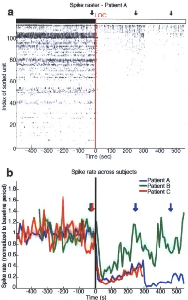
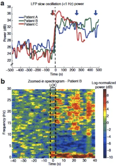
profound shift in brain state. We find that LOC is marked by the abrupt onset of slow oscillations (0.1-1 Hz) in the local field potential. Power in the slow oscillation band rises sharply at LOC and maintains this increase throughout the post-LOC period. Neuronal spiking becomes coupled to the local slow oscillation within seconds of LOC: spiking occurs only in short intervals of activity that are interspersed with suppression lasting hundreds of milliseconds, periodically interrupting information processing. These periods
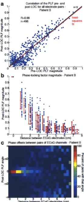
measures remain similar to the conscious state and neuronal spike rates can recover to baseline levels after LOC despite continued unresponsiveness. This demonstrates that short periods of normal spike dynamics can still occur during unconsciousness. We conclude that the slow oscillation is a fundamental component of propofol-induced unconsciousness, marking a functional isolation of cortical regions while significant connectivity is preserved within local networks.
2.3 Results
2.3.1 Rapid loss of consciousness after propofol bolus
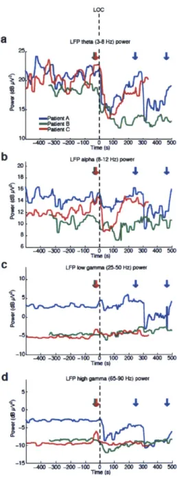
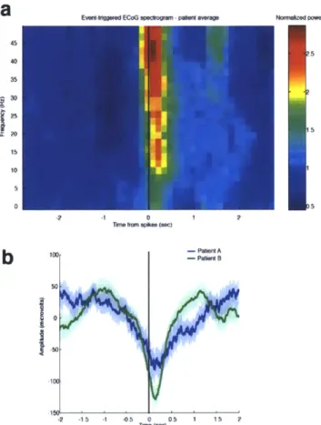
We recorded single units (n=198), local field potentials (LFP), and intracranial electrocorticograms (ECoG) in three patients undergoing intracranial monitoring for surgical treatment of epilepsy. Single units and LFPs were recorded from a 96-channel microelectrode array (Truccolo et al., 2011) implanted in temporal cortex for research purposes. We recorded throughout induction of general anesthesia by bolus administration of propofol, before planned neurosurgery to remove the electrodes. Patients performed an auditory task requiring a button press in response to stimuli. All patients completely ceased responding to the task within 40 seconds of propofol administration and remained unresponsive for the remainder of the recording period, lasting 5-10 minutes post-LOC. LOC was defined as the onset of this period of unresponsiveness to auditory stimuli. To acknowledge the fact that LOC could have occurred at any point between the last response and the failure to make the next response, LOC was defined as the interval beginning one second before the first missed stimulus, up until the second missed stimulus (5 seconds total). We then compared spectra across all ECoG channels in the pre- and post-LOC periods. In agreement with previous scalp EEG studies of healthy subjects (Murphy et al., 2011), we found that average spectra in the post-LOC period differed significantly from that in the pre-LOC period: slow (0.1-1 Hz) and gamma (25-40 Hz) power increased in the unconscious state (Fig. 2.1e). These results suggested that propofol acted as expected in these patients, and did not reveal any gross disruption of GABA networks.
LOC LFP Oum (34 Hz) pow -400 -300 -200 -10 0 100 200 300 400 500 This (S) LFP a~rel (5-12 Hz) pww 20 15 16 114
'12
10 18 16 It 4 50 e40 .30 ~20 10 -400 -300 -200 -10 0 100 200 300 400 50W LFP low grM (050 Hz) pao-400 -.5o 0 -Ito d 10 200A 360 400 'Wo
This (8)
LF-P tqft gurun (65-90 Hz) pow
Avage specun acwosn al ECoG channeb
0 10 20 30 Frequency (Hz) 40 4 4 -4W -30 M20 -100 0 10 20 400 500 km (0)
Figure 2.1 Bandpower changes relative to LOC. Power in different frequency bands from a representative microelectrode LFP, for each patient. None of the bands show a strong change at LOC that is then maintained throughout the post-LOC period. Dashed line indicates LOC and arrows are times of propofol delivery (+/- 20 seconds). A) Theta power transiently decreases after LOC. B) Alpha power transiently decreases after LOC.
I4 C 10 I 5 0 -5 d 5 -10