Publisher’s version / Version de l'éditeur:
Journal of Immunological Methods, 362, 1-2, pp. 161-167, 2010-10-01
READ THESE TERMS AND CONDITIONS CAREFULLY BEFORE USING THIS WEBSITE. https://nrc-publications.canada.ca/eng/copyright
Vous avez des questions? Nous pouvons vous aider. Pour communiquer directement avec un auteur, consultez la première page de la revue dans laquelle son article a été publié afin de trouver ses coordonnées. Si vous n’arrivez pas à les repérer, communiquez avec nous à [email protected].
Questions? Contact the NRC Publications Archive team at
[email protected]. If you wish to email the authors directly, please see the first page of the publication for their contact information.
NRC Publications Archive
Archives des publications du CNRC
This publication could be one of several versions: author’s original, accepted manuscript or the publisher’s version. / La version de cette publication peut être l’une des suivantes : la version prépublication de l’auteur, la version acceptée du manuscrit ou la version de l’éditeur.
For the publisher’s version, please access the DOI link below./ Pour consulter la version de l’éditeur, utilisez le lien DOI ci-dessous.
https://doi.org/10.1016/j.jim.2010.09.027
Access and use of this website and the material on it are subject to the Terms and Conditions set forth at
New ELISA approach based on coiled-coil interactions
Liberelle, Benoît; Bartholin, Laurence; Boucher, Cyril; Murschel, Frédéric;
Jolicoeur, Mario; Durocher, Yves; Merzouki, Abderrazzak; De Crescenzo,
Gregory
https://publications-cnrc.canada.ca/fra/droits
L’accès à ce site Web et l’utilisation de son contenu sont assujettis aux conditions présentées dans le site LISEZ CES CONDITIONS ATTENTIVEMENT AVANT D’UTILISER CE SITE WEB.
NRC Publications Record / Notice d'Archives des publications de CNRC:
https://nrc-publications.canada.ca/eng/view/object/?id=ca2e232f-148e-47d1-a241-0c678651ab40
https://publications-cnrc.canada.ca/fra/voir/objet/?id=ca2e232f-148e-47d1-a241-0c678651ab40
Research paper
New ELISA approach based on coiled-coil interactions
Benoît Liberelle
a,b, Laurence Bartholin
a, Cyril Boucher
a, Frédéric Murschel
a, Mario Jolicoeur
a,b,
Yves Durocher
c, Abderrazzak Merzouki
b, Gregory De Crescenzo
a,b,⁎
aDepartment of Chemical Engineering, Bio-P2Research Unit,
bInstitute of Biomedical Engineering, Groupe de Recherche en Sciences et Technologies Biomédicales (GRSTB), Ecole Polytechnique de Montréal, P.O. BOX 6079,
Station Centre-ville, Montréal (QC) Canada H3C 3A7
cAnimal Cell Technology Group, Bioprocess Centre, Biotechnology Research Institute, National Research Council Canada, Montréal (Qc), Canada H4P 2R2
a r t i c l e
i n f o
a b s t r a c t
Article history: Received 22 June 2010
Received in revised form 2 September 2010 Accepted 16 September 2010
Available online 1 October 2010
The de novo designed heterodimeric E/K coiled-coil system has been previously demonstrated to be an excellent capture/dimerization system applicable to various needs in both biotechnology and pharmaceutical fields. Those include controlled protein dimerization, capture, purification and Western-blot detection. We here report the development of a new generation of ELISA test based on coiled-coil interactions for the direct quantitation of coil-tagged epidermal growth factor (EGF). The new approach was evaluated for its specificity, plate storability and reusability as well as for convenience when compared to commercially available systems. Our results show a similar affinity/sensitivity to standard capturing antibody-based ELISA systems and an improved affinity/ sensitivity when compared to the commercially available Ni-NTA capture system. The E/K coiled-coil ELISA system was validated with respect to recovery, intra- and inter-assay variations. The practical working range was estimated to be between 5.2 and 34,000 pM. Furthermore, the storability and reusability of the plates was greater than the two aforementioned systems, suggesting that the E/K coiled-coil system is a good alternative to traditional tags such as poly-histidine for the development of ELISA tests aiming at quantitating coil-tagged proteins.
© 2010 Elsevier B.V. All rights reserved.
Keywords: EGF
E/K coiled-coil interactions ELISA test
1. Introduction
α-helical coiled-coil is an oligomerization motif being naturally present in a wide variety of proteins such as transcription factors, motor and viral fusion proteins (Burkhard et al., 2001). In fact, protein data base analysis revealed that approximately 10% of all proteins contain coiled-coil domains, therefore making it one of the most abundant motif found in proteins (Mewes et al., 2000; Walshaw and Woolfson, 2001). Coiled-coil motif results from the wrapping of two or more amphipathic α-helices around each other in a left handed supercoil fashion; the helices being aligned in either a parallel or anti-parallel manner (Lupas, 1996). Each peptide involved in
this domain possesses a characteristic signature, i.e. a heptad repeat denoted (abcdefg)n, in which hydrophobic residues at
the (a) and (d) positions pack in a characteristic ‘knobs-into-holes’ manner to form the hydrophobic core of the coiled-coil structure (Crick, 1953). The structural properties of coiled-coils have been extensively characterized by high-resolution crys-tallography and NMR-based studies. The influence of each residue of the heptads upon coiled-coil stoichiometry, stability as well as relative alignment of α-helices composing the motif (i.e., parallel or anti-parallel) are now well understood (Parry et al., 2008). This knowledge has opened the way to the design of de novo heterodimeric coiled-coils for protein engineering and biotechnological applications. Of particular interest, the de novo designed heterodimeric E/K coiled-coil system has been demonstrated to be a promising capture/dimerization tool for various applications (Tripet et al., 1996; Chao et al., 1998). Within the E/K coiled-coil complex, each peptide is composed of a distinct heptad that is repeated five times (Ecoil: [VSALEKE]5;
⁎ Corresponding author. Department of Chemical Engineering, Ecole Polytechnique de Montréal, P.O. BOX 6079, Station Centre-ville, Montréal (Québec) Canada H3C 3A7. Tel.: +1 514 340 5121x7428; fax: +1 514 340 2991.
E-mail address:[email protected](G. De Crescenzo).
0022-1759/$ – see front matter © 2010 Elsevier B.V. All rights reserved. doi:10.1016/j.jim.2010.09.027
Contents lists available atScienceDirect
Journal of Immunological Methods
Kcoil: [VSALKEK]5), in which hydrophobic residues at the (a) and
(d) positions (V and L, respectively) are common to both peptides, while residues at positions (e) and (g) are distinct for each peptide (E or K). These residues further stabilize the E/K coiled-coil structure through electrostatic interactions while increasing specificity. Among the applications of the E/K peptides are the stable capture of fully bioactive proteins on biosensor (De Crescenzo et al., 2003b) and cell culture-compatible surfaces (Boucher et al., 2009) or virus pili (Le et al., 2009), the artificial dimerization of growth factors (Nakaji-Hirabayashi et al., 2009) or receptor ectodomains (De Crescenzo et al., 2004) for increased biological activities and the detection of coil-tagged protein by Western blot (Boucher et al., 2010). Furthermore, this coiled-coil system was demonstrated to be promising for the development of affinity chromatography strategies (Tripet et al., 1996; Chao et al., 1998).
We here report another application for the E/K system, namely, the development and the validation of a novel enzyme linked immunosorbent assay (ELISA) approach for the detec-tion of coil-tagged proteins. In that endeavor, a chimeric protein corresponding to the epidermal growth factor (EGF) fused to both Ecoil and His tags at its N-terminus was used as a model protein. We compared the detection sensitivity of our novel ELISA approach that relied on coiled-coil mediated protein capture to those of standard ELISAs based on His tag- or antibody-mediated capture. Secondly, we evaluated the stor-ability, reusability and regeneration of the system and found many advantages when compared to current commercial ones. The E/K coiled-coil system was also shown to be efficient for the determination of coil-tagged EGF concentration being present in complex mixtures such as cell culture medium. Altogether, our results demonstrated that the novel E/K coiled-coil-based ELISA approach is more efficient than commercially available affinity-based systems at a lower cost. Furthermore, the newly developed system showed improved storability and enhanced reusability when compared to both Ni-NTA systems and standard ELISAs.
2. Materials and methods 2.1. Material and reagents
Aminated microplates (96 Well Clear Polystyrene Amine Surface Stripwell Microplate, Corning catalog #2388) were purchased from Fisher Scientific Co. (Ottawa, ON). Bare (standard) microplates (96 Well Clear Polystyrene Stripwell Microplates, Costar catalog #2592) were obtained from R&D Systems (Minneapolis, MN). Ni-NTA microplates (Ni-NTA HisSorb plate, Qiagen catalog #35061) were purchased from Qiagen (Mississauga, ON). Dulbecco's Phosphate Buffered Saline (modified PBS, without calcium chloride and magne-sium chloride), Tween 20, cysteine (99 + % purity), guanidine hydrochloride (Gnd-HCl) and sodium chloride (99.99% purity) were purchased from Sigma-Aldrich Canada Ltd. (Oakville, ON). Sulfuric acid (98% purity) was purchased from VWR International Inc (Mont-Royal, QC). Succinimidyl 6-[30-(2-pyridyldithio)-propionamido]hexanoate (LC-SPDP, 95 + % purity) was obtained from Pierce Biotechnology, Inc. (Rockford, IL). MilliQ quality water (18.2 MΩ cm; total organic compounds (TOC) = 4 ppb) was generated with a Millipore Gradient A 10 purification system. Cysteine-tagged Kcoil peptides (De Crescenzo et al., 2003a) were synthesized by the peptide facility at University of Colorado (Denver, CO). Commercially available DuoSet ELISA kit containing mouse anti-human EGF antibody (capture antibody), biotinylated goat anti-human EGF antibody (detection antibody), strepta-vidin-horseradish peroxidase (streptavidin-HRP), bovine serum albumin (BSA), substrate solution (hydrogen perox-ide/tetramethylbenzidine) and untagged human EGF were purchased from R&D Systems (Minneapolis, MN).
2.2. Plasmid
The pTT5-Ecoil-EGF plasmid coding for Ecoil-(His)8-EGF, a
recombinant protein corresponding to human EGF N-termi-nally fused to the Ecoil (5 heptad repeats) and to an (His)8
affinity tag (MW=12,711 Da), was constructed by in-frame ligation of Ecoil, (His)8tag and human EGF cDNA sequences as
previously described (Boucher et al., 2008). 2.3. Ecoil-(His)8-EGF production and purification
Ecoil-(His)8-EGF was produced in HEK 293-6E cells and
purified by immobilized metal ion affinity chromatography (IMAC) as previously reported (Boucher et al., 2008). Protein concentration was determined by Bradford assay and analyzed by SDS-PAGE. Purified tagged EGF was then aliquoted and stored at −80 °C until use. Note that, within this chimeric protein, the EGF moiety was demonstrated to be as active as untagged EGF in in vitro cell assay (Boucher et al., 2008).
For small-scale Ecoil-(His)8-EGF production, HEK293-6E
cells were seeded in 6-well cell culture plates (1.67 × 10⁶cells/
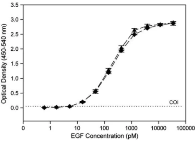
ml in 1.8 mL medium). 2 μg of pTT5-Ecoil-EGF plasmid were diluted in 100 μL of F17 medium to which 100 μL of F17 medium containing 4 μg of 25 kDa linear polyethylenimine was added. The mixture was immediately vortexed and incubated for 15 min at room temperature prior to cell addition. Cells were fed 24 h post-transfection (hpt) with 0.5% TN1 peptone (w/v, final) (Pham et al., 2005). Following harvest at different Fig. 1. Dose–response curves corresponding to the detection of (♦) untagged
human EGF and (■) Ecoil-(His)8-EGF incubated on capture antibody-coated
bare plates. Optical density recorded at 450 nm was corrected at 540 nm. Results are the means ± standard deviation of five plates.
time points (up to 120 hpt), the medium was clarified by centrifugation (3500×g, 10 min). Ecoil-(His)8-EGF present in
the supernatant was then quantified by ELISA (see below).
2.4. Plate preparation
2.4.1. Capture antibody coating on standard microplates Capture antibody was diluted to 4.0 μg/mL in 10 mM PBS without carrier protein. 100 μL of the dilute solution were transferred to the bare microplate wells and allowed to incubate overnight at room temperature (RT). The plates were then washed three times with 200 μL of wash buffer, consisting of 10 mM PBS, 0.05% Tween 20 (PBS-T). In order to block the plates, 200 μL of PBS containing 1% bovine serum albumin (PBS-BSA) was added into each well and then incubated for 1 h at RT. The plates were washed three times with PBS-T.
2.4.2. Kcoil peptides grafting on aminated microplates Kcoil layers were generated on aminated plates via LC-SPDP linker by adapting the protocol from Boucher et al. (Boucher et al., 2009). Briefly, 100 μL of 2 mM LC-SPDP in 10 mM phosphate buffer (10% DMSO) were added to each well for 1 h at RT. The plates were rinsed four times using 200 μL of MilliQ water. Then, the thiol-reactive wells were incubated with 100 μL of 10 μM cysteine-tagged Kcoil peptides (5 heptad repeats) in 10 mM PBS for 2 h at RT. The plates were rinsed two times using 200 μL of 10 mM PBS and finally four times using 200 μL of MilliQ water. Unreacted LC-SPDP sites were blocked using 100 μL of 50 mM cysteine solution (1 M NaCl in 0.1 M sodium acetate, pH 4.0) followed by rinsing with MilliQ water. The Kcoil-coated plates dried in air can be stored at 4 °C until use.
2.4.3. Ni-NTA microplates
Ni-NTA-coated plates were used without any pretreatment, as recommended by the manufacturer.
2.5. Assay procedure
In the subsequent ELISA procedure, samples or standards refer to untagged EGF for capture antibody-coated plates and Ecoil-(His)8-EGF for Kcoil-coated or Ni-NTA-coated plates.
100 μL of sample or standard diluted in PBS-BSA via direct dilutions were added into the corresponding wells. Plates were allowed to incubate for 1 h at RT. After washing the plates three times with PBS-T, 100 μL of detection antibody (50 ng/mL in PBS-BSA) were added to the plates followed by incubation for 1 h at RT. After a second washing with PBS-T, 100 μL of streptavidin-HRP (diluted 200 times with the PBS-BSA) was added to the plates and the solution was incubated for 20 min at RT. Following final washing with PBS-T, the reaction was revealed with 100 μL of substrate solution (1:1 mixture of hydrogen peroxide/tetramethylbenzidine). After an incubation time of 17 min in the dark at RT, the colorimetric reaction was stopped by adding 50 μL of 2 N H2SO4. The optical density (OD)
at 450 nm and 540 nm (for correction) was measured using an ELISA plate reader (Victor3V Multilabel Counter, PerkinElmer
Inc., Woodbridge, ON).
Table 1
Impact of BSA pretreatment on the non-specific adsorption of untagged EGF. Optical densities were obtained by incubating 34 nM of untagged EGF on cysteine-and Kcoil-functionalized plates. Results correspond to the mean±stcysteine-andard deviation (n=4).
BSA blocking treatment
Covalently grafted layers
Cysteine Kcoil peptide
– 1.031 ± 0.042 0.014 ± 0.003
+ 0.158 ± 0.002 0.006 ± 0.002
Fig. 2. Schematic illustration of Ecoil-(His)8-EGF capture onto Kcoil-coated wells. Thiol-reactive LC-SPDP linker attached on aminated well allowed for the covalent
grafting of cysteine-tagged Kcoil peptides. Remaining active linkers were blocked using cysteine. Ecoil-(His)8-EGF were captured to the well surface via reversible
E/K coiled-coil interactions.
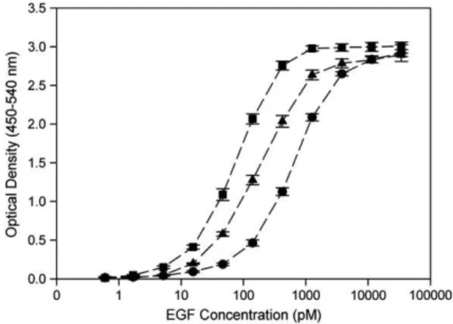
Fig. 3. Dose–response curves corresponding to Ecoil-(His)8-EGF incubation on
(■) capture antibody-coated bare plates, (▲) Kcoil-coated aminated plates and (●) Ni-NTA plates. Optical density recorded at 450 nm was corrected at 540 nm. Results are the means ± standard deviation of five plates.
2.6. Regeneration of Kcoil-coated microplates
The detachment of the coil-tagged EGF from the well surface was performed by covering each well with 200 μL of 5 M guanidine hydrochloride solution in a plate shaker for 5 min, followed by rinses using 200 μL of 10 mM PBS and finally MilliQ water. This cleaning cycle was repeated 10 times.
2.7. Cut-off index and working range determination
The cut-off index (COI) value was determined by an ELISA sensitivity test. Briefly, untagged EGF (34 nM) was incubated in the Kcoil-coated wells of modified plates and ELISA test was repeated 5 times. The value of the blank average OD (0.014) added to the value of the OD standard deviation multiplied by two (0.003 × 2) was determined to be the cut-off index value (COI=0.02). Above this value, ODs are considered to be significant. The working range was determined by triplicate dilutions of recombinant Ecoil-(His)8-EGF at concentrations
ranging from 0.6 pM to 34,000 pM. The final working range was determined by combined evaluation of plotting ‘measured over mean’ as a function of concentration. Intra- and inter-assay variations were determined in order to verify the reproducibil-ity and specificreproducibil-ity of our assay. Intra- and inter-assay variations were determined by dilutions of the Ecoil-(His)8-EGF and
triplicate testing on either the same plate or different plates, according to the ELISA conditions described above.
2.8. Statistical analysis
Values are given as mean value± standard deviation (s.d.). The data were expressed as means ± s.d. and analyzed using Statistica 9.0 (StatSoft, Inc., Tulsa, USA). Statistical significance was determined by one-way ANOVA and main effect ANOVA followed by Tukey HSD post-hoc test. Differences were considered significant for pb 0.05.
3. Results and discussion
While small affinity tags such as poly-histidine tails are now broadly used for the purification and detection of recombinant proteins, de novo engineered coil peptide tags have also proven to be promising for affinity purification and excellent in many other biotechnology applications including Western-blot de-tection (Boucher et al., 2010) protein capture (De Crescenzo et al., 2003b; Boucher et al., 2009) as well as controlled protein dimerization (De Crescenzo et al., 2004; Noirclerc-Savoye et al., 2005). While coil tags can be added to peptides via chemical reaction (Tripet et al., 2003), the production of more-complex coil-tagged proteins in bacteria (Le et al., 2009) and mamma-lian cells has also been reported (De Crescenzo et al., 2003b; Boucher et al., 2008). In that endeavor, the development of efficient expression vectors containing cDNA coding for signal peptide (to ease protein secretion) and cDNA coding for coil tag have already been designed and validated for the production of fully bioactive complex proteins (De Crescenzo et al., 2003b). The present work further extends the use of this new tag system to the development of ELISA test aiming at quantitating coil-tagged recombinant proteins that are produced by these new expression platforms. In that endeavor, Ecoil-(His)8-EGF,
expressed by transiently transfected HEK293-6E cells was used as a model protein.
More specifically, antibody/antigen, coiled-coil and His/Ni-NTA interactions have been explored to capture Ecoil-(His)8
-EGF proteins onto anti--EGF antibody, Kcoil and Ni-NTA-coated ELISA plates, respectively. The amount of EGF protein attached to the wells via those three different approaches was then evaluated by applying a standard ELISA test, i.e. addition of biotinylated anti-EGF antibody followed by the attachment of streptavidin-HRP and colorimetric reaction. In order to com-pare the different approaches, detection antibody, streptavidin-HRP and streptavidin-HRP substrate were added to the wells at the same concentration and the colorimetric oxidation reaction was allowed to proceed for the same duration in each case (17 min). First, antibody-mediated capture of Ecoil-(His)8-EGF was
evaluated using conventional ELISA technique with antibody-Fig. 4. Dose–response curves corresponding to Ecoil-(His)8-EGF incubation
(▲) with or (♦) without BSA pretreatment on Kcoil-coated aminated plates. Results are the means ± standard deviation of four wells.
Table 2
Intra- and inter-assay coefficients (optical density variation in %) for Ecoil-(His)8-EGF captured on antibody-coated, Kcoil-coated and Ni-NTA-coated plates.
Results are means of five plates.
Concentration (pM)
34,000.0 11,333.3 3777.8 1259.3 419.8 139.9 46.6 15.5 5.2
Capture antibody Intra 1.9 1.9 1.8 1.7 1.9 3.5 3.1 4.4 4.4
Inter 1.6 1.9 1.6 1.1 2.1 3.1 7.0 6.2 12.1
Kcoil Intra 2.3 2.8 2.7 3.3 3.3 3.7 5.4 4.1 9.3
Inter 1.2 1.2 2.2 3.4 3.9 6.2 6.2 5.0 10.9
Ni-NTA Intra 2.1 3.0 2.2 2.9 3.5 5.2 7.8 8.1 10.3
coated standard plates and compared to that of untagged EGF. As seen inFig. 1, the concentration curves corresponding to both tagged and untagged EGF overlapped, thus indicating that neither Ecoil nor His tag impacted Ecoil-(His)8-EGF capture by
anti-EGF antibody. This result is in excellent agreement with our previous study that had demonstrated that both Ecoil and His tags did not affect Ecoil-(His)8-EGF bioactivity as deduced
from in vitro cell assays (Boucher et al., 2008).
Secondly, Kcoil-coated microplates were generated by adapting the optimized grafting conditions we previously described (Boucher et al., 2009). To do so, commercially available aminated plates were reacted with bifunctional LC-SPDP linker to create a stable amide bond. The other extremity of LC-SPDP linking agent was then reacted with the thiol group of the cysteine-terminated Kcoil peptide to form a covalent disulfide bond. After Kcoil grafting, cysteine was used to block the unreacted thiol-reactive groups of the LC-SPDP in order to prevent the subsequent attachment of molecules
containing thiol groups. Ecoil-tagged EGF proteins were then captured onto Kcoil-modified surface via reversible coiled-coil interactions by simple incubation (Fig. 2).
The specificity of Kcoil-coated plates towards Ecoil-(His)8
-EGF was then evaluated by incubating untagged -EGF on aminated wells that had been coated with Kcoil peptide or with cysteine only as a reference. A concentration of 34 nM of untagged EGF was incubated in the modified wells. Our results showed that a higher optical density (OD) value was obtained on cysteine-covered wells (1.031) when compared to Kcoil-cysteine-covered wells (0.014) (seeTable 1). This result indicated that a large amount of untagged EGF peptide could adsorb non-specifically on cysteine-grafted wells whereas Kcoil grafting at the surface of the wells was efficient to block non-specific EGF adsorption. This further suggests that Kcoil grafting, using our experimental protocol, resulted in complete and very efficient microplate surface coverage. This conclusion is further supported by the fact that Kcoil-functionalized microplate pretreatment using bovine serum albumin (BSA) as blocking agent did not drastically reduce the non-specific adsorption of untagged EGF since OD only decreased from 0.014 to 0.006 (Table 1). In stark contrast, the same BSA pretreatment significantly reduced untagged EGF non-specific adsorption onto cysteine-covered wells (OD decrease from 1.031 to 0.158,Table 1).
In order to test the efficiency of our coiled-coil based ELISA system, we compared dose–response standard curves corresponding to various concentrations of Ecoil-(His)8-EGF
being incubated onto antibody-coated, Kcoil-grafted and commercially available Ni-NTA plates. As seen inFig. 3, the standard dose–response curves shifted to higher concentration values for the tag-specific plates compared to capture antibody-coated plates by a factor 4 and 10 for Kcoil- and Ni-NTA-antibody-coated plates, respectively (Fig. 3). These shifts corresponded to apparent thermodynamic dissociation constants, KD, equal to
70, 185 and 675 pM for antibody/Ecoil-(His)8-EGF,
Kcoil/Ecoil-(His)8-EGF and Ni-NTA/Ecoil-(His)8-EGF interactions,
respec-tively, as deduced from fitting Fig. 3 data with a simple interaction model (Langmuir isotherm). The apparent KD
corresponding to Kcoil/Ecoil-(His)8-EGF interactions, as
deter-mined by ELISA (i.e. 185 pM) was in good agreement with that determined for Ecoil/Kcoil interactions using a surface plasmon resonance biosensing approach (KD= 63 pM) (De Crescenzo
et al., 2003a). Altogether, our results thus indicate that the Kcoil system is more efficient than the commercially available Ni-NTA affinity-based system for Ecoil-(His)8-EGF capture.
The influence of BSA pretreatment prior to Ecoil-(His)8-EGF
incubation on Kcoil-coated plates was also evaluated. As shown inFig. 4, superimposable dose–response curves were obtained with or without BSA pretreatment, confirming that the latter is not required for Kcoil plate usage as previously deduced (Table 1), as long as BSA is present in the PBS solution used for diluting samples.
To determine the practical working range of the new system (i.e., without BSA pretreatment), we performed OD reading for various dilutions of Ecoil-(His)8-EGF (from 0.6 to 34,000 pM).
Intra-assay OD variations of less than 10% were obtained within the 5.2-to-34,000 pM range (Table 2), which thus defines the practical working range of our coiled-coil-based ELISA. In addition, inter-assay variations in that working range were also acceptable (≤10%) thus making our system as efficient as commercially available ones (Table 2).
Fig. 5. Dose–response curves corresponding to Ecoil-(His)8-EGF incubation on
fresh (▲) and aged (♦) Kcoil-grafted aminated plates. Fresh plates were generated at least 2 h before Ecoil-(His)8-EGF loading. Artificially aged plates were generated
4 weeks before use and stored in a dried state at 4 °C. Optical density recorded at 450 nm was corrected at 540 nm. Results are the means±standard deviation of five and two plates for freshly made and aged plates, respectively.
Fig. 6. Dose–response curves corresponding to Ecoil-(His)8-EGF incubation on
(▲) fresh and (■) regenerated Kcoil-coated aminated plates. Regeneration of Kcoil plates was performed using guanidine hydrochloride to detach captured Ecoil-(His)8-EGF, as described in theMaterial and Methodssection. Remaining
Ecoil-(His)8-EGF after washing protocol is shown as (●). Optical density
recorded at 450 nm was corrected at 540 nm. Each data point corresponds to the mean± standard deviation of four wells.
Kcoil-coated microplate storage stability and reusability were then assayed. Freshly made plates and aged plates that had been stored for 4 weeks at 4 °C gave comparable dose– response curves for Ecoil-(His)8-EGF detection, thus indicating
that our Kcoil coating was stable, and thus suitable for commercial purposes (Fig. 5). For Kcoil-coated plate reusability, guanidine hydrochloride (Gnd-HCl, 5 M), a strong chaotropic agent, was used to promote Ecoil-(His)8-EGF/Kcoil complex
dissociation. Gnd-HCl was selected based on our previous results that indicated that coiled-coil complexes could be disrupted with such denaturing agent (De Crescenzo et al., 2003b). Ecoil-(His)8-EGF amounts remaining in the wells were
then revealed with the same detection antibody as that we used in previous experiments, since its binding to EGF is not dependent upon the conformation of the latter (Gnd-HCl treatment might have unfolded the EGF moiety of the fusion protein). As seen inFig. 6, a complete detachment of captured Ecoil-(His)8-EGF was obtained following ten Gnd-HCl washing
cycles when the tagged growth factor had been incubated at concentrations lower than 420 pM (i.e. approximately 70% well saturation).
Higher amounts of captured Ecoil-(His)8-EGF resulting from
incubations at concentrations higher than 420 pM, were not completely stripped with such a treatment; increasing the number of washing cycles did not further enhance Ecoil-(His)8
-EGF removal (data not shown), indicating a potential limitation of the method. Following Kcoil microplate stripping, its reusability was assessed by reloading fresh Ecoil-(His)8-EGF
in the wells for which complete removal had been attained. As shown inFig. 6, standard dose–response curves corresponding to stripped and fresh plates overlapped, thus unambiguously demonstrating that our washing protocol did not affect the capture properties of the Kcoil microplates. Our Kcoil-coated ELISA plates are reusable when moderate amounts of Ecoil-(His)8-EGF (corresponding less than 70% well saturation) are
captured; within that range, i.e. 0 b [Ecoil-(His)8-EGF] b 420 pM,
the calibration curve was observed to be linear.
We then tested the applicability of our coiled-coil-based ELISA system for the quantification of Ecoil-(His)8-EGF in cell
culture medium. To do so, small-scale transient transfections of
HEK293-6E cells with plasmid encoding Ecoil-(His)8-EGF were
performed in 6-well culture plates. Cell culture supernatants were harvested everyday for 5 days post-transfection and Ecoil-(His)8-EGF concentrations were determined using our
coiled-coil- and antibody-based standard ELISA tests. As seen in
Fig. 7, both ELISA tests gave identical results, hence unambig-uously demonstrating that our coiled-coil-based approach is adequate for Ecoil-(His)8-EGF quantification in complex
mix-tures such as cell culture medium. 4. Conclusion
We have demonstrated the validity of a coiled-coil-based ELISA approach for coil-tagged EGF detection. We have also highlighted its advantages such as plate stability upon storage and reusability and its improved affinity/sensitivity when compared to that of commercially available Ni-NTA-based ELISA. The unique features of the new assay make it ideally suited for Ecoil-tagged recombinant protein detection and quantification. Furthermore, this novel assay has the potential to be optimized for coil-tagged protein interaction studies and high throughput protein quantification.
Acknowledgement
This work was supported by the Canada Research Chairs on Protein-enhanced Biomaterials (GDC), on Applied Metabolic Engineering (MJ), by the Natural Sciences and Engineering Research Council of Canada (GDC, MJ) and by the Groupe de Recherche en Sciences et Technologies Biomédicales (GRSTB) through a post-doctoral fellowship to BL.
References
Boucher, C., St-Laurent, G., Loignon, M., Jolicoeur, M., De Crescenzo, G., Durocher, Y., 2008. The bioactivity and receptor affinity of recombinant tagged EGF designed for tissue engineering applications is defined by the nature and position of the tags. Tissue Eng. A 14, 2069.
Boucher, C., Liberelle, B., Jolicoeur, M., Durocher, Y., De Crescenzo, G., 2009. Epidermal growth factor tethered through coiled-coil interactions induces cell surface receptor phosphorylation. Bioconjugate Chem. 20, 1569. Boucher, C., St-Laurent, G., Jolicoeur, M., De Crescenzo, G., Durocher, Y., 2010.
Protein detection by Western blot via coiled-coil interactions. Anal. Biochem. 399, 138.
Burkhard, P., Stetefeld, J., Strelkov, S.V., 2001. Coiled coils: a highly versatile protein folding motif. Trends Cell Biol. 11, 82.
Chao, H., Bautista, D.L., Litowski, J., Irvin, R.T., Hodges, R.S., 1998. Use of a heterodimeric coiled-coil system for biosensor application and affinity purification. J. Chromatogr. B. Biomed. Sci. App. 715, 307.
Crick, F.H.C., 1953. The packing of alpha-helices: simple coiled coils. Acta Crystallogr. 6, 689.
De Crescenzo, G., Litowski, J.R., Hodges, R.S., O'Connor-McCourt, M.D., 2003a. Real-time monitoring of the interactions of two-stranded de novo designed coiled-coils: Effect of chain length on the kinetic and thermodynamic constants of binding. Biochemistry (Mosc). 42, 1754.
De Crescenzo, G., Pham, P.L., Durocher, Y., O'Connor-McCourt, M.D., 2003b. Transforming growth factor-beta (TGF-beta) binding to the extracellular domain of the type II TGF-beta receptor: receptor capture on a biosensor surface using a new coiled-coil capture system demonstrates that avidity contributes significantly to high affinity binding. J. Mol. Biol. 328, 1173. De Crescenzo, G., Pham, P.L., Durocher, Y., Chao, H., O'Connor-McCourt, M.D., 2004. Enhancement of the antagonistic potency of transforming growth factor-beta receptor extracellular domains by coiled coil-induced homo-and heterodimerization. J. Biol. Chem. 279, 26013.
Le, P.U., Lenferink, A.E., Pinard, M., Baardsnes, J., Massie, B., O'Connor-McCourt, M.D., 2009. Escherichia coli expression and refolding of E/K-coil-tagged EGF generates fully bioactive EGF for diverse applications. Protein Expr. Purif. 64, 108.
Fig. 7. Ecoil-(His)8-EGF quantification in transiently transfected HEK296-6E
cell culture medium after. Culture media were collected at different times (24 to 120 h) post-transfection and Ecoil-(His)8-EGF concentrations were
determined using coiled-coil-based (▲) and standard (■) ELISA (1/100 and 1/500 sample dilution respectively).
Lupas, A., 1996. Coiled coils: new structures and new functions. Trends Biochem. Sci. 21, 375.
Mewes, H.W., Frishman, D., Gruber, C., Geier, B., Haase, D., Kaps, A., Lemcke, K., Mannhaupt, G., Pfeiffer, F., Schuller, C., Stocker, S., Weil, B., 2000. MIPS: a database for genomes and protein sequences. Nucleic Acids Res. 28, 37. Nakaji-Hirabayashi, T., Kato, K., Iwata, H., 2009. Surface-Anchoring of
Spontaneously Dimerized Epidermal Growth Factor for Highly Selective Expansion of Neural Stem Cells. Bioconjugate Chem. 20, 102. Noirclerc-Savoye, M., Le Gouellec, A., Morlot, C., Dideberg, O., Vernet, T., Zapun,
A., 2005. In vitro reconstitution of a trimeric complex of DivIB, DivIC and FtsL, and their transient co-localization at the division site in Streptococcus pneumoniae. Mol. Microbiol. 55, 413.
Parry, D.A., Fraser, R.D., Squire, J.M., 2008. Fifty years of coiled-coils and alpha-helical bundles: a close relationship between sequence and structure. J. Struct. Biol. 163, 258.
Pham, P.L., Perret, S., Cass, B., Carpentier, E., St-Laurent, G., Bisson, L., Kamen, A., Durocher, Y., 2005. Transient gene expression in HEK293 cells: peptone addition posttransfection improves recombinant protein synthesis. Bio-technol. Bioeng. 90, 332.
Tripet, B., Yu, L., Bautista, D.L., Wong, W.Y., Irvin, R.T., Hodges, R.S., 1996. Engineering a de novo-designed coiled-coil heterodimerization domain off the rapid detection, purification and characterization of recombinantly expressed peptides and proteins. Protein Eng. 9, 1029.
Tripet, B., De Crescenzo, G., Grothe, S., O'Connor-McCourt, M., Hodges, R.S., 2003. Kinetic analysis of the interactions between troponin C (TnC) and troponin I (TnI) binding peptides: evidence for separate binding sites for the “structural” N-terminus and the "regulatory" C-terminus of TnI on TnC. J. Mol. Recognit. 16, 37.
Walshaw, J., Woolfson, D.N., 2001. Socket: a program for identifying and analysing coiled-coil motifs within protein structures. J. Mol. Biol. 307, 1427.