Biochemical Characterization
of the E. coli
Very Short Patch Repair Pathway and its Coordination with
Methyltransferase Repair of 0
6-Methylguanine
by
Peter Thomas Rye
B.S., Biochemistry (1999)
Syracuse University in New York
Submitted to the Department of Chemistry in Partial
Fulfillment of the Requirements for the Degree of
Doctor of Philosophy in Biological Chemistry
at the
Massachusetts Institute of Technology
June 2006
© 2006 Massachusetts Institute of Technology. All Rights Reserved.
The author hereby grants to MIT permission to reproduce
and to distribute publicly paper and electronic
copies of this thesis document in whole or in part.
Signature of Author:
Department of Chemistry
April 7, 2006
Certified by:
Jo~haM. Essigmann
William and Betsy Leitch Professor of Chemistry and Toxicology
Thesis Supervisor
Accepted by:
-Robert W. Field
.TalaLm
o,.A
nThwr,
,,
3.
o
.Li.UrI
ti.
jm;
,rChairman, Department Committee on Graduate Students
MASSACHUSETTS INSTITUTEOF TECHNOLOGY
Gerald
N. Wogan.,
-Professor of Chemisfry ancdBiological Engineering
Chairman
John M. Essigmann
William and Betsy LeitchffRessor ohemistry
and Toxicology
Thesis Supervisor
Barbara Imperiali
Biochemical Characterization of the E. coli
Very Short Patch Repair Pathway and its Coordination with
Methyltransferase Repair of 06-Methylguanine
by
Peter Thomas Rye
Submitted to the Department of Chemistry on April 28, 2006 in Partial Fulfillment of the Requirements for the Degree of
Doctor of Philosophy in Biological Chemistry
The E. coli Very Short Patch Repair (VSPR) system corrects T:G mismatches that arise through Dcm-mediated methylation and subsequent deamination of the underlined cytosine residue in the palindromic sequence 5'-CCWGG-3' (W is an adenine or thymine). Vsr initiates VSPR by producing a single stranded nick on the 5' side of the mismatched T. The MutS and MutL mismatch recognition proteins stimulate this activity, as cells lacking either of these proteins display diminished VSPR. Genetic studies also indicate that Pol I is responsible for removing and replacing a short tract of nucleotides downstream of the incision site and that DNA Ligase seals the nick to complete the repair event. However, until now, biochemical investigation of the repair steps downstream of Vsr incision have been lacking.
Herein, we describe two novel in vitro assays used to probe the biochemical events of VSPR. The first was used to verify the reconstitution of VSPR using purified
E. coli Vsr, Pol I, and DNA Ligase enzymes, while the second was used to measure the
distribution of VSPR patch sizes in whole cell extracts. By monitoring the loss of radiosignal from a series of substrates that contained the label at prescribed distances downstream of the T:G mismatch, we were able to determine that VSPR patches are distributed around 2 to 4 deoxynucleotides in length. Interestingly, under certain reaction conditions, the addition of DNA Ligase improved the efficiency of repair initiation by Vsr, suggesting that VSPR may be optimal in the context of a multi-protein complex.
Lastly, we investigated the effect of VSPR proteins on methyltransferase (MTase) repair of O6-methylguanine (6mG). MTase repair of O6mG opposite T results in a G:T mismatch that must be further processed to yield the native G:C base pairing. The G:T mismatch is therefore an intersection of the two pathways and led us to hypothesize that MTase and VSPR proteins might interact. Indeed, cells lacking the functions of MutS, MutL, or Vsr proteins displayed decreased MTase repair in vivo, revealing a previously unknown interaction. The cooperation between proteins of these two repair systems may shed light on the biological significance of the VSPR system. Thesis Supervisor: John M. Essigmann
I would like to thank my advisor, Dr. John Essigmann, for his guidance and support throughout the years. John was consistently receptive to my ideas and very helpful in thinking them through. His energy in his work provided a great model to follow and inspired me to achieve the standards of excellence he set for himself. He is a brilliant scientist and has an amazing talent to communicate ideas. I thank him for his time and persistence in teaching me those skills, and providing, all the while, an enjoyable and productive environment in which to work. John is an extremely kind and selfless individual, and he has taught me more than he will ever be know.
I would also like to thank my thesis committee members, Drs. Gerald Wogan and Barbara Imperiali, for taking the time and energy to assess my work and provide valuable feedback. I am especially indebted to Dr. Wogan for continually inspiring me to seek a greater understanding of my work, the implications it may have, and how to take it to the next level.
Everyone in the Essigmann Laboratory has positively impacted me in some way. I need to thank Sarah, Kyle, Jim, Lauren, and Sreeja for being such great friends and reading my many thesis drafts and suggesting how to make them better. Your many comments were appreciated and helped enormously in defining the quality of my work. Many thanks go to Kim for her attention and kindness in dealing with administrative matters. I am convinced no other could take her place. I would also like to thank many individuals (past and present) for their friendship and scientific discussions. Aida, Alfio, Annie, Bob, Bogdan, Charles, Denise, Eunsuk, Francis, Jeannette, Jen, Jim, John, Kaushik, Maryann, Michelle, Neena, Nicole, Paul, Pei-sze, Shawn, Uday, Will, Yuri, and Zoran were all excellent people with whom to work. I hope I stay in touch with you all and I look forward to seeing where your careers lead you. I consider all of you my friends, and I hope that I had impacted your life in a positive way like you have impacted mine.
Special thanks go to the numerous undergraduates I have had the privilege to mentor. Lucien, Meghan, Ai-ris, Jenny, Dana, and Jelly were all amazing students to teach and also to learn from. The saying that "there's no greater way to learn than by teaching" holds very true. You all kept me on my toes by demanding an unparalleled understanding of our work. I owe a great amount of what I understand to our discussions. I also owe a great amount of what we accomplished in the lab to your efforts. I truly appreciate the devotion you all displayed in your work and the passion you had towards accumulating meaningful data. I hope that you feel great value, as I do, in the UROP
experience we shared.
Many thanks go to fellow chemistry graduate students. I remember the great friendships formed in my first semester at MIT. I thank Aaron, Abi, Fran, Jen, Lyn, Mary, Matt, Shawn, Woody, and Zarixia for helping to make MIT the right choice. Our first year was one of the best. I look forward to seeing where you all end up. To Hector,
thanks for everything. You are an amazing person and friend. I hope we never lose touch.
I would like to thank the "Merrimack Crew" for keeping me grounded, cheering me on, and always being around. I will forever be grateful for the friendship of Andy, Cory, Derek, Erin, Greg, Holly, Jamie, Jen, Jennie, Jesse, Jessica, Mandy, Mark, Matt, Meredith, Mike, Mike, Salstrom, Sarah, and Steve. I feel blessed every day that I have such wonderful people in my life who I can count on for anything. Thanks to you, and your significant others, for everything in the past, present, and (most exciting) the future. One word, LIFE. I love you all.
I am also thankful for Syracuse folks including Aleta, Ben, Bry, Cindy, Edi, Green, Hosmer, Jill, Jordan, Kyoko, Larissa, Maria, Meth, Mooney, Morgan, Moran, Rachael, Rob, Steve, and Zander. You all had a positive impact on my life and helped me to remember the things that are truly important. May we continue to keep the good times rolling.
I would also like to acknowledge friends that I have made along the way. I feel fortunate to have had the support from people such as Cara, Carolyn, Kristen, Molly, Paul, Sylvia, and TK. You all have made the last years of my life more enjoyable. Special thanks goes to Sarah for her irreplaceable friendship. She has always helped me to enjoy life and appreciate its many graces. I will forever be touched by her unyielding optimism and the friendship we share.
Lastly, I would like to extend the largest thank you possible to my wonderful family. I have received so much love and support from my two sisters, Elise and Heide, their husbands, Paul and Randy, and their families. I am deeply touched by how much they selflessly helped me through the tough times and celebrated with me through the good ones. It was also amazing to have received copious amounts of letters, drawings, pictures, phone calls, and hugs from my wonderful nieces and nephews Ben, Billy, Claire, Hope, Jane, Katie, Kay, Morgan, and Sara. You all are amazing people and I
thank you for the love and support that you gave me.
To my parents, the two most generous, kind, wise, fun, and loving people in my life, thank you for everything. You guys are always such an amazing source of direction, strength, and inspiration. I cannot thank you enough for the endless support you give me. Your attitude and advice keep me going through the rocky times and your excitement during the highs is incomparable. I thank God that I have such wonderful parents to push, pull, and stand by when needed. I also thank you for your talents in knowing just how to help me see things through. I know, from experience, that I can always look to your teachings for clarity and your example for guidance. By far and away, you contribute to my life's work the most. Therefore, I dedicate this thesis to you. Remember, this is only the beginning.
COMMITTEE PAGE 2 ABSTRACT 3 ACKNOWLEDGMENTS . 4 TABLE OF CONTENTS . 6 ABBREVIATIONS 9 LIST OF FIGURES . . . 12 LIST OF TABLES . . . 15 LIST OF SCHEMES . . . 16
CHAPTER
1. Mismatch Repair in Escherichia coli.
. . . .17
1.1. Mutation Avoidance . . . 18
1.2. The Very Short Patch Repair System. . . . . 19
1.2.1. Dcm - in Vivo and in Vitro Properties. . . . 22
1.2.2. Vsr - in Vivo and in Vitro Properties. . . . 25
1.2.3. Structural Data for Vsr . . . 28
1.2.4. Catalytic Mechanism of Vsr .
.
.
.
.
34
1.3. Methyl-Directed Mismatch Repair - Lessons From Streptococcus
Pneumoniae 36
1.4. Methyl-Directed Mismatch Repair in E. coli. . . . 37
1.4.1. Dam - in Vivo and in Vitro Properties. . . . 38
1.4.2. MutS - in Vivo and in Vitro Properties . . . 39
1.4.3. MutL - in Vivo and in Vitro Properties . . . 41
1.4.4. MutH - in Vivo and in Vitro Properties . . . 42
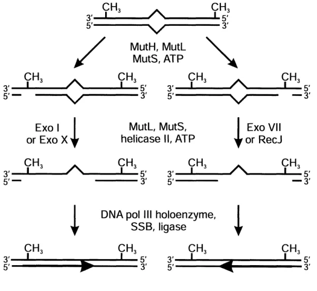
1.4.5. Mechanism of Methyl-Directed Mismatch Repair . . 44
1.5. Mismatch Repair in Eukaryotes . . . 45
1.6. Interaction Between the VSPR and MMR Systems . . . 46
1.7. Mechanism of VSPR . . . 51
1.8. Repair of T:G Mismatches in Eukaryotes . . . . 52
1.9. References . . . 69
Purified Proteins Abstract Introduction Experimental Procedures Results. Discussion References
The Size of Repair Tracts Created by E. coli Very Short Patch Repair System. Abstract Introduction Experimental Procedures Results. Discussion References
06-Methylguanine - Formation, Consequences, and Repair . 06-Methylguanine - A Biologically Important Adduct 06-Methylguanine is Mutagenic
06-Methylguanine is Cytotoxic
The Adaptive Response to Alkylating Agents Ada - in Vivo and in Vitro Properties . Ogt - in Vivo and in Vitro Properties
Protection Against Alkylation Induced Toxicity by Ada and Ogt Repair of 06-Methylguanine by Nucleotide Excision Repair. References
Effect of Mismatch Repair Proteins on Methyltransferase Repair of 06-Methylguanine in Vivo Abstract Introduction Experimental Procedures Results. 2.1. 2.2. 2.3. 2.4. 2.5. 2.6. CHAPTER 3. 3.1. 3.2. 3.3. 3.4. 3.5. 3.6. CHAPTER 4. 4.1. 4.2. 4.3. 4.4. 4.5. 4.6. 4.7. 4.8. 4.9. CHAPTER 5. 5.1. 5.2. 5.3. 5.4. 87 88 89 91 97 101 115 118 119 120 122 127 130 144 146 147 148 152 153 156 161 163 164 172 186 187 188 191 197
5.6. Future Directions 5.7. References
CHAPTER 6. Cloning, Expression, and Purification of Truncated Hormone Receptors
6.1. Introduction
6.2. Nuclear Hormone Receptors . 6.3. Experimental Procedures 6.4. References
CHAPTER 7. A Rationally Designed Genotoxin that Selectively Destroys Estrogen Receptor-Positive Breast Cancer Cells
7.1. Abstract 7.2. Manuscript 7.3. References
CHAPTER 8. Design, Synthesis, and Evaluation of Estradiol-linked Genotoxicants as Anti-cancer Agents.
8.1. Abstract 8.2. Introduction
8.3. Experimental Procedures 8.4. Results and Discussion 8.5. Conclusion 8.6. Acknowledgments 8.7. References CURRICULUM VITAE 203 224 229 230 230 232 242 244 245 246 253 256 257 258 260 261 264 265 270 273
ABBREVIATIONS
ImA 1-methyladenine
2D-NMR two dimensional nuclear magnetic resonance
3mA 3-methyladenine 3mC 3-methylcytosine 3mG 3-methylguanine 5mC 5-methylcytosine 7mA 7-methyladenine 7mG 7-methylguanine Aa amino acid A adenine Amp Ampicillin AP apurinic AR androgen receptor
ATP adenosine triphosphate
BCNU 1,3-bis(2-chloroethyl)- 1 -nitrosourea
BER base excision repair
,3ME beta-mercaptoethanol bp base pair C cytosine Cam Chloroamphenicol CCNU N-(2-chloroethyl)-\P-cyclohexyl-N-nitrosourea Da Dalton
DBD DNA binding domain
DMS dimethylsulphate
DMSO dimethylsulfoxide
DNA deoxyribonucleic acid
DTT 1,4-dithio-DL-threitol
E. coli
Escherichia coli
EMSA electrophoretic mobility shift assay
ENU N-ethylnitrosourea
ER estrogen receptor
FAD flavin adenine dinucleotide
g gravity
G guanine
GR glucocorticoid receptor
h hour
HEPES 4-(2-hydroxyethyl)piperazine- 1-ethanesulfonic acid
HTH helix-turn-helix
IDL insertion-deletion loop
Kb kilo-base
kDa kilo Daltons
Kan Kanamycin
KOH potassium hydroxide
LB Luria Broth
LBD ligand binding domain
M molar
MCS multiple cloning sites
MGMT 06-methylguanine DNA methyltransferase
Mel methyl iodide
MMR methyl-directed mismatch repair
MMS methyl methanesulphonate
MNNG N-methyl-N' -nitro-N-nitrosoguanidine
MNU N-methlynitrosourea
MTase methyltransferase
NaCl sodium chloride
NEB New England Biolabs (Ipswich, MA)
NEN New England Nuclear
NHR nuclear hormone receptor NTD N terminal domain 02mC L-methylcytosine &OmG 06-methylguanine O2mT &-methylthymine O4mT 04-methylthymine ODN oligodeoxynucleotide
PAGE poly-acrylamide gel electrophoresis
PCNA proliferating cell nuclear antigen
PCR polymerase chain reaction
PNK polynucleotide kinase
PR progesterone receptor
RM restriction modification
RT room temperature
Rpm revolutions per minute
SAM S-adenosylmethionine
SDS Sodium Dodecyl Sulfate
T thymine
T4PNK T4 polynucleotide kinase
TAE Tris-Acetate-EDTA
TBE Tris-Borate-EDTA
TE Tris-EDTA
TLC thin layer chromatography
Tris 2-amino-2-(hydroxymethyl)- 1,3-propanediol
U uracil
UV ultraviolet
VSPR Very Short Patch Repair
W adenine or thymine
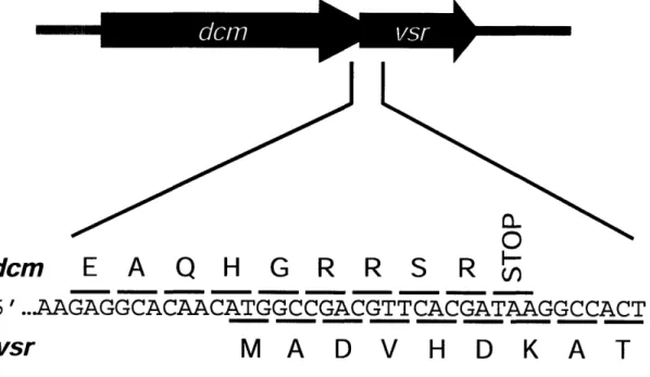
Figure 1.1. Organization of the dcm and vsr genes 53
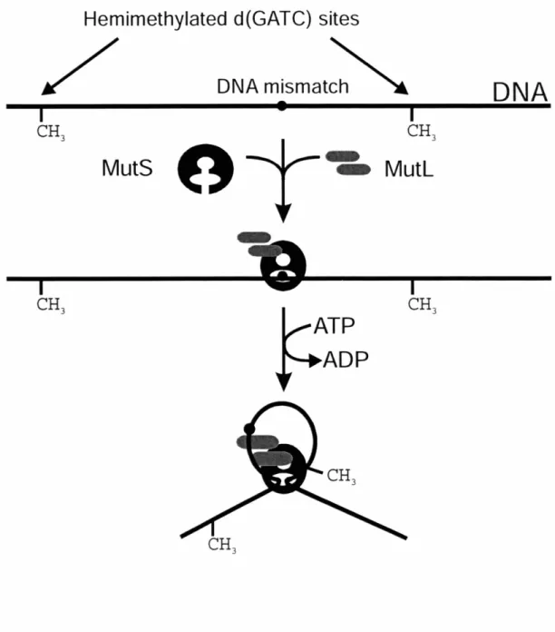
Figure 1.2. Repair of mismatches that arise through methylation and subsequent
deamination of cytosine 54
Figure 1.3. Unmethylated and hemimethylated Vsr substrates . 55
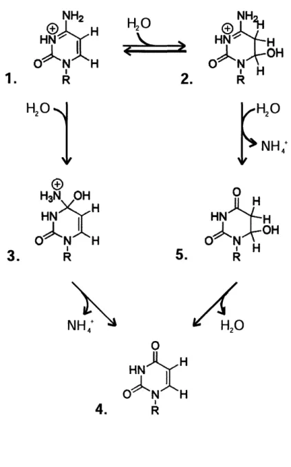
Figure 1.4. Proposed mechanisms for the hydrolytic deamination of cytidine 56
Figure 1.5. Chemistry behind C:G to T:A transition mutations . 57
Figure 1.6. Model for how VSPR could be shaping the E. coli genome . 58
Figure 1.7. Structure of vsr bound to its product - minor groove side 59
Figure 1.8. Structure of vsr bound to its product - major groove side 60
Figure 1.9. Geometric characteristics of C:G and T:G base pairs. 61
Figure 1.10. Methyl direction of mismatch repair . 62
Figure 1.11. Methyl-directed mismatch repair is bidirectional 63
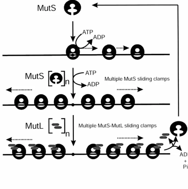
Figure 1.12. ATP-hydrolysis dependent translocation model for MMR . 64
Figure 1.13. Molecular switch model for MMR, part 1 65
Figure 1.14. Molecular switch model for MMR, part 2 66
Figure 1.15. Molecular switch model for MMR, part 3 67
Figure 2.1. Unmethylated and hemimethylated Vsr substrates . 105
Figure 2.2. PhosphorImager results from Vsr nicking assays on unmethylated,
hemimethylated, and control substrates 106
Figure 2.3. Graphical representation of unmethylated and hemimethylated
substrate nicking by Vsr 107
Figure 2.4. PhosphorImagery results from incubating T:G containing duplexes
with VSPR proteins 108
Methodology for assessing VSPR efficiency in vitro. 110 Representative PhosphorImagery results from TLC separation of
mononucleotides at interrogation site. . . . 111
Percentage of VSPR events initiated that went to completion 112 Method for detecting VSPR by monitoring the incorporation of
radioactivity into substrate DNA 135
Extracts from cells over-expressing recombinant Vsr incorporate
[a32P]-dCTP into short duplex substrates via VSPR . 136
Graphical representation of results from measuring VSPR via
radioactivity incorporation 137
Method for detecting VSPR by measuring the depletion of
radioactivity signal from substrate DNA 138
Distribution of VSPR patch lengths created in vitro . 139
Addition of surplus Ligase to VSPR reactions 140
Addition of Ligase to VSPR reactions containing excess substrate . 141
Sites of methylation on the DNA bases 165
Conversion of MNNG to the active methanediazonium ion by
cysteine 166
The R and S stereoisomer of methylphosphotriesters. . . 167
Base pairing models for O6mG:T and O6mG:C 168
Genetic structure and regulation of the adaptive response in E. coli . 169
Direct reversal of base damage by Ada 170
Sequence of ogt and location of PCR primers 206
Sequence of ada/alkB and location of PCR primers . 207
Sequence of mutHand location of PCR primers 208
Sequence of mutL and location of PCR primers 209
Figure 2.6. Figure 2.7. Figure 2.8. Figure 3.1. Figure 3.2. Figure 3.3. Figure 3.4. Figure 3.5. Figure 3.6. Figure 3.7. Figure 4.1. Figure 4.2. Figure 4.3. Figure 4.4. Figure 4.5. Figure 4.6. Figure 5.1. Figure 5.2. Figure 5.3. Figure 5.4.
Sequence of vsr and location of PCR primers
Relative efficiencies of MTase repair in MR defective strains PCR mediated gene replacement
In vivo assay for MTase repair
Hypothetical mechanism for stimulation of MTase repair by VSPR proteins
Bacterial two-hybrid system utilizing domains of adenylate cyclase. Model for mechanism of bifunctional toxicants
Restriction map of pSK278
Restriction map of pSK278-hER-LBD Restriction map of pSK278-hAR-LBD Restriction map of pSK278-hPR-LBD Restriction map of pSK278-hGR-LBD
(A) Piperidine treatment of 5'-d(AATATTGGCCAATATT) treated with compound 1. (B) Retarded mobility of oligonucleotide-1 in the presence of ER-LBD illustrated by EMSA
Survival of MCF-7 (ER+) and MD-MB231 (ER-) cells treated with 1 or chlorambucil
Structure and molecular features of lead compound .
Survival of MCF-7 (ER+) and MDA-MB231 (ER-) breast cancer cells after 2 h exposure to estradiol-linked toxicants .
Figure Figure Figure Figure Figure Figure Figure Figure Figure Figure Figure Figure Figure 5.6. 5.7. 5.8. 5.9. 5.10. 5.11. 6.1. 6.2. 6.3. 6.4. 6.5. 6.6. 7.1. 211 212 213 214 215 216 236 237 238 239 240 241 251 252 266 269 Figure 7.2. Figure 8.1. Figure 8.2.
LIST OF TABLES
Table 1.1. E. coli and S. cerevisiae Proteins Required for MMR 68
Table 2.1. Oligodeoxynucleotides used for cloning Vsr into pET28a and
construction of nicking assay and reconstitution assay substrates 113
Table 2.2. Quantitative results from reconstitution assays containing
Vsr, Pol I, and DNA Ligase protein . 114
Table 3.1. Oligodeoxynucleotides used to construct duplexes containing internal radiolabels at prescribed distances away from the mismatched T 142
Table 3.2. DNA strands containing radiolabels at prescribed distances
away from the T to be mismatched 143
Table 4.1. Relative amounts of DNA alkylated products following reaction of
double stranded DNA with MMS and MNU alkylating agents 171
Table 5.1. PCR primers used for construction of recombination substrates 217
Table 5.2. PCR primers used for analyzing strain genotypes 218
Table 5.3. Sizes of genes and PCR amplification products 219
Table 5.4. Initial plating results from MTase assay on C215 derivatives 220
Table 5.5. Initial plating results from MTase assay on C216 derivatives 221
Table 5.6. Initial plating results from MTase assay on C217 derivatives 222
Table 5.7. Initial plating results from MTase assay on C218 derivatives 223
Table 8.1. RBA= relative binding affinity for the rabbit uterine ER as compared
Scheme 7.1. Synthesis of compound 1 250
CHAPTER
1
1.1. MUTATION AVOIDANCE
The formation of a base pair other than the canonical Watson-Crick G:C or A:T by a polymerase error, or by a chemical or hydrolytic reaction of an existing base pair, will create a mismatch and lead to a mutation if not repaired. Systems for mutation avoidance are present in both prokaryotic and eukaryotic cells (1-15). These mismatch repair (MR) systems are highly conserved from bacteria to humans and play an essential role in maintaining the integrity of the genetic code and health of the host. Cells deficient in MR exhibit a mutator phenotype, in which the spontaneous mutation rate is increased, and the cells are hyper-recombinogenic. In mammals, loss of MR is related to an increased susceptibility to cancer (6,;16). Understanding mismatch avoidance is therefore paramount to human health.
Mismatched base pairs can arise in DNA through multiple processes (17). Errors in replication can cause an incorrect deoxynucleotide to be incorporated into the newly synthesized strand. In fact, in Escherichia coli (E. col), MR contributes almost 1000-fold to the fidelity of DNA replication (4;18). Though, under normal conditions of replication, DNA polymerase III is responsible for proper base selection and insertion
(19) and has associated proofreading activity (20), mistakes are made with a frequency of
10-9 to 10-1°0 (21). Additionally, slippage during replication can result in the formation of
insertion-deletion loops (IDLs), which are composed of bases that are not matched at all. IDLs of less than four nucleotides have been shown to be good substrates for MR
(22-24). IDLs of up to 4 or 5 bases can also be repaired by MR, albeit at lower efficiencies (25). In contrast, heteroduplexes containing large regions of nonhomology are not MR
substrates (26). Recombination between two DNA strands that do not share complete sequence homology can also result in the formation of IDLs and mismatches that are subject to MR (27;28). Base pairs containing a chemically modified nucleobases can also substrates for MR (29). In particular, one mismatched base pair that can arise in DNA naturally is through the deamination of 5-methylcytosine (5mC). Small amounts of 5mC are present in both prokaryotic (30) and eukaryotic (31) DNA, and the increased propensity of 5mC to hydrolytically deaminate elevates the level of T:G mismatches.
- Chapter
1
-The next sections will describe two mismatch repair pathways in E. coli, giving particular attention to the very short patch repair (VSPR) pathway and the cellular components it shares with the methyl-directed mismatch repair (MMR) pathway.
1.2. THE VERY SHORT PATCH REPAIR SYSTEM
In E. coli, the underlined cytosine of the pentanucleotide sequence 5' CCWGG -3' (where W is an A or T) is methylated at the 5-position by a DNA cytosine methyltransferase (32;33) which is the protein product of the dcm gene (34). The 5-methylcytosines (5mC) present in Dcm methylation recognition sites are prone to spontaneous deamination, which results in a T:G mismatch (35;36). If left unrepaired, a C:G -- T:A transition mutation will arise following the next round of DNA replication. To avoid the formation of these mutations, the very short patch repair system removes
and replaces the mismatched T with the proper base (37).
The first indication that such a system existed was observed in 1981 by Margaret Lieb when, in multifactor crosses, the recombination frequencies of certain amber mutations in the repressor (cI) gene of bacteriophage X were higher than predicted by the physical distance separating them (38). Further study of the am6 mutation revealed that these events arose from C -+ T transition mutations in a glutamine codon (5'- CAG -3') located within the Dcm recognition sequence 5'- CCAGG -3' (39), and recombinants had the C:G genotype (37;40). Additionally, Lieb demonstrated that a high frequency of these unexpected recombinants included co-repair of markers within 10 nucleotides of the mismatch but not in areas further than 20 nucleotides (39). Taken together, these findings led Lieb to define a very short repair (VSPR) system that acts on the sequence CTWGG -3' (where T is mismatched with G) to preferentially restore it to 5'-CCWGG -3'. The significance of the 5'- 5'-CCWGG -3' sequence, being that for Dcm recognition and methylation, was not overlooked.
Additional evidence for the existence of VSPR and a proposed role in maintaining Dcm methylation sites came from experiments with heteroduplex DNA (41-43). Substrate containing a T:G mismatch in the sequence 5'- CTAGG -3', which could result from the deamination of the Dcm product, was indeed subject to repair with a high (if not complete) bias in favor of replacing the mismatched T. Increasingly, it was accepted that VSPR functioned to reduce the mutation level associated with the spontaneous
deamination of 5mC created by Dcm.
This hypothesis was corroborated by Lieb in 1991 when she observed that mutation at a particular hot spot was dependent upon Dcm and VSPR activity (44) In lysogens where only Dcm was present, the mutation frequency at the hot spot was 10-fold higher than in wt bacteria. This hot spot disappeared when both Dcm and Vsr were removed, while addition of Vsr to wild type (wt) bacteria caused a 4-fold decrease in mutation frequency. It is now accepted that a primary role of VSPR is to reduce the number of mutations that result from cytosine methylation and deamination (45).
In the years following the discovery of the VSPR system, a number of genetic and biochemical studies were conducted to implicate the proteins involved in the repair process. In 1987, Lieb reported (46) that the protein products of mismatch repair genes
mutS and mutL were involved in VSPR on the basis that the frequency of in vivo crosses
in cells lacking either one of these genes was greatly diminished. Strains lacking mutHor
uvrD exhibited no difference from wt. Interestingly, the recombination frequency was
diminished in dcm6 cells lacking the Dcm methylase, leading Lieb to conclude that the protein product of dcm had a repair function in addition to its methylase function. At the time, however, there was no explanation for this mixed phenotype.
In a report by Dzidic and Radman in 1989 (47) the genetic requirements for hyper-recombination by VSPR were extended to include polA (the gene for Pol I). In fact, the recombination frequency was diminished in cells lacking the polA gene sequences for either its 5' --+ 3' exonuclease or polymerase domains. This observation
- Chapter
I
-led the authors to conclude that both activities of Pol I were paramount for effective VSPR. In contrast, xth-, nth- and nfo- strains showed wt recombination frequencies.
In 1990, Sohail reported (48) that the two phenotypes of the dcm mutants, lacking both methylation and repair activities, had been genetically pinpointed by mapping. Additionally, they cloned genes that complemented the chromosomal mutation for methylation, but not repair, and vice versa. The gene essential for methylation, dcm, was predicted to code for a 473-amino-acid protein and was not required for VSPR, while the second gene, vsr, was predicted to code for a 156-amino-acid protein which was required for VSPR but not for methylation. The two genes were transcribed from a common promoter even though they were found in different translational registers. The 5' end of
vsr overlaps the 3' end of dcm by 7 codons in a +1 reading frame (Figure 1.1). It was
hypothesized that this genetic arrangement assures Vsr is always produced along with Dcm to minimize the mutagenic propensity of cytosine methylation. Sohail also noted
(48) that although the two genes are transcribed from a common promoter, two separate
proteins are produced. Thus, the genetic arrangement of dcm and vsr was the source of confusion regarding initial experiments conducted on the dcm6 strain, as they lack vsr in addition to dcm. This misunderstanding led to the incorrect inference (46) that the dcm product is required for VSPR.
One year later, Hennecke showed that the vsr gene product was an 18 kDa mismatch endonuclease that produced a single strand nick 5' to the underlined T within duplex DNA sequences 5'- CTWGN -3' or 5'- NTWGG -3' (where N is A, T, C, or G) when the T was opposite a G (49). The incision was mismatch-dependent and strand-specific. These results therefore illustrated that Vsr was the protein that initiates VSPR. Consistent with these observations, strains lacking vsr are completely defective in VSPR and have a very high frequency of C -- T transitions at 5mC sites (38;50).
Altogether, these experiments provided a model for the mechanism of VSPR (Figure 1.2). Evidence suggested that Vsr initiates repair by producing a nick 5' to the mismatched T. The previous implication of MutS and MutL in mismatch recognition
suggested these proteins stimulated VSPR by assisting Vsr in finding or incising substrate mismatches. The exact mechanism of Vsr stimulation, however, is still unknown (see below). Subsequent to incision, Pol I removes the T residue with its 5' -, 3' exonuclease activity and commences repair synthesis in the usual template-directed fashion, with the short synthesis tracts typical for the enzyme (51). DNA Ligase is reasonably assumed seal the nicked strand on which repair has been performed.
1.2.1. DCM- IN VIVOAND IN VITROPROPERTIES
Dcm is the 53 kDa product of dcm, which is located at 43 minutes on the E. coli chromosome (34). The protein methylates the underlined cytosine residue in the sequence 5'- CCWGG -3' (where W is A or T) at the C5 position of the pyrimidine ring
(32). The recognition sequence is palindromic and thus also results in methylation at the
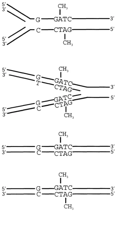
corresponding position of the opposite strand, as well (32). The deamination of a 5mC in fully dcm methylated DNA gives rise to a T:G mismatch in a hemimethylated duplex. If the G containing strand contains an unmethylated C, the mismatched duplex is said to be unmethylated (Figure 1.3). In E. coli, approximately 1% of all cytosine residues are methylated by Dcm (52). This translates roughly into approximately 10,000 Dcm recognition sites in the E. coli genome, or on average one recognition site every 400-500 base pairs.
Not surprisingly, the role of Dcm has been a topic of much discussion. However, no obvious biologically interesting phenotype of dcm mutants has been reported (52), and the reason for the existence of the dcm-vsr operon remains unclear (53). The closest characteristic to a phenotype for dcm mutants is the ability of their isolated genomic DNA to be cut by EcoRII endonuclease, which is part of a restriction modification (RM)
system and recognizes the same sequence as Dcm (54;55). The EcoRII
methyltransferase and Dcm share a high degree of sequence similarity (48), are functionally very similar, and are likely derived from a common ancestor.
- Chapter
I
-An EcoRII RM system was found in drug resistance transfer plasmids (56), and therefore the function of Dcm could be to protect the host's DNA from invasion (53). In support of this theory, cells bearing Dcm were shown to be resistant to the virulence of a parasitic EcoRII gene complex (53). It is also interesting to note that 5'- CCWGG -3' is one of the most commonly utilized recognition sequences for RM systems (53). As of March 20th, 2006, the REBASE database (57) identified 169 of the total 3773 RM systems as recognizing the 5'- CCWGG -3' sequence.
It has also been suggested that methylation by Dcm could be involved in gene regulation (52). A Dcm methylation site has been identified in the promoter region for
lexA (58), however no relationship between the Dcm methylation status and expression
has been established. Interestingly, some Dcm sites were found to remain unmethylated in dcm+ cells (59), suggesting that some sites are protected by DNA binding molecules or by DNA topology, and could play a role in a yet to be discovered biological process.
The resultant 5mC residues are subject to "spontaneous" hydrolytic deamination
(60;61), analogous to the process that converts cytosine to uracil (62). Two chemical
mechanisms have been proposed for the hydrolytic deamination of cytosine (62;63). One mechanism involves direct attack at the 4-position of the pyrimidine ring by a hydroxyl ion, and the other involves an addition-elimination reaction with the formation of dihydrocytosine as an intermediate (Figure 1.4). While the mechanism involving direct attack has the advantage of simplicity, relatively little direct evidence has been reported to support its occurrence in reactions involving nucleophilic displacement of the amino group of cytosine. On the other hand, the route involving addition to the C=C double bond is similar to that established for the reaction between hydroxylamine and cytosine derivatives (64). Additionally, it has been directly demonstrated that nucleophiles can add reversibly to the 5,6 double bond of a uracil ring (65). The deamination of dihydrocytosine derivatives (resembling intermediate 2 in Figure 1.4) was also shown to be a rapid process (66). Therefore, evidence suggests that an addition-elimination reaction is a probable mechanism for deamination of cytosine and 5mC in DNA.
Deamination of either cytosine or 5mC gives rise to products that yield C:G --T:A transition mutations upon the next round of replication (Figure 1.5). It is noteworthy, however, that the extent of cytosine and 5mC deamination is not equivalent. The higher rate of deamination for 5mC as compared to C (60;61), coupled to the finding that T:G mismatches are repaired less efficiently than U:G mispairs (67) results in cytosine methylation sequences being hot spots for C -, T mutations. Interestingly, the accumulation of these hotspots in E. coli, depends on the phase of growth.
In exponentially growing cells (67), VSPR reduced the 5mC - T mutations by a
factor of four. However, cells deficient in Dcm displayed more than 10-fold decrease in the 5mC -- T mutation level. Therefore, the inefficiency of VSPR during exponential growth allows Dcm sequences to be hotspots for C -+ T mutations. In contrast, cells in stationary phase (68) are completely protected by VSPR from Dcm sequences being hotspots. It was observed that the 5mC - T mutation rate in cells that were stored for long periods of time was equivalent to that for cells lacking cytosine methylation. It was also observed that the 5mC - T mutation rate for cells lacking VSPR was consistent
with the rate of 5mC deamination at 37 C. Subsequently, it was shown that the concentration of Vsr in the cell is low during exponential phase and increases when cells start to enter stationary phase (69). To summarize, the ability of VSPR to protect against 5mC -+ T mutations is linked to the amount of Vsr that is available to initiate repair.
Possible explanations for how the amount of Vsr is controlled in the cell (70;71), and why VSPR was designed to be less efficient in exponential phase have been proposed
(72), and are discussed below.
In eukaryotes, methylation of cytosine occurs within CpG dinucleotides (73), and has been implicated, among other functions, in the regulation of gene expression (74). A large number of genetic diseases involve C to T changes at CpG sites (75), including 13-thalassemia, hemophilia, and cancer (76-79), therefore establishing the maintenance of the sites as critical.
- Chapter
1
-1.2.2. VSR- IN VIVO AND IN VITRO PROPERTIES
Vsr is the 18 kDa product of vsr, located at 43 minutes on the E. coli chromosome
(48). This 156 amino acid protein functions as a strand-specific endonuclease to initiate
repair of T:G mismatches in the sequence 5'- CTWGG -3' (where T is mismatched with a G) (49). These T:G mismatches arise through the methylation of the underlined C in the sequence 5'- CCWGG -3' by Dcm, followed by deamination of that methylated residue. Vsr, therefore, helps to reduce the mutation level associated with the spontaneous deamination of 5mC created by Dcm.
In 1995, Lieb and Rehmat (80) reported recombination experiments conducted on derivatives of Dcm sequences. Their findings showed that VSPR was most efficient in the pentanucleotide sequence 5'- CTAGG -3', however there was also significant correction when either the 5' most C or the 3' most G was replaced by another base. In
fact, some VSPR activity was observed in all of the sequences tested.
The same year, Glasner confirmed the broad sequence specificity of Vsr biochemically (81). Using fluorescence-labeled oligodeoxynucleotide (ODN) substrates, 14 substrates were found to be processed by the enzyme, with varying efficiencies. The substrates differed at one or two positions from the canonical pentanucleotide substrate sequence CTWGG -3'. Not surprisingly, the sequences CTAGG -3' and 5'-CTTGG -3' were the most reactive substrates with relative rates of 100 and 68% respectively. Sequence derivatives where the first or last base was varied (5' NTWGG -3' or 5'- CTWGN --3') were all repaired with relative rates of 22% or higher. Therefore, Vsr will act efficiently on T:G mismatches in many sequence contexts.
This same study (81) investigated potential relationship between the efficiency of VSPR on individual sequences and their underrepresentation in the genome. It was previously hypothesized (82;83) that if a T:G mismatch resulted from misincorporation of a G opposite a T in the template DNA strand, it would be a potential substrate for both the methyl directed mismatch repair MMR and the VSPR systems. Further, if such a
mismatch evaded correct repair by MMR, VSPR might incorrectly carry out "repair" by removing and replacing the T. Therefore, over time, VSPR could operate on mismatches not intended for the system and would provide a driving force in depleting the genome of the T-containing sequences (for example 5'- CTAG -3') and enriching it for the corresponding C-containing sequences (for example 5'- CCAG -3'). Ultimately, VSPR could have a hand in modifying the E. coli genome (Figure 1.6).
Interestingly, Glasner's data (81) reflected a high quality of positive correlation between the relative second-order rate constants on VSPR removal of mismatched T and the underrepresentation rank for the 14 substrates studied. It was hypothesized that this process would lead to corresponding sequence polymorphisms in the total E. coli K-12 population. In a case where two genomes that differ in one or more such polymorphic sites combine, the T:G mismatches that are substrates for Vsr would be repaired in favor of G and thereby transfer the polymorphism to the other genome involved. Additionally, because VSPR replaces a short stretch of DNA downstream of the T:G mismatch, any mismatches within the reach of the DNA synthesis tract would also be repaired in favor of the G containing strand. In conclusion, Glasner postulated that VSPR might assist in the patchwise unidirectional transfer of genetic information from one DNA molecule to another, and contribute to the overall fitness of E. coli K- 12.
Another function for VSPR appears to be to repair U:G mismatches in Dcm sequences that arise by deamination of cytosine (84). It was found that deamination of cytosine can be promoted by the Dcm methylase. In the absence of S-adenosylmethionine, DNA cytosine methylases promote the formation of a 5-dihydro intermediate that is much more susceptible to deamination than cytosine (85-88). Normally, uracils that arise by the deamination of C are recognized by uracil DNA glycosylase and are repaired by the base excision repair pathway. However, genetic reversion assays (84) revealed that although VSPR is not as efficient as the glycosylase system at correcting U:G mismatches to C:G, the VSPR system does contribute to the correction of U:G mismatches in Dcm sites.
- Chapter
-The ability of Vsr to nick 5' to U:G mismatches in Dcm sequences was also studied biochemically (89). Results indicated that the single turnover rate for the U:G mismatch was about half that for the T:G mismatch. These biochemical parameters, however, were determined using protein that was not fully active. Turner observed that much of the Vsr purified from over-expressing E. coli strains is catalytically inactive, and less than 1% of the protein bound DNA (89). In fact, it was not until direct titration experiments were conducted (89;90), in which increasing amounts of Vsr were added to a constant amount of substrate DNA, that the activity of an individual protein preparation could be determined. Ultimately, the authors concluded that the U:G mismatch had a specificity constant (kst/KD) 10-fold lower than that for a T:G mismatch, due to an increase in equilibrium dissociation constant (KD) and a reduction in the rate constant for single turnover (kst). Similarly, the specificity constant for a T:G mismatch in an unmethylated duplex (Figure 1.3) was 10-fold less than that for a T:G mismatch in a hemimethylated duplex (Figure 1.3). Crystallographic data containing Vsr bound to duplex DNA (91;92) later suggested that the methyl group present in the hemimethylated duplex may pack against an aliphatic region of the protein (Phe-77, Lys-78, Val-79), and assist binding at the protein DNA interface. These findings support the notion that the biological role of Vsr is to initiate repair of T:G mismatches that arise from deamination of the Dcm product. Even so, the ability of Vsr to incise unmethylated substrates has also been confirmed in vivo (46).
To obtain the highest activity from Vsr protein, it has been reported (93) that current preparations should include freshly transforming E. coli with the plasmid that directs Vsr over-expression, purifying Vsr from the over-expressing strains, and using the purified protein within two weeks of preparation. Furthermore, loss of Vsr activity due to aggregation of protein during storage has been reported (94). Therefore, for best results, Vsr protein must be prepared fresh each time. While many changes have been made to the current methods for preparing Vsr, the reasons for the discrepancy in activity are not understood. Perhaps, as discussed below, the low activity of some Vsr preparations can be attributed to the loss of 14 residues from N-terminus of Vsr upon storage. Whatever
the case, adhering to these precautions has allowed fully active Vsr to be isolated and used to measure the binding and kinetic constants for its interaction with substrate DNA.
The binding affinity of Vsr for its hemimethylated substrate (Figure 1.3) was measured using competition gel-retardation assays (93). Complexes of Vsr and a 32p_
labeled duplex were formed (in the presence of Ca2 +) and increasing amounts of unlabelled competitor DNA were added. Utilizing these methods, a KD of 10.9 nM was determined. Single turnover reaction conditions were utilized to saturate the substrate and reflect rates of turnover after substrate binding yet before product release. Under these conditions, the kst was 7.5 min'. For the unmethylated substrate (Figure 1.3), a kst of 2.8 min-' for Vsr was independently reported (94). A comparison of the kinetic measurements between the hemimethylated and unmethylated substrates suggests that the activity of Vsr is roughly 2.5-fold more efficient on the hemimethylated substrate. The kinetics of Vsr on these two substrates, however, have never been tested in parallel with fully active protein.
Determination of both KD and kt values (93) allowed the computation of the single turnover specificity constant (kst/KD), which was 0.7 min-' nM-1 for Vsr. This constant is generally agreed to be the most suitable metric for comparing the ability of different proteins to bind DNA substrates (95-97). In reactions where the steady state rate of hydrolysis is measured, the specificity constants oftentimes reflect the product release step as this step is commonly rate limiting. In such cases, the steady state constants are not particularly informative for comparisons. In light of the observation that Vsr produces a very stable enzyme product complex (89), the single turnover constants are indeed more useful than the steady state values.
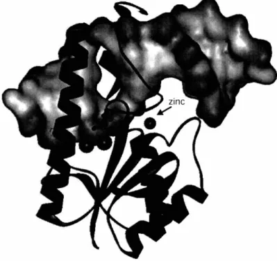
1.2.3. STRUCTURAL DATA FOR VSR
To date, three structures of Vsr have been solved by X-ray diffraction and are available on the RCSB Protein Data Bank. The first structure, IVSR, (98) was solved to
- Chapter
1
-1.80 A and contains residues 21 to 156. The second structure, 1CWO, (91) was solved to 2.30 A and contains residues 2 to 156 of the Vsr protein bound to product duplex DNA. The third structure, 10ODG, (92) was solved to 2.80 A and contains residues 22-155 of the Vsr protein bound to its reaction product site in duplex DNA. Both of the Vsr-DNA complexes contain hydrolyzed substrate. The following discussion is derived from the data presented in these three publications, and will go into the finer details of the structures and what can be learned upon their inspection.
Vsr has a three-layered a3/la fold that is stabilized by a structural zinc site (98). The Zn2+ is coordinated by four protein residues (Cys-66, His-71, Cys-73, Cys-117) in a tetrahedral arrangement, three of which are on one loop. This coordination is distinct from the majority of zinc sites found in proteins. Typically, the four coordinating residues are divided equally on two strands (99). While Vsr shares no sequence homology with other endonucleases (49), the overall topology of Vsr resembles the type II restriction endonucleases, which have been extensively reviewed elsewhere (100).
Members of this family include a growing list of restriction enzymes, as well as the DNA repair nucleases MutH (101), X exonuclease (102), and archeal Holliday junction resolvase (103). However, more specific comparison reveals that Vsr is not a standard type II endonuclease, as previously expected.
The central f3 sheet of Vsr is composed of two short and four long P strands, and braced on either side by a-helicies. The active site of this type II restriction endonuclease family is located in a niche formed in the sheet and is composed of the catalytic motif PDX(6_30)(D/E)XK (104;105). For this family of enzymes, these conserved residues are superimposable from enzyme to enzyme and are essential for catalytic activity. Typically, the first two residues are acidic and bind an essential divalent cation. The last residue, typically Lys, is the most variant amino acid between enzymes, and has been proposed to stabilize the transition state (106;107) or orient the attacking water (108;109). Superimposition of the 13 sheet of Vsr onto members of the type II restriction enzyme family shows that Asp-51 (D) of Vsr (a catalytically essential residue) is the first conserved Asp of the catalytic motif. In keeping with the motif, this
residue does bind an essential divalent cation. The rest of the catalytic motif for type II restriction enzymes, however, is not conserved in Vsr.
Phe-62 in Vsr superimposes onto the second catalytic residue of the motif, however this residue can not engage in metal coordination. The third residue in the motif correlates to a partially conserved His or Asp 64 in Vsr, and while this residue is important for activity, it is not essential. Additionally, His 69, which is close in vicinity to His 64 but positionally distinct from the catalytic residues of type II restriction enzymes, was found to be absolutely required for Vsr activity. These findings have disqualified Vsr from strict conservation with the type II restriction enzyme catalytic motif, and imply that the mechanism for Vsr differs from that of typical type II restriction enzymes. These structural data helped to clarify the discrepancies between how the motif is used for typical type II restriction enzymes and Vsr.
In contrast to typical type II restriction enzymes which utilize highly coordinated metal ions, Vsr coordinates two Mg2+ions through extensive metal water clusters and has very little direct coordination with the Mg2+ions. Besides Asp-51, the only other direct metal coordination includes the main chain carbonyl of Thr-63, one residue away from the predicted Phe-62. His-64 and Glu-25 coordinate water molecules in the water metal clusters. Alanine scanning has shown each of these residues to be important for Vsr activity. The crystal structure also revealed that the product DNA duplex, containing a 5' phosphate on the mismatched thymine and a 3' hydroxyl on the neighboring cytosine, was involved in Mg2 + coordination. The oxygen on the C3' of the cytosine coordinates one metal ion. Additionally, the 5' phosphate of the Vsr reaction product was coordinated with both metal ions. Interestingly, His-69 was coordinating one of the terminal phosphate oxygens. This coordination is atypical since Mg2 +generally prefers the harder oxygen atoms of carboxyl residues as ligands.
In addition to the structural involvement of zinc, Vsr requires Ca2+ or Mg2+ for substrate binding (90). This observation is in contrast to a number of restriction enzymes including EcoRI and BamHI, which require metal ions for catalysis but not for binding
Chapter 1
-(100). This distinction implies that the metal ions in Vsr play a structural role in addition
to a catalytic role. Consistent with this idea, it was observed that Vsr can bind substrate, but not cleave it, in the presence of Ca2+ (89). This property allows the binding and
cleavage activities of Vsr to be separated, and has been useful for a number of biochemical studies (89;90;93).
In conclusion, the first structure of the Vsr protein alone revealed many important features of the molecule including its topology in relation to type II restriction enzymes, certain conserved elements in its catalytic site, its novel metal binding coordination, and why certain residues are important or critical to catalysis. The second structure of Vsr contained the protein bound to duplex DNA product, and provided valuable information on how the two molecules interact.
The N-terminus of Vsr, which was partially removed in the truncated structure, does not form a single alpha helix as was predicted by secondary structure analysis of the primary sequence (91). Rather, the N-terminus is composed of two short alpha helices separated by a flexible linker (Figure 1.7). These results are in good agreement with limited proteolysis studies which suggested that Vsr had exposed turn-arounds at amino acids 14 and 20 (91;98). The main core of Vsr binds the DNA duplex from the major groove while the most N-terminal helix (Lys-7 to Arg 15) pins down the DNA from the minor groove side. A large opening has been formed in the center of the DNA duplex via intercalation of multiple side chains between the DNA strands. The protein surface shows a strong electrostatic and steric complementarity to the DNA duplex. The basic regions follow along the phosphate backbone of the DNA and a strong acidic patch is found close to the metal binding sites in the catalytic center. Met-14 and Ile-17 project from the minor groove side and leave only enough space for the phosphate backbone to pass on either side.
Remarkably, three aromatic residues (Phe-67, Trp-68, and Trp 86) insert into the DNA duplex from the major groove side (Figure 1.8). Phe-67 stacks with the A:T base pair in the recognition sequence, Trp-68 stacks with the mispaired T, and Trp-86 stacks with two sugars on the strand opposite the mismatched T. The major groove is opened
up, the DNA is significantly unwound, and the duplex is kinked by approximately 44 degrees. It is surmised then that these residues recognize and enhance the structural distortion in DNA resulting from the T:G mismatch (99). Not surprisingly, these three residues are invariantly conserved in the Vsr family (91).
Two of the intercalating residues (Phe-67 and Trp-68), are adjacent to His-69, which is absolutely required for activity and proposed to activate the attacking water. Additionally, all these residues are part of the loop that contains three (Cys-66, His-71, and Cys-73) of the four residues that coordinate the structural Zn2+ion. Therefore, these residues are constrained conformationally and precisely aligned for mismatch recognition and DNA strand incision.
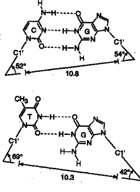
Vsr recognizes its mismatch within the DNA helix, primarily by the structural distortion in DNA caused by wobble pairing between T and G (Figure 1.9). In a T:G wobble pair, the 06 and N1 of guanine hydrogen bond to the N3 and the 02 of thymine, respectively. This pairing is preferred by Vsr for a number of reasons. Firstly, the geometry of a T:G mispair is such that it can enter the active site of Vsr. A normal G:C base pair would encounter steric clash between the O2 of cytosine and Thr- 19. However, because the 02 of T in a T:G mispair is pushed into the major groove, these mismatches can fit into the Vsr active site. Secondly, the wobble pairing achieves a shape such that the scissile phosphate of the mismatched thymine is in proximity to the catalytic residues in the active site. In other words, normal base pairs would be situated such that their phosphate group would not be proximal to the Vsr catalytic residues. Thirdly, the wobble pairing disrupts the DNA stacking between the mismatched G and the base on the 5' side such that it causes (1) an opening of the DNA by unwinding, (2) increased distance between G and its 5' neighbor, and (3) the ability of Vsr to recognize and bind the G:T mismatch by intercalating within the recognition site. In addition to the ability of Vsr to recognize T:G mismatches via the drastically different geometry, there are a few hydrogen bonding contacts between the mismatched base pair and Vsr residues. The invariant Asn-93 binds to the 04 of thymine and presumably plays a role in cytosine discrimination. Guanine is bound at its 06 position by Lys-89 and at its N\ by the main
- Chapter
1
-chain of Met-14. Biochemical analysis of mutants in these residues will be necessary to determine the extent to which these interactions are required for recognition.
The five base pair recognition sequence occurs through an interlaced network of direct and water mediated hydrogen bonding interactions. Two notable features are that (1) Arg-120 has the only direct contact with the major groove, and it binds to the G:C base pair that is required for recognition, and (2) the third base pair in the middle of the recognition sequence (which can be A:T or T:A) has the most interactions. The sequence specificity of Vsr may therefore lie primarily in the ability of duplex DNA to tolerate intercalation.
The third structure of Vsr was obtained using a DNA duplex that generated a site corresponding to the hemimethylated cleavage product of Vsr (92). This second protein/DNA structure is quite similar to the co-crystal structure previously reported (91), but some differences are worth mentioning. The structure shows a 60° angle along the minor groove as compared 44° in the previous structure solved by Tsutakawa. Additionally, the middle A:T base pair (in the sequence 5'- CTAGG -3') was in a Hoogsteen conformation, with the A being in the syn conformation. This pairing is believed to exist in bulk DNA at low levels (in equilibrium with the Watson-Crick form) but has not, until recently, been observed in a protein-DNA complex (110). To explain why this Hoogsteen pairing was first observed in this structure, it is relevant to note that, in this structure, Vsr is bound to a hemimethylated DNA substrate. The methyl group on the C5 position of C would potentially clash with the C5 methyl group of T if the thymine residue was not positioned closer to the minor groove through a Hoogsteen pair. Therefore, it was hypothesized that, in this structure, the Hoogsteen A:T base pair is more energetically favorable than a normal A:T base pair that would introduce methyl-methyl clash.
1.2.4. CATALYTIC MECHANISM OF VSR
Taken together, these structural data (91;98) lead Tsutakawa to propose a catalytic mechanism for Vsr (99). When Vsr binds the mismatch-containing DNA duplex, the scissile phosphate is coordinated and stabilized by both metal water clusters. His-69 abstracts a proton from one of the waters in the metal water clusters and the resulting activated water attacks the phosphate group between the mismatched thymine and its 5' cytosine neighbor. The orientation of the His-69, terminal phosphate, and deoxyribose oxygen groups suggests an inline attack. The lack of significant side chain conservation in the area surrounding the deoxyribose oxygen suggests that its protonation is from a water molecule and not the protein.
Additional support for His-69 acting as a catalytic base comes from the observation that replacement of this residue with alanine results in a variant with dramatically reduced activity (91;98). Additionally, a hydrogen bond between His-69 and Asp-97 would increase the pKa of His-69, facilitating its role as a base (99). Several endonucleases including DNaseI (111), exonuclease III (112), and HAP1 (113) use a similar Asp-His pair to provide basic catalysis.
The two magnesium water clusters are also probably required for stabilizing the additional negative change that develops on the scissile phosphate in the transition state. It has also been hypothesized that one of the magnesium ions may have a role in correctly aligning the activated water molecule for attack, while the other magnesium ion may provide Lewis acid catalysis by coordinating to the leaving group and neutralize the developing negative charge on the scissile phosphate (93).
Furthermore, the possibility of His-69 involvement with direct attack of the scissile phosphate to form an enzyme-DNA covalent intermediate, subsequently hydrolyzed by water, was excluded by Elliot and coworkers on the basis of stereochemical analysis of the phosphate group (93;114,115). They deduced that the formation and subsequent hydrolysis of an enzyme-DNA intermediate would proceed
Chapter 1
-with two displacements at the phosphorus atom and therefore two inversions of configuration. Ultimately, the resulting product would have overall retention of stereochemistry (115;116). Conversely, if His-69 deprotonates a water molecule which then attacks the scissile phosphate directly, only one displacement occurs at the phosphate and the resulting product would have overall inversion of stereochemistry. Experiments conducted using H2180 and Vsr substrates that contained the Rp phosphorothioate isomer at the scissile phosphate site showed that the Vsr reaction proceeds with inversion of configuration at the scissile phosphate, and, therefore, His-69 functions as a base to deprotonate the attacking water molecule.
The final catalytic requirement for nucleases is either a Lewis or a proton acid to interact with, and thereby stabilize, the developing negative charge on the leaving group
(114;117). In the case of Vsr, the leaving group is the 3'-bridging oxygen atom at the
scissile phosphate. To probe if stabilization of the leaving group is achieved through coordination with a metal center, this group was modified to contain a 3'-phosphorothioate (which coordinates Mn2+ better than Mg2+) and binding and hydrolysis rates were measured (93). While binding of Vsr to this modified substrate was unchanged, hydrolysis in the presence of Mg2 +was strongly reduced. However, cleavage was fully restored to wild type levels when Mg2+ was replaced with Mn2+, strongly suggesting that one Mg2+serves as a Lewis acid and stabilizes the negative charge on the leaving group as it develops.
In summary, when Vsr binds to a T:G mismatch in duplex DNA, the scissile phosphate is coordinated and stabilized by two Mg2+ water clusters. His-69, acts as a general base and abstracts a proton from a water in the metal water cluster. The resulting activated water conducts a direct inline attack on the phosphate group between the mismatched thymine and its 5' cytosine neighbor. The increased negative charge in the transition state is localized on the two non-bridging oxygen atoms and is stabilized by the two Mg2 + water clusters. One of the Mg2+ions also acts as a Lewis acid to labilize the leaving group by neutralizing the negative charge that develops as the reaction proceeds.
1.3. METHYL-DIRECTED MISMATCH REPAIR
-LESSONS FROM STREPTOCOCCUS PNEUMONIAE
Evidence of the existence of mismatch repair in prokaryotes and the idea of strand discrimination originated from early work involving transformation studies in
Streptococcus pneumoniae (S. pneumoniae) (118). In a S. pneumoniae wt strain, the
efficiencies to which different transformants integrated was varied. Mismatch repair was proposed to be responsible for this phenomenon because heteroduplexes formed during integration would have contained mismatches, since the regions involved in recombination were not completely homologous. It was also observed that the transformation efficiency was dependent not only upon the system's preference for undertaking repair on the donor strand, underscoring strand discrimination, but also on its preference for the type of mismatch on which repair was carried out. In other words, different mismatches were repaired with different efficiencies.
Treatment of S. pneumoniae wt cells with mutagens led to the isolation of mutants that did not discriminate between high and low efficiency markers (119). The mutations responsible for this phenotype were designated hex (originally for "high efficiency unknown [x]", now for heteroduplex repair deficiency) (118). Interestingly, hex mutants showed an elevated level of mutation accumulation (120), suggesting that the hex-dependent MMR system could play a role in mutation avoidance.
It was hypothesized that strand discrimination was established by the ability of the system to recognize that donor strands possessed strand breaks at their ends (121). In support of this hypothesis, low efficiency markers were sensitive to donor DNA that had been UV irradiated (and thus contained a high number of strand breaks) in a fashion that required the hex genes (118;122). Therefore, the mutator phenotype of the hex mutants was likely due to their inability to recognize strand breaks and discriminate between strands (123-125). Since the lagging strand of DNA is continuously forming breaks at the ends of its Okazaki fragments during replication (124;126) it is believed that hex