HAL Id: hal-02339909
https://hal.archives-ouvertes.fr/hal-02339909
Submitted on 31 Oct 2019HAL is a multi-disciplinary open access archive for the deposit and dissemination of sci-entific research documents, whether they are pub-lished or not. The documents may come from teaching and research institutions in France or abroad, or from public or private research centers.
L’archive ouverte pluridisciplinaire HAL, est destinée au dépôt et à la diffusion de documents scientifiques de niveau recherche, publiés ou non, émanant des établissements d’enseignement et de recherche français ou étrangers, des laboratoires publics ou privés.
Impact of a Model Soil Microorganism and of Its
Secretome on the Fate of Silver Nanoparticles
Elise Eymard-Vernain, Cećile Lelong, Ana-Elena Pradas del Real, Romain
Soulas, Sarah Bureau, Vanessa Tardillo Suarez, Benoit Gallet, Olivier Proux,
Hiram Castillo-Michel, Géraldine Sarret
To cite this version:
Elise Eymard-Vernain, Cećile Lelong, Ana-Elena Pradas del Real, Romain Soulas, Sarah Bureau, et al.. Impact of a Model Soil Microorganism and of Its Secretome on the Fate of Silver Nanoparti-cles. Environmental Science and Technology, American Chemical Society, 2017, 52 (1), pp.71-78. �10.1021/acs.est.7b04071�. �hal-02339909�
1
Impact of a model soil microorganism and of its secretome on the
2
fate of silver nanoparticles
3
Eymard-Vernain Elise 1,2, Lelong Cécile 2, Pradas del Real Ana-Elena 1 3, Soulas Romain 4, 4
Bureau Sarah 1, Tardillo Suarez Vanessa 5, Gallet Benoit 6, Proux Olivier 7 8 , Castillo-Michel 5
Hiram 3 and Sarret Géraldine 1* 6
7
1 ISTerre (Institut des Sciences de la Terre), Université Grenoble Alpes, CNRS, Grenoble, 8
France 9
2 Université Grenoble Alpes, CEA Grenoble, Lab Chim & Biol Met, UMR CNRS CEA UJF, 10
ProMD Team, BIG, Grenoble, France 11
3 ID21, ESRF-The European Synchrotron, CS40220, 38043 Grenoble, France 12
4 LITEN, CEA Grenoble, 17 Rue des Martyrs, 38054 Grenoble, France 13
5 ID16b, ESRF-The European Synchrotron, CS40220, 38043 Grenoble, France 14
6 Université Grenoble Alpes, CEA, CNRS, IBS, F-38000 Grenoble, France 15
7 Observatoire des Sciences de l'Univers de Grenoble (OSUG), UMR CNRS 832, Université 16
Grenoble-Alpes, F-38041 Grenoble Cedex 9, France 17
8 BM30B/CRG-FAME, ESRF, Polygone scientifique Louis Néel, 71 avenue des Martyrs, 18
38000 Grenoble, France 19
21
Abstract
22
Sulfidation is a key process for silver nanoparticles released from consumer products in the 23
environment. This study focuses on the impact of a model soil microorganism, Bacillus subtilis, 24
on the fate of pristine and already sulfidized Ag-NPs. The nanoparticles were incubated with 25
the initial growth medium, isolated secretome and living bacteria, and characterized for their 26
size and morphology, agglomeration state, structure and Ag speciation. No Ag internalization 27
or sorption on the cell wall was detected. A partial sulfidation, leading to an Ag-Ag2S core-28
shell structure, was observed in the presence of the secretome, and the rate limiting step of the 29
reaction was the oxidation of Ag0, and it was favored near the crystal dislocations. The 30
sulfidation was complete in the presence of the living bacteria and followed an indirect 31
pathway. Both crystalline Ag2S and amorphous Ag2S and/or Ag-thiol were identified. At the 32
opposite, the bacteria had no impact on Ag2S. These results suggest that microorganisms 33
participate to the sulfidation of Ag-NPs in aerobic systems such as unsaturated soils, and thus 34
affect the bioavailability of Ag. It is important to take these transformations into account during 35
exposure experiments, since they drastically change the exposure conditions. Finally, the 36
secretome of B. subtilis might be used for the green synthesis of Ag-Ag2S core-shell 37 nanoparticles. 38 39 40 41 42
43
Introduction
44
Because of their broad-spectrum antimicrobial properties, silver nanoparticles (Ag-NPs) 45
are increasingly produced and used in consumer products (cosmetics and personal care 46
products, food and food packaging, textile, electronics) and also in medicine and agriculture.1 47
This increased use has raised some concerns, both from a toxicological point of view for 48
consumers during the use of these products, and from an ecotoxological point of view since 49
Ag-NPs and their secondary products are expected to end up in environmental compartments, 50
especially in soils, at the end of their life cycle.2-3 Indeed, Ag-NPs released in wastewater 51
accumulate in sewage sludge during wastewater treatment.4 In Europe, 40 to 50% of this 52
biosolid is applied on agricultural soils as amendment.5 53
Since the last decade, the ecotoxicity of Ag-NPs has been studied at various levels of 54
complexity, from single organisms in growth media to complex systems such as soils and 55
natural waters6-7. The impact on bacteria has been the focus of many studies since Ag-NPs are 56
primarily used as biocides. This property results from several effect of Ag-NPs on bacteria 57
including the generation of reactive oxygen species (ROS), the disruption of the membrane 58
integrity, the interaction of Ag ions with proteins and disruption of their regular function, and 59
the interference with DNA replication, causing DNA damage.8-9 These effects are supposed to 60
result mainly from the release of Ag ions by oxidative dissolution of the Ag-NPs. As a 61
consequence, they are influenced by the physicochemical characteristics of the nanoparticles 62
including the size, the shape and the nature of the coating.10 The agglomeration state is another 63
factor influencing the toxicity of Ag-NPs. The formation of agglomerates tends to decrease 64
biocidal activity, possibly due to a decreased dissolution. Finally, chemical transformations 65
likely modify the toxicity of Ag-NPs.11 Sulfidation is a key process taking place in sewage 66
systems12and wastewater treatment plants.4 This transformation decreases Ag-NPs toxicity, 67
thanks to the low solubility of Ag2S.13-15 Sulfidation might also occur in growth media during 68
in vitro exposure experiments, due to the composition of the growth medium itself, and to the
69
release of thiol-containing molecules by the living organisms. To our knowledge, the impact of 70
microorganisms’ activity on the speciation of Ag-NPs in growth media has never been studied. 71
Thus, it is important to assess the extent of these transformations, and to identify the agents 72
responsible for these changes (the growth medium itself vs. the bacterial activity). Bacteria may 73
also influence the chemical transformations of Ag-NPs in natural systems such as soils, 74
especially in the rhizosphere. Studies on simplified systems can help to decipher the role of the 75
various soil components in the NPs transformations. 76
The objective of this study was to characterize the physicochemical changes of Ag-NPs 77
induced by a classical growth medium for bacteria (LB), by the molecules secreted by the 78
bacteria, and by a living bacterial culture. The chosen strain is Bacillus subtilis, a model soil 79
bacterium ubiquitous in the environment16. In soils, it plays a role in rhizospheric processes as 80
a symbiotic organism for plants17. This Gram-positive bacterium secretes large amounts of 81
proteins, peptides and amino acids.18-19 It is actually used by the industry to produce 82
biosurfactant, antifungal compounds, protease and poly-γ-glutamate.19 Exposure conditions 83
were chosen as close as possible to environmentally relevant conditions: Pristine and aged (i.e., 84
sulfidized) Ag-NPs (referred to as Ag2S-NPs), as well as Ag lactate as a control for ionic Ag 85
were used, at a dose of 1 mg Ag L-1. These various Ag forms were incubated in the initial 86
growth medium, in the secretome without bacteria (supernatant after culture), and in the 87
bacterial culture during the stationary phase. Although most exposure experiments are 88
conducted during the exponential phase (e.g., 20-21). The stationary phase, during which the 89
medium is depleted in nutrients and when biopolymers and other exudates are present, is closer 90
to the physiological state of bacteria in the environment.22-23 A combination of analytical and 91
physical techniques were used, including dynamic light scattering (DLS), transmission electron 92
microscopy coupled with energy dispersive X-ray spectrometry (TEM-EDX), nano X-ray 93
fluorescence, bulk and nano X-ray absorption spectroscopy, and inductively coupled plasma – 94
atomic emission spectrometry (ICP-AES). 95
96
Material and methods
97
Nanoparticles
98
The silver nanoparticles and silver lactate were purchased at Sigma: PVP-coated Ag-99
NPs (ref 758329, 5 wt% in ethylene glycol, <100 nm by TEM) and silver lactate (ref 85210, 100
CH3CH(OH)COOAg, Molecular Weight: 196.94). Silver lactate was preferred to silver nitrate 101
to avoid the formation of AgCl in the medium. Ag2S-NPs were obtained by sulfidation of PVP-102
Ag-NPs as described in Levard et al.24. More details and characterization are provided in 103
Figures S1 and S2. 104
105
Bacterial strain and culture media
106
The B. subtilis strain used was the 3610 strain (wild type) (personal gift, Maria 107
Laaberki). The growth medium was LB: 10 g.L-1 tryptone (Becton Dickinson ref: 212750), 5 g 108
L-1 yeast extract (Becton Dickinson ref: 21705) and 5 g L-1 NaCl (Sigma: S9888). Isolation of 109
the secretome: Bacillus subtilis strain 3610 was grown in Erlenmeyer flasks (250 mL) 110
containing LB medium with an initial OD600 of 0.01 with shaking at 190 rpm at 37 °C. After 111
16h, the medium was depleted in nutrients from LB, and contained essentially the molecules 112
secreted by the bacteria (> 10 g L-1 poly-γ−glutamate25, 360 mg L-1 exopolysaccharides, 1 g L -113
1 DNA, and many proteins). Although the stationary phase was established since several hours, 114
we did not observe significant cell mortality (as evaluated by lactate dehydrogenase assay). The 115
culture was centrifuged at 8000 rpm for 15 min, and the cell-free supernatant (SN), considered 116
as the secretome, was recovered and used for incubations. 117
118
Incubations with Ag-NPs, Ag2S-NPs and Ag lactate
119
Incubations with living bacteria: Cells were grown at 37°C in LB medium in 250 mL
120
Erlenmeyer flasks during 16h with an initial OD600 of 0.01. After 16h of growth, the cells 121
culture were further incubated alone (for the control) or with 1mg L-1 of Ag-NPs, or Ag
2S-NPs 122
or 1.8 mg L-1 of silver lactate ((= 1mg L-1 of Ag) during 5h under shaking. Viability and central 123
metabolism were not modified after exposure in these conditions (Eymard Vernain et al., subm). 124
Incubations in LB and SN without bacterial cells: SN and LB media were incubated without 125
(for the control) and with 1mg L-1 of Ag-NPs or Ag2S-NPs or silver lactate during 5h at 37°C 126
in 250 mL Erlenmeyer flasks filled up to 50 mL with shaking (referred to as “aerated condition”, 127
A, same as the bacterial culture), and in closed 50 mL falcon tubes (referred to as “unaerated 128
condition”, NA) at 25°C. These two conditions allowed us to test the influence of the aeration 129
on the transformations. 130
131
Sample preparation after incubation
132
All samples were obtained after centrifugation at 8000 rpm for 30 min. Different sample 133
preparations were used depending on the technique. 134
For TEM-EDX, samples without bacteria were deposited on a TEM copper grid (Ted Pella, ref 135
01824) and dried in inert atmosphere, and bacterial samples were fixed by high pressure 136
freezing, freeze-substitution and resin embedding (Supporting Information), prepared as 400 137
nm sections and deposited on a TEM copper grid. 138
For nanoXRF/nanoXANES analyses, 400 nm sections of embedded bacteria were deposited on 139
a grid with a Si3N4 window (Ted Pella, ref 21500-10). Other samples were prepared by 140
deposition of a drop of the fresh pellet on an ultralene film, drying in inert atmosphere, and 141
fixation of this film on a PEEK sample holder. 142
For bulk EXAFS analysis, pellets were mixed with 20% glycerol as cryoprotectant, placed in 143
the 5 mm diameter holes of the cryo-sample holder, plunged in liquid N2, and introduced in 144
frozen hydrated state in the cryostat. For Ag lactate samples, Ag could not be recovered by 145
centrifugation, so the whole medium after incubation was freeze dried, diluted in BN and 146
pressed into 5 mm diameter pellets. 147
148
TEM-EDX
149
Sample were observed using a Tecnai OSIRIS (FEI) in STEM mode operating at 200 150
kV with a probe current close to 0.7 nA. Elemental analysis was performed using four 151
windowless energy dispersive X-ray detectors (Bruker) offering almost 1 srad of solid angle. 152
Hyperspectral EDX maps were obtained, from which elemental profiles for S and Ag were 153
extracted, and Ag/S peak intensity ratios were calculated. Diffraction patterns were recorded in 154
TEM mode using selected area diffraction (SAD) technique. 155
NanoXRF
156
NanoXRF and XANES analyses were performed on beamline ID16b at the European 157
Synchrotron Radiation Facility (ESRF, Grenoble, France). The optics used were a double white 158
mirror with Pt coating and a KB mirrors system which focus the beam down to 62 (vertical) × 159
83 (horizontal) nm2. Two fluorescence detectors (three-elements SDD arrays from SGX 160
Sensortech) were used. The photon flux was 5.8 × 10⁹ ph s-1 at 25.61 keV. NanoXRF maps 161
were recorded at 25.61 keV, with a step size of 50 x 50 nm, and a dwell time of 100 ms. 162
X-ray absorption spectroscopy
164
Ag speciation was studied by Ag K-edge (25.5 keV) bulk EXAFS and XANES 165
spectroscopy on FAME (BM30B) beamline at the ESRF and Samba beamline at SOLEIL 166
synchrotron (Paris, France) at 15-20°K using a liquid He cryostat, by Ag LIII-edge (3.35 keV) 167
bulk XANES spectroscopy on ID21 beamline at the ESRF at 80°K using a liquid N2 cryostat, 168
and by Ag K-edge nanoXANES spectroscopy at room temperature on ID16b beamline at the 169
ESRF. Experimental details have been described previously for Ag K-edge bulk EXAFS on 170
FAME26 and Ag LIII-edge XANES on ID2127. For FAME and Samba data, a Si(220) 171
monochromator, a 30- and 35-element, respectively, Ge detector were used. For Ag K-edge 172
NanoXANES spectroscopy on ID16b, a Si(111) double crystal monochromator was used and 173
spectra were recorded using the full XAS mapping mode.28 S speciation was also studied by S 174
K-edge XANES spectroscopy on ID21 (details in supporting information). 175
XANES and EXAFS data treatment was done with ATHENA from the Demeter software 176
package.29 After normalization, spectra were analyzed by linear combination fitting using 177
libraries of Ag K- and L-edge and S K-edge references spectra detailed in Supporting 178 Information. 179 180 ICP-AES 181
The intracellular silver was determined by ICP-AES after cell lysis (details in
182
Supporting Information. 183
184
Dynamic light scattering (DLS)
185
Nanoparticles at 1 mg L-1 were incubated in ultrapure water (referred to as H2O), LB 186
medium or supernatant during 5 h in NA conditions before the DLS (dynamic light scattering) 187
measurement. The DLS apparatus was a DynaPro NanoStar (Wyatt Technology). Each 188
measurement was performed at least on biological triplicates, and with four technical replicates. 189
For each DLS analysis, between 2 and 5 hydrodynamic diameters were obtained. The results of 190
the various measurements were plotted on the same figure, with no averaging. The absence of 191
detectable particle in H2O, LB and SN media was validated. 192
Statistical tests
193
Statistical tests (Tukey post hoc after ANOVA) were done on the EDX data (S/Ag peak heights) 194
using the software SPSS. 195
196
Results
197
Distribution of silver in presence of Bacillus subtilis
198
Bacteria exposed to Ag-NPs, Ag2S-NPs and Ag lactate were studied by nanoXRF in 199
order to evaluate the possible internalization of Ag, or binding to the cell wall. Figure 1 shows 200
the nanoXRF maps obtained on 400 nm sections of bacteria. Cells appear on the Os maps, 201
thanks to OsO4 staining (Os in Figure 1, details on freeze-substitution in Supporting 202
information). For B. subtilis after exposure to Ag-NPs, Ag-rich spots of a few tens of nm, not 203
in contact with bacteria, were observed. No concentrated or diffuse Ag was detected in the 204
cytoplasm or cell wall of the bacteria. The detection limit for Ag in our experimental conditions 205
is estimated to 27 ± 7 mg Ag L-1 (Supporting information). For B. subtilis after exposure to 206
Ag2S-NPs, aggregates of several micrometers of Ag2S-NPs were observed in the medium. 207
Beside this effect, there was no correlation between Ag and Os localization, suggesting no 208
internalization of Ag. This finding was confirmed by analyzing bacteria far from the Ag2S-NPs 209
aggregate, which shows the absence of silver (fourth row in Figure 1). Finally, for bacteria after 210
exposure to Ag lactate, no Ag was detected inside and outside bacteria. 211
The absence of accumulation of silver in the bacteria for all conditions was confirmed 212
by ICP-AES after cell lysis. Indeed, for all conditions, silver concentration in the samples after 213
digestion was below the quantification limit of 3 µg L-1, corresponding to 4.5 10-2 µg Ag per 214
15 mL tube. This amount corresponds to 0.018% of the initial Ag (1 mg L-1 in 250 mL of 215
bacterial culture), to 4.5 attograms Ag per cell (considering about 1010 cells in 250 mL), and to 216
an intracellular concentration of 8.6 mg L-1. Bondarenko et al.21 determined an intracellular 217
concentration of 0.7 mg L-1 at the EC50 of B. subtilis exposed to Ag-NPs. The absence of toxicity 218
observed in the present study suggests that the intracellular Ag concentration is below this 219
value. A more sensitive method would be necessary to confirm this point. To sum up, for the 220
three conditions, no internalization or association with the cell wall was observed, which is 221
consistent with the absence of effects on the bacterial growth in our experimental conditions 222
(Eymard Vernain et al., subm.). 223
After evaluating the possible internalization of Ag, we investigated the effects of Bacillus 224
subtilis on the physicochemical state of the nanoparticles, and possible chemical
225
transformations, by characterizing the Ag-NPs after incubation with the bacteria, and with the 226
growth medium (LB) alone and the secretome (SN) alone. 227
Size and morphology of the NPs
228
The nominal size and morphological characteristics of Ag-NPs and Ag2S-NPs were observed 229
by transmission electron microscopy (TEM), after incubation for 5 h in H2O, LB and SN (in 230
NA conditions) and cell culture. Figure S3 shows representative images for these conditions, 231
and compares the size distribution for Ag-NPs. A decrease in nominal size was observed 232
between H2O (majority of the nanoparticles between 40 and 80 nm) and LB or SN (majority of 233
NPs between 10 and 60 nm)(Figure S4). This observation suggests some partial dissolution of 234
the Ag-NPs in LB and SN. In addition, TEM observation showed that the original and partly 235
dissolved Ag-NPs were multi-twinned crystals (Figure S3 A-C). For the nanoparticles 236
incubated with B. subtilis, there was no significant change in diameter compared to H2O, but a 237
dramatic change in morphology. Particles were actually aggregates of small (5 to 20 nm in 238
diameter) single crystal nanoparticles (Figure S3). 239
Contrary to the Ag-NPs, Ag2S-NPs produced by sulfidation with Na2S formed aggregates 240
between 350 nm and 10 µm, in which it was not possible to distinguish individual nanoparticles 241
and to determine their nominal size (Figure S5). 242
Agglomeration state of Ag-NPs
243
The study of the agglomeration state by DLS was restricted to Ag-NPs since Ag2S-NPs 244
particles were not in a nano state (see below). Suspensions of Ag-NPs after incubation in H2O, 245
LB or SN in NA conditions were analyzed. For H2O and LB, the DLS measurements were 246
highly reproducible, and showed two populations: one with a diameter between 5 and 20 nm 247
and the other one with diameter of about 100 nm (Figure S6). For SN, a variety of diameters, 248
from a few nm to 1 µm, were obtained. Large diameters suggest an agglomeration due to SN, 249
but the agglomeration state was not stable. Indeed, results were not reproducible. Figure S6
250
shows the various diameters obtained on the triplicate samples, with four analyses per sample. 251
There results suggest that some molecules secreted by Bacillus subtilis, present in SN, induce 252
an agglomeration of the silver nanoparticles. 253
The combination of DLS and TEM results suggest a partial dissolution of the individual 254
nanoparticles in LB and SN, and their agglomeration in SN only. 255
256
TEM-EDX analyses
The Ag-NPs after incubation in the various media (H2O, LB, SN, and bacterial culture) 258
were then studied by TEM-EDX. This technique allowed imaging the distribution of S and Ag 259
within the nanoparticles (Figure 2A). Different patterns were evidenced for Ag-NPs depending 260
on the medium. With Bacillus subtilis, Ag and S distribution was uniform, with a Ag/S 261
elemental ratio of 2/1, and SAED and HRTEM showed that the aggregates were composed of 262
single crystals of acanthite (Ag2S, Figure S7). Thus, Ag-NPs were sulfidized in the presence of 263
bacteria. For Ag-NPs incubated in SN in aerated conditions, a coating of Ag2S was observed, 264
leading to an Ag-Ag2S core-shell structure. The Ag2S coating had a thickness of a few nm 265
(Figure 2A and S8A), and was thicker near the dislocations of the multi-twinned Ag-NPs 266
(Figure S9). HRTEM analyses showed that the Ag2S shell was crystalline but surrounded by 267
amorphous or organic phases (Figure S9). These observations suggest that the sulfidation 268
occurs at the surface of the NPs, preferentially near the crystal defaults, possibly driven by some 269
organic compounds. These observations suggest that the sulfidation progresses from the surface 270
to the inner part of the NPs. This coating was less developed for SN in non-aerated conditions, 271
even less for LB in aerated conditions (Figure 2A and Figure S8B), and almost absent for LB 272
in non-aerated condition (Figure 2A). For Ag2S-NPs after incubation of the various media 273
(H2O, SN, B. subtilis), a homogeneous distribution of Ag and S was observed, except in some 274
localized regions with only Ag (Figure 2B). These areas are probably due to electron beam 275
damage, since they increased in size with beam exposure. Moreover, bulk XRD analyzes of 276
Ag2S-NPs before incubation (Figure S1) and bulk XAS analyses after incubation (see next 277
section) showed the absence of Ag0 in these samples. 278
In order to compare more quantitatively the extent of sulfidation between the conditions, we 279
extracted the EDX spectra of the whole NPs, and compared the height of the S peak after 280
normalization to the height of the Ag peak for each condition (Figure 2C). As expected from 281
the EDX maps, the S/Ag peak height increased in the order LB (NA) < SN (NA) < LB (A) < 282
SN (A) < B. subtilis. The difference was not significant for the first three, and significant (p < 283
0.05) for the latter two. The S/Ag peak was slightly lower for Ag-NPs + B. subtilis than for the 284
Ag2S-NPs reference. Thus, SN was more efficient at sulfidizing the NPs than LB, and aeration 285
enhanced the sulfidation, but in all these conditions the sulfidation remained partial. Only the 286
living bacteria led to an almost complete transformation to Ag2S. 287
X-ray absorption spectroscopy 288
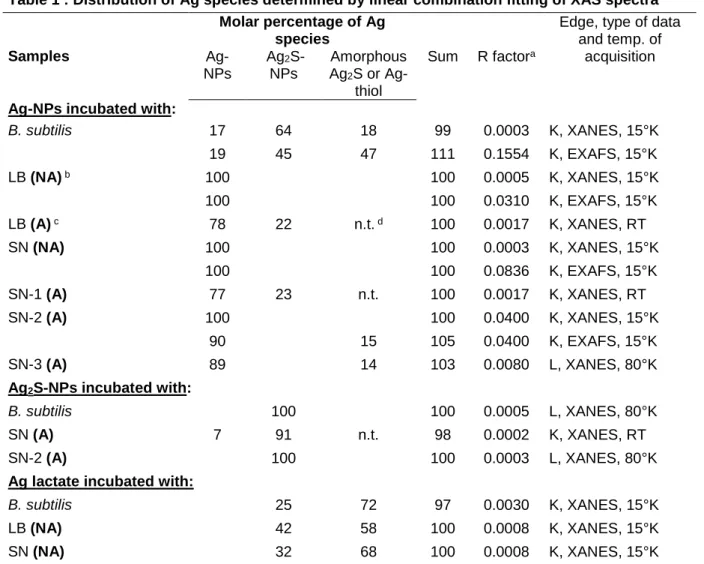
The nature and proportions of Ag species in each sample were then studied by X-ray 289
absorption spectroscopy. XANES and EXAFS spectra, or XANES spectra only, were treated 290
by LCFs using model compound spectra (Figure 3, Table 1). Principal component analysis was 291
not used because the set of spectra at each edge was too small. Among the reference spectra of 292
our K-edge database, four standards including Ag-NPs, Ag2S-NPs, amorphous Ag2S and Ag-293
thiol were sufficient to reproduce the spectra. Because of their similarity, the last two species 294
were merged into a single pool. Although the LB medium contains 5 g L-1 NaCl, AgCl was not 295
detected in any sample. No significant change in speciation was detected for Ag-NPs after 296
incubation in LB or SN in NA conditions. By contrast, in A conditions, about 22% of sulfidation 297
was detected for LB and 14 to 23% for SN (Table 1). The nature of the secondary species, either 298
crystalline Ag2S or amorphous Ag2S/Ag-thiol, remains unclear for these samples because it is 299
a minor phase (the error bar for this type of analysis is estimated at ± 10%). In the presence of 300
B. subtilis, about 80% of Ag-NPs were sulfidized. The proportion of the secondary forms
301
slightly differ between XANES and EXAFS LCFs (64 and 45% for crystalline Ag2S, 302
respectively, and 18 and 47% for amorphous Ag2S/Ag and/or Ag-thiol, respectively, Table 1), 303
but it remains clear that both species were present. The fit quality was clearly weaker (R factor 304
increased by 20%) when only crystalline Ag2S was used. 305
For silver lactate, the sulfidation was total in LB and SN, even in NA conditions. This 306
is not surprising since Ag is already present as Ag+, so there is no oxidation step required before 307
sulfidation. The final form was a mixture of crystalline Ag2S and amorphous Ag2S/Ag-thiol. A 308
similar result was obtained for Ag lactate in the presence of B. subtilis. Again, fits were clearly 309
weaker (R factor doubled) if only crystalline Ag2S was used. Concerning Ag2S-NPs, there was 310
no significant change in speciation after incubation with B. subtilis and with SN in aerated 311
conditions. The other conditions were not tested. 312
The speciation of sulfur in the LB medium before culture and in the supernatant after 313
culture (in absence of silver) were studied by sulfur K-edge XANES spectroscopy (Figure S10). 314
For both media, a mixture of thiols (R-SH), alkyl sulfides (R-S-R), sulfoxides (R-S=O) and 315
sulfate were observed. The proportion of thiols was comparable in both media (26 to 32% of 316
total S). 317
Discussion
318
Concerning the fate of pristine Ag-NPs in the various media, the molecules secreted by 319
B. subtilis (SN medium) increased the agglomeration of Ag-NPs as compared to pure water,
320
whereas the molecules of the initial growth medium (LB) did not. Meanwhile, a decrease of the 321
particle nominal diameter, suggesting a partial dissolution, was observed after incubation with 322
SN and LB. Although the toxicity of Ag-NPs strongly relies on their dissolution 21, 30-31, in our 323
experimental conditions (1 mg L-1 Ag applied during the stationary phase), no impacts in terms 324
of cell mortality or changes in the central metabolism of Bacillus subtilis were observed. This 325
may be related to the presence of exopolymers produced by the bacteria known to decrease 326
metals availability32-33, and to other types of molecules excreted by the bacteria. This absence 327
of toxicity is consistent with the absence of internalization of silver inside cells, as observed by 328
nanoXRF and ICP-AES coupled with cell lysis. In addition, some transformations of Ag-NPs 329
were observed, likely affecting Ag availability. Ag-NPs were partly sulfidized (about 20% of 330
total Ag), and the secondary species (either crystalline Ag2S or amorphous Ag2S/Ag-thiol) 331
formed a homogeneous shell on the NPs, leading to an Ag-Ag2S core-shell structure. Based on 332
TEM-EDX results, this surface sulfidation was slightly higher in the medium containing the 333
secretome (SN) than in the growth medium (LB). The aeration of the suspension, i.e., the 334
presence of dissolved oxygen, clearly enhanced the reaction. The sulfidation of ionic silver (Ag 335
lactate) in LB and SN was tested in non-aerated conditions. In that case the reaction was 336
complete, with formation of crystalline Ag2S and amorphous Ag2S/Ag-thiol. These results 337
allowed us to conclude that the limiting step of the sulfidation of Ag-NPs under our conditions 338
was not the availability of reduced sulfur, but of oxygen, necessary for the oxidation of Ag0 339
into Ag+. After incubation with Bacillus subtilis, the sulfidation was almost complete (80%) 340
and again, both crystalline Ag2S (acanthite) and amorphous Ag2S/Ag-thiol were formed, based 341
on bulk XANES and EXAFS analysis. These results are in contradiction with those obtained 342
by Hsueh et al.34, who identified Ag2O in a bacterial pellet of B. subtilis after exposure to 100 343
mg L-1 Ag-NPs. However, to our opinion these results are not valid since Ag2S and Ag2O 344
reference spectra shown in this article are almost identical (which is not the case, e.g., 35), and 345
the first peak for the various references (Ag2S, Ag2O, Ag-NPs) are surprisingly at the same 346
position. 347
The changes in speciation occurred without internalization or sorption on bacterial cell 348
walls, as observed both from TEM-EDX and nanoXRF. The composite structure of the Ag2S 349
aggregates, composed of Ag2S nanocrystals much smaller than the original Ag-NPs (Figure 350
S3), suggest an indirect sulfidation, with release of Ag+ ions followed by reaction with sulfides. 351
On the contrary, a direct sulfidation was observed by Thalmann et al.36 for Ag-NPs in the 352
presence of HS-, with the formation of hollow Ag2S spheres attributed to the Kirkendall effect. 353
Sulfidation of Ag-NPs can be realized by dissolved inorganic sulfide species including H2S, 354
HS− and S2−, and can be direct or indirect depending on the S/Ag ratio.37 Inorganic sulfides are 355
likely from the aerobic culture medium of a chemoheterotrophic strain like B. subtilis. Thus, 356
thiol-containing molecules present in the medium (as observed from S XANES spectroscopy) 357
are the most likely sources of sulfide. This pathway would involve a cleavage of the S-C bond. 358
Such cleavage has been documented in synthesis routes of nanosized Ag2S using cysteine as S 359
donor38-39. The proposed sulfidation mechanism is schematized in Figure S11. 360
These results show that living bacteria are more efficient at sulfidizing the Ag-NPs than 361
the growth medium or secreted molecules alone. A possible explanation of this observation 362
could be a modification of the secretome under Ag-NPs exposure. Indeed, the secretome used 363
for the SN conditions is produced by the bacteria in the absence of Ag-NPs, and may differ 364
from the one produced in the presence of Ag-NPs. Based on our conclusion on the rate limiting 365
step of the sulfidation, we can hypothesize that the bacteria under Ag-NPs exposure excrete 366
more oxidizing molecules. They might also excrete more thiol-containing molecules. These 367
hypotheses could be tested by mass spectrometry proteomics experiments. Anyhow, an 368
important result of this study is the demonstration that the bacterial activity has a strong impact 369
on the fate of Ag-NPs. 370
Methodological and Environmental implications
371
This study shows that the bacterial activity has a major impact on the fate of Ag-NPs, 372
by enhancing their sulfidation. The results suggest that the bacterial activity favors the first step 373
of the reaction, which is the oxidation of Ag0. Concerning the second step, the sulfidation itself, 374
thiol-containing proteins and peptides are the most likely source of reduced S. These results 375
suggest that microorganisms may participate to the sulfidation of Ag-NPs in aerobic systems in 376
the environment, such as unsaturated soils. On the contrary, B. subtilis does not modify the 377
speciation of Ag2S-NPs. 378
This study has some implications for nano(eco)toxicology studies. In most studies, the 379
NPs are characterized before the exposure experiment. Our study evidenced various extents of 380
chemical transformations of Ag-NPs after 5 hours of incubation, depending on the media. These 381
changes clearly modify the exposure conditions. Thus, it is very important to monitor the 382
speciation of NPs during exposure experiments. This type of information would clearly help 383
the interpretation, and the inter comparison between studies. This point is valid for silver, and 384
other NPs prone to transformations and/or redox reactions such as ZnO, CuO, CeO2 for 385
example. 386
In this study, Ag2S-NPs produced by reacting Ag-NPs and Na2S, was used as a proxy 387
of aged Ag-NPs. These Ag2S-NPs formed large aggregates (several tens of micrometers) of 388
crystalline Ag2S (acanthite), which were not altered in the presence of Bacillus subtilis or with 389
their secreted molecules. This form clearly differed from the secondary products obtained after 390
incubation of Ag-NPs with bacteria, which included both crystalline and amorphous Ag2S 391
and/or Ag-thiol, with particle size still in the nanometer range. In soils amended with sewage 392
sludge, a mixture of crystalline and amorphous Ag2S and/or Ag-thiol, present as micro and 393
nanosized particles and as diffuse concentrations, was observed26. Thus, Ag2S produced by 394
reacting Ag-NPs and Na2S may not be the most relevant material for toxicity studies since it is 395
highly aggregated and fully crystalline. Sulfidation protocols involving thiol-containing 396
molecules might provide more representative aged Ag-NPs. 397
The development of safer-by-design nanoparticles is a growing research topic. In the 398
case of Ag-NPs, a decrease of the release of Ag from products (which can reach large amounts, 399
for example for textiles40-41) while keeping the antibacterial properties, should be targeted. 400
Partly sulfidized Ag-NPs, with an Ag-Ag2S core-shell structure, such as those produced in this 401
study, might be a material to test in this perspective. The secretome of Bacillus subtilis may 402
have some potential for the green synthesis of these nanocomposites. 403
Supporting Information
405
Materials and Methods: Nanoparticles characterization, high pressure freezing and freeze-406
substitution, Ag reference compounds for XAS analyses, nanoXRF detection limit, ICP-AES 407
after cell lysis. Results: DLS analyses, TEM coupled with EDX, electron diffraction and high 408
resolution, S K-edge XANES and proposed mechanism of sulfidation of Ag-NPs in the 409
presence of B. subtilis (Figures S3 to S11). All data are presented in a single PDF document. 410
411
Acknowledgments
412The authors thank the French program LabEx Serenade (11-LABX-0064) for providing a PhD 413
fellowship and EquipEx NanoID (ANR-10-EQPX-39) for funding the TEM Osiris used in this 414
study. ISTerre is also part of Labex OSUG@2020 (ANR10 LABX56). The authors thank ESRF 415
and Soleil synchrotrons for providing beam time. The beamline staff of FAME, ID16b and ID21 416
(ESRF) and Samba (Soleil) are acknowledged for their help in collecting data. Giulia Veronesi 417
is acknowledged for the S XANES measurements on ID21, and Alexandre Gelabert, Morgane 418
Desmau and Clément Levard are acknowledged for recording the data on Samba beamline. We 419
thank Christine Moriscot for her help on TEM sample preparation, realized on the platforms of 420
the Grenoble Instruct-ERIC Center (ISBG : UMS 3518 CNRS-CEA-UGA-EMBL) with 421
support from FRISBI (ANR-10-INSB-05-02) and GRAL (ANR-10-LABX-49-01) within the 422
Grenoble Partnership for Structural Biology (PSB). The electron microscope facility used for 423
TEM sample preparation is supported by the Rhône-Alpes Region, the Fondation Recherche 424
Medicale (FRM), the fonds FEDER and the GIS-Infrastrutures en Biologie Sante et Agronomie 425
(IBISA). 426
References
428
(1) Pulit-Prociak, J.; Stoklosa, K.; Banach, M., Nanosilver products and toxicity. Environmental 429
Chemistry Letters 2015, 13 (1), 59-68.
430
(2) Gottschalk, F.; Sonderer, T.; Scholz, R. W.; Nowack, B., Modeled environmental concentrations 431
of engineered nanomaterials (TiO2, ZnO, Ag, CNT, fullerenes) for different regions. Environ. Sci. 432
Technol. 2009, 43, 9216-9222.
433
(3) Sun, T. Y.; Gottschalk, F.; Hungerbuehler, K.; Nowack, B., Comprehensive probabilistic 434
modelling of environmental emissions of engineered nanomaterials. Environ. Pollut. 2014, 185, 69-76. 435
(4) Kaegi, R.; Voegelin, A.; Sinnet, B.; Zuleeg, S.; Hagendorfer, H.; Burkhardt, M.; Siegrist, H., 436
Behavior of metallic silver nanoparticles in a pilot wastewater treatment plant. Environ. Sci. & Technol. 437
2011, 45 (9), 3902-3908. 438
(5) Kelessidis, A.; Stasinakis, A. S., Comparative study of the methods used for treatment and final 439
disposal of sewage sludge in European countries. Waste Management 2012, 32 (6), 1186-1195. 440
(6) Navarro, E.; Baun, A.; Behra, R.; Hartmann, N. B.; Filser, J.; Miao, A. J.; Quigg, A.; Santschi, P. 441
H.; Sigg, L., Environmental behavior and ecotoxicity of engineered nanoparticles to algae, plants, and 442
fungi. Ecotoxicology 2008, 17 (5), 372-386. 443
(7) Maillard, J.-Y.; Hartemann, P., Silver as an antimicrobial: facts and gaps in knowledge. Crit. 444
Rev.Microbiol. 2013, 39 (4), 373-383.
445
(8) Niazi, J. H.; Gu, M. B., Toxicity of Metallic Nanoparticles in Microorganisms- a Review. In 446
Atmospheric and Biological Environmental Monitoring, Kim, Y. J.; Platt, U.; Gu, M. B.; Iwahashi, H., Eds.
447
Springer Netherlands: Dordrecht, 2009; pp 193-206. 448
(9) Duran, N.; Duran, M.; de Jesus, M. B.; Seabra, A. B.; Favaro, W. J.; Nakazato, G., Silver 449
nanoparticles: A new view on mechanistic aspects on antimicrobial activity. Nanomedicine-450
Nanotechnology Biology and Medicine 2016, 12 (3), 789-799.
451
(10) Marambio-Jones, C.; Hoek, E. M. V., A review of the antibacterial effects of silver nanomaterials 452
and potential implications for human health and the environment. J. Nanopart. Res. 2010, 12 (5), 1531-453
1551. 454
(11) Reidy, B.; Haase, A.; Luch, A.; Dawson, K.; Lynch, I., Mechanisms of Silver Nanoparticle Release, 455
Transformation and Toxicity: A Critical Review of Current Knowledge and Recommendations for Future 456
Studies and Applications. Materials 2013, 6 (6), 2295. 457
(12) Kaegi, R.; Voegelin, A.; Ort, C.; Sinnet, B.; Thalmann, B.; Krismer, J.; Hagendorfer, H.; Elumelu, 458
M.; Mueller, E., Fate and transformation of silver nanoparticles in urban wastewater systems. Water 459
Research 2013, 47 (12), 3866-3877.
(13) Levard, C.; Hotze, E. M.; Lowry, G. V.; Brown, G. E., Environmental Transformations of Silver 461
Nanoparticles: Impact on Stability and Toxicity. Environ. Sci. Technol. 2012, 46 (13), 6900-6914. 462
(14) Levard, C.; Hotze, E. M.; Colman, B. P.; Dale, A. L.; Truong, L.; Yang, X. Y.; Bone, A. J.; Brown, G. 463
E.; Tanguay, R. L.; Di Giulio, R. T.; Bernhardt, E. S.; Meyer, J. N.; Wiesner, M. R.; Lowry, G. V., Sulfidation 464
of silver nanoparticles: Natural antidote to their toxicity ? Environ. Sci. Technol. 2013, 47 (23), 13440-465
13448. 466
(15) Yu, S. J.; Yin, Y. G.; Liu, J. F., Silver nanoparticles in the environment. Environmental Science-467
Processes & Impacts 2013, 15 (1), 78-92.
468
(16) Earl, A. M.; Losick, R.; Kolter, R., Ecology and genomics of Bacillus subtilis. Trends in 469
Microbiology 16 (6), 269-275.
470
(17) Pandey, A.; Palni, L. M. S., Bacillus species: The dominant bacteria of the rhizosphere of 471
established tea bushes. Microbiological Research 1997, 152 (4), 359-365. 472
(18) Marvasi, M.; Visscher, P. T.; Casillas Martinez, L., Exopolymeric substances (EPS) from Bacillus 473
subtilis : polymers and genes encoding their synthesis. FEMS Microbiology Letters 2010, 313 (1), 1-9. 474
(19) van Dijl, J.; Hecker, M., Bacillus subtilis: from soil bacterium to super-secreting cell factory. 475
Microbial Cell Factories 2013, 12 (1), 3.
476
(20) Kim, S. W.; Baek, Y.-W.; An, Y.-J., Assay-dependent effect of silver nanoparticles to Escherichia 477
coli and Bacillus subtilis. Applied Microbiology and Biotechnology 2011, 92 (5), 1045-1052. 478
(21) Bondarenko, O.; Ivask, A.; Kakinen, A.; Kurvet, I.; Kahru, A., Particle-Cell Contact Enhances 479
Antibacterial Activity of Silver Nanoparticles. Plos One 2013, 8 (5). 480
(22) Völker, U.; Hecker, M., From genomics via proteomics to cellular physiology of the Gram-481
positive model organism Bacillus subtilis. Cellular Microbiology 2005, 7 (8), 1077-1085. 482
(23) Overkamp, W.; Ercan, O.; Herber, M.; van Maris, A. J. A.; Kleerebezem, M.; Kuipers, O. P., 483
Physiological and cell morphology adaptation of Bacillus subtilis at near-zero specific growth rates: a 484
transcriptome analysis. Environmental Microbiology 2015, 17 (2), 346-363. 485
(24) Levard, C.; Reinsch, B. C.; Michel, F. M.; Oumahi, C.; Lowry, G. V.; Brown, G. E., Sulfidation 486
Processes of PVP-Coated Silver Nanoparticles in Aqueous Solution: Impact on Dissolution Rate. 487
Environ. Sci. Technol. 2011, 45 (12), 5260-5266.
488
(25) Zeng, W.; Chen, G.; Wu, H.; Wang, J.; Liu, Y.; Guo, Y.; Liang, Z., Improvement of Bacillus subtilis 489
for poly‐γ‐glutamic acid production by genome shuffling. Microbial Biotechnology 2016, 9 (6), 824-833. 490
(26) Pradas del Real, A. E.; Castillo-Michel, H.; Kaegi, R.; Sinnet, B.; Magnin, V.; Findling, N.; 491
Villanova, J.; Carriere, M.; Santaella, C.; Fernandez-Martinez, A.; Levard, C.; Sarret, G., Fate of Ag-NPs 492
in sewage sludge after application on agricultural soils. Environ. Sci. Technol. 2016, 50 (4), 1759-1768. 493
(27) Pradas del Real, A. E.; Vidal, V.; Carrière, M.; Castillo-Michel, H.; Levard, C.; Chaurand, P.; 494
Sarret, G., Silver Nanoparticles and Wheat Roots: A Complex Interplay. Environ. Sci. Technol. 2017, 51 495
(10), 5774-5782. 496
(28) Castillo-Michel, H.; Larue, C.; Pradas del Real, A.-E.; Cotte, M.; Sarret, G., Practical review on 497
the use of synchrotron based micro- and nano- X-ray fluorescence mapping and X-ray absorption 498
spectroscopy to investigate the interactions between plants and engineered nanomaterials. Plant 499
Physiol. Biochem. 2017, 110, 13-32.
500
(29) Ravel, B.; Newville, M., ATHENA and ARTEMIS: Interactive graphical data analysis using IFEFFIT. 501
J. Synchr. Rad. 2005, 12, 537-541.
502
(30) Ivask, A.; Kurvet, I.; Kasemets, K.; Blinova, I.; Aruoja, V.; Suppi, S.; Vija, H.; Käkinen, A.; Titma, 503
T.; Heinlaan, M.; Visnapuu, M.; Koller, D.; Kisand, V.; Kahru, A., Size-Dependent Toxicity of Silver 504
Nanoparticles to Bacteria, Yeast, Algae, Crustaceans and Mammalian Cells In Vitro. PLOS ONE 2014, 9 505
(7), e102108. 506
(31) Agnihotri, S.; Mukherji, S.; Mukherji, S., Size-controlled silver nanoparticles synthesized over 507
the range 5-100 nm using the same protocol and their antibacterial efficacy. RSC Advances 2014, 4 (8), 508
3974-3983. 509
(32) Li, C. C.; Wang, Y. J.; Dang, F.; Zhou, D. M., Mechanistic understanding of reduced AgNP 510
phytotoxicity induced by extracellular polymeric substances. J. Hazardous Mat. 2016, 308, 21-28. 511
(33) Koser, J.; Engelke, M.; Hoppe, M.; Nogowski, A.; Filser, J.; Thoming, J., Predictability of silver 512
nanoparticle speciation and toxicity in ecotoxicological media. Environmental Science-Nano 2017, 4 513
(7), 1470-1483. 514
(34) Hsueh, Y.-H.; Lin, K.-S.; Ke, W.-J.; Hsieh, C.-T.; Chiang, C.-L.; Tzou, D.-Y.; Liu, S.-T., The 515
Antimicrobial Properties of Silver Nanoparticles in <italic>Bacillus subtilis</italic> Are Mediated by 516
Released Ag<sup>+</sup> Ions. PLOS ONE 2015, 10 (12), e0144306. 517
(35) Yin, L.; Cheng, Y.; Espinasse, B.; Colman, B. P.; Auffan, M.; Wiesner, M.; Rose, J.; Liu, J.; 518
Bernhardt, E., More than ions: the effects of silver nanoparticles on Lolium multiflorum. Environ. Sci. 519
Technol. 2011, 45, 2360-2367.
520
(36) Thalmann, B.; Voegelin, A.; Morgenroth, E.; Kaegi, R., Effect of humic acid on the kinetics of 521
silver nanoparticle sulfidation. Environmental Science-Nano 2016, 3 (1), 203-212. 522
(37) Liu, J. Y.; Pennell, K. G.; Hurt, R. H., Kinetics and Mechanisms of Nanosilver Oxysulfidation. 523
Environ. Sci. Technol. 2011, 45 (17), 7345-7353.
524
(38) Xiang, J.; Cao, H.; Wu, Q.; Zhang, S.; Zhang, X.; Watt, A. A. R., l-Cysteine-Assisted Synthesis and 525
Optical Properties of Ag2S Nanospheres. The Journal of Physical Chemistry C 2008, 112 (10), 3580-526
3584. 527
(39) Banerjee, S.; Show, B.; Kundu, A.; Ganguly, J.; Gangopadhyay, U.; Saha, H.; Mukherjee, N., N-528
acetyle cysteine assisted synthesis of core-shell Ag2S with enhanced light transmission and diminished 529
reflectance: Surface modifier for c-SiNx solar cells. Journal of Industrial and Engineering Chemistry 530
2016, 40, 54-61. 531
(40) Lorenz, C.; Windler, L.; von Goetz, N.; Lehmann, R. P.; Schuppler, M.; Hungerbuhler, K.; 532
Heuberger, M.; Nowack, B., Characterization of silver release from commercially available functional 533
(nano)textiles. Chemosphere 2012, 89 (7), 817-824. 534
(41) Mitrano, D. M.; Rimmele, E.; Wichser, A.; Erni, R.; Height, M.; Nowack, B., Presence of 535
Nanoparticles in Wash Water from Conventional Silver and Nano-silver Textiles. ACS Nano 2014, 8 (7), 536
7208-7219. 537
539
Table 1 : Distribution of Ag species determined by linear combination fitting of XAS spectra Molar percentage of Ag
species
Edge, type of data and temp. of acquisition Samples Ag-NPs Ag2 S-NPs Amorphous Ag2S or Ag-thiol Sum R factora
Ag-NPs incubated with:
B. subtilis 17 64 18 99 0.0003 K, XANES, 15°K 19 45 47 111 0.1554 K, EXAFS, 15°K LB (NA) b 100 100 0.0005 K, XANES, 15°K 100 100 0.0310 K, EXAFS, 15°K LB (A) c 78 22 n.t. d 100 0.0017 K, XANES, RT SN (NA) 100 100 0.0003 K, XANES, 15°K 100 100 0.0836 K, EXAFS, 15°K SN-1 (A) 77 23 n.t. 100 0.0017 K, XANES, RT SN-2 (A) 100 100 0.0400 K, XANES, 15°K 90 15 105 0.0400 K, EXAFS, 15°K SN-3 (A) 89 14 103 0.0080 L, XANES, 80°K Ag2S-NPs incubated with: B. subtilis 100 100 0.0005 L, XANES, 80°K SN (A) 7 91 n.t. 98 0.0002 K, XANES, RT SN-2 (A) 100 100 0.0003 L, XANES, 80°K
Ag lactate incubated with:
B. subtilis 25 72 97 0.0030 K, XANES, 15°K
LB (NA) 42 58 100 0.0008 K, XANES, 15°K
SN (NA) 32 68 100 0.0008 K, XANES, 15°K
Fits were done in the [E0-20; E0+80] eV range for XANES spectra, and in the [2-12] Å-1 range for EXAFS spectra. Up to three components were used, except for XANES spectra recorded at RT, for which only two-components (Ag-NPs and Ag2S-NPs) were used. a Fit quality criterion provided by ATHENA (R factor = ∑[ k 2 χexp – k2 χfit]2 ⁄∑[k2 χexp]2 for EXAFS spectra, and ∑[ µexp – µfit]2 ⁄∑[µexp]2 for XANES spectra. b Non-aerated conditions, c Aerated conditions. d Not tested because of the absence of these reference spectra in the K-edge database at RT.LB: initial growth medium. SN: supernatant (three replicates analyzed for Ag-NPs, aerated conditions).
540 541
542
Figure captions 543
544
Figure 1: Nano-XRF analysis of Bacillus subtilis after incubation with Ag-NPs, Ag2S-NPs, and 545
Ag lactate. The bacteria are imaged using the Os signal. Max number of counts for Os and Ag 546
are given for each map. Scale bar: 1µm. step size: 0.05*0.05 µm, dwell time: 500ms 547
548
Figure 2: Ag (red), S (green) and Ag + S elemental maps obtained by TEM-EDX for the Ag-549
NPs after incubation in H2O, LB, SN, and with bacteria in aerated (A) or non-aerated (NA) 550
condition (Fig. 2A) and for Ag2S-NPs after 5h incubation in H2O, SN and with bacteria (Fig. 551
2B). Scale bar: 50 nm. (C) Comparison of the S/Ag peak height of the EDX spectra for 552
individual NPs, extracted from the maps. Significant differences according to the Tukey test 553
(p< 0.05) are indicated by letters. 554
555
Figure 3: XAS spectra for Ag reference compounds and samples, recorded in various 556
conditions: A, B: Bulk Ag K-edge XANES (A) spectra and EXAFS (B) spectra recorded at 557
15°K. C: nano Ag K-edge XANES spectra recorded at room temperature. D: bulk Ag LIII-edge 558
XANES spectra recorded at 80°K. For the samples, the color of the spectra correspond to the 559
initial Ag form: Ag-NPs in red, Ag2S-NPs is green, and Ag lactate in grey. Linear combination 560
fitting are shown in blue dashed lines. 561
562
Figure 1
564
Figure 2
566
Figure 3
567 568