HAL Id: hal-01789569
https://hal.archives-ouvertes.fr/hal-01789569
Submitted on 11 May 2018
HAL is a multi-disciplinary open access
archive for the deposit and dissemination of
sci-entific research documents, whether they are
pub-lished or not. The documents may come from
teaching and research institutions in France or
abroad, or from public or private research centers.
L’archive ouverte pluridisciplinaire HAL, est
destinée au dépôt et à la diffusion de documents
scientifiques de niveau recherche, publiés ou non,
émanant des établissements d’enseignement et de
recherche français ou étrangers, des laboratoires
publics ou privés.
De novo telomere addition at chromosome breaks:
Dangerous Liaisons
Dmitri Churikov, Vincent Géli
To cite this version:
Dmitri Churikov, Vincent Géli.
De novo telomere addition at chromosome breaks: Dangerous
Liaisons.
Journal of Cell Biology, Rockefeller University Press, 2017, 216 (8), pp.2243-2245.
JCB JCB: Spotlight
T
H
E J
O
U
R
N
A
L O
F C
E
L
L B
IO
L
O
G
Y
2243The Rockefeller University Press J. Cell Biol. Vol. 216 No. 8 2243–2245 https://doi.org/10.1083/jcb.201705156
All eukaryotic cells with linear chromosomes face the problem of terminal DNA sequence loss that occurs as a result of either incomplete replication of the DNA strand synthesized by the lagging strand replication machinery or accidental collapse of the replication forks. If left unrepaired, these losses will even-tually trigger the DNA damage response (DDR) and cell cycle arrest. Most eukaryotes use the enzyme telomerase that special-izes in supplementing lost sequences at the chromosome ends to counteract this problem. Telomerase is minimally composed of the catalytic subunit, a telomerase reverse transcription, and the RNA component that serves as a scaffold for telomerase subunit assembly and also carries a region that templates the synthesis of telomeric DNA repeats. The template region of telomerase RNA determines both the guanine-rich sequence of telomeric repeats and the specificity of telomerase for chromosome ends because the template region has to anneal to the single-strand DNA (ssDNA) tail exposed at the site of its action. To further reinforce the specificity of telomerase for chromosome ends, a telomerase recruitment mechanism has evolved that relies on specialized proteins that bind telomeric ssDNA with high af-finity and sequence specificity such as Cdc13 in budding yeast and POT1 in mammalian cells. However, in spite of these ad-aptations, telomerase does interfere with repair of DNA double strand breaks (DSBs) and may occasionally add telomeric re-peats to either spontaneous or induced DSBs, a process known as chromosome healing by de novo telomere addition (Penna-neach et al., 2006). In this issue, Ouenzar et al. describe a novel mechanism that restricts the action of telomerase by spatial ex-clusion from sites of DNA repair.
Although de novo telomere addition at internal non-telo-meric sites stabilizes the end of a broken chromosome, it leads to the loss of large portions of chromosome arms that usually endangers cell viability. Previous work in budding yeast identi-fied molecular mechanisms responsible for curbing the action of
telomerase on DSBs and uncovered that these mechanisms are intimately linked to DDR signaling (Makovets and Blackburn, 2009; Zhang and Durocher, 2010). Upon detection of DSBs, Mec1 kinase, a budding yeast orthologue of ATR, initiates a signaling cascade by phosphorylating multiple targets. One of these targets phosphorylated in a MEC1-RAD53-DUN1– dependent manner is a Pif1 helicase, a known telomerase inhib-itor that dislodges telomerase from its DNA substrate (Boulé et al., 2005). Importantly, this phosphorylation of Pif1 specifically mediates telomerase inhibition at DNA breaks, but not at telo-meres (Makovets and Blackburn, 2009). In addition, activated Mec1 that accumulates at resected DNA ends directly phos-phorylates Cdc13 on Ser 306, and this phosphorylation event prevents accumulation of Cdc13 at DNA ends with very short telomere-like sequence seeds, thereby suppressing telomere addition to accidental DSBs (Zhang and Durocher, 2010). Al-though these sophisticated mechanisms elegantly incorporated into the DDR diminish the action of telomerase on DSBs, it appears to not be the whole story. Indeed, Ouenzar et al. (2017) add a new layer of regulation to previous results by showing that telomerase action at DNA breaks is restricted by the se-questration of its RNA component in the nucleolus.
Ouenzar et al. (2017) have used FISH to quantify intracel-lular distribution of TLC1 RNA, a telomerase RNA in budding yeast, at different phases of the cell cycle. They found that in-dividual telomerase RNA molecules formed foci in the nucleo-plasm during the G1 and S phases of the cell cycle, but became sequestered in the nucleoli in the G2/M phase. Interestingly, the cell cycle–dependent trafficking of TLC1 RNA into the nucleoli required Pif1 activity in the nucleoplasm, which removes telo-merase from telomeres in late S/G2. The majority of DSBs in budding yeast are repaired by homologous recombination (HR), which is active in G2 phase and occurs exclusively in the nuc-leoplasm, suggesting that nucleolar localization of TLC1 RNA could serve to reduce the interference of telomerase with HR in DSB repair. To interrogate a functional significance of nucleolar localization of TLC1 RNA in the context of DSB repair, Ouen-zar et al. (2017) examined the distribution of TLC1 RNA in the cells treated with bleomycin, a radiomimetic drug that gener-ates DSBs. In bleomycin-treated cells in the G2/M phase, TLC1 RNA redistributed slightly into the nucleoplasm, indicating that its trafficking between nucleoplasm and nucleolus is affected
Telomerase counteracts the loss of terminal DNA sequences from chromosome ends; however, it may erroneously add telomeric repeats to DNA double-strand breaks. In this issue, Ouenzar et al. (2017. J. Cell Biol. https ://doi .org /10 .1083 /jcb .201610071) uncover cell cycle–dependent sequestration of the telomerase RNA in nucleoli, a process that excludes telomerase from DNA repair sites.
De novo telomere addition at chromosome breaks:
Dangerous Liaisons
Dmitri Churikov1,2 and Vincent Géli1,2
1Marseille Cancer Research Center, U1068 Institut National de la Santé et de la Recherche Médicale, UMR7258 Centre National de la Recherche Scientifique,
Aix Marseille University, Marseille, France
2Institut Paoli-Calmettes, Marseille, France
© 2017 Churikov and Géli This article is distributed under the terms of an Attribution– Noncommercial–Share Alike–No Mirror Sites license for the first six months after the publication date (see http ://www .rupress .org /terms /). After six months it is available under a Creative Commons License (Attribution–Noncommercial–Share Alike 4.0 International license, as described at https ://creativecommons .org /licenses /by -nc -sa /4 .0 /).
Correspondence to Vincent Géli: [email protected]
on May 10, 2018 jcb.rupress.org
Downloaded from
http://doi.org/10.1083/jcb.201705156 Published Online: 14 July, 2017 | Supp Info:
JCB • Volume 216 • NumBer 8 • 2017
2244
by DNA damage. Surprisingly, nucleolar localization of TLC1 RNA was greatly reduced and most of the cells accumulated
TLC1 RNA foci in the nucleoplasm when HR was abolished by deletion of RAD52. This loss of nucleolar localization was a result of redistribution of TLC1 RNA from the nucleolus into the nucleoplasm rather than its retention in the nucleoplasm because the cells arrested in G2/M with nocodazole still redis-tributed TLC1 RNA into the nucleoplasm upon treatment with bleomycin. Importantly, TLC1 RNA molecules that left nucle-olus partially colocalized with persistent DSBs in the nucleo-plasm highlighted by either Rfa1 or H2AX immunostaining. Whether the remaining TLC1 RNAs that did not colocalize with DSBs were associated with telomeres remains to be determined.
To investigate the role of DDR in TLC1 RNA traffick-ing, Ouenzar et al. (2017) inactivated several factors that act upstream of Rad52. DSB resection is initiated by the MRX complex, and deletion of either the MRX component MRE11 or XRS2 completely suppressed the redistribution of TLC1 RNA into the nucleoplasm in rad52Δ cells upon DNA damage.
This suggests that the exit of TLC1 RNA from the nucleolus could be triggered by DSB processing and the accumulation of ssDNA in the absence of Rad52. In support of this notion, in-activation of Tel1, which positively influences MRX activity at DSBs, also diminished accumulation of the TLC1 RNA foci in the nucleoplasm, whereas inactivation of Mec1 had no effect. These results also suggest involvement of the ssDNA binding protein Cdc13, which recruits telomerase to both telomeres and DSB via its interaction with the Est1 subunit of telomerase complex (Bianchi et al., 2004). Indeed, Cdc13 that is normally undetectable by immunofluorescence formed visible foci upon induction of DSBs, and these foci further increased in size in
rad52Δ cells. Direct observation of Cdc13-GFP confirmed that
it colocalizes with DSBs marked by Rfa1-mCherry in the ma-jority of G2/M cells. Most importantly, a cdc13-2 mutant that is proficient in ssDNA binding but fails to recruit telomerase completely suppressed the exit of TLC1 RNA from nucleolus in bleomycin-treated rad52Δ cells in G2/M. This observation
strongly implicated Est1–Cdc13 interaction in the nucleoplas-mic accumulation of TLC1 RNA after DNA damage.
Although excessive accumulation of ssDNA at the sites of DNA breaks and its enhanced binding by Cdc13 in the absence of Rad52 may trigger TLC1 RNA redistribution into the nucle-oplasm, the situation might be more complex. Time course ex-periments in rad52Δ cells demonstrated that initially only few TLC1 RNA molecules exit the nucleolus and rapidly localize to DSBs in agreement with the aforementioned model, but later on the remaining TLC1 RNA pool leaves the nucleolus and accu-mulates in the nucleoplasm but not at DSBs. What causes this second wave of TLC1 RNA exit remains unknown, but possi-bly these TLC1 RNA molecules associate with telomeres. An-other puzzling finding is the effect of RAD51 deletion, which resulted in disappearance of the Cdc13 foci and retention of the
TLC1 RNA in the nucleolus even in the rad52Δ cells,
suggest-ing that somehow Rad51 promotes accumulation of Cdc13 at DSBs. Reduced binding of Cdc13 to irreparable HO-induced DSB in rad51Δ cells has been previously reported (Oza et al.,
2009), but the reason behind it remains unknown. One pos-sibility is that the presence of Rad51 may have an impact on the competition between RPA and Cdc13 for ssDNA binding in favor of the latter.
In search of the regulators of the cell cycle–dependent
TLC1 RNA trafficking, Ouenzar et al. (2017) turned to SUMO- ylation, a well-known modulator of the DDR, which affects both the intranuclear distribution and function of several HR
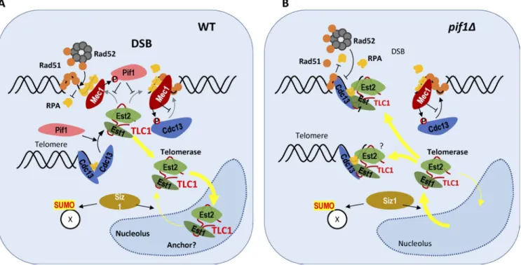
Figure 1. Several mechanisms promote telomerase exclusion from DSBs in G2/M. (A) In wild-type cells, Mec1 activated at DSBs phosphorylates both Pif1 and Cdc13. Phosphorylated Pif1 specifically inhibits telomerase action at DSBs, whereas phosphorylation of Cdc13 limits its accumulation at resected DSBs. In addition, most of the telomerase (Est2-TLC1-Est1) is sequestered in the nucleolus by a yet unknown anchor. These three mechanisms prevent de novo telomere addition at DSBs. (B) In Pif1-deficient cells, telomerase exits the nucleolus in a Siz1-dependent manner and telomerase action is no longer prevented at DSBs. This increases the chance of telomerase recruitment to DSBs via an Est1–Cdc13 interaction.
Telomerase rNA sequestration • Churikov and Géli 2245
and telomere proteins. Although deletion of either SIZ1 or SIZ2 genes encoding two homologous SUMO E3 ligases had no ef-fect on cell cycle–dependent TLC1 RNA trafficking between the nucleoplasm and nucleolus in the absence of DNA dam-age, it did so when DNA damage was induced with bleomycin. Specifically, the deletion of SIZ1 strongly decreased accumu-lation of TLC1 RNA in the nucleoplasm of rad52Δ cells, thus
implicating Siz1 in the control of spatial distribution of TLC1 RNA after DNA damage (Fig. 1). Further analysis revealed that Siz1 is neither involved in DSB processing nor Cdc13 accu-mulation at resected DSBs but rather acts downstream of these events. The identity of downstream targets of Siz1-dependent SUMOylation, which promote TLC1 RNA exit from nucleolus in response to DNA damage, remains to be established.
To address a functional role of TLC1 RNA trafficking in de novo telomere addition at spontaneous DSBs, Ouenzar et al. (2017) performed a series of gross chromosomal rearrangement (GCR) assays in a set of mutants that predictably affect TLC1 RNA trafficking, and then quantified the number of GCR events corresponding to telomere healing. The results of these assays demonstrated that Siz1-dependent SUMOylation is indeed re-quired for de novo telomere addition in the absence of Rad52, but it becomes largely dispensable when Pif1 activity in the nucleoplasm is reduced. Finally, using paired-end sequencing, Ouenzar et al. (2017) revealed that de novo telomere addition in bleomycin-treated yeast occurred downstream of short (<10 nucleotides) TG-rich sequences and that RAD52 deletion in-creases de novo telomere addition. They also found that a high percentage of de novo telomere addition occurred at the ribo-somal DNA locus, but it remains unclear whether this reflects telomerase activity in the nucleolus or not because broken ri-bosomal DNA relocalizes to the nucleoplasm for HR-mediated repair (Torres-Rosell et al., 2007).
The study by Ouenzar et al. (2017) clearly demonstrates that spatial segregation of the telomerase and HR activities is an-other mechanism that minimizes the odds of telomere addition to accidental DSBs (Fig. 1). It also raises a number of questions requiring further investigation. It is not yet clear what triggers TLC1 exit from the nucleolus in the absence of Rad52. Possi-bly, DNA damage persists when HR is dysfunctional, leading to increased SUMOylation and eventually Siz1-dependent exit of TLC1. Another intriguing question is the mechanism respon-sible for G2/M phase–specific sequestration of TLC1 RNA in the nucleolus. The nucleolus is now recognized as a multifunc-tional cellular compartment, with functions beyond its role in ribosome subunit assembly. It exerts its multiple functions in part via sequestration of the regulatory proteins; a prominent example in yeast being the cell cycle regulator Cdc14. Because the nucleolus is not membrane bound and nucleolar localiza-tion signals are not well defined, many molecules freely enter and leave the nucleolus by diffusion. It is believed that retention or sequestration of molecules occurs primarily as a function of their affinity for the anchored resident proteins, such as nucleo-lin and nucleophosmin. These affinities, and hence the stability
of the interactions, could be regulated by posttranslational modifications in a cell cycle–dependent manner. TLC1 RNA retention in the nucleolus is clearly cell cycle regulated, and its retention in the nucleolus might result from anchoring via other subunits of the telomerase complex. Interestingly, both telomer-ase components Est2 and Est1 localize to the nucleolus when overexpressed but become nucleoplasmic upon overexpression of TLC1 (Teixeira et al., 2002). Human telomerase also local-izes to the nucleolus where its assembly takes place, but in con-trast to its yeast counterpart remains in the nucleolus during most of the cell cycle and is released only when telomeres are replicated (Wong et al., 2002). Nevertheless, the nucleolar as-sociation of human telomerase is enhanced after DNA damage, thus the aspect of spatial separation of telomerase and HR activ-ity appears to be conserved from yeast to humans.
Acknowledgments
We thank Marie-Noëlle Simon for her comments.
Work in the Gelí laboratory is supported by the Ligue Contre le Cancer (Equipe Labéllisée).
The authors declare no competing financial interests.
References
Bianchi, A., S. Negrini, and D. Shore. 2004. Delivery of yeast telomerase to a DNA break depends on the recruitment functions of Cdc13 and Est1. Mol. Cell. 16:139–146. http ://dx .doi .org /10 .1016 /j .molcel .2004 .09 .009 Boulé, J.-B., L.R. Vega, and V.A. Zakian. 2005. The yeast Pif1p helicase removes
telomerase from telomeric DNA. Nature. 438:57–61. http ://dx .doi .org /10 .1038 /nature04091
Makovets, S., and E.H. Blackburn. 2009. DNA damage signalling prevents deleterious telomere addition at DNA breaks. Nat. Cell Biol. 11:1383– 1386. http ://dx .doi .org /10 .1038 /ncb1985
Ouenzar, F., M. Lalonde, H. Laprade, G. Morin, F. Gallardo, S. Tremblay-Belzile, and P. Chartrand. 2017. Cell cycle–dependent spatial segregation of telomerase from sites of DNA damage. J. Cell Biol. http ://dx .doi .org /10 .1083 /jcb .201610071. http ://dx .doi .org /10 .1083 /jcb .201610071 Oza, P., S.L. Jaspersen, A. Miele, J. Dekker, and C.L. Peterson. 2009.
Mechanisms that regulate localization of a DNA double-strand break to the nuclear periphery. Genes Dev. 23:912–927. http ://dx .doi .org /10 .1101 /gad .1782209
Pennaneach, V., C.D. Putnam, and R.D. Kolodner. 2006. Chromosome healing by de novo telomere addition in Saccharomyces cerevisiae. Mol. Microbiol. 59:1357–1368. http ://dx .doi .org /10 .1111 /j .1365 -2958 .2006 .05026 .x Teixeira, M.T., K. Forstemann, S.M. Gasser, and J. Lingner. 2002. Intracellular
trafficking of yeast telomerase components. EMBO Rep. 3:652–659. http ://dx .doi .org /10 .1093 /embo -reports /kvf133
Torres-Rosell, J., I. Sunjevaric, G. De Piccoli, M. Sacher, N. Eckert-Boulet, R. Reid, S. Jentsch, R. Rothstein, L. Aragón, and M. Lisby. 2007. The Smc5–Smc6 complex and SUMO modification of Rad52 regulates recombinational repair at the ribosomal gene locus. Nat. Cell Biol. 9:923– 931. http ://dx .doi .org /10 .1038 /ncb1619
Wong, J.M.Y., L. Kusdra, and K. Collins. 2002. Subnuclear shuttling of human telomerase induced by transformation and DNA damage. Nat. Cell Biol. 4:731–736. http ://dx .doi .org /10 .1038 /ncb846
Zhang, W., and D. Durocher. 2010. De novo telomere formation is suppressed by the Mec1-dependent inhibition of Cdc13 accumulation at DNA breaks. Genes Dev. 24:502–515. http ://dx .doi .org /10 .1101 /gad .1869110