ARCHNES
fMASSAC 'WyQET IN! TIfl)TE I OF Cr"LJL3
Cooperative Behaviors in the
JUN30 2015Evolution of Antibiotic Resistance
LIBRARIES
by
Evgene Yurtsev
B.S., University of California, Santa Barbara (2008) Submitted to the Department of Physics
in partial fulfillment of the requirements for the degree of Doctor of Philosophy in Physics
at the
MASSACHUSETTS INSTITUTE OF TECHNOLOGY
June 2015
@
Massachusetts Institute of Technology 2015. All rights reserved.Signature of Author
...
Signature redacted
artment of Physics May 14, 2015
Signature redacted
Certified by ... ... Jeff Gore Assistant Professor Thesis SupervisorSignature redacted
Accepted by ... . Nergis Mavalvala Associate Department Head for EducationCooperative Behaviors in the
Evolution of Antibiotic Resistance
by
Evgene Yurtsev
Submitted to the Department of Physics on May 14, 2015, in partial fulfillment of the
requirements for the degree of Doctor of Philosophy in Physics
Abstract
Through a combination of experiments and modeling, I explored how inactivation of antibiotics by antibiotic-resistant bacteria affects the evolution of antibiotic resistance in two simple microbial communities. First, I examined the interaction between a re-sistant strain and a sensitive strain of the bacteria Escherichia coli in the presence of the -lactam antibiotic ampicillin. Second, I investigated whether two strains of
Escherichia coli can form a cross-protection mutualism in a multi-drug environment
containing the antibiotics ampicillin and chloramphenicol. In both experimental sys-tems, I found that inactivation of antibiotics by resistant bacteria is an important cooperative behavior which enables microbes to help each other survive in otherwise lethal antibiotic concentrations. The rich dynamical behaviors that arise even in these simple systems highlight the inherent challenge in deciphering the workings of more complex microbial communities.
Thesis Supervisor: Jeff Gore Title: Assistant Professor
Acknowledgments
During the course of my PhD, I have had the pleasure to meet and interact with many wonderful people. In one way or another, all these people helped to make the past several years of my life very happy. Anyway, since I did not want people to start gossiping or constructing wild conspiracy theories, I tried to list all names alphabetically to make it a bit more difficult to decipher whom I liked the best! I most certainly forgot to include someone important in the acknowledgments section below. If so, please let me know; there is always the possibility of being included in the acknowledgments section of my next dissertation.
To my advisor Jeff: thank you for training me as a scientist over the past several years. You showed me time and time again that by asking the correct questions, it is possible to extract meaningful information even from a mess. One just needs to maintain a consistent (and hopefully correct) representation of how the world works, and then all else follows! You taught me that to do good science I need to take time to ask all the basic questions. And that when things get complex, then even if I fail to grasp all the details, I can still verify that the basic ideas make sense and that they work as advertised! In guiding me through the land of science, you taught me how to tackle problems and how to communicate; two skills that will benefit me for many years to come. Lastly, I wanted to thank you for your consistent enthusiasm and optimism - your support helped me power through many things that simply did not work!
I want to thank my thesis committee members, Prof. Kardar and Prof. Mirny, for both their time and patience! While I have the opportunity, I also want thank both of them for teaching one of the best classes I have taken at MIT! (Just in case you're wondering, it wasn't introduction to modern dance.) I also wanted to thank my teachers and mentors from over the years who made it possible for me to get to MIT in the first place: Mr. Birdsong, Dr. Byrum, Prof. Cannell, Prof. Forti, Prof. Hansma, Dr. May, Mr. Newton, Dr. Neri.
in dodgeball, others in ping-pong or squash, but all of you consistently made the day-to-day in the lab fun! I wanted to give special thanks to Sherry and Arolyn with whom I worked very closely over the years. And if I'm already here, I'll throw an extra extended round of special thanks to Yonatan and Andrea who both served as mentors to me (usually against their wills).
To my former roommates Ben, Diego, Enrique, Jordi, Kat, Laura, Paula and Roberto: despite having lived with me, you still chose to remain friends with me long after we stopped sharing a roof. (As if deciding to live with me was not a bad enough decision to begin with?) Anyway, in this moment of emotional weakness, I want to apologize to you (the Spanish-speaking ones) for intentionally misleading you over multiple years into thinking that I was picking up Spanish from only listening to your conversations. I was secretly taking Spanish at MIT all this time.
To the friends I have made during my time at MIT: thank you for all the wonderful experiences. Of course, the end of my PhD does not mark any special turning point in our friendships - I suspect those will last for many years to come!
Peter, Patty and Rachael, thank you for being my second family in Boston and for providing me with a second home here!
Mom, dad, wonderful sibling units, grandpa and grandma, thank you keeping me
in mind even though I am not always nearby!
Heather, of course, I did not forget about you! You have been by my side through-out the duration of my entire PhD, sharing many wonderful moments with me during the past few years. I wanted to thank you especially for taking care of me during the last few months when I was locked in my bat-cave night and day. In a sense this achievement is as much mine as it is yours!
I would like to dedicate this dissertation to my parents and
grand-parents: despite all the challenges you had to face, you always put the needs of your children ahead of your own.
A1 rOCBSIIaIo 3Ty qHccepTaImo MOHM pOAHTeJIiIM, 6a6ymKe H ge-qymKe: HeCMOTp5I Ha Bce BbIiiaaBIHe TpyAHOCTH, BbI Bcera CTaBH-JIH HHTepeCbI qeTem- BbIIIIe co6CTBeHHbIX.
Contents
List of Figures 9
List of Tables 12
1 Introduction 13
1.1 Mechanisms of Antibiotic Resistance . . . . 14
1.2 The Evolution of Antibiotic Resistance . . . . 15
2 Bacterial Cheating Drives the Population Dynamics of Cooperative Antibiotic Resistance Plasmids 19 2.1 O verview . . . . 19
2.2 Introduction . . . . 20
2.3 R esults . . . . 21
2.3.1 Population dynamics of antibiotic resistance plasmids . . . . . 21
2.3.2 Using difference equation maps to study population dynamics 21 2.3.3 A simple model captures the population dynamics . . . . 25
2.3.4 Addition of a 0-lactamase inhibitor selects for resistance . . . 31
2.4 D iscussion . . . . 33
2.5 Materials and Methods . . . . 36
2.5.1 Strains . . . . 36
2.5.2 Competition Experiments . . . . 36 3 Seasonality Gives Rise to Oscillatory Dynamics in a Bacterial
3.1 O verview . . . .
3.2 Introduction . . . .
3.3 R esults . . . .
3.3.1 A cross-protection mutualism . . . . 3.3.2 Seasonality gives rise to strong oscillatory dynamics
3.3.3 Oscillations can destabilize the mutualism . . . . . 3.3.4 Evolutionary stability of the mutualism . . . . 3.4 Discussion . . . .
A Modeling antibiotic resistance
A.1 Overview . . . .
A.2 Definition of Parameters . . . .
A.3 Analytic Solutions for the Equilibrium Fraction . . . .
A.4 Summary of Models . . . .
A.4.1 Model 1 . . . . A.4.2 Model 4 . . . . A.4.3 Model 7 . . . .
A.5 Sample Derivation of Equilibrium Fractions . . . . A.5.1 Equilibrium Relations . . . .
A.5.2 Solving Model 1 . . . . A.5.3 Solving Model 7 . . . .
A.6 Fitting Experimental Data . . . . A.6.1 Qualitative Behavior of the Equilibrium Fraction . A.6.2 Parameter Values for Simulations . . . .
B Supporting Figures for Chapter 2 C Supporting Materials for Chapter 3
C.1 Oscillations in models of mutualisms . . . . C.1.1 Phenomenological model . . . . C.1.2 Cross-feeding mutualism . . . . 37 38 39 39 40 46 48 51 56 . . . . 56 . . . . 56 . . . . 58 . . . . 59 . . . . 59 60 . . . . 60 . . . . 61 . . . . 61 62 . . . . 64 . . . . 66 . . . . 66 . . . . 67 69 97 97 98 98
C.1.3 Cross-protection mutualism . . . . 100
C.1.4 A model incorporating experimental features . . . . 101
C.2 Mapping the separatrix . . . . 103
List of Figures
2-1 In the presence of resistant cells, sensitive cells can survive at otherwise
lethal antibiotic concentrations . . . . 24
2-2 A simple model describes the population dynamics of a cooperative antibiotic resistance plasmid in the 0-lactam antibiotic ampicillin . . 26
2-3 Experimental difference equations confirm model predictions regarding the equilibria and dynamics of resistant and sensitive bacteria. .... 30
2-4 As predicted by the model, addition of the -lactamase inhibitor tazobac-tam increases the fraction of resistant cells in the population. . . . . . 32
3-1 Cross-protection mutualism in a multi-drug environment . . . . 41
3-2 Oscillatory dynamics appear across a broad range of antibiotic concen-trations . . . . 43
3-3 "Seasonality" gives rise to oscillations . . . . 45
3-4 Limit cycle oscillations in the mutualism . . . . 47
3-5 Oscillations cause the mutualism to collapse in harsher environments 49 3-6 Oscillations vanish upon invasion by a double resistant strain . . . . . 51
B-1 Using flow cytometry to measure relative abundances of bacteria . . . 70
B-2 Comparison of flow cytometry versus CFU counts . . . . 71
B-3 Growth of sensitive cells in ampicillin . . . . 72
B-4 Final cell density is independent of ampicillin concentration . . . . 73
B-5 Final cell density is independent of tazobactam concentration . . . . . 74
B-7 Comparison of first day against subsequent day dynamics . . . . 76
B-8 Intra-day growth dynamics . . . . 77
B-9 Growth curves in ampicillin obtained by counting CFUs . . . . 79
B-10 Growth of resistant cells in ampicillin . . . . 80
B-11 Relative fitness measurements . . . . 81
B-12 Difference maps measured with another plasmid . . . . 83
B-13 Difference maps obtained at different dilution factors . . . . 84
B-14 The fraction of resistant and sensitive cells that maximizes overall grow th rate . . . . 85
B-15 Oscillatory dynamics around the unstable fixed point . . . . 86
B-16 Selection for resistance in ampicillin and tazobactam . . . . 87
B-17 Difference maps measured in the presence of tazobactam . . . . 88
B-18 Selection for resistance in ampicillin and sulbactam . . . . 89
B-19 Effect of the cost of resistance on the time to reach equilibrium . . . . 90
B-20 Dynamics with bactericidal versus bacteriostatic antibiotics . . . . 91
B-21 Non-monotonic selection in piperacillin . . . . 92
B-22 Swapping CFP/YFP fluorescence controls . . . . 93
B-23 Difference maps in the presence of kanamycin . . . . 94
I-24 L kIopulLation dyniaicIsn in UifereItI -laULaml ailLiOtiCS . . . . ... C-i Obligatory region in two independent experiments at 100x dilution . . 108
C-2 Survival of the mutualism over time . . . . 109
C-3 Calibration between flow cytometry and CFU counting . . . . 110
C-4 Model simulations at 100x dilution . . . 111
C-5 Model simulations in (pseudo)-continuous regime . . . . 113
C-6 "Generic" cross-protection mutualism subject to periodic dilution . . . 114
C-7 Cell death destabilizes the cross-protection mutualism . . . . 115
C-8 Smooth dynamics observed in a (pseudo)-continuous regime . . . . . 116
C-10 Population dynamics across different antibiotic concentrations at a
di-lution strength of 10x . . . . 118
C-11 Oscillations appear even outside the region of obligatory mutualism . 119 C-12 Population dynamics across different antibiotic concentrations at a di-lution strength of 100x . . . . 120
C-13 Trajectories approach the separatrix as the environment deteriorates . 121 C-14 Period 3 oscillations in obligatory region . . . . 122
C-15 Loss of periodicty before collapse . . . . 123
C-16 Survival region of sensitive DH5a strain . . . . 124
C-17 Adding sensitive DH5a cells does not significantly change survival region125 C-18 Emergence of sensitive bacteria . . . . 126
C-19 Maximum likelihood to estimate the smoothing parameter . . . . 128
C-20 Interpolated probability surface with experimental data . . . . 129
C-21 Driven phenomenological model of obligatory mutualism . . . . 130 C-22 A phenomenological model of obligatory mutualism with periodic dilution 131
List of Tables
Chapter 1
Introduction
The discovery of penicillin in 1928 marked a significant advancement in humanity's fight against disease. However, shortly after the discovery of antibiotics, researchers were already concerned about the ability of microbes to adapt and develop antibiotic resistance. It is not clear whether anyone could have anticipated just how fast this adaptation would occur. Indeed, just a few years after penicillin was made com-mercially available, bacterial infections resistant to treatment with penicillin were observed in patients 11, 2]. Ever since, humanity has been locked in an arms race against bacteria, striving to find new antibiotics as older antibiotics lose their
po-tency.
The emergence of antibiotic resistance in bacteria has become a significant health concern 12, 3]. Many pathogenic bacteria that were once susceptible to antibiotic
treatment have since acquired genes for antibiotic resistance. In the U.S., at least 2 million people get infected each year by bacteria that are resistant to one or more antibiotics [4]. These infections directly result in the death of at least 23,000 people [4]. Moreover, as a result of tougher drug regulations and high costs associated with developing new antibiotics, the rate at which new antibiotics have been brought into the market has been declining steadily 12, 5]. On the whole, humanity's arsenal of
1.1
Mechanisms of Antibiotic Resistance
Significant effort has been devoted to understanding how bacteria become resistant. The efficacy of many antibiotics depends on their ability to bind either the cell wall or a target inside the cell, and disrupt the normal operations of the cell. Sensitive bacteria can become resistant by "finding" a way of preventing the drug from binding to its target.
Researchers have uncovered an abundance of mutations that enable the bacteria to achieve this goal. Some mutations can modify the permeability of the cell wall, preventing the antibiotic from ever entering the cell 16, 7]. Other mutations can
modify the structure of the target site, preventing the antibiotic from binding to the target even if the antibiotic manages to get inside the cell [8, 9]. Yet another strategy involves creating more of the target - in essence, creating "back up" options for the cell 11, 10]. Some of these mutations involve no more than a few nucleotide changes
in the genome, making such mutations relatively easy to acquire.
However, bacteria have been evolving in the presence of antibiotics for millions of years, and have developed a much richer arsenal of resistance mechanisms than just the "passive" mechanisms of antibiotic resistance mentioned above 111, 121. Two examples of such mechanisms include efflux pumps and antibiotic-resistance enzymes; both mechanisms are extremely common and clinically relevant [13-15]. Bacteria can use efflux pumps to simply pump the antibiotic outside of the cell [16J. Enzymes that grant resistance to antibiotics usually work by modifying the antibiotic, rendering the antibiotic dysfunctional 113, 14]. However, antibiotic-resistance enzymes can also restructure the target of the antibiotic, preventing the antibiotic from binding to this
target 112, 17].
One would rightly suspect that such elaborate mechanisms are too complicated to evolve de-novo on a short notice. Unfortunately, it turns out that there is no need to evolve them de-novo. Amongst the many important discoveries in the field of microbial genetics was the discovery of horizontal gene transfer - that genes could be transmitted between unrelated bacteria. Bacteria can pick up DNA from their
surroundings on their own or acquire DNA from viruses or extra-chromosomal pieces of DNA called plasmids that are exchanged between cells [18, 191. Therefore, as long as the genes encoding the necessary mechanisms are present nearby, sensitive bacteria can gain access to the genes via horizontal gene transfer. An important implication of this discovery is that human pathogens can use the soil as a large reservoir of antibiotic resistance genes 120-231.
As our understanding of antibiotic resistance advances, increasingly intricate mech-anisms of antibiotic resistance are being discovered. Only relatively recently did we understand that genetically identical cells can exhibit large variation in their behav-ior 124]. Such heterogeneous behavior has been implicated in the ability of bacterial populations sensitive to antibiotics to survive antibiotic treatment 125]. What hap-pens is that a small fraction of the cells in a bacterial population randomly chooses to enter a dormant state. In this dormant state, the cells do not attempt to divide, which makes them less vulnerable to environmental threats. Thus, upon exposure to antibiotics, such cells are more likely to survive antibiotic treatment than the rest of the population. Eventually these dormant cells resume growth, but by that time the antibiotic may be already gone. The failure of antibiotic treatment in this scenario is particularly interesting because none of the cells is genetically resistant to antibi-otics. Specifically, dormant cells that "wake up" and resume growth are as sensitive
to antibiotics as they were before entering the dormant state.
1.2
The Evolution of Antibiotic Resistance
Tremendous progress was made in understanding how various processes contribute to the evolution of resistance. Loosely speaking, evolution is the set of processes that determine which new variants (mutants) can appear and the processes that determine how the frequency of different variants changes with time. Thus, one avenue of research has been concerned with understanding the availability of mutations that increase the level of resistance while a complementary avenue of research has been focused on understanding the spread of resistance variants.
In studying which mutations were available to bacteria, researchers found that the set of available mutations seemed to be context specific. For example, because mutations can interact with one another constructively and destructively, the order in which mutations are acquired can affect the likelihood of particular paths to increased resistance 126, 27]. Overall, the starting genotype, the environment and the strength of the selective pressure exerted by antibiotics can all affect the likelihood of different evolutionary paths to increased resistance 128-331.
Many clinical professionals worry that the misuse of antibiotics has increased the rate at which resistance evolves 13, 34]. For example, many patients stop taking antibiotics once the symptoms of the infections disappear. By terminating the treat-ment too early, the antibiotics may fail to properly eradicate the infection, requiring additional treatment later. The prolonged use of antibiotics could provide a longer window of time during which antibiotic resistance could evolve. Hence, a major line of research has been focused on understanding which antibiotics should be prescribed, for how long those antibiotics should be prescribed for, and how to get patients to better comply with treatment procedures [35].
In many studies, an implicit working assumption is that once a resistant mutant emerges, this mutant quickly increases in abundance until it dominates the entire bacterial population. This assumption is reasonable in many situations because the selective pressures exerted by antibiotics are very strong. Therefore, without any additional information, one would rightly expect that only the fittest (most resistant) bacteria would survive antibiotic treatment.
However, evidence from a multitude of microbial studies suggests that microbes live in communities [361. The aggregation of bacteria in groups allows bacteria to help each other in unfavorable environments. One particularly important group behavior
is the formation of bacterial communities called bio-fi1ms. Bio-iimis are spatially
structured communities in which the cells "stick" to each other 137-391. Because bio-films form on and stick to surfaces, they are a particular nuisance in bio-medical instrumentation. Antibiotics often fail to penetrate the core of bio-films, so cells at the core are exceedingly likely to survive antibiotic treatment whether they are
resistant or sensitive. As a result, bio-films are responsible for many cases of chronic
infections, where treatment by antibiotics helps to alleviate the infections, but fails to remove the bio-film that causes them.
Bio-films are not the only cooperative behavior in which microbes engage. The scientific community has come to appreciate a variety of mechanisms by which bac-teria collectively "resist" antibiotics [40, 41]. A simple example of another collective behavior involves the deactivation of antibiotics by resistant bacteria. Bacterial cul-tures that contain more cells can clear antibiotics quickly and resume growth faster. In the medical community, this effect is referred to as the inoculum effect, where in-fections composed of more cells can withstand higher antibiotic concentrations. The inoculum effect can be a problem when treating infections using #-lactam antibi-otics 142]. Collective behaviors in bacteria range from the aforementioned biofilms to coordinated group responses mediated by the exchange of signaling molecules for communication 140, 41, 43].
The ability of bacteria to behave collectively is predicated on the presence of pos-itive interactions between microbes. Pospos-itive interactions are interactions in which one microbe helps to increase the fitness of another microbe. Such interactions may help relatively unfit microbes to survive antibiotic treatment. Hence, these interac-tions may have a significant role in shaping microbial communities in the presence of antibiotics.
The study of positive interactions has long fascinated researchers. These inter-actions are fundamental in many complex systems, ranging from the organization of human societies to the evolution of multi-cellular life [44]. A long lasting debate in evolutionary biology has been concerned with how cooperative behaviors evolve and persist in populations. Because cooperative behaviors are amenable to exploitation
by cheaters, it was not immediately clear which factors could maintain cooperative
behaviors. The cumulative effort of many studies has resulted in tremendous progress in understanding these factors in general terms [451. However, these factors must be worked out in any specific system if one needs to understand its behavior.
of enzymes that modify and deactivate antibiotics 113, 14]. Because resistant cells clear the antibiotic from the environment, they may be able to allow sensitive cells to survive in otherwise lethal concentrations 146-48]. Although this is a common mechanism of resistance, the question of how it might affect the evolution of antibiotic resistance has received little attention.
In this thesis, we will explore how inactivation of antibiotics by resistant cells shapes the dynamics of simple microbial communities growing in the presence of an-tibiotics. The thesis is composed of two experimental case studies. In Chapter 2, we examine the interaction between a resistant strain and a sensitive strain of the bacte-ria Escherichia coli in the presence of the -lactam antibiotic ampicillin. In Chapter
3, we investigate whether two strains of resistant bacteria can form a cross-protection
mutualism in a multi-drug environment containing the antibiotics ampicillin and chlo-ramphenicol. In both studies, we find that inactivation of antibiotics by resistant cells is an important interaction, enabling different bacterial strains to coexist and survive in otherwise lethal antibiotic concentrations. The rich dynamical behaviors that arise even in these simple systems highlight the inherent challenge in deciphering the work-ings of more complex microbial communities. We expect that our results may help provide insight into the evolution of antibiotic resistance and perhaps into how an-tibiotic resistance spreads during the course of anan-tibiotic treatment.
Chapter 2
Bacterial Cheating Drives the
Population Dynamics of Cooperative
Antibiotic Resistance Plasmids
2.1
Overview
Inactivation of #-lactam antibiotics by resistant bacteria is a "cooperative" behavior that may allow sensitive bacteria to survive antibiotic treatment. However, the fac-tors that determine the fraction of resistant cells in the bacterial population remain unclear, indicating a fundamental gap in our understanding of how antibiotic resis-tance evolves. Here, we experimentally track the spread of a plasmid that encodes a #-lactamase enzyme through the bacterial population. We find that independent of the initial fraction of resistant cells, the population settles to an equilibrium fraction proportional to the antibiotic concentration divided by the cell density. A simple model explains this behavior, successfully predicting a data collapse over two orders of magnitude in antibiotic concentration. This model also successfully predicts that adding a commonly used -lactamase inhibitor will lead to the spread of resistance, highlighting the need to incorporate social dynamics into the study of antibiotic re-sistance.
2.2
Introduction
A frequent mechanism of antibiotic resistance involves the production of an enzyme
that inactivates the antibiotic 113, 14]. The acquisition of such an enzyme through a plasmid often imposes a metabolic cost on the individual cell 149-511; however, since resistant cells inactivate the antibiotic, reducing its extracellular concentration, they help protect the entire bacterial population [52, 53]. Hence, antibiotic inactivation can be viewed as a cooperative behavior, suggesting that sensitive "cheater" bacteria that do not help to break down the antibiotic may be able to survive antibiotic treatment
when in the presence of resistant cells.
Previous studies have provided valuable insight into the evolutionary processes that govern the spread of antibiotic resistance 13, 26, 30, 31, 34]. However, despite
the clinical importance of antibiotic resistance phenotypes, there has been a relative dearth of quantitative analysis of cooperative bacterial growth in the presence of an-tibiotics. Many microbiologists have observed the presence of "satellite colonies" sur-rounding a resistant colony on an agar plate containing the O-lactam ampicillin. The presence of satellite colonies, which are composed of cells that are in principle unable to grow in ampicillin, is evidence of the extremely cooperative nature of ampicillin re-sistance. Indeed, recent experiments have detected coexistence between resistant and sensitive cells using a resistance enzyme that was genetically modified to inactivate the antibiotic outside the cell [46, 54]. Furthermore, it is known in the clinic that bacteria carrying even wild-type enzymes may provide protection to pathogenic but otherwise sensitive bacteria [52, 55, 56]. The ability of sensitive bacteria to survive antibiotic treatment suggests that the spread of plasmids that encode cooperative antibiotic resistance genes should exhibit non-trivial population dynamics.
2.3
Results
2.3.1 Population dynamics of antibiotic resistance plasmids
To probe the population dynamics of such plasmids, we co-cultured a sensitive strain of E. coli bacteria with an isogenic strain containing an additional plasmid encoding a -lactamase enzyme. The enzyme hydrolytically inactivates the antibiotic 157],
providing high-level resistance against ampicillin. In our experiments, the bacterial culture was grown to saturation over 23 hours in the presence of ampicillin. The saturated culture was then diluted (initially by 100x) into fresh media containing the same initial antibiotic concentration, serving as the starting culture for the following day. Using flow cytometry, we were able to track how the fraction of resistant cells
changed over time (Materials and Methods, Fig. B-1,B-2).
We found that in the presence of resistant bacteria, sensitive bacteria survived and even thrived at a clinically relevant 1581 antibiotic concentration of 100 pg/mL, which is fifty-fold larger than their minimum inhibitory concentration (MIC) (Fig. 2-1A, Fig. B-3). A bacterial population with a high fraction of resistant cells inactivated the antibiotic quickly, allowing its sensitive cells to increase in frequency. Over time, the resistant fraction decreased until finally settling to a value of ~0.25. To test whether this fraction corresponded to an equilibrium fraction, we started a culture at a fraction below the supposed equilibrium. One might have expected the resistant fraction to gradually converge to the equilibrium value. Instead, the resistant fraction initially overshot the equilibrium, jumping to -0.95, and only then proceeded to decay to the equilibrium. The resistant fraction at the end of the day therefore depends non-monotonically on the resistant fraction at the beginning of the day.
2.3.2 Using difference equation maps to study population
dy-namics
Since the final cell density after 23 hours of growth was approximately constant regard-less of the starting conditions (Fig. B-4,B-5,B-6), the only parameter that changed
from day-to-day was the fraction of resistant cells. To examine how the final resistant fraction depended on the initial resistant fraction on a given day, we used the time course data (Fig. 2-1A) to generate a "difference equation" map (Fig. 2-1) relating the fraction of resistant cells at the end and beginning of each day. As expected, the difference equation is non-monotonic as a result of the "overshoot" discussed previ-ously, and the equilibrium fraction can be obtained by finding where the difference equation map crosses the 45-degree line. In principle, if the underlying difference equation is known, one can estimate the dynamics of the population over time by repeated application of the difference equation (or by the process of cobwebbing
il-lustrated in Fig. 2-1).
In an attempt to map the difference equation using data from a single day (instead of the eight-day time-course used in Fig. 2-1A, B), we started cultures at a range of different initial resistant fractions and measured the resulting final resistant fractions after a single day of growth (Fig. 2-1C). Such maps obtained over a single day of growth recapitulated the dynamics observed over multiple days, but with a slight overestimate of the equilibrium resistant fraction (Fig. 2-1B, Fig. B-7). As might be expected, cultures grown at higher antibiotic concentrations had a larger equilibrium fraction of resistant cells (Fig. 2-1C). However, the difference equations revealed thiat over a broad range of conditions, the sensitive cells could invade when present at low frequency. Starting with a resistant fraction below the equilibrium leads to an initial overshoot in the fraction of resistant cells in the population. After the overshoot, the resistant fraction proceeds to evolve to the equilibrium fraction, which is independent of the initial composition of the population. The resistant cells are not driven extinct by the sensitive "cheater" cells because
#-lactamase
is largely contained within the periplasmic space of the resistant cells [53, 59, 60], thereby giving them some preferential access to e 'Deneits of' Lheir "cooperative" behavior [611. Sinceboth resistant and sensitive cells can invade the population when present at low frequency, we observe coexistence of the two strains even in our well-mixed liquid cultures 161-631. This coexistence between "cooperators" and "cheaters" is similar to what is observed when individuals are playing the cooperative "snowdrift" game
161], although it is important to note that our experimentally observed overshoot in resistant fraction over time (Fig. 2-1) indicates that the interactions between different cell types here is much richer than is assumed in the standard models in game theory.
1.0 100 pg/mI arpicillin 00.8 -.6 - V m0.4- 0.2-0.01 2 3 4 5 6 7 8 Time (days)
B
C
8 1.0 ol.0 U U 200 0.8 0.8 -- - 150 4# 0.6 -- 0.6 3 ' -100,* A 0.4 - 0.4 - -'@ 0.2 - 0.2- 0 .C A S : i. -0 0.2 0.4 0.6 0.8 1.0 u- 00 0.2 0.4 0.6 0.8 1.0Initial resistant fraction Initial resistant fraction
Figure 2-1: In the presence of resistant cells, sensitive cells can survive at
otherwise lethal antibiotic concentrations. (A) Experimental time traces show-ing the evolutionary dynamics between sensitive E. coli and an isogenic strain that is resistant as the result of a plasmid containing a /-lactamase gene. A single resistant and a single sensitive colony were used to create 3 cultures with a different initial fraction of resistant cells. These 3 cultures were then grown
for one day in the absence of ampicillin to make sure that resistant and
sensi-tive cells experienced the same growth conditions (see Materials and Methods). Then, every 23 hours, the fraction of resistant cells was measured using flow cytometry, and the cultures were diluted by a factor of 100x into fresh media
containing 100 pg/mL ampicillin. Each data point represents a single flow
cy-tometry measurement. (B) The orange time trace that starts at 10that shows
how the resistant fraction on day n+1 depends on the fraction on day n. The light orange line is an estimation of the difference equation. A simple trick to
estimate the time dynamics with a difference equation is to use cobwebbing
(dark orange lines), in which the daily dynamics are obtained by bouncing back and forth between the data line and the dashed diagonal line. (C) For each antibiotic concentration (indicated adjacent to each curve), a difference
equa-tion map was obtained experimentally by starting populaequa-tions at 24 different
initial fractions and measuring the final fraction after 23 hours of growth. The
intersection of a given difference equation map with the diagonal line represents the equilibrium fraction for that particular condition.
2.3.3
A simple model captures the population dynamics
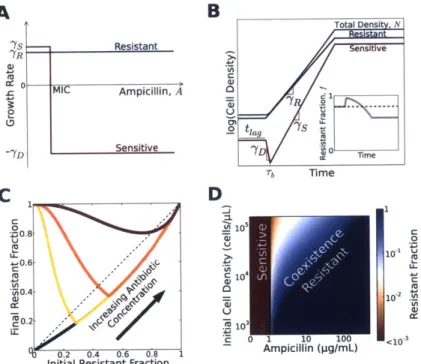
To better understand the population dynamics, we developed a simple model that describes the growth of the bacteria in the presence of antibiotics (Fig. 2-2A, B, Fig. B-8). For the range of antibiotic concentrations we probed, the resistant cells were essentially unaffected and grew at a constant rate of yR (Fig. B-8, B-9B, B-10). We assumed that sensitive cells grow at a rate ys > -yR for antibiotic concentrations below
their MIC, but die at a rate -yD for higher concentrations (Fig. B-3,B-8,B-9). Plating experiments showed that, in addition to cell death, we should incorporate a short lag phase that follows after inoculation of the bacteria into fresh media, during which bacteria neither divide nor die (Fig. B-9). We modeled antibiotic degradation phe-nomenologically using Michaelis-Menten kinetics with a maximum rate per cell Vmax and an effective Michaelis constant Km (A). While this model clearly neglects many aspects of bacterial growth in antibiotics, it successfully captures the key features of the dynamics (Fig. 2-iC, Fig. 2-2C) and predicts conditions that enable coexistence between resistant and sensitive cells (Fig. 2-2D).
A B Total Densi, N --- Resistant ns ve 'YR CU C MIC Ampicillin, A o0 U -2 tag 7s Sensitive '0 0 T Tb Time C DI 0 1 Z0.8 U -1 .0 ~0.6 rp -' A 0.4 10 40.2 1r 0.2 0.4 0.6 0.8 2
mpicillin (pg/mL) Initial Resistant Fraction
Figure 2-2: A simple model describes the population dynamics of a
cooper-ative antibiotic resistance plasmid in the /3-lactam antibiotic ampicillin. (A) Growth rates of resistant (blue) and sensitive (red) bacteria as a function of antibiotic concentration. Free of the metabolic cost associated with resistance, sensitive cells grow faster than resistant cells ('7s > 7y2) at antibiotic concen-trations below the MIC of the sensitive bacteria. Above the MIC, sensitive cells die at a rate of 7YD- (B) The population dynamics within a single com-petition cycle (one day). During the lag phase (t < tiag), neither cell type divides nor dies, but the antibiotic is constantly hydrolyzed by resistant cells. After the lag phase, each sub-population grows at a rate that depends on the extracellular antibiotic concentration. At time Tbs, the extracellular antibiotic concentration drops below the MIC of the sensitive cells. Cell growth ceases when the total population density reaches saturation. Inset: The time trace of the resistant fraction within a single day. (C) The model gives rise to dif-ference equations that resemble experimental data (Fig. 2-iC, Fig. 2-3A, B).
(D) The equilibrium resistant fraction predicted by our model as a function of the antibiotic concentration and the initial cell density. According to the model, coexistence between resistant and sensitive cells is possible at antibiotic concentrations above the MIC of sensitive cells.
We obtained an exact analytic solution of this model that describes the dependence of the equilibrium resistant fraction, fR, on the initial antibiotic concentration, Aj, and initial cell density, Ni. The model predicts that the equilibrium fraction scales in the following manner:
Ai + Km ln(Ai/MIC) - MIC Ai>KmMIC Ai
VmaxNi Vmax Ni
This relationship is surprisingly insensitive to many parameters, including the length of the lag phase, rate of cell death, and cost associated with resistance (A). In particular, our analytic solution of the model predicts that the resistant fraction at equilibrium increases approximately linearly with the antibiotic concentration, a prediction borne out in experimental difference maps obtained at multiple antibiotic concentrations (Fig. 2-3A, B, C). Moreover, the model predicts that the equilib-rium fraction is inversely proportional to the starting cell density. This prediction was experimentally confirmed by measuring the difference equations at four differ-ent starting cell densities. In each case, the equilibrium resistant fraction increases linearly with antibiotic concentration, but with slopes that decrease with increasing initial cell density (Fig. 2-3A, B, C). We therefore find a surprising simplicity to the population dynamics of the antibiotic resistance plasmid in the population, despite the biological complexity of the interaction between the cells and the antibiotic.
In addition to providing significant insight into the population dynamics, the model can quantitatively describe the experimental data. To acquire realistic
param-eters for the model, we measured the growth rate of resistant bacteria (_YR=1.1/hr,
Fig. B-9) and the relative growth rate of sensitive bacteria (Ys/7R=1.15, Fig. B-11). Together these allowed us to deduce the overall metabolic cost of carrying the plasmid
(7s - 7R=~0.17/hr), which includes the cost of plasmid maintenance, of expressing the -lactamase enzyme, and of expressing a red-fluorescent protein used for tracking the resistant fraction (Fig. B-1). Control experiments using another plasmid that did not express a fluorescent protein exhibited similar population dynamics (Fig. B-12). We proceeded to measure the death rate of sensitive bacteria in the presence of the
antibiotic (2.8/hr, Fig. B-9) and the lag time before cell growth/death (1 hr, Fig. B-9).
Using these experimentally measured parameters, we then fit our 30 measured equilibrium fractions (in Fig. 2-3C) to obtain estimates of MIC = 1.1 pg/mL, Vma,
= 106 molecules
/
(CFU-sec), and Km = 6.7 pg/mL. This MIC is slightly lower than our measured value (-2 pg/mL, Fig. B-3) because antibiotic concentrations below the measured MIC already partially inhibit the growth of sensitive bacteria (Fig.B-3). In addition, our fitted value for the maximum rate of hydrolysis per cell Vmax
is reasonable since a single enzyme can hydrolyze as many as ~ 103 molecules per second [601. Although the estimate of Km agrees with literature values (from 4.9 to 26.5 pg/mL [64-66]), we note that the KM in our model is a phenomenological parameter because antibiotic hydrolysis occurs both inside and outside the cells
[59,
651. The resistant fraction at equilibrium in our model increases linearly with the
antibiotic concentration for A > Km, but deviates slightly from linearity for A < KM due to the Michaelis-Menten kinetics of antibiotic degradation (Fig. 2-3C). This simple model not only captures the behavior of the equilibrium fractions, but also successfully predicts the experimental difference equations using the same parameter values (Fig. 2-3A, B, Fig. B-13).
Another way to think about the scaling predicted by the model is that, at equi-librium, the number of resistant cells is proportional to the antibiotic concentration
(NRi - fA.Ni ~ Ai). Indeed, a plot of the equilibrium density of resistant cells against
the antibiotic concentration revealed a striking collapse of the data extending over two orders of magnitude in the antibiotic concentration (Fig. 2-3D). Intuitively, more resistant cells would be required to deactivate larger amounts of the antibiotic within a fixed period of time. Non-intuitively, the model predicts that the time necessary for a bacterial population to saturate in the presence of the antibiotic is minimized at a resistant fraction that corresponds neither to the equilibrium fraction nor to a fully resistant population (Fig. B-14). Given the similarity between our experimental difference equations and the well-known "logistic equation" from theoretical ecology
to become unstable, leading to oscillations around the equilibrium. We found that the equilibrium fractions should become unstable as the antibiotic concentration de-creases; however, the size of the oscillations does not become large enough to observe experimentally (Fig. B-15).
100x Dilution
100 "
0.2 0.4 0.6 0.8 1.0 initial Resistant Fraction
0 50 100 150 200 Amnpicillin (pg/mL) B C 0 'U 2 C D 200x Dilution 00 0 100 15/ 5 0.2 0.4 0.6 0.8 1.0 Initial Resistant Fraction
Dilution E104 100x C 200x ,6 400x 1 * 800x 100 101 102 Ampicillin (pg/mL)
Figure 2-3: Experimental difference equations confirm model predictions
re-garding the equilibria and dynamics of resistant and sensitive bacteria. (A-B) Experimental difference equations obtained at two dilution factors (100x and 200x) and different antibiotic concentrations. At a given antibiotic concentra-tion, an increase in the dilution ratio leads to stronger selection for resistance. Each difference equation plotted in a, b includes data obtained on 3 different days. Measurement error from flow cytometry was typically smaller than sym-bol size. (C) The equilibrium fractions as a function of ampicillin concentration at four different dilution factors (see Fig. B-13 for difference equations). The relationship is approximately linear for antibiotic concentrations higher than
Km. The equilibrium fractions were extracted from the difference equation plots by determining the intersection between the difference equations and the diagonal line (dashed line in (A)). Error bars represent standard error of the mean (n=3). (D) Plotting the initial density of resistant cells at equilibrium as a function of antibiotic concentration reveals a data collapse that extends over two orders of magnitude in the concentration. (A-D) Solid curves show a single fit of the model to all the experimental data.
A 1.0 0.8 0.6 u0.4 0.2 0 CC 0 'U 'U E C0 L" 1.0 0.8 0.6 0.4 0.2 0.0 1.0 0.8 0.6 0.4 0.2 01
2.3.4
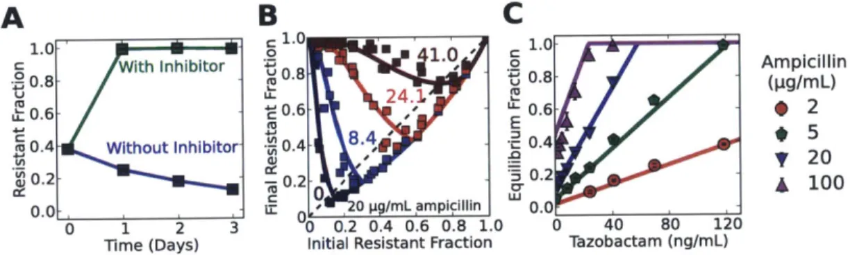
Addition of a -lactamase inhibitor selects for resistance
Given the predictive power of the model, we explored the expected consequences of adding a O-lactamase inhibitor such as tazobactam, which is used clinically together with many 0-lactam antibiotics [57, 59, 68]. Tazobactam competitively binds
#-lactamase enzymes [68, 69] and prevents them from hydrolyzing the antibiotic, leading to an increase in the effective Michaelis constant KM. A sufficiently large increase in the Michaelis constant (Km) can significantly compromise the ability of resistant cells to degrade the antibiotic, leading to complete inhibition of bacterial growth (Fig. B-16). However, if the increase in Km is not sufficiently large, the resistant cells may survive the treatment, but the larger KM would hinder their ability to protect sensitive cells against the antibiotic. Specifically, since the equilibrium fraction of resistant cells is proportional to Km, the model predicts that adding a -lactamase inhibitor will lead to an increase in the resistant fraction. We have tested this prediction and found that the addition of tazobactam can indeed result in a completely resistant population (Fig. 2-4A, Fig. B-17).
Not only does the model provide qualitative insight, it also makes surprisingly accurate quantitative predictions about the population dynamics that take place in the presence of the inhibitor. Although the actual mechanism of inhibition is more complicated
[571,
we modeled tazobactam as a competitive inhibitor, which increases the Michaelis constant KM to Keff = KM - (1 + [I]/K1), where [I] and K, are theinhibitor concentration and dissociation constant, respectively. Since the equilibrium fraction increases linearly with KM, the model predicts that it should also increase linearly with the inhibitor concentration [I]. To probe this predicted dependence of the equilibrium fraction on the inhibitor concentration, we measured the equilibrium fractions from maps of difference equations obtained at varying tazobactam concen-trations (Fig. 2-4B, C). We successfully fit the new 31 equilibrium fractions (Fig. 2-4C) using one additional free parameter KI, confirming the predicted linear de-pendence on the inhibitor concentration. The K, from the fit (4.6 ng/mL) was well within literature values (3 to 11.4 ng/mL [69-71]). Remarkably, using the value of
A
B
C
1.0-' 1.0 1.0 With Inhibitor 0 , o Ampicillin 0.8 - 0.8 0.8 (pg/mL /. - ' -0.6
-*
2 0.4 Without Inhibitor 0.4 4' u- 0.4 _ :3 52 LO. -022 0.2-0 0.2 100 . , 3 - 0 uigp/mL ampicillin 0.0 0 3 ) 0.2 0.4 0.6 0.8 1.0 0 40 80 120 Time (Days) Initial Resistant Fraction Tazobactam (ng/mL)Figure 2-4: As predicted by the model, addition of the -lactamase inhibitor
tazobactam increases the fraction of resistant cells in the population. (A) Sen-sitive E. coli cells increase in frequency when grown in 20 pg/mL ampicillin in the absence of tazobactam; however, the addition of the inhibitor at a concen-tration of 1000 ng/mL results in a completely resistant bacterial population. Cultures were diluted daily by a factor of 100x into fresh media containing 20 pg/mL ampicillin. Error bars represent standard error of the mean of 4 different bacterial cultures. (B) Experimental difference equation maps for 4 different concentrations of the inhibitor tazobactam (in ng/mL) at a background of 20 pg/mL ampicillin and dilution factor of 100x (see Fig. B-17 for more difference equations). Each difference equation map contains data obtained on 3 different days. (C) As predicted by the model, the equilibrium fractions depend linearly on the concentration of the inhibitor tazobactam with a slope that depends on the ampicillin concentration. The equilibrium fractions were extracted from the difference equation plots by determining the intersection between the difference equations and the diagonal line (dashed line in (A)). Error bars represent stan-dard error of the mean (n=3). (B-C) Solid curves show a fit of the model to all the experimental data with a single free parameter of K, = 4.6 ng/mL (other parameters held fixed).
K, obtained from the fits to the equilibrium fractions successfully recapitulated the
To verify that our conclusions were not limited to tazobactam, we tried the 3-lactamase inhibitor sulbactam, which is often administered together with ampicillin clinically 158, 68, 69]. We found that at least for our experimental conditions (E.
coli bacteria inoculated at an initial cell density - 10' cells/PL), the addition of
sulbactam can lead to the accelerated spread of resistant bacterial cells in a range of clinically relevant antibiotic concentrations (Fig. B-18).
2.4
Discussion
We have presented a quantitative analysis of the population dynamics that stem from the cooperative nature of antibiotic inactivation, and which can lead to coexistence between sensitive cells and resistant cells. Our analysis was based on two key fea-tures: (1) the presence of a metabolic cost associated with being resistant, and (2) the inactivation of the antibiotic by resistant cells. When both features apply, our model suggests that resistant and sensitive cells may coexist at high concentrations of the antibiotic, with the fraction of resistant cells approximately proportional to the antibi-otic concentration divided by the cell density. We found that this simple dependence on antibiotic concentration and cell density successfully predicts the equilibrium frac-tion of resistant cells over two orders of magnitude in antibiotic concentrafrac-tion (Fig.
2-3D).
This model not only agrees quantitatively with experimental data, but it also provides insight into the conditions that enable coexistence between resistant and sensitive cells. For example, a recent study observed coexistence with a mutated /-lactamase enzyme that inactivated the antibiotic outside of the cell [54], allowing resistant cells to efficiently "share" their resistance with the bacterial population to support coexistence. However, in our study, we were able to observe coexistence even with a wild-type /3-lactamase enzyme, which is primarily periplasmic [591. To properly interpret these results, it is important to recognize that the site of antibiotic inactivation determines the degree of preferential protection offered to resistant cells. Furthermore, as long as resistant cells are sufficiently protected to be unaffected by
the antibiotic, only the overall rate of antibiotic inactivation is important in deter-mining the dynamics between resistant and sensitive cells. Hence, even if antibiotic inactivation occurs inside the cell, it is still a cooperative behavior that may allow sensitive cells to survive.
The interplay between initial cell density and antibiotic concentration is often im-portant in determining growth dynamics in antibiotics [42, 72]. Likewise, our model suggested that the key parameter in governing the population dynamics was not the antibiotic concentration, but the ratio between the antibiotic concentration and the initial cell density. Specifically, we found that at high cell densities, resistant cells could protect sensitive cells against antibiotic concentration as high as 200 pg/mL (Fig. 2-3A), which is a hundred-fold higher than the minimum inhibitory concentra-tion of sensitive cells. Given the cooperative nature of antibiotic inactivaconcentra-tion, it is likely that other ecological factors will be important to consider when attempting to understand the evolution of antibiotic resistance [73-75].
One might worry that our conclusions may be limited to laboratory strains since natural strains would be better adapted to plasmids found in the wild. However, our model and experiments argue that the equilibrium fraction depends only weakly on the fitness cost of carrying the resistance plasmid (Fig. B-19). Compensatory mutations that alleviate the cost of resistance [49-51] will increase the time it takes the population to settle into its equilibrium fraction, but will not significantly change that fraction. Since our model only uses a few key phenotypic traits to characterize the outcome of bacterial growth in the antibiotic, it should be broadly applicable in
describing both intra-species [54] and inter-species [46] dynamics.
Within the framework of our model an important qualitative difference between using a bactericidal versus a bacteriostatic antibiotic is that the overshoot of the resistant fraction above the equilibrium fraction should only appear when using a bactericidal antibiotic (Fig. 2-1A, Fig. B-20). The lower the initial resistant fraction is, the longer it takes for the antibiotic to be inactivated, and the more opportunity there is for a bactericidal antibiotic to kill the sensitive strain and promote the growth of the resistant strain.
Throughout our experiments, we limited ourselves to antibiotic concentrations which do not affect the growth of resistant cells. However, at high enough concentra-tions, a bactericidal antibiotic may lead to lysis of resistant cells and the subsequent release of their beta-lactamase enzymes into the extra-cellular space
[761.
Since these enzymes inactivate the antibiotic even faster extracellularly, the death of resistant cells may further increase the cooperative nature of bacterial growth in the antibiotic177]. Such a scenario may explain the observed non-monotonic selection for resistance
and difference equation maps that deviate from our model at high concentrations of the 1-lactam antibiotic piperacillin (Fig. B-21).
Understanding how the fraction of resistant bacteria changes with time is a central goal in studying antibiotic resistance. This already difficult task is further complicated
by cooperative behaviors that allow resistant microbes to "share" their resistance with
the rest of the bacterial population. The cooperative nature of antibiotic inactivation causes the fitness of resistant cells to decrease as their fraction in the bacterial popula-tion increases (i.e., it leads to negative frequency dependent selecpopula-tion [54], Fig. 2-3A, B). Overall, this enables coexistence between resistant and sensitive cells, even in the absence of the spatial structure present in biofilms
178-80],
interactions between bac-teria and antibiotic degradation products [811, bacbac-terial persistence 1821, and indole production [831. Since antibiotic inactivation is a frequent mechanism of antibiotic resistance [14], similar population dynamics may appear with other classes of antibi-otics (e.g., macrolides, aminoglycosides) and with chromosomally encoded enzymes. However, despite the potential ubiquity of cooperative antibiotic resistance, the social aspect of antibiotic resistance remains under-appreciated, highlighting the importance of quantitatively characterizing social interactions to gain a thorough understanding of the maintenance of phenotypic and genotypic diversity within populations.2.5
Materials and Methods
2.5.1
Strains
All strains are derived from Escherichia coli DH5a. The resistant strain contained the
pFPV-mCherry plasmid [84] (also see Addgene plasmid 20956), expressing a TEM-1 ,3-lactamase enzyme and an mCherry fluorescent protein. In addition, the resistant and sensitive strains expressed cerulean and yellow fluorescent protein genes, respec-tively, under the promoter PiacUV5, and a kanamycin resistant gene, both carried on the plasmid pZS2501+11 [85, 86] (origin of replication: pSC101). Control experi-ments in which the cerulean and yellow fluorescent markers were swapped gave nearly identical difference equation maps (Fig. B-22).
2.5.2
Competition Experiments
All cultures were grown in a shaker at 500 rpm and 37 C. Before the competition
experiments, single colonies of resistant and sensitive strains were grown separately in 5 mL lysogeny broth (LB) together with antibiotics for selection for 23 hours. The saturated cultures (corresponding to a density of ~ 107 cells/pL) were diluted by a
factor of lOnx and co-cultured at different fractions in 96 we--ptefonaTnPgLB
and 5 pg/mL of kanamycin for another 23 hours to synchronize the growth state of both strains (see Fig. B-11). All competition experiments were carried out using syn-chronized mixed cultures. The cultures were diluted into 96 well-plates containing 5 pg/mL of kanamycin, LB, and appropriate concentrations of ampicillin, tazobactam and sulbactam, and grown for another 23 hours. In multi-day experiments, cultures were serially diluted into 96-well plates containing freshly prepared media with appro-priate concentrations of antibiotics. Control experiments showed that the population dynamics were similar regardless of whether kanamycin was absent or present at 5 pg/mL (Fig. B-23). In addition, control experiments showed that similar growth dynamics apply in other -lactam antibiotics (Fig. B-24). Fractions were determined using flow cytometry on a BD-LSR II and confirmed by plating (Fig. B-1,B-2).
Chapter 3
Seasonality Gives Rise to Oscillatory
Dynamics in a Bacterial
Cross-Protection Mutualism
3.1
Overview
Understanding how bacteria survive and respond to antibiotic exposure is important both clinically and ecologically. A common mechanism of antibiotic resistance in-volves the inactivation of antibiotics by resistant bacteria. Inactivation of antibiotics is a cooperative behavior, which can allow resistant bacteria to protect sensitive bac-teria against antibiotics, altering the dynamics of how antibiotic resistance spreads. However, despite the prevalence of antibiotic inactivation as a mechanism of antibi-otic resistance, how it affects the population and evolutionary dynamics of microbial populations remains poorly understood, particularly in the presence of more than one antibiotic. Here, we investigate whether two Escherichia coli strains can protect each other in the presence of chloramphenicol and ampicillin. Our experiments reveal that the two strains can form an effective cross-protection mutualism, helping each other survive in antibiotic concentrations that inhibit growth of either strain alone. More-over, we find that "seasonality" (introduced by periodic dilution into fresh media
sup-plemented with antibiotics) gives rise to large oscillations in the relative abundances of the two strains, with an oscillation period longer than the period between succes-sive antibiotic exposures. While the mutualism remains stable in modest antibiotic concentrations, the mutualism collapses at high antibiotic concentrations due to the oscillations which destabilize the mutualism. The ability of the two strains to form a successful cross-protection mutualism without requiring a period of co-evolution indicates that similar mutualisms may frequently arise in course of the antibiotic treatment and in natural environments such as the soil.
3.2
Introduction
Mutualisms are reciprocal positive interactions between two species. Because mu-tualisms are thought to be fundamentally important in many ecosystems, ecologists have devoted significant time studying their evolution, stability, and ecological func-tion
[87-94].
One of the best studied mutualisms is the one formed between flowering plants and their pollinators. In this mutualism, the pollinator mediates the repro-duction of the plant and in return receives nutrition194].
More broadly, positive interactions are abundant in nature and can take on many forms: one species can in-crease the fitness of another by providing nutrition, protection, or transportation [94,951, and the benefits from a positive interaction may arise immediately or at a later
time.
The desire to understand the basic forces that shape microbial communities has created a recent surge of interest in microbial mutualisms [96-102]. So far the majority of studies of microbial mutualisms have focused on cross-feeding [97-99, 103] due to the apparent abundance of this type of interaction. However, another potentially frequent microbial interaction involves the protection of one microbe by another [40, 41, 83], which could lead to the formation of cross-protection mutualisms. Protective interactions are especially interesting in the context of antibiotic resistance, where they may influence the spread of antibiotic resistance genes [47, 104].