Control of the mitotic exit network during meiosis
The MIT Faculty has made this article openly available. Please share how this access benefits you. Your story matters.Citation Attner, M. A., and A. Amon. “Control of the Mitotic Exit Network During Meiosis.” Molecular Biology of the Cell 23.16 (2012): 3122– 3132. Copyright © 2012 by The American Society for Cell Biology As Published http://dx.doi.org/10.1091/mbc.E12-03-0235
Publisher American Society for Cell Biology Version Final published version
Citable link http://hdl.handle.net/1721.1/72485
Terms of Use Article is made available in accordance with the publisher's policy and may be subject to US copyright law. Please refer to the publisher's site for terms of use.
Control of the Mitotic Exit Network during meiosis. Michelle A. Attner1 and Angelika Amon1,*
1David H. Koch Institute for Integrative Cancer Research and Howard Hughes Medical
Institute, Massachusetts Institute of Technology, 76-543, 500 Main Street, Cambridge, MA 02139, USA.
* To whom correspondence should be addressed: e-mail: [email protected]
ABSTRACT
The Mitotic Exit Network (MEN) is an essential GTPase signaling pathway that triggers exit from mitosis in budding yeast. We show here that during meiosis, the MEN is dispensable for exit from meiosis I, but contributes to the timely exit from meiosis II. Consistent with a role for the MEN during meiosis II, we find that the signaling pathway is only active during meiosis II. Our analysis further shows that MEN signaling is
modulated during meiosis in several key ways. Whereas binding of MEN components to spindle pole bodies (SPBs) is necessary for MEN signaling during mitosis, during
meiosis MEN signaling occurs off SPBs, and does not require the SPB recruitment factor Nud1. Furthermore, unlike during mitosis, MEN signaling is controlled through the regulated interaction between the MEN kinase Dbf20 and its activating subunit Mob1. Our data lead to the conclusion that a pathway essential for vegetative growth is largely dispensable for the specialized meiotic divisions and provide insights into how cell cycle regulatory pathways are modulated to accommodate different modes of cell division. INTRODUCTION
In the final phase of the cell cycle, after chromosomes segregate in anaphase, cells exit from mitosis. In the budding yeast Saccharomyces cerevisiae this cell cycle transition is brought about by the inactivation of mitotic cyclin-dependent kinases, known as Clb-CDKs (Stegmeier and Amon, 2004). Central to the precipitous inactivation of Clb-Clb-CDKs at the end of mitosis is the protein phosphatase Cdc14 (Jaspersen et al., 1998; Visintin et al., 1998; Zachariae et al., 1998). Cdc14 triggers the degradation of Clb cyclins, the up-regulation of a Clb-CDK inhibitor and the dephosphorylation of Clb-CDK substrates to rapidly reset the cell to a low CDK state. This resetting in turn causes the events of mitotic exit: mitotic spindle disassembly, chromosome decondensation and cytokinesis.
Given the central importance of Cdc14 to exit from mitosis, it is not surprising that its activity is tightly controlled (Shou et al., 1999; Visintin et al., 1999). Cdc14 is bound to its nucleolar-localized inhibitor Cfi1/Net1 throughout most of the cell cycle. During anaphase, the phosphatase is released from its inhibitor, freeing it to dephosphorylate its targets in the nucleus and cytoplasm. This release occurs in two waves and by two pathways: the Cdc14 early anaphase release (FEAR) network and the Mitotic Exit Network (MEN). The FEAR network is not essential for viability and promotes a transient burst of Cdc14 release early in anaphase (Pereira et al., 2002; Stegmeier et al., 2002; Yoshida et al., 2002; Rock and Amon, 2009), which contributes to the co-ordination of anaphase events. The MEN is required for sustained Cdc14 release which is essential for exit from mitosis (Stegmeier and Amon, 2004).
The MEN is a Ras-like GTPase signaling cascade in which the GTPase is encoded by TEM1 (Shirayama et al., 1994). Localization of Tem1 to the spindle pole body (SPB, yeast equivalent of the centrosome), is essential for MEN activation (Valerio-Santiago and Monje-Casas, 2011). Tem1 is recruited to SPBs during metaphase and remains there until the completion of anaphase (Bardin et al., 2000; Pereira et al., 2000). During metaphase, Tem1 is kept inactive at SPBs by the two-component GTPase activating protein (GAP) Bub2-Bfa1 (Geymonat et al., 2002). During anaphase, Tem1 is activated by multiple signals, including spindle position. Tem1-GTP then recruits the kinase Cdc15 to SPBs, which is thought to activate Cdc15 (Asakawa et al., 2001). Cdc15 in turn recruits Dbf2, the founding member of the NDR (nuclear Dbf2-related) protein kinase family, and its regulatory subunit Mob1 to SPBs and activates the Dbf2-Mob1 kinase complex (Mah et al., 2001; Visintin and Amon, 2001). Budding yeast harbor two Dbf2-like kinases, Dbf2 and Dbf20. Dbf2 provides the predominant activity during
vegetative growth and is active only during anaphase. Dbf20 is expressed at low-levels during vegetative growth but is induced during sporulation (Chu et al., 1998). Regulation of its activity in mitosis is not understood in detail, but it has been shown that Dbf2 is more active than Dbf20 in vitro (Toyn and Johnston, 1994).
MEN components are thought to assemble into signaling modules by a scaffold protein Nud1. The Nud1 protein localizes to SPBs and is required for the association of Tem1, Cdc15, and Dbf2-Mob1 with SPBs (Adams and Kilmartin, 1999; Gruneberg et al., 2000;
Visintin and Amon, 2001; Valerio-Santiago and Monje-Casas, 2011). This function is essential for exit from mitosis. Cells harboring a temperature sensitive allele of NUD1 arrest in anaphase with MEN components not localized to SPBs (Adams and Kilmartin, 1999; Bardin et al., 2000; Gruneberg et al., 2000; Visintin and Amon, 2001). This requirement of NUD1 for exit from mitosis is at least in part due to its function in recruiting MEN components to SPBs because binding of Tem1 and Cdc15 to SPBs is essential for MEN activity (Rock and Amon, 2011; Valerio-Santiago and Monje-Casas, 2011).
The MEN integrates multiple cellular events, such as onset of chromosome segregation, Polo kinase activity and spindle position. This ensures that exit from mitosis only occurs when chromosome segregation has been completed yielding two daughter cells each containing a complete complement of the genome (Stegmeier and Amon, 2004). Regulation of the MEN by spindle position is understood best. The MEN is only activated when the nucleus has been pulled into the daughter cell and a MEN component-carrying SPB has entered the bud (Yeh et al., 1995; Bardin et al., 2000; Pereira et al., 2000). Spindle position control of the MEN is accomplished by a system composed of a MEN-inhibitory and a MEN-activating zone, and a sensor that moves between them. The MEN inhibitor Kin4 is located in the mother cell, the Kin4 inhibitor Lte1 in the bud, and the MEN GTPase Tem1 is localized to the SPB that will migrate into the bud (Bardin et al., 2000; Pereira et al., 2000; D'Aquino et al., 2005; Maekawa et al., 2007; Chan and Amon, 2010; Bertazzi et al., 2011; Falk et al., 2011). Only when the MEN-bearing SPB escapes the zone of the MEN inhibitor Kin4 in the mother cell and moves into the bud where the Kin4 inhibitor (and hence MEN activator) Lte1 resides, can exit from mitosis occur. In this manner, spatial information is sensed and translated to regulate MEN activity.
While the function of the MEN has been characterized in mitosis, it has not been well characterized in meiosis, a specialized cell division. During meiosis, a diploid cell undergoes two rounds of chromosome segregation following one round of DNA replication, and results in the formation of four haploid gametes called spores in yeast (Marston and Amon, 2004). Whereas S. cerevisiae divides by budding during
membranes growing around the four meiotic products after both meiotic divisions have been completed. These changes in the chromosome segregation pattern and in the morphological constraints on chromosome segregation require the basic cell cycle machinery to be changed in fundamental ways.
Here, we investigate the function and regulation of the Mitotic Exit Network during meiosis. Previous studies showed that Cdc14 is essential for progression through meiosis (Sharon and Simchen, 1990). As during mitosis, the phosphatase is released from the nucleolus during anaphase I and anaphase II. However, the FEAR network rather than the MEN appears to be critical for the activation of Cdc14, at least during anaphase I (Buonomo et al., 2003; Marston et al., 2003). The MEN in fact appears to be dispensable for exit from meiosis I (Kamieniecki et al., 2005; Pablo-Hernando et al., 2007). This is perhaps not surprising as one of the major functions of the MEN,
coordinating exit from mitosis with spindle position, is less important during meiosis, as both nuclear divisions occur within the confines of a single cell. Together, these
observations raise the question of whether a pathway, which is absolutely essential for progression through mitosis, is also required for progression through meiosis and, if it is, which signals regulate it. We find that the MEN is dispensable for exit from meiosis I, and contributes to the timely release of Cdc14 from the nucleolus during anaphase II and exit from meiosis II. Consistent with a role of the MEN only during meiosis II, we find that the signaling pathway is only active during meiosis II. Our analysis further revealed that the MEN pathway is regulated differently during meiosis and mitosis. Meiotic MEN signaling does not require the Nud1 scaffold protein and relies instead on the regulated interaction between Dbf20 and its regulatory subunit Mob1. Our data show that the MEN, a signaling cascade essential for mitotic exit, is dispensable for the meiotic divisions and shed light on how MEN signaling is adapted to function during a symmetric cell division, meiosis.
RESULTS
The Mitotic Exit Network is required for the timely exit from meiosis II.
The MEN is essential for viability and serves the important function of ensuring that exit from mitosis only occurs when the nucleus has been threaded through the bud neck into the daughter cell. In contrast, during meiosis the two nuclear divisions occur within the
mother cell and are followed by membrane growth around the meiotic products (Neiman, 2011). This difference in cell division pattern and morphology raises the question of whether the MEN, a pathway critical to cell division by budding, functions during meiosis and if it does, how its activity is regulated. To address these questions we examined the consequences of loss of MEN function during meiosis and
investigated the regulation of MEN signaling.
To generate cell cultures that progress through the meiotic divisions in a synchronous manner we employed a previously developed synchronization protocol termed the “Ndt80 block-release” system (Carlile and Amon, 2008). Briefly, NDT80 encodes a transcription factor that is essential for entry into the meiotic divisions. Cells in which NDT80 is expressed from the GAL1-10 promoter and that also contain a fusion between the GAL1-10 transcription factor Gal4 and the estrogen receptor (GAL4-ER) arrest prior to entry into meiosis I (in pachytene) in the absence of estrogen, but progress
synchronously through the meiotic divisions upon estrogen addition to the medium. To determine whether the MEN is required for the meiotic divisions we examined the consequences of inactivating various MEN components. We employed an allele of CDC15 (cdc15-as1) (Bishop et al., 2000; D'Aquino et al., 2005) that can be specifically inhibited upon addition of an ATP analog, 1-NA-PP1, while leaving other cellular kinase activities intact. Treatment of vegetatively growing cdc15-as1 cells with inhibitor
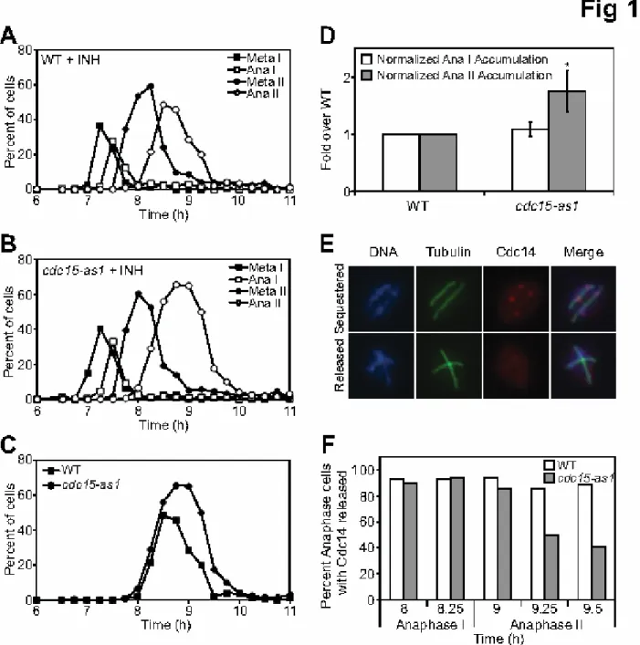
prevented exit from mitosis causing an anaphase arrest (see Figure 6F). In contrast, exit from meiosis I was not delayed in cdc15-as1 cells and exit from meiosis II was only subtly delayed (Figure 1A–C). This is best seen when time spent in anaphase II is measured by integrating the area under the line, representing the number of cells harboring anaphase II spindles. In the presence of inhibitor, cdc15-as1 cultures
harbored 1.75 fold more cells with anaphase II spindles than wild-type cultures (Figure 1D; n=6 independent experiments). These findings are consistent with previous reports analyzing the effect of depleting Cdc15 and Tem1 in meiosis (Kamieniecki et al., 2005; Pablo-Hernando et al., 2007).
Similar results were obtained in cells lacking the MEN component Mob1. Mob1 was selectively depleted during meiosis by placing the MOB1 gene under the control of the mitosis-specific CLB2 promoter (MOB1-mn). Lack of transcription during the meiotic
divisions effectively depletes the Mob1 protein (Figure S1A). MOB1-mn cells
progressed through the meiosis I-meiosis II transition with wild-type kinetics, but were delayed in exit from meiosis II (Figure S1B-D). We were not able to deplete the
essential component Dbf2 (data not shown), preventing us from examining the
consequences of losing both Dbf2 and Dbf20 activity on meiotic progression. However, deleting DBF20 alone did not affect progression through meiosis (data not shown), presumably because low-levels of Dbf2 present in meiotic cells support the timely exit from meiosis I. We also have not been able to deplete the essential component Tem1 during meiosis. However, other studies have shown that like other MEN components, TEM1 is not required for the meiosis I-meiosis II transition (Kamieniecki et al., 2005). We conclude that the Mitotic Exit Network, essential for cell cycle progression during vegetative growth, is dispensable for progression through meiosis.
The essential function of the MEN during mitosis is to release Cdc14 from the nucleolus (Shou et al., 1999; Visintin et al., 1999). Cdc14 is also released from the nucleolus during exit from meiosis I and meiosis II and is essential for meiosis (Buonomo et al., 2003; Marston et al., 2003). The observation that the MEN was dispensable for the meiotic divisions therefore predicts that Cdc14 release from the nucleolus during exit from meiosis I and II must occur independently of MEN function. This is indeed what we observe. Cdc14 was released with wild type kinetics during exit from meiosis I in cdc15-as1 and mob1-mn mutants. However, we observed a slight impairment in Cdc14 release during exit from meiosis II, consistent with a minor contribution of the MEN to this cell cycle transition (Figure 1E-F; Figure S1E; (Pablo-Hernando et al., 2007)). This requirement for Cdc14 release was particularly evident in later time points raising the possibility that the MEN is needed to maintain Cdc14 in the released state during exit from meiosis II. We conclude that while the MEN is essential for Cdc14 release from the nucleolus during exit from mitosis, other pathways must be responsible for bringing about this event during the meiotic divisions. The FEAR network likely plays this role. Meiotic cells lacking FEAR network components exhibit a severe defect in releasing Cdc14 from the nucleolus during meiosis I and exhibit a phenotype remarkably similar to that of cells lacking CDC14 function (Buonomo et al., 2003; Marston et al., 2003).
MEN activity is restricted to meiosis II.
During mitosis, MEN activity is controlled by nuclear position. The pathway is only activated when the nucleus moves into the bud (Bardin et al., 2000; Pereira et al., 2000). In contrast, the meiotic nuclear divisions occur within the mother cell, raising the question of how, if at all, the MEN is controlled during the meiotic divisions. To address this question we first determined whether MEN components are expressed during the meiotic divisions. Consistent with the observation that the MEN is needed for the timely exit from meiosis II, we found that all MEN components analyzed were expressed during meiosis. The GTPase Tem1 and the effector kinase Cdc15 were present at constant levels throughout meiosis and the proteins exhibited no noticeable mobility shifts (Figure S2A-D). Mob1 and Nud1 were also found throughout meiosis but their mobility changed. Slower migrating forms of the proteins appeared concomitantly with meiosis I entry (Figure S2E-H). This change in mobility is likely due to phosphorylation (Luca and Winey, 1998; Gruneberg et al., 2000; Keck et al., 2011) but the significance of this mobility shift is at present unclear.
Of the two Dbf2-like kinases present in S. cerevisiae only Dbf20 was expressed at appreciable levels during meiosis (Figure S2I-L). Dbf20 mobility also changed during meiosis. Concomitant with entry into meiosis II, slower migrating forms of the protein became apparent. The appearance of this form of Dbf20 correlated well with Dbf20 activity. Dbf20 immunoprecipitated from cells progressing through meiosis in a
synchronous manner exhibited low activity during meiosis I but showed robust activity during anaphase of meiosis II (Figure 2A-B, Figure S3A; note that slower migrating forms of Dbf20 can only be detected with the Myc fusion and not with the Dbf20-ProA fusion).
As during mitosis where Dbf2 activity depends on CDC15 (Visintin and Amon, 2001), we found that Dbf20 activity depended on Cdc15 kinase activity during meiosis. Dbf20 kinase activity was greatly reduced in cdc15-as1 cells progressing through meiosis (Figure 2C; Figure S3B-C). We conclude that the MEN is only active during exit from meiosis II. This observation is consistent with the finding that the MEN is required for the timely exit from meiosis II but not for exit from meiosis I. We have thus far not been able to determine the mechanisms that restrict MEN activity to exit from meiosis II, but
as described below, our analyses of MEN regulation during meiosis II revealed that the pathway is regulated in fundamentally different ways during meiosis II than during mitosis.
MEN components are not detected on spindle pole bodies in meiosis
During mitosis, association of MEN components with SPBs is essential for MEN activity (Rock and Amon, 2011; Valerio-Santiago and Monje-Casas, 2011). The MEN
components Bub2-Bfa1, Tem1, Cdc15 and Dbf2-Mob1 localize to SPBs in a manner that depends on the spindle pole body component Nud1 (Bardin et al., 2000; Gruneberg et al., 2000; Visintin and Amon, 2001). We examined the ability of these MEN
components to bind to SPBs during meiosis. The MEN components Tem1, Cdc15, or Mob1, whether tagged with GFP, Myc or HA, were never detected at SPBs in any stage of meiosis (Figure S4A-D). Additionally, despite the role of Cdc15 in spore wall
formation (Kamieniecki et al., 2005; Pablo-Hernando et al., 2007), Cdc15-eGFP was not detected on SPBs during spore formation (Figure S4B, bottom panels). However, the protein required for tethering these proteins to SPBs during mitosis, Nud1, was present at all SPBs throughout meiosis (Figure S5A-B).
Bfa1 localized to both SPBs during anaphase I as judged by co-localization with the SPB component Spc42. During anaphase II Bfa1 localized to only two of the four SPBs (Figure S5C-D). This anaphase II localization pattern is reminiscent of that found in mitotically dividing cells: Bub2-Bfa1 is concentrated at the SPB that migrates into the bud during anaphase (Pereira et al., 2000; Molk et al., 2004). This asymmetric
localization is brought about by bud-specific cell cortex proteins (Monje-Casas and Amon, 2009). It will be interesting to determine whether these cell cortex proteins are also asymmetrically localized in symmetrically shaped meiotic cells. Taken together, these results indicate that although the MEN is active during anaphase II, most MEN components are not detected at spindle pole bodies.
NUD1 is not required for Dbf20 kinase activity in meiosis II
The absence of MEN components from SPBs during meiosis raises the interesting possibility that MEN signaling does not require localization of its components to spindle pole bodies during meiosis II. A prediction of this hypothesis is that NUD1 is not
kinase activity in cells carrying the temperature sensitive nud1-44 allele (Adams and Kilmartin, 1999). We first established 34°C as a restrictive temperature for the nud1-44 allele, which is a temperature that is still permissive for progression through meiosis. To analyze cells progressing through the mitotic cell cycle in a synchronous manner, 44 cells were arrested in G1 with pheromone. Upon release from the G1 arrest, nud1-44 cells arrested in anaphase and failed to activate Dbf2 kinase at this temperature (Figure 3A-C). Thus, nud1-44 cells are defective in MEN signaling at 34°C during vegetative growth. In contrast to mitosis, nud1-44 cells progressed through the meiotic divisions at 34°C and activated Dbf20 kinase activity with kinetics indistinguishable from that of wild-type cells (Figure 3D-F). To address the possibility that Dbf2 and Dbf20 exhibit a differential requirement for Nud1 in their activation we expressed DBF2 in meiosis from the copper inducible CUP1 promoter (to induce sufficient amounts of Dbf2) and examined the effects of inactivating NUD1 on Dbf2 kinase activity. nud1-44 cells progressing through meiosis at 34°C harbored wild-type levels of Dbf2 kinase activity (Figure S6A-C).
We were unable to tag nud1-44 in order to examine the fate of the nud1-44 protein at elevated temperatures during meiosis. We therefore cannot exclude the formal
possibility that 34°C is not a restrictive temperature for the nud1-44 allele during meiosis. However, given that Dbf2 kinase activity is completely abolished in nud1-44 cells at 34°C during vegetative growth, yet Dbf20 or Dbf2 exhibit wild-type levels of kinase activity in nud1-44 cells progressing through meiosis at 34°C, we consider it more likely that during meiosis Dbf2 and Dbf20 kinase activity do not require NUD1. We were also not able to assess the requirement for NUD1 in spore formation because spore formation is greatly impaired at 34°C even in wild type cells (data not shown). We conclude that an SPB component essential for MEN activation in mitosis is dispensable for MEN activation in meiosis. Furthermore, unlike in mitosis, when SPB binding is essential for MEN signaling, SPB binding is not a prerequisite for MEN activity during meiosis.
Dbf2 and Dbf20 are differentially regulated.
Our results indicate that SPB association is not important for MEN regulation during meiosis. Are other aspects of MEN signaling also differentially regulated between
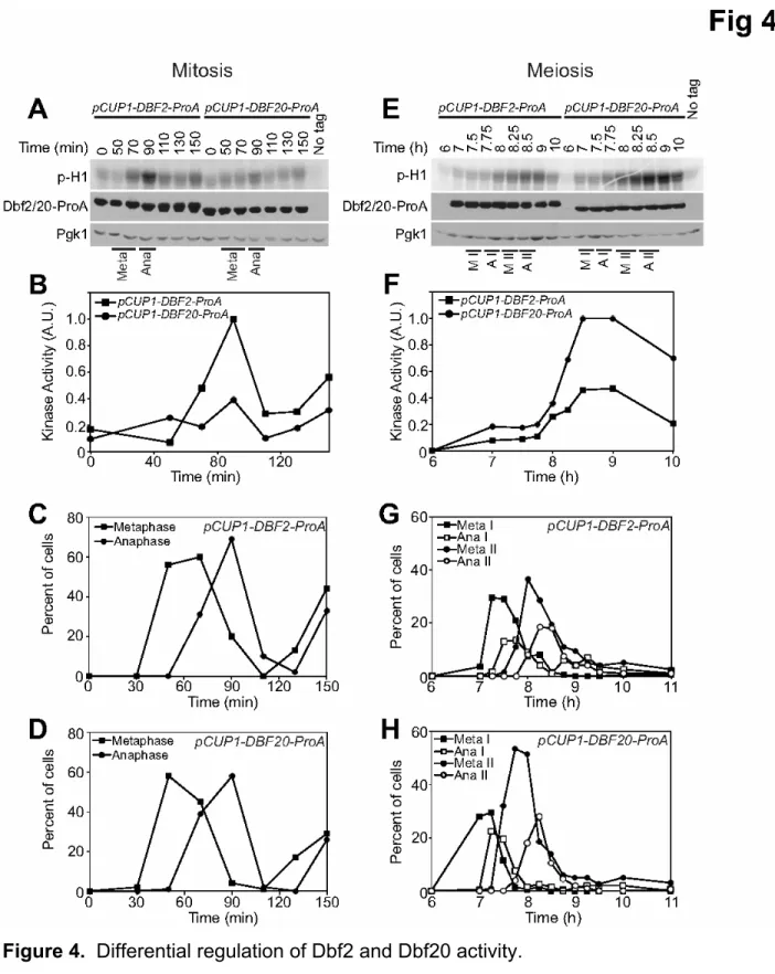
mitosis and meiosis? One obvious difference between the two types of divisions is the use of different Dbf2 family members, Dbf2 in mitosis and Dbf20 during meiosis. Are additional differential controls operative on Dbf2 and Dbf20 during mitosis and meiosis? To address this possibility, we placed both DBF2 and DBF20 under the control of the CUP1 promoter, allowing us to express equal levels of both proteins during mitosis and meiosis (Figure 4A, E).
We first compared Dbf2- and Dbf20-associated kinase activity in cells progressing through the mitotic cell cycle in a synchronous manner. Whereas Dbf2 kinase activity peaked during anaphase, Dbf20-associated kinase activity remained at low levels throughout the cell cycle (Figure 4A-D). Expression of Dbf2 during the meiotic divisions led to Dbf2 kinase activity that is regulated similarly to that of Dbf20 (Figure 4E-H). Our results indicate that, when expressed, both Dbf2 and Dbf20 are active during meiosis and the activity of both kinases is restricted to exit from meiosis II. In contrast, during mitosis, the kinases are differentially regulated. Dbf2 is active during exit from mitosis (Figure 4A-D, (Visintin and Amon, 2001)), but Dbf20 is largely inactive throughout the mitotic cell cycle (Figure 4A-D). The basis for this phenomenon is unknown.
Furthermore, we note that expression of DBF2 has no adverse effects on progression through meiosis and spore formation. Thus, it is unclear why Dbf2 expression is down-regulated during the meiotic divisions.
The Dbf20 - Mob1 interaction peaks at exit from meiosis II and depends on CDC15.
During mitosis, Cdc15 is activated by Tem1 and Cdc5 at SPBs and phosphorylates Dbf2 to activate the Dbf2-Mob1 complex (Mah et al., 2001; Rock and Amon, 2011). The observation that MEN activation does not require NUD1 during meiosis II raised the question of whether MEN signaling was differently wired during meiosis II than during mitosis. We first examined the association of Dbf2 and Dbf20 with its activating subunit Mob1 by co-immunoprecipitation in cells progressing through mitosis in a synchronous manner. The interaction between Dbf2 and Mob1 was low during G1 but steadily
increased as cells progressed through the cell cycle reaching peak levels during mitosis (Figure 5A-B), indicating that the interaction between Dbf2 and Mob1 is subtly cell cycle regulated, being higher during mitosis.
To facilitate the comparison of the Dbf20-Mob1 interaction between meiosis and mitosis we expressed DBF20 from the CUP1 promoter. We found that Dbf20 and Mob1 bind to each other throughout the cell cycle during vegetative growth, as judged by
co-immunoprecipitation (Figure 5C-D). This result shows that the binding between Dbf20 and Mob1 is not regulated during the mitotic cell cycle. Furthermore, since we do not detect Dbf20-associated kinase activity in mitosis (Figure 4A), we also conclude that the Dbf20-Mob1 interaction is not sufficient for Dbf20 kinase activity during vegetative growth. During the meiotic divisions the Dbf20-Mob1 interaction fluctuated. The interaction between the two proteins was low from pachytene until metaphase I,
somewhat increased during anaphase I and significantly increased during anaphase II (Figure 5E-F).
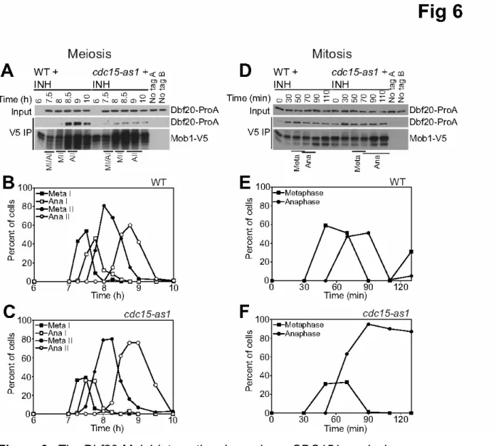
Given that the Dbf20-Mob1 interaction was especially high during anaphase II we next asked whether the interaction between the two proteins was regulated by CDC15. Interestingly, the interaction between Dbf20 and Mob1 was dramatically reduced in cdc15-as1 cells treated with inhibitor in meiosis (Figure 6A-C). In contrast, the Dbf20-Mob1 interaction occurred independently of CDC15 during mitosis (Figure 6D-F). These results indicate that during mitosis, Dbf20-Mob1 and Dbf2-Mob1 complexes form before MEN activation and that Cdc15 activates the kinase complex specifically during
anaphase. During meiosis, Cdc15 activation during anaphase II is required for Dbf20-Mob1 complex formation and kinase activation. Our results indicate that MEN signaling is regulated in a substantially different manner during meiosis than during mitosis. In both cell division types the two pathways regulate Cdc14 activity, but signal
transmission through the pathway has evolved perhaps so that the pathway can respond to different cellular signals.
DISCUSSION
Our studies of the MEN during meiosis led to two remarkable conclusions. First, the MEN, a signaling pathway essential for vegetative growth contributes little to meiotic cell cycle regulation. Second, MEN regulation changes dramatically during meiosis. Thus, MEN signaling serves as a paradigm for understanding how signaling pathways are rewired to serve different functions in different biological contexts.
MEN functions in meiosis.
In mitosis, the MEN is essential for the release of Cdc14 from the nucleolus and hence its activation during anaphase in mitosis. In contrast, during meiosis, release of Cdc14 from the nucleolus during anaphase is also essential (Buonomo et al., 2003; Marston et al., 2003), but surprisingly, the MEN components Cdc15 and Mob1 do not appear to contribute to the release of Cdc14 during anaphase I at all and only plays a minor maintenance role in Cdc14 release during anaphase II. Instead, it appears that the FEAR network, which plays only a minor role in promoting Cdc14 activation during mitosis, is essential for Cdc14 activation during anaphase I (Pereira et al., 2002; Stegmeier et al., 2002; Yoshida et al., 2002; Buonomo et al., 2003; Marston et al., 2003). A role for the FEAR network in anaphase II activation of Cdc14 has not been described yet, but given that the MEN has only a minor function in Cdc14 activation, it is likely that the FEAR network also functions during meiosis II to bring about Cdc14
release from the nucleolus.
Why does the FEAR network rather than the MEN bring about the activation of Cdc14 during meiosis? Cdc14 regulation must be responsive to different cellular signals during gametogenesis than during vegetative growth. During vegetative growth Cdc14 activity is controlled by spindle position, which is essential because of the inherently
asymmetric manner by which budding yeast divides (Fraschini et al., 2008). The MEN couples Cdc14 activation to spindle position (Bardin et al., 2000; Pereira et al., 2000). In contrast, meiosis is a symmetric division, as both meiotic divisions occur within the confines of a single cell (Neiman, 2011), rendering the control of Cdc14 activation during meiosis I and II by spindle position unimportant. Consistent with this idea is our finding that a gene essential for the regulation of the MEN by spindle position, KIN4, (D'Aquino et al., 2005; Pereira and Schiebel, 2005; Chan and Amon, 2010) does not function during the meiotic divisions. Cells harboring a deletion of KIN4 undergo meiosis with wild-type kinetics (M.A.A., unpublished observations). However, exit from meiosis I and II as well as spore wall formation must still be coordinated with chromosome
segregation. This dependence is established by the FEAR network (Stegmeier and Amon, 2004) and the Polo kinase Cdc5 (Rock and Amon, 2011). Indeed their activity is
essential for meiotic progression (Buonomo et al., 2003; Clyne et al., 2003; Lee and Amon, 2003; Marston et al., 2003).
During meiosis, the MEN has lost its essential function to promote the final stages of chromosome segregation, but at least CDC15 has been co-opted to fulfill a novel meiosis-specific function, spore-wall morphogenesis ((Kamieniecki et al., 2005; Pablo-Hernando et al., 2007); M.A.A. unpublished observations). As spore wall formation must occur only after the completion of the two meiotic divisions it is perhaps not surprising that the MEN is only active during meiosis II. What keeps the MEN inactive during exit from meiosis I is thus an important question that remains to be addressed. Despite intense efforts we have not been able to prematurely activate the MEN during meiosis I. Genetic alterations known to hyper-activate the MEN during mitosis failed to do so in meiosis (M.A.A., unpublished observations). This suggests that it is important to keep the MEN inactive in meiosis I, possibly to ensure that cells do not initiate spore wall formation prematurely.
Signaling through the MEN differs between mitosis and meiosis II in multiple ways.
Our analyses indicate that signal transmission through the MEN is modified in at least three ways during meiosis II: (1) Dbf2 is replaced by Dbf20, (2) Dbf20 binding to its activating subunit Mob1 is regulated differently and (3) MEN signaling does not occur at SPBs, nor does it require the SPB scaffolding subunit Nud1.
During mitosis, Dbf2 is the predominant kinase required for MEN signaling. In fact in several strain backgrounds including SK1, Dbf2 is essential. Even in S288C, where Dbf2 is not essential, Dbf2 is the predominant kinase (Toyn and Johnston, 1994). In contrast, during meiosis II Dbf20 takes over this role. This differential requirement for the two homologs is in part due to their expression levels, with Dbf2 levels being low during the meiotic divisions (Figure S2I). When we expressed Dbf2 from an inducible promoter in meiosis, the protein was readily expressed and was active as a kinase (Figure 4E). Curiously, this was not the case for Dbf20. Dbf20 expressed from the CUP1 promoter readily accumulated in vegetative cells, bound to Mob1 but did not exhibit kinase activity. These results indicate that Dbf20 is differentially regulated from Dbf2 at least during mitosis. The mechanism for this differential regulation remains to be
discovered. Perhaps an activator in addition to Cdc15 is needed for Dbf20 to be active, and this activator is absent during mitosis. Alternatively, Dbf20 may be inhibited during vegetative growth. Why cells use DBF20 rather than DBF2 in meiosis II remains
unclear. However we note that even though Dbf2 is active as a kinase when expressed during meiosis its activity level is lower than that of Dbf20 (Figure 4E). Perhaps Dbf2 cannot be as effectively activated off SPBs during meiosis II as Dbf20.
Dbf2 and Dbf20 not only show differential expression during mitosis and meiosis, their association with their common activator Mob1 appears to be differentially regulated between mitosis and meiosis. Whereas Dbf2 and Dbf20 bind to Mob1 throughout the cell cycle during vegetative growth, Dbf20 binding to Mob1 fluctuated during the meiotic divisions. Complex formation was significantly increased during meiosis II. Furthermore, whereas Dbf20 binding to Mob1 did not require CDC15 during vegetative growth, it depended on the upstream kinase during meiosis II. These results suggest that during mitosis a mechanism exists that can promote Dbf20 – Mob1 binding that is absent during meiosis II. During meiosis II, this mechanism is either replaced by a CDC15-dependent mechanism or, more likely, CDC15 can also promote the interaction
between Dbf2 and Mob1 during mitosis, but is not required for this, because of a second redundant mechanism that appears operative throughout the cell cycle. Taken together, our data are consistent with a model where Cdc15 is activated during meiosis II, which then promotes the interaction and activity of the Dbf20-Mob1 kinase complex, and maintaining Cdc14 in its released form. These events may require posttranslational modifications on Dbf20 that are restricted to meiosis II and independent of CDC15 (Figure S2K; M.A.A., unpublished observations).
In addition to multiple forms of differential regulation of the Dbf2-Mob1 family of protein kinases during meiosis, signal transmission as a whole appears very different between meiosis II and mitosis. In mitosis, loading of MEN components onto SPBs is critical for MEN activation (Valerio-Santiago and Monje-Casas, 2011). Furthermore, MEN
signaling in mitosis requires the SPB component Nud1, the putative scaffold for MEN signaling (Gruneberg et al., 2000; Visintin and Amon, 2001). In contrast, MEN
components are not found at SPBs during meiosis II. The spindle pole body undergoes a major restructuring of its outer plaque during meiosis II to initiate spore wall formation
(Neiman, 2011). It is possible that the altered structure of the outer plaque of the SPB during meiosis II precludes detection of MEN components. However, the fact that Bfa1 can be detected at SPBs during meiosis and that Nud1 is dispensable for MEN
signaling during meiosis II indicates that MEN signaling does not occur in the context of the SPB during meiosis II. Nud1 is present at SPBs during meiosis II, but for reasons that we do not understand does not recruit MEN components to SPBs at detectable levels. The altered architecture of this organelle may impede Nud1-dependent
recruitment of MEN components to SPBs. How MEN signaling modules are assembled during meiosis II off SPBs is not known. There is no obvious Nud1 homolog encoded in the S. cerevisiae genome, but functional homologs could exist. Proteomic-based screens could address this possibility in the future.
Why signaling through the MEN is rewired during meiosis II is an important question that remains to be addressed. CDC15 fulfills a novel spore-wall formation function (Kamieniecki et al., 2005; Pablo-Hernando et al., 2007) which may necessitate employing Dbf20 rather than Dbf2 in signal transmission. Signaling without the MEN scaffold and outer plaque component Nud1 may be necessary as to not interfere with Nud1’s function in spore wall formation (Gordon et al., 2006).
Parallels in other organisms
The MEN is a conserved signaling pathway. In Schizosaccharomyces pombe, the pathway is known as the septation initiation network (SIN) and regulates cytokinesis. There are several similarities between MEN and SIN function during the meiotic
divisions. Like MEN mutants, SIN mutants do not exhibit defects in meiotic progression, but fail to form spore walls (Krapp et al., 2006). This indicates that co-opting of a
pathway essential for the mitotic but not meiotic divisions to perform a novel function, spore wall formation, has occurred early during fungal evolution. S. pombe also contains two Dbf2 homologs, Sid2 and Slk1. During mitosis, Sid2 functions in SIN signaling (Sparks et al., 1999), but as in budding yeast, the other Dbf2 homolog, Slk1 is up-regulated and active in meiosis (Ohtaka et al., 2008; Perez-Hidalgo et al., 2008; Yan et al., 2008). It thus appears that the utilization of MEN/SIN signaling in spore wall formation is conserved between budding and fission yeast.
The core Cdc15-Dbf2-Mob1 signaling module of the MEN is conserved in mammals. In mammalian cells these components are MST1/2 (mammalian sterile 20-like kinase 1/2), NDR kinase (nuclear Dbf2-related kinase), and hMob1 respectively and function
together to control a number of cellular processes including cell proliferation (Hergovich et al., 2006; Pan, 2010). The core signaling module may be regulated similarly between mammalian cells and the MEN in meiosis. The interaction between Mob1 and NDR kinase family members is promoted when Mob1 is phosphorylated (Hirabayashi et al., 2008; Praskova et al., 2008), suggesting that regulation of the NDR kinase-Mob1 interaction may be a common mechanism for regulating the activity of this pathway in the absence of spatial control. Finally, it will be interesting to determine whether meiosis-specific modules also exist in mammals and whether they regulate late stages of germ cell formation.
MATERIALS AND METHODS Yeast strains
All yeast strains used in this study are derivatives of SK1 and listed in Table S1. TEM1-9MYC, CDC15-TEM1-9MYC, MOB1-13MYC, LTE1-13MYC, NUD1-13MYC, pCLB2-3HA-MOB1, BFA1-GFP, TEM1-GFP, CDC15-eGFP, MOB1-GFP, DBF20-ProA, DBF2-ProA, pCUP1-DBF20-ProA, pCUP1-DBF2-ProA, and MOB1-3V5 strains were made using a PCR-based method (Longtine et al., 1998). 3MYC-DBF2 and 3MYC-DBF20 were created using a URA3-popout based approach (Schneider et al., 1995). The nud1-44 allele from strain A1920 was backcrossed 5X to SK1.
Sporulation
Strains were grown overnight on YEPG plates (3% glycerol) and then transferred to 4% YEPD (4% glucose) plates in the morning. Cells were cultured in YEPD medium to saturation (approximately 24 hours), and then diluted into buffered YTA medium (1% yeast extract, 2% tryptone, 1% potassium acetate, 50mM potassium phthalate) to OD600=0.35. Cells were grown overnight and then resuspended in sporulation medium
(1% potassium acetate pH 7) at an OD600=1.9. Sporulation experiments were
performed at 30°C unless otherwise noted. Meiotic divisions were synchronized with the Ndt80 block-release protocol. Cells contain NDT80 under the GAL1-10 promoter and a Gal4-estrogen receptor fusion. Cells were transferred to sporulation medium at
time=0 hours. Owing to the lack of NDT80, GAL-NDT80 cells will arrest in pachytene. At t=6 hours, when most cells had reached pachytene, 1μM β-estradiol was added to the medium allowing cells to progress through the meiotic divisions in a synchronous manner. For cdc15-as1 experiments, 10μM 1-NA-PP1 (4-Amino-1-tert-butyl-3-(1’-naphthyl)pyrazolo[3,4-d]pyrimidine; Toronto Research Chemicals, North York ON) was added to the medium at the same time as β-estradiol.
Fluorescence microscopy
Indirect in situ immunofluorescence of tubulin was performed as described previously (Kilmartin and Adams, 1984). Cdc14-3HA immunofluorescence was performed as described in (Marston et al., 2003). To perform immunofluorescence of Nud1-13Myc, cells were subjected to a 15 minute fixation with 3.7% formaldehyde in 0.1M potassium phosphate (KPi pH 6.4) buffer and prepared for staining as described by (Marston et al., 2003). Cells were incubated with an anti-Myc (9E10 epitope, Covance, Princeton, NJ) primary antibody at 1:2000 and anti-mouse-Cy3 (Jackson Laboratory, Bar Harbor, ME) secondary antibody at 1:500 for at least two hours each. For Bfa1-GFP imaging, cells were fixed for 15 minutes with 3.7% formaldehyde in KPi. Cells were resuspended in 0.1M KPi/1.2M sorbitol/1% Triton. Cells were resuspended in 0.05μg/ml DAPI in
KPi/Sorbitol. Cells were imaged with a Zeiss Axioplan 2 microscope and a Hamamatsu ORCA-ER digital camera.
Immunoblot analysis
For immunoblot analysis of Dbf20-ProA, Dbf2-ProA, Tem1-9Myc, Cdc15-9Myc, 3Myc-Dbf2, 3Myc-Dbf20, Mob1-13Myc, Nud1-13Myc, Lte1-13Myc, 3HA-Mob1, and Pgk1, cells were incubated for at least 10 minutes in 5% trichloroacetic acid. Cell pellets were washed once in acetone and dried overnight. Cells were lysed in 100μl lysis buffer (50mM Tris-Cl at pH 7.5, 1mM EDTA, 2.75 mM DTT, complete protease inhibitor cocktail [Roche]) with a bead mill. After sample buffer was added, cell lysates were boiled. Myc-tagged proteins were detected with anti-Myc (9E10 epitope, Covance, Princeton, NJ) antibodies at a dilution of 1:500. HA-tagged proteins were detected with anti-HA (HA.11, Covance, Princeton, NJ) antibodies at a dilution of 1:1000. ProA-tagged proteins were detected by incubation with rabbit IgG (Sigma-Aldrich, St. Louis, MO) at a concentration of 1:5000. Pgk1 was detected with an anti-Pgk1 antibody
(Invitrogen, Carlsbad, CA) using a 1:5000 dilution. Quantification was performed using ImageQuant software.
Dbf2 and Dbf20 kinase assays
Dbf20 kinase assays were performed as previously described (Visintin and Amon, 2001) with several modifications. Cells were lysed in a cold block in a bead mill, and 1-2mg of lysate was used in immunoprecipitations (total volume of 150μl). Dbf20-ProA was immunoprecipitated using IgG-coupled Dynabeads and incubated for one hour at 4°C. To prepare IgG-coupled beads, Dynabeads (Invitrogen Dynal AS, Oslo, Norway) were incubated with 0.33 mg/ml rabbit IgG (Sigma-Aldrich, St. Louis, MO) and 1M ammonium sulfate in 0.1M sodium phosphate pH 7.4 buffer at 37°C overnight. Phosphorylation of histone H1 was quantified with the PhosphorImaging system. Co-immunoprecipitation assays
Cells were resuspended in 200μl of NP40 lysis buffer (150mM NaCl, 50mM Tris-Cl pH 7.5, 1% NP40, 60mM β-glycerophosphate, 0.1mM sodium orthovanadate, 15mM p-Nitrophenylphosphate, 1mM DTT, complete protease inhibitor cocktail [Roche,
Mannheim, Germany]) and lysed in a bead mill. 1-2mg (meiosis) or 500μg (mitosis) of extract were used in immunopreciptiation experiments (volume of 150μl) with agarose beads directly conjugated to the V5 epitope (Sigma-Aldrich, St. Louis, MO). Lysates were incubated for 45 minutes at 4°C, washed, and beads were resuspended in sample buffer, boiled, and proteins were analyzed by Western blot analyses.
ACKNOWLEDGMENTS
We thank Frank Solomon and members of the Amon laboratory for comments on the manuscript, and V. Guacci for plasmids. This work was supported by the National Institutes of Health (GM62207 to A.A.). A.A. is also an investigator of the Howard Hughes Medical Institute.
References
Adams, I.R., and Kilmartin, J.V. (1999). Localization of core spindle pole body (SPB)
components during SPB duplication in Saccharomyces cerevisiae. The Journal of cell biology 145, 809-823.
Asakawa, K., Yoshida, S., Otake, F., and Toh-e, A. (2001). A novel functional domain of Cdc15 kinase is required for its interaction with Tem1 GTPase in Saccharomyces cerevisiae. Genetics 157, 1437-1450.
Bardin, A.J., Visintin, R., and Amon, A. (2000). A mechanism for coupling exit from mitosis to partitioning of the nucleus. Cell 102, 21-31.
Bertazzi, D.T., Kurtulmus, B., and Pereira, G. (2011). The cortical protein Lte1 promotes mitotic exit by inhibiting the spindle position checkpoint kinase Kin4. The Journal of cell biology 193, 1033-1048.
Bishop, A.C., Ubersax, J.A., Petsch, D.T., Matheos, D.P., Gray, N.S., Blethrow, J., Shimizu, E., Tsien, J.Z., Schultz, P.G., Rose, M.D., Wood, J.L., Morgan, D.O., and Shokat, K.M. (2000). A chemical switch for inhibitor-sensitive alleles of any protein kinase. Nature 407, 395-401.
Buonomo, S.B., Rabitsch, K.P., Fuchs, J., Gruber, S., Sullivan, M., Uhlmann, F., Petronczki, M., Toth, A., and Nasmyth, K. (2003). Division of the nucleolus and its release of CDC14 during anaphase of meiosis I depends on separase, SPO12, and SLK19. Developmental cell 4, 727-739. Carlile, T.M., and Amon, A. (2008). Meiosis I is established through division-specific
translational control of a cyclin. Cell 133, 280-291.
Chan, L.Y., and Amon, A. (2010). Spindle position is coordinated with cell-cycle progression through establishment of mitotic exit-activating and -inhibitory zones. Molecular cell 39, 444-454.
Chu, S., DeRisi, J., Eisen, M., Mulholland, J., Botstein, D., Brown, P.O., and Herskowitz, I. (1998). The transcriptional program of sporulation in budding yeast. Science 282, 699-705. Clyne, R.K., Katis, V.L., Jessop, L., Benjamin, K.R., Herskowitz, I., Lichten, M., and Nasmyth, K. (2003). Polo-like kinase Cdc5 promotes chiasmata formation and cosegregation of sister centromeres at meiosis I. Nature cell biology 5, 480-485.
D'Aquino, K.E., Monje-Casas, F., Paulson, J., Reiser, V., Charles, G.M., Lai, L., Shokat, K.M., and Amon, A. (2005). The protein kinase Kin4 inhibits exit from mitosis in response to spindle position defects. Molecular cell 19, 223-234.
Falk, J.E., Chan, L.Y., and Amon, A. (2011). Lte1 promotes mitotic exit by controlling the localization of the spindle position checkpoint kinase Kin4. Proceedings of the National Academy of Sciences of the United States of America 108, 12584-12590.
Fraschini, R., Venturetti, M., Chiroli, E., and Piatti, S. (2008). The spindle position checkpoint: how to deal with spindle misalignment during asymmetric cell division in budding yeast. Biochemical Society transactions 36, 416-420.
Geymonat, M., Spanos, A., Smith, S.J., Wheatley, E., Rittinger, K., Johnston, L.H., and Sedgwick, S.G. (2002). Control of mitotic exit in budding yeast. In vitro regulation of Tem1 GTPase by Bub2 and Bfa1. The Journal of biological chemistry 277, 28439-28445.
Gordon, O., Taxis, C., Keller, P.J., Benjak, A., Stelzer, E.H., Simchen, G., and Knop, M. (2006). Nud1p, the yeast homolog of Centriolin, regulates spindle pole body inheritance in meiosis. The EMBO journal 25, 3856-3868.
Gruneberg, U., Campbell, K., Simpson, C., Grindlay, J., and Schiebel, E. (2000). Nud1p links astral microtubule organization and the control of exit from mitosis. The EMBO journal 19, 6475-6488.
Hergovich, A., Stegert, M.R., Schmitz, D., and Hemmings, B.A. (2006). NDR kinases regulate essential cell processes from yeast to humans. Nature reviews. Molecular cell biology 7, 253-264.
Hirabayashi, S., Nakagawa, K., Sumita, K., Hidaka, S., Kawai, T., Ikeda, M., Kawata, A., Ohno, K., and Hata, Y. (2008). Threonine 74 of MOB1 is a putative key phosphorylation site by MST2 to form the scaffold to activate nuclear Dbf2-related kinase 1. Oncogene 27, 4281-4292.
Jaspersen, S.L., Charles, J.F., Tinker-Kulberg, R.L., and Morgan, D.O. (1998). A late mitotic regulatory network controlling cyclin destruction in Saccharomyces cerevisiae. Molecular biology of the cell 9, 2803-2817.
Kamieniecki, R.J., Liu, L., and Dawson, D.S. (2005). FEAR but not MEN genes are required for exit from meiosis I. Cell cycle 4, 1093-1098.
Keck, J.M., Jones, M.H., Wong, C.C., Binkley, J., Chen, D., Jaspersen, S.L., Holinger, E.P., Xu, T., Niepel, M., Rout, M.P., Vogel, J., Sidow, A., Yates, J.R., 3rd, and Winey, M. (2011). A cell cycle phosphoproteome of the yeast centrosome. Science 332, 1557-1561.
Kilmartin, J.V., and Adams, A.E. (1984). Structural rearrangements of tubulin and actin during the cell cycle of the yeast Saccharomyces. The Journal of cell biology 98, 922-933.
Krapp, A., Collin, P., Cokoja, A., Dischinger, S., Cano, E., and Simanis, V. (2006). The
Schizosaccharomyces pombe septation initiation network (SIN) is required for spore formation in meiosis. Journal of cell science 119, 2882-2891.
Lee, B.H., and Amon, A. (2003). Role of Polo-like kinase CDC5 in programming meiosis I chromosome segregation. Science 300, 482-486.
Longtine, M.S., McKenzie, A., 3rd, Demarini, D.J., Shah, N.G., Wach, A., Brachat, A.,
Philippsen, P., and Pringle, J.R. (1998). Additional modules for versatile and economical PCR-based gene deletion and modification in Saccharomyces cerevisiae. Yeast 14, 953-961.
Luca, F.C., and Winey, M. (1998). MOB1, an essential yeast gene required for completion of mitosis and maintenance of ploidy. Molecular biology of the cell 9, 29-46.
Maekawa, H., Priest, C., Lechner, J., Pereira, G., and Schiebel, E. (2007). The yeast centrosome translates the positional information of the anaphase spindle into a cell cycle signal. The Journal of cell biology 179, 423-436.
Mah, A.S., Jang, J., and Deshaies, R.J. (2001). Protein kinase Cdc15 activates the Dbf2-Mob1 kinase complex. Proceedings of the National Academy of Sciences of the United States of America 98, 7325-7330.
Marston, A.L., and Amon, A. (2004). Meiosis: cell-cycle controls shuffle and deal. Nature reviews. Molecular cell biology 5, 983-997.
Marston, A.L., Lee, B.H., and Amon, A. (2003). The Cdc14 phosphatase and the FEAR network control meiotic spindle disassembly and chromosome segregation. Developmental cell 4, 711-726.
Molk, J.N., Schuyler, S.C., Liu, J.Y., Evans, J.G., Salmon, E.D., Pellman, D., and Bloom, K. (2004). The differential roles of budding yeast Tem1p, Cdc15p, and Bub2p protein dynamics in mitotic exit. Molecular biology of the cell 15, 1519-1532.
Monje-Casas, F., and Amon, A. (2009). Cell polarity determinants establish asymmetry in MEN signaling. Developmental cell 16, 132-145.
Neiman, A.M. (2011). Sporulation in the budding yeast Saccharomyces cerevisiae. Genetics 189, 737-765.
Ohtaka, A., Okuzaki, D., and Nojima, H. (2008). Mug27 is a meiosis-specific protein kinase that functions in fission yeast meiosis II and sporulation. Journal of cell science 121, 1547-1558. Pablo-Hernando, M.E., Arnaiz-Pita, Y., Nakanishi, H., Dawson, D., del Rey, F., Neiman, A.M., and Vazquez de Aldana, C.R. (2007). Cdc15 is required for spore morphogenesis independently of Cdc14 in Saccharomyces cerevisiae. Genetics 177, 281-293.
Pan, D. (2010). The hippo signaling pathway in development and cancer. Developmental cell 19, 491-505.
Pereira, G., Hofken, T., Grindlay, J., Manson, C., and Schiebel, E. (2000). The Bub2p spindle checkpoint links nuclear migration with mitotic exit. Molecular cell 6, 1-10.
Pereira, G., Manson, C., Grindlay, J., and Schiebel, E. (2002). Regulation of the Bfa1p-Bub2p complex at spindle pole bodies by the cell cycle phosphatase Cdc14p. The Journal of cell biology 157, 367-379.
Pereira, G., and Schiebel, E. (2005). Kin4 kinase delays mitotic exit in response to spindle alignment defects. Molecular cell 19, 209-221.
Perez-Hidalgo, L., Rozalen, A.E., Martin-Castellanos, C., and Moreno, S. (2008). Slk1 is a meiosis-specific Sid2-related kinase that coordinates meiotic nuclear division with growth of the forespore membrane. Journal of cell science 121, 1383-1392.
Praskova, M., Xia, F., and Avruch, J. (2008). MOBKL1A/MOBKL1B phosphorylation by MST1 and MST2 inhibits cell proliferation. Current biology : CB 18, 311-321.
Rock, J.M., and Amon, A. (2009). The FEAR network. Current biology : CB 19, R1063-1068. Rock, J.M., and Amon, A. (2011). Cdc15 integrates Tem1 GTPase-mediated spatial signals with Polo kinase-mediated temporal cues to activate mitotic exit. Genes & development 25, 1943-1954.
Schneider, B.L., Seufert, W., Steiner, B., Yang, Q.H., and Futcher, A.B. (1995). Use of polymerase chain reaction epitope tagging for protein tagging in Saccharomyces cerevisiae. Yeast 11, 1265-1274.
Sharon, G., and Simchen, G. (1990). Mixed segregation of chromosomes during single-division meiosis of Saccharomyces cerevisiae. Genetics 125, 475-485.
Shirayama, M., Matsui, Y., and Toh, E.A. (1994). The yeast TEM1 gene, which encodes a GTP-binding protein, is involved in termination of M phase. Molecular and cellular biology 14, 7476-7482.
Shou, W., Seol, J.H., Shevchenko, A., Baskerville, C., Moazed, D., Chen, Z.W., Jang, J., Charbonneau, H., and Deshaies, R.J. (1999). Exit from mitosis is triggered by Tem1-dependent release of the protein phosphatase Cdc14 from nucleolar RENT complex. Cell 97, 233-244. Sparks, C.A., Morphew, M., and McCollum, D. (1999). Sid2p, a spindle pole body kinase that regulates the onset of cytokinesis. The Journal of cell biology 146, 777-790.
Stegmeier, F., and Amon, A. (2004). Closing mitosis: the functions of the Cdc14 phosphatase and its regulation. Annual review of genetics 38, 203-232.
Stegmeier, F., Visintin, R., and Amon, A. (2002). Separase, polo kinase, the kinetochore protein Slk19, and Spo12 function in a network that controls Cdc14 localization during early anaphase. Cell 108, 207-220.
Toyn, J.H., and Johnston, L.H. (1994). The Dbf2 and Dbf20 protein kinases of budding yeast are activated after the metaphase to anaphase cell cycle transition. The EMBO journal 13, 1103-1113.
Valerio-Santiago, M., and Monje-Casas, F. (2011). Tem1 localization to the spindle pole bodies is essential for mitotic exit and impairs spindle checkpoint function. The Journal of cell biology 192, 599-614.
Visintin, R., and Amon, A. (2001). Regulation of the mitotic exit protein kinases Cdc15 and Dbf2. Molecular biology of the cell 12, 2961-2974.
Visintin, R., Craig, K., Hwang, E.S., Prinz, S., Tyers, M., and Amon, A. (1998). The phosphatase Cdc14 triggers mitotic exit by reversal of Cdk-dependent phosphorylation. Molecular cell 2, 709-718.
Visintin, R., Hwang, E.S., and Amon, A. (1999). Cfi1 prevents premature exit from mitosis by anchoring Cdc14 phosphatase in the nucleolus. Nature 398, 818-823.
Yan, H., Ge, W., Chew, T.G., Chow, J.Y., McCollum, D., Neiman, A.M., and Balasubramanian, M.K. (2008). The meiosis-specific Sid2p-related protein Slk1p regulates forespore membrane assembly in fission yeast. Molecular biology of the cell 19, 3676-3690.
Yeh, E., Skibbens, R.V., Cheng, J.W., Salmon, E.D., and Bloom, K. (1995). Spindle dynamics and cell cycle regulation of dynein in the budding yeast, Saccharomyces cerevisiae. The Journal of cell biology 130, 687-700.
Yoshida, S., Asakawa, K., and Toh-e, A. (2002). Mitotic exit network controls the localization of Cdc14 to the spindle pole body in Saccharomyces cerevisiae. Current biology : CB 12, 944-950. Zachariae, W., Schwab, M., Nasmyth, K., and Seufert, W. (1998). Control of cyclin
ubiquitination by CDK-regulated binding of Hct1 to the anaphase promoting complex. Science 282, 1721-1724.
FIGURE LEGENDS
Figure 1. The MEN is dispensable for exit from meiosis I but is required for the timely exit from anaphase II. (A - D) Wild-type (A, A14201) or cdc15-as1 (B, A19440) cells harboring a GAL-NDT80 fusion as the sole source of NDT80 were induced to sporulate. 1μM β-estradiol and 10μM 1-NA-PP1 was added to cultures six hours after transfer into sporulation medium. The percentage of cells with metaphase I (closed squares),
anaphase I (open squares), metaphase II (closed circles), anaphase II (open circles) was determined at the indicated times. 200 cells were analyzed at each time point. (C)
Comparison of the percentage of anaphase II cells in wild-type (closed squares) and cdc15-as1 mutants (closed circles). (D) Quantification of the accumulation of anaphase I and anaphase II spindles in wild-type and cdc15-as1 mutants (n=6 experiments). The area under the lines for anaphase I and anaphase II spindles was determined and expressed as a fold change of wild-type (anaphase I: mean=1.083, SD=0.122, p=0.598 (two-tailed, paired t-test); anaphase II: mean=1.757, SD=0.362, p=0.005 (two-tailed, paired t-test)). (E - F) Examples of anaphase II cells with Cdc14 sequestered in the nucleolus (top panels; E) or released into the nucleus and cytoplasm (bottom panels; E). Cdc14-HA is shown in red, DNA in blue and microtubules in green. (F)
Quantification of Cdc14 release in anaphase II cells. WT (A22130; white bars) or cdc15-as1 (A22129; gray bars) cells carrying CDC14-HA fusions were induced to sporulate as in (A). At the time points indicated, the percentage of anaphase I or anaphase II cells with Cdc14 released was determined (n=100 for each time point except for wild-type 8.25 hr, n=35 and wild-type 9 hr, n=36).
Figure 2. Dbf20 kinase activity peaks in anaphase II and depends on Cdc15 kinase activity.
(A) Cells containing a DBF20-ProA fusion (A23162) were sporulated as described in Figure 1A except 1-NA-PP1 was not added. Samples were taken at the indicated times to determine associated kinase activity and ProA protein levels. Dbf20-associated kinase activity was assessed by phosphorylation of the substrate Histone H1 (p-H1). Pgk1 was used as a loading control. The peak of each stage of meiosis is indicated below the blot (MI - metaphase I, AI - anaphase I, MII - metaphase II, AII - Anaphase II.)
(B) Quantification of Dbf20-associated kinase activity (closed circles) and anaphase II spindles (closed squares) are shown (n=200 cells counted).
(C) Wild-type (A23162) or cdc15-as1 (A23733) cells containing a DBF20-ProA fusion were induced to sporulate as described in Figure 1A. Dbf20-associated kinase activity and Dbf20-ProA protein levels were determined at the indicated times.
Figure 3. MEN signaling occurs in a NUD1-independent manner during meiosis. (A - C) 34°C is a restrictive temperature for nud1-44. Wild-type (A8499) and nud1-44 (A28757) cells containing a 3MYC-DBF2 fusion were arrested in G1 with α-factor
pheromone (5μg/ml) in YEPD medium for 2 hours at room temperature and then shifted to 34°C for 30 minutes. Cells were released into pheromone-free YEPD medium at 34°C thereafter. Dbf2 protein and kinase activity (A) and the percentage of cells in metaphase and anaphase (B, C) were analyzed at the indicated times (n=100 cells per
ProA fusion were induced to sporulate via the Ndt80-block release protocol. Cells were incubated in sporulation medium at room temperature and shifted to 34°C after five hours. At six hours, cultures were induced with 1μM β-estradiol. Cells were maintained at 34°C throughout the rest of the experiment. Samples were taken at the indicated times to determine Dbf20 protein levels and Dbf20 kinase activity (D) and meiotic progression (E, F; n=100 cells per time point).
Figure 4. Differential regulation of Dbf2 and Dbf20 activity.
(A - D) pCUP1-DBF2-ProA (A25020) or pCUP1-DBF20-ProA (A25193) cells were arrested in G1 with α-factor pheromone (5μg/ml) in YEPD medium. 50μM CuSO4 was
30 minutes), cells were released into pheromone-free YEPD medium containing 50μM CuSO4. Dbf2 and Dbf20 kinase activity, Dbf2 and Dbf20 protein (A, B) and mitotic
progression (C, D; n=100 cells per time point) were examined at the indicated times. Quantifications of Dbf2 and Dbf20 kinase activity are shown in the graph in (B). (E - H) pCUP1-DBF2-ProA (A25028) or pCUP1-DBF20-ProA (A25195) cells were sporulated via the Ndt80 block-release protocol. 1μM β-estradiol and 50μM CuSO4
were added to the medium six hours after transfer into sporulation medium. Dbf2 and Dbf20 kinase activity, Dbf2 and Dbf20 protein (E, F) and meiotic progression (G, H; n=100 cells per time point) were examined at the indicated times. Quantifications of Dbf2 and Dbf20 kinase activity are shown in the graph in (F).
Figure 5. The Dbf20-Mob1 interaction is under cell cycle control in meiosis but not mitosis.
(A, B) Cells containing pCUP1-DBF2-ProA and a MOB1-V5 fusion (A27687) or MOB1 (A25020, no tag control) were arrested in G1 with α-factor pheromone (5μg/ml) in YEPD medium. 50μM CuSO4 was added to the medium two hours into the arrest. When the
arrest was complete (2 hours 30 minutes), cells were released into pheromone-free YEPD medium containing 50μM CuSO4. Western blots in (A) show total Dbf2-ProA
the indicated timepoints. Quantification of co-immunoprecitation expressed as the amount of Dbf2 co-immunoprecipitated over the amount of Mob1-V5
immunoprecipitated are shown in (B). Values were normalized so that the maximum value was set to 1.0 (closed triangles). For comparison, the percentage of cells in metaphase (open squares) and anaphase (open circles) is shown. (C, D) Cells
containing pCUP1-DBF20-ProA and a MOB1-V5 fusion (A27367) or MOB1 (A25191, no tag control) were grown and analyzed as described in (A, B). (E, F) Cells containing pCUP1-DBF20-ProA and a MOB1-V5 fusion (A27370) or MOB1 (A25195, no tag control) were sporulated via the Ndt80 block-release protocol. Cells were induced with 1μM β-estradiol and 50μM CuSO4 six hours after transfer to sporulation medium.
Western blots show immunoprecipitated Mob1-V5, co-immunoprecipitated Dbf20-ProA, and total Dbf20 protein (input) at the indicated timepoints (E). Quantification of the amount of Dbf20 co-immunoprecipitated with Mob1-V5 is shown as a ratio of the two values. Values were normalized so that the maximum value was set to 1.0. For comparison, meiotic progression was also analyzed.
Figure 6. The Dbf20-Mob1 interaction depends on CDC15 in meiosis.
(A - C) Cells containing pCUP1-DBF20-ProA, a MOB1-V5 fusion, and CDC15 (A27371) or cdc15-as1 (A29149) were induced to sporulate via the Ndt80 block-release protocol as in Figure 1A with the addition of 10μM NA-PP1 and 50μM CuSO4 six hours after
transfer to sporulation medium. Western blots in (A) show total Dbf20-ProA protein (input), immunoprecipitated Mob1-V5 and co-immunoprecipitated Dbf20-ProA at the indicated timepoints. The genotype of no tag A is pCUP1-DBF20-ProA (A25195) and the genotype of no tag B is pCUP1-DBF20-ProA cdc15-as1 (A29150). Meiotic progression in the two strains is shown in (B, C). (D - F) Cells containing pCUP1-DBF20-ProA and a MOB1-V5 fusion, and CDC15 (A27367) or cdc15-as1 (A29125) were arrested in G1 with α-factor pheromone (5μg/ml) in YEPD medium as in Figure
4A. 50μM CuSO4 was added to the medium 2 hours into the arrest. When the arrest
was complete (2 hours 30 minutes), cells were released into YEPD medium containing 10μM NA-PP1 and 50μM CuSO4. Total Dbf20-ProA protein (input),
Co-immunoprecipitated Dbf20-ProA, and immumoprecipitated Mob1-V5 are shown for the indicated time points in (D). Mitotic progression of the two strains is shown in (E, F). The genotype of no tag A is pCUP1-DBF20-ProA (A25193) and the genotype of no tag B is pCUP1-DBF20-ProA cdc15-as1 (A29124).