1 2 3 4 5 6 7 8 9 10 11 12 13 14 15 16 17 18 19 20 21 22 23 24 25 26 27 28 29 30 31 32 33 34 35 36 37 38 39 40 41 42 43 44 45 46 47 48 49 50 51 52 53 54 55 56 57
Supporting Information
�
Copyright Wiley-VCH Verlag GmbH & Co. KGaA, 69451 Weinheim, 2020
Detection of HER2
+
Breast Cancer Cells using Bioinspired
DNA-Based Signal Amplification
Sarah D. Rafiee
+, Samet Kocabey
+, Michael Mayer, Jonathan List,* and Curzio Rüegg*© 2020
The Authors. Published by Wiley-VCH Verlag GmbH & Co. KGaA.
This is an open access article under the terms of the Creative Commons Attribution
Non-Commercial License, which permits use, distribution and reproduction in any medium,
provided the original work is properly cited and is not used for commercial purposes.
Wiley VCH Freitag, 24.01.2020
Author Contributions
S.R. Data curation:Equal; Formal analysis:Equal; Investigation:Equal; Methodology:Equal; Validation:Equal; Writing - Original Draft:Equal
S.K. Data curation:Equal; Formal analysis:Equal; Investigation:Equal; Methodology:Equal; Validation:Equal; Writing - Original Draft:Equal
M.M. Conceptualization:Supporting; Formal analysis:Supporting; Funding acquisition:Supporting; Methodology:Sup-porting; Resources:SupMethodology:Sup-porting; Supervision:SupMethodology:Sup-porting; Writing - Review & Editing:Equal
J.L. Conceptualization:Lead; Data curation:Equal; Formal analysis:Equal; Investigation:Supporting; Methodology:-Supporting; Supervision:Methodology:-Supporting; Writing - Original Draft:Equal
C.R. Conceptualization:Equal; Data curation:Equal; Formal analysis:Equal; Funding acquisition:Lead; Project admin-istration:Lead; Resources:Lead; Supervision:Equal; Validation:Equal; Writing - Original Draft:Equal
Experimental Procedures
Materials
Native and biotin-modified DNA strands (HPLC purified) used for the screening on streptavidin coated beads (Cy5 labeled and HPLC purified) were purchased from Biomers (Ulm, Germany). Alexa-647 modified H1 and H2 DNA strands (HPLC purified) used in cell experiments were purchased from Eurofins MWG Operon (Ebersberg, Germany). The detailed sequences of the DNA strands are given in Table S1. Anti-human HER2/ERBB2 antibody (trastuzumab) was purchased from R&D Systems (United Kingdom). Streptavidin–Alexa Fluor 488 conjugate was purchased from Thermo Fisher Scientific (Basel, Switzerland).
Screening the hairpins with streptavidin beads by flow cytometry
2.5 µL of 0.5% w/v beads corresponding to 2.5 × 10⁵ streptavidin coated polystyrene beads (4-4.9 µm diameter,
Spherotech, Illinois, USA) were transferred to U-bottom 96-well microplate (Thermo Fisher Scientific (Basel, Switzerland)). Beads were incubated sequentially with double initiator (DI) (5 nM) for 20 minutes, H1-AF647 (500 nM) and H2 (500 nM) for different time intervals (30 minutes, 2 hours, 4 hours and 24 hours) in 100 µL of FACS buffer (5 mM ethylenediaminetetraacetic acid (EDTA), 3% FCS in 1x PBS) at 4 °C. Between each step, cells were rinsed with 200 µL FACS buffer and centrifuged at 260g (Awel MF48-R) for 2 minutes to wash unbound molecules away. Before adding DI, H1 and H2, oligonucleotides were incubated at 95 °C for 90 seconds and rapidly cooled on ice. After incubation for the respective times, the beads were analyzed using Flow Cytometry using MACSQuant Analyzer 10 flow cytometer (Miltenyi Biotec) and data analyzed with FlowJo Software (v10, FlowJo LLC). Median fluorescence intensity (MFI) for all samples were background corrected by subtracting the MFI values from the appropriate controls.
Cell culture
Human breast cancer cell lines BT-474 (high HER2/ERBB2 expression) was kindly provided by Dr. Mohamed Bentires-Alj (University of Basel, Switzerland). MDA-MB-468 (no detectable ERBB2 expression) and U937 (leukemia-derived monocytic cell line) were obtained from ATCC. Cells were cultured in Dulbecco's modified Eagle's medium (DMEM) supplemented with 10% FCS, 100 U/mL Penicillin and 100 μg/mL Streptomycin at 37 °C in 5% CO2. All cell culture reagents were purchased from Invitrogen Life Technologies (Basel, Switzerland).
HCR detection on cancer cells by flow cytometry
Cells cultured overnight were harvested with 1x trypsin (10x trypsin diluted in 1x PBS; Sigma-Aldrich (Buchs, Switzerland) and 2.5 × 10⁵ cells were transferred to V-bottom 96-well microplate (Thermo Fisher Scientific (Basel, Switzerland)). Cells
were washed with FACS buffer (5 mM ethylenediaminetetraacetic acid (EDTA), 3% FCS in 1x PBS) by centrifuging at 900 g, 4 °C for 5 minutes. Then, the supernatant was removed and cells were incubated sequentially with biotinylated trastuzumab (100ng/mL) for 30 minutes, streptavidin-AF488 (10µg/mL) for 20 minutes, double initiator (DI) (5 nM) for 20 minutes, H1-AF647 (500 nM) and H2 (500 nM) for different time intervals (30 minutes, 2 hours and 4 hours) in 100 µL of FACS buffer at 4 °C. Between each step, cells were rinsed with 200 µL FACS buffer and centrifuged at 700 g (Awel MF48-R) for 5 minutes to wash unbound molecules away. Before adding DI, H1 and H2, oligonucleotides were incubated at
95 °C for 90 seconds and rapidly cooled on ice. After incubation for the respective times, the cells were analyzed by flow cytometry using MACSQuant Analyzer 10 flow cytometer (Miltenyi Biotec) and data analyzed with FlowJo Software (v10, FlowJo LLC). Median fluorescence intensity (MFI) for all samples were background corrected by subtracting the MFI values from the appropriate controls.
HCR detection in co-cultured cancer cells by confocal microscopy
To visualize the selective targeting of HER2+ overexpressing BT-474 cells, MDA-MB-468 cells (HER2-) were stained with
1 µM Cell Tracker Green CMFDA (chloromethyl fluorescein diacetate, Invitrogen) for 30 minutes prior to mix with BT-474 cells according to the manufacturers protocol. Then, BT-474 cells and MDA-MB-468 cells were seeded into 24-well plates at a density of 2x10⁴ cells per well overnight. 13 mm glass coverslips were coated with 200 µL poly-L-lysine (Sigma Aldrich
(Buchs, Switzerland)) for the adhesion of the cells. Next day cells were washed with 1x PBS and HCR was performed using biotinylated trastuzumab (100ng/ml), DI (5 nM), H1-Alexa647 (500 nM) and H2 (500 nM) for 2 hours. After washing with PBS, cells were fixed for 10 minutes using 4% formaldehyde solution (Sigma Aldrich (Buchs, Switzerland)). Fixed cells then washed with 1x PBS and mounted on glass slides using antifade mounting medium (Invitrogen). Confocal imaging was performed on a Leica TCS SP5 confocal microscope (Leica Microsystems GmbH, Mannheim, Germany).
HCR detection on cancer cells mixed with PBMC by flow cytometry
Peripheral blood mononuclear cells (PBMC) were isolated from the blood sample of a 25-years old healthy female donor. Ethical committee approval was obtained from the Commission Cantonale de la recherche sur l’être humain of Canton Vaud, Switzerland (023-CER-FR). Mononuclear cells were isolated from the blood using Histopaque-1077 (Sigma-Aldrich, (Buchs, Switzerland)) according to manufacturer’s protocol. Briefly, 3 mL of whole blood was carefully layered on top of 3 mL Histopaque-1077 reagent in a 15 mL tube (9 mL blood was used in total) and centrifuged at 400 g (Awel C48) for 30 minutes at RT. After centrifugation, the opaque layer containing mononuclear cells were carefully aspirated and transferred to new tube. Then, the cells mixed with 10 mL 1x PBS and centrifuged again at 250 g (Awel C48) for 10 minutes. After several washing steps, the cell pellet was resuspended in 1x PBS and number of cells were counted. To investigate the sensitivity of the detection, BT-474 cells were mixed with PBMCs (105 cells/well) or U937 cells (105 cells/well) and seeded
into V-bottom 96-well plates (Thermo Fisher Scientific (Basel, Switzerland)) at the following ratios: 1:1, 1:4, 1:10, 1:100. 2 hours after HCR, cells were washed and analyzed by Flow Cytometry as mentioned above.
HCR detection on cancer cells that directly spiked into the blood by flow cytometry
The blood sample was donated by a 31-years old healthy male donor.Ethical committee approval was obtained from the Commission Cantonale de la recherche sur l’être humain of Canton Vaud, Switzerland (023-CER-FR).BT-474 cells were counted and resuspended in 1x PBS and serially diluted. Then they mixed directly with 1 or 2 mL blood sample and further diluted at the following estimated ratios :1:10, 1:20, 1:30, 1:60, 1:100 and 1:200. Mononuclear cells mixed with BT-474 cells were isolated from the blood using Histopaque-1077 (Sigma-Aldrich, Buchs, Switzerland) according to manufacturer’s protocol as briefly described above. All of the isolated cells were washed and analysed by Flow Cytometry after 2 hrs of HCR by choosing count limit as 3 x 10⁶ cells per sample.
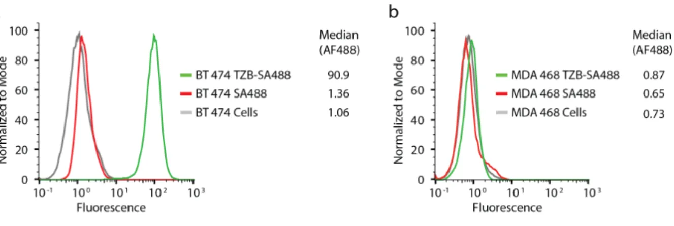
Figure S1. Association of biotinylated trastuzumab to a. HER2+ breast cancer cells BT-474 and b. HER2- breast cancer cells
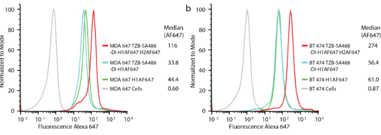
Figure S2. Hybridization Chain Reaction (HCR) at 37°C on (a) HER2- breast cancer cells MDA-MB-468 and (b) HER2+ breast
Figure S4. Time-dependent HCR amplification on BT-474 and MDA-MB-468 cells. (TZB: trastuzumab, DI: double initiator,
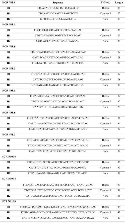
Table S1. Double Initiator (DI) and hairpin sequences used in the present work. All sequences are listed 5’ to 3’.
HCR N6L4 Sequence 5'-Mod. Length
DI TTCATAGTTCCGTTGTTCCGGTTC Biotin 24 H1 TTGAACCGGAACCATAGTTCCG Cyanine 5 22 H2 GTTCCGGTTCCGGAACTATG None 20 HCR N6L6 DI TTCTTCTACCTCACTTCCTCACTATCAC Biotin 28 H1 TTGTGATAGTGAGGCTTCTACCTCAC Cyanine 5 26 H2 CCTCACTATCACGTGAGGTAGAAG None 24 HCR N6L8 DI TTCTCTACTCCACCTCTTCACCTCACAATTAC Biotin 32 H1 CACCTCACAATTACGAGGTGGAGTAGAG Cyanine 5 28 H2 TTGTAATTGTGAGGTGCTCTACTCCACCTC None 30 HCR N7L7 DI TTCTTCATTCATCTCCTTCATCTCCACTCTAC Biotin 32 H1 CATCTCCACTCTACGGAGATGAATGAAG Cyanine 5 28 H2 TTGTAGAGTGGAGATGCTTCATTCATCTCC None 30 HCR N8L6 DI TTCACACTCAATCACCTTCAATCACCTCCAAC Biotin 32 H1 TTGTTGGAGGTGATTGCACACTCAATCACC Cyanine 5 30 H2 CAATCACCTCCAACGGTGATTGAGTGTG None 28 HCR N8L8 DI TTCTTAACTCCATCTCACTTCATCTCACCATTACAC Biotin 36 H1 TTGTGTAATGGTGAGATGCTTAACTCCATCTCAC Cyanine 5 34 H2 CATCTCACCATTACACGTGAGATGGAGTTAAG None 32 HCR N9L7 DI TTCCACTCACATCTCACCTTCATCTCACCTACATCC Biotin 36 H1 TTGGATGTAGGTGAGATGCCACTCACATCTCACC Cyanine 5 34 H2 CATCTCACCTACATCCGGTGAGATGTGAGTGG None 32 HCR N10L6 DI TTCACCTCCACTTCACTCTTCACTTCACTCTTACTC Biotin 36 H1 CACTTCACTCTTACTCGAGTGAAGTGGAGGTG Cyanine 5 32 H2 TTGAGTAAGAGTGAAGTGCACCTCCACTTCACTC None 34 HCR N10L8 DI TTCACCTCACCATCCAACTCTTCATCCAACTCAACTCCAC Biotin 40 H1 TTGTGGAGTTGAGTTGGATGCACCTCACCATCCAACTC Cyanine 5 38 H2 CATCCAACTCAACTCCACGAGTTGGATGGTGAGGTG None 36 HCR N12L8 DI TTCTCATTCTCACTTACCTACCTTCACTTACCTACCATCCTCAC Biotin 44 H1 TTGTGAGGATGGTAGGTAAGTGCTCATTCTCACTTACCTACC Cyanine 5 42 H2 CACTTACCTACCATCCTCACGGTAGGTAAGTGAGAATGAG None 40
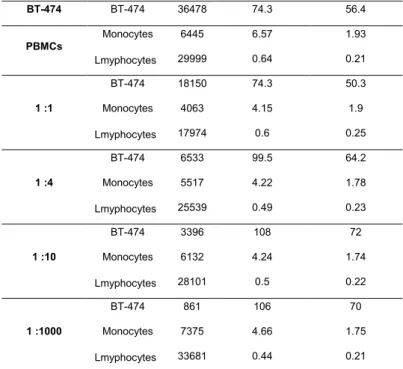
Table S2. Corresponding number of gated cells and the median fluorescence intensities at AF488 and AF647 channels in
Figure 4.
Sample Name Subset Name Count Median (AF647) Median (AF488)
BT-474 BT-474 36478 74.3 56.4 PBMCs Monocytes 6445 6.57 1.93 Lmyphocytes 29999 0.64 0.21 1 :1 BT-474 18150 74.3 50.3 Monocytes 4063 4.15 1.9 Lmyphocytes 17974 0.6 0.25 1 :4 BT-474 6533 99.5 64.2 Monocytes 5517 4.22 1.78 Lmyphocytes 25539 0.49 0.23 1 :10 BT-474 3396 108 72 Monocytes 6132 4.24 1.74 Lmyphocytes 28101 0.5 0.22 1 :1000 BT-474 861 106 70 Monocytes 7375 4.66 1.75 Lmyphocytes 33681 0.44 0.21
Table S3. Corresponding number of gated cells and the median fluorescence intensities at AF647 channel in Figure 5.
Sample Name Subset Name Count Median (AF647)
1 :10 BT-474 146865 242 Monocytes 182397 3.21 Lmyphocytes 834000 0.25 1 :20 BT-474 101356 280 Monocytes 241295 2.51 Lmyphocytes 929000 0.14 1 :30 BT-474 49168 264 Monocytes 193288 3.6 Lmyphocytes 940000 0.31 1 :60 BT-474 34117 301 Monocytes 197742 2.94 Lmyphocytes 985000 0.24 BT-474 23760 150 1 :100 Monocytes 146412 3.61 Lmyphocytes 735000 0.33 BT-474 18301 490 1:200 Monocytes 162794 2.99 Lmyphocytes 852000 0.24