HAL Id: hal-02415223
https://hal.archives-ouvertes.fr/hal-02415223
Submitted on 11 Nov 2020HAL is a multi-disciplinary open access archive for the deposit and dissemination of sci-entific research documents, whether they are pub-lished or not. The documents may come from teaching and research institutions in France or abroad, or from public or private research centers.
L’archive ouverte pluridisciplinaire HAL, est destinée au dépôt et à la diffusion de documents scientifiques de niveau recherche, publiés ou non, émanant des établissements d’enseignement et de recherche français ou étrangers, des laboratoires publics ou privés.
Heart on a chip: Micro-nanofabrication and microfluidics
steering the future of cardiac tissue engineering
Maria Kitsara, Dimitrios Kontziampasis, Onnik Agbulut, Yong Chen
To cite this version:
Maria Kitsara, Dimitrios Kontziampasis, Onnik Agbulut, Yong Chen. Heart on a chip: Micro-nanofabrication and microfluidics steering the future of cardiac tissue engineering. Microelectronic Engineering, Elsevier, 2019, 203-204, pp.44-62. �10.1016/j.mee.2018.11.001�. �hal-02415223�
1
Heart on a chip: Micro-nanofabrication and Microfluidics Steering the Future of Cardiac Tissue
Engineering
Maria Kitsaraa*, Dimitrios Kontziampasisb, Onnik Agbuluta, Yong Chenc
a Sorbonne Université, Institut de Biologie Paris-Seine (IBPS), UMR CNRS 8256, Biological Adaptation and
Ageing, 75005, Paris, France.
b School of Biomedical Sciences, Faculty of Biological Sciences, University of Leeds, LS2 9JT, Leeds, W.
Yorkshire, UK.
c Ecole Normale Supérieure-PSL Research University, Sorbonne Universités, UPMC Univ Paris 06, UMR CNRS
8640, PASTEUR, 75005, Paris, France.
*Corresponding author contact details:
[email protected], [email protected]
Sorbonne Université, Institut de Biologie Paris-Seine (IBPS), UMR CNRS 8256 7, quai St Bernard, PC 75005 Paris-FRANCE
Tel: +33 (0) 144273507
Abstract
The evolution of micro and nanofabrication approaches significantly spurred the advancements of cardiac tissue engineering over the last decades. Engineering in the micro and nanoscale allows for the rebuilding of heart tissues using cardiomyocytes. The breakthrough of human induced pluripotent stem cells expanded this field rendering possible the development of human tissues from adult cells, thus avoiding the ethical issues of the usage of embryonic stem cells but also creating patient-specific human engineered tissues. In the case of heart, the combination of cardiomyocytes derived from human induced pluripotent stem cells and micro/nano engineering devices gave rise to new therapeutic approaches of cardiac diseases. In this review, we survey the micro and nanofabrication methods used for cardiac tissue engineering, ranging from clean room-based patterning (such as photolithography and plasma etching) to electrospinning and additive manufacturing. Subsequently, we report on the main approaches of microfluidics for cardiac culture systems, the so-called “Heart on a Chip”, and we assess their efficacy for future development of cardiac disease modeling and drug screening platforms.
Keywords: cardiac tissue engineering, micro/nanofabrication, polymeric scaffold, microfluidics, heart-on-a-chip
1. Introduction
In 1988 Joseph Vacanti’s and Robert Langer’s groups [1] transplanted biodegradable polymer-cell artificial three-dimensional (3D) scaffolds into animals. Their approach, named “chimeric neomorphogenesis”, was based on the idea of remodeling and self-regulation of tissue. The transplanted patch would only provide a means of initial support (similar to a scaffold) for cells and then dissolve. Later on [2], Vacanti explained the reasons why there is a huge and increasing need of artificial organs, and why surgeons struggle to address the problem of lack of donors especially for infants and very young children. Introducing how nature copes with problematic situations on pathologies of organs, he proposed the biomimetic approach of transplanting
2
a “chimeric” composite patch directly on problematic areas of organs. This patch was a composite material made from polymers and seeded in vitro with cells. Polymers, he claimed, could be modified in terms of composition to achieve desired mechanical and chemical properties, and promote not only biocompatibility, but additionally cell attachment with the aid of surface modification methods. Langer and Vacanti renamed their approach, and the idea of “tissue engineering” was introduced [3]. This altered approach expanded even more, to the use of synthetic and natural polymers as well as composite materials and mixtures. Their idea signalled the need for true interdisciplinary research, novel advancements in materials and polymer science, pioneer fabrication methods having dual or triple scale capabilities that extend from nano to centimeter scales, if this idea of engineering a tissue or an organ would have any chances to be materialised and be successful.
On the other hand, advances in micro-nanofabrication and microfluidics have opened the repertoire of applications in life sciences including not only diagnosis but also disease modeling and therapeutic approaches. The field of microfluidics and lab-on-a-chip (LoC) devices stems from the areas of molecular analysis, biodefence, molecular biology, and microelectronics according to Whitesides [4]. Regarding the LoC devices development, the first ones were fabricated by standard photolithographic techniques in silicon, adapted from microelectronics and MEMS (micro electro mechanical systems) research. Over the years, silicon and later glass were gradually replaced by polymeric materials, taking advantage of their low cost and customizable material characteristics, enabling their manufacturing with a plethora of methods [5]. Starting from optical lithography and clean room facilities for fabrication of chips based on SU-8, the development of rapid prototyping techniques stirred their evolution in a more affordable and fast manner. Likewise, cell culture substrates benefited from microelectronics: while in transistors fabrication of a rough surface due to reactive ion etching was fatal, this “failure” or “unwanted” side effect of a technique, was proven to be beneficial in cell cultures were nano and microtopography acts as a spatial cue for promoting cell adhesion and proliferation [6].
Consequently, tissue engineering and regenerative medicine have significantly flourished over the last decades, due to the progress of micro and nano-engineering. The area of “Organ on a Chip” consists the most recent and challenging topic of LoC devices, where microfluidics and cell cultures are combined in providing an in vivo-like environment. Towards this direction, in this review we emphasize in the advances for the development of “Organ on a Chip” systems focusing on heart, the so called “Heart on Chip”, which importance is underlined by the high evolution in research concerning cardiac tissue engineering and regenerative medicine. The first part covers the main micro-nano fabrication methods that have been utilized for the development and improvement of cardiac tissue engineering. The second part focuses on presenting the last advances of combining the in vitro cardiac tissues with microfluidics for the creation of higher degree-integrated “Heart on a Chip” systems for cardiac disease modeling and drug screening. The advent of induced pluripotent stem cells (iPSCs) from adult cells by Shinya Yamanaka in 2006 [7] gave a further boost in the field, rendering possible the generation of cardiomyocytes (CMs) derived from human iPSCs (hiPSC-CMs), thus providing with patient-specific cardiac cells.
2. Micro and nanoengineering methods used for cardiac tissue engineering
The extracellular matrix (ECM) environment plays a significant role in cell behaviour, functionalities and viability [8–10]. Cardiac cells are no exception to the rule [11,12]. Substrates with controlled architectures can provide critical information on the study of the cell-material interactions on adhesion and on other (relevant to adhesion) properties such as orientation, shape, viability, migration. By understanding and decoding the dominant mechanisms on cell-material interactions we could then provide the necessary properties to mimic the microenvironment of the heart [10,13,14]. It is obvious that in order to be able to study the aforementioned interactions, a total control on the micron and nano size features of the surface of the culture interface, between cells and materials must be set. Micro and nanofabrication and materials engineering have traditionally tackled with controlling and fabricating structures at these scales and they are the ones that have been called out to give the answers on providing artificial platforms with a top level of control on the chemistry, topography, elasticity, conductivity and other useful properties [5,15–20].
3 2.1 Optical Lithography
Optical lithography or photolithography is the most widespread micro/nanofabrication method. It has been used in micro/nanoelectronics and MEMS, as well for the fabrication of LoC devices using a top-down approach [5]. It uses light (UV, or DUV) going through a mask and transfers a desired pattern on a photosensitive material [21] [15]. The patterned polymer is subsequently used as a mask for transferring this pattern on a substrate with plasma etching. Although this is a multistep process, it is very well controlled and defined, and can be versatile in terms of combination with other techniques. It has been found that cells can be cultured directly on top of electrodes, printed circuit boards, as well as other micro/nanoelectronic elements, since SiO2, Cu, Au, Pt, glass, quartz, Kapton, polyimide and most of the materials of the substrates
of micro/nanoelectronic and MEMS proved to be not only compatible to the cells but in some cases were favorable towards specific functionalities [22].
In the case of cardiomyocytes (CMs) and cardiac tissue, the most profound example is the one of the microelectrode array (MEA) that is used for evaluating arrhythmias [23,24], while similar apparatus has been used for single cell detection of cellular activity [25]. Perfectly aligned arrays of long silicon nanowires fabricated using a complementary metal oxide semiconductor (CMOS) compatible approach and configured as field-effect transistors, demonstrated an ability for real‐time extracellular recording of cellular bioelectricity, enabling non-invasive, high‐sensitivity, high‐throughput, long‐term electrophysiology measurements at the single‐cell, tissue and organ levels [26]. The use of MEAs for the examination of cardiac responses was aimed to be used for drug screening purposes.
Optical lithography is also used to direct the attachment and the shape of cells which are grown on surfaces. It has been found that when cardiac cells are cultured on surfaces that have been patterned into having typically 20 μm wide lines, cells tend to align and stretch their ECM and their nucleus towards the length of the line [27]. Aligning CMs is crucial for the contraction and propagation along the long axis [28].
Jain et al. fabricated micron sized ridges on a silicon surface as a model to study cardiac failure of diverse origin, achieving stretching and alignment of cultured cells [29]. Yamaguchi et al. have selectively modified and patterned APTES-COOH surfaces with the help of Vacuum UV photolithography. The selective linear local surface chemical modification, created a patterned culture of primary rat cardiac cells [30].
Khademhosseini group developed various systems based on gelatin methacryloyl (GelMA) hydrogels [31]. GelMA hydrogels are photocrosslinkable and their mechanical properties can be adjusted to native ECM ones, while they can be microfabricated with various methods, rendering them a versatile material for tissue engineering. For example, they developed a coating method of PDMS-based microfluidics with GelMA and methacrylated tropoelastin (MeTro) for improving CMs attachment and organization inside the channels [32]. By adjusting the injection flow rate of the prepolymer solution during UV crosslinking, a controllable coating thickness was achieved. Nevertheless, culture of neonatal rat CMs showed that they are better attached and beating on the MeTro coatings.
2.2 Soft lithography
Soft lithography is a name attributed to a plethora of related techniques. These techniques are used for micro/nanopatterning and their common characteristic is that they involve fabrication or replication of a “master”. This master can either be used as a stamp or as a mold to pattern an elastomer, which is later cross-linked. After cross-linking, the patterned elastomer is used as a stamp/mold for the micro/nanopatterning of other materials. The name of the method was initially introduced due to the fact that it uses elastomeric materials, with the most notable one being polydimethylsiloxane (PDMS) [5]. It should be noted that optical lithography, electron beam, laser patterning or other microfabrication methods are used for the fabrication of the initial stamp or mold. Since cells and tissue are not grown in direct contact with the masks but with the materials that are patterned at later steps of this process, they do not provide any additional functionality and thus they are not categorised separately in this study. An exception to this rule is the work by Badie et al. which used diffusion tensor magnetic resonance imaging (DTMRI) to image
4
and translate the directionality of cardiac tissue into a lithography photomask [33]. Optical lithography followed for the transfer of this pattern on a photoresist over a silicon wafer. This was used as a mold for a PDMS by a soft lithography approach to pattern fibronectin on a coverslip, which would be used as a surface for the control of culture of CMs. This approach can aid in the understanding of the complex role of cardiac microarchitecture on healthy or remodelled systems in macroscopic impulse.
In the case of cardiac cells and tissue engineering, soft lithography is by far the most widely employed technique for the micro/nanopatterning of surfaces. Beussman et al. fabricated PDMS micropost arrays, functionalised with ECM protein to measure the contractility of stem cell derived CMs [34]. Others modified the surface of posts or pillars to achieve a better response or a desired control on the attachment of cardiac cells [35–38].
Figure 1. Formation of realistic cell alignments in micropatterned cultures. Schematic representation of the fabrication method (left) and immunostaining results (right). Red: sarcomeric a-actinin, green: connexin-43, blue: nuclei. Scale bars: (a–c) 250 μm; (d) 50 μm. Reprinted with permission from [33].
Micro-scale grooves fabricated with soft lithography can significantly enhance cell alignment as was the case with optical lithography [39,40]. Additionally, alignment of CMs lead to a greater expression of cardiac genes and CMs markers, and gap junctional proteins such as connexion-43 [41,42]. Kim et al. [43] fabricated nanosized arrays using polyethylene glycol (PEG) hydrogel in an effort to reconstruct aligned fibrils. CMs were found to align along the direction of the topography, with analysis showing that this was due to the alignment of the focal adhesions. Many other studies followed similar approaches to promote desired functionalities for CMs by either micro/nanopatterning hydrogels and elastomers, or by printing a thin layer of micro/nanopatterned natural polymers to guide cardiac cells (such as collagen, gelatin, fibronectin, laminin, agarose or similar) [44,45,54–57,46–53].
Feinberg and Parker used soft lithography to tailor a nanofabric containing fibronectin, laminin, collagen type I and collagen type IV, which mimics the ECM. They have shown that cardiac cells cultured on the fabric could functionally organise cells into anisotropic tissue, including papillary threads, without any degradation [58]. Boateng et al. demonstrated that arrays of microposts (which they called micropegs) having 10µm tall extruded cylinder provided a vertical surface for cell attachment and greatly hindered the proliferation of fibroblasts comparing to smooth flat substrates [59]. This is a very interesting finding due to the well-known problem of separating CMs from fibroblasts and other non-myocyte cells, from the primary cell source. The reason behind this behaviour of fibroblasts was determined to be the extensive elongation of their nuclei, which resulted in a decreased proliferation rate [60].
Stem cell differentiation was also found to be controlled by the effect of nanotopography (nanopillars and nanogrooves) created by soft lithography. This nanotopography-induced differentiation showed that with the control of the size and the distance of the nanostructures, the stem cells differentiated into different kind of cells including beating CMs. This is a promising but still very immature study, which faces many problems
5
that need to be addressed (for example homogeneity) [61–63]. A very recent study used microsized pillars with different elasticity for
2.3 Laser based fabrication
The use of laser based fabrication techniques is becoming more widespread over the years. Lasers have the ability to modify a variety of materials in the micro and nano scale in a very controlled manner with the aid of robotic arms and computer controllers [64]. This part of the paper will only focus on planar fabrication techniques and thus will narrow down mainly to laser ablation. Laser ablation as a technique uses a focused laser beam to etch away material from the bulk, by directly “writing” the desired pattern [65]. It is common for use in various biological applications due to the fact that the laser beam causes vaporisation of the material with no sign of any thermal effects [66]. Patz et al. used excimer laser ablation to directly fabricate channels on agarose, and showed improved attachment, alignment, proliferation and differentiation of C2C12 myoblasts [67]. Similar approaches have been used for the modification of other soft matter [68,69]. The same approach was used by Engelmayr et al. for the production of multi-layered elastomeric scaffolds which had an “accordion-like honeycomb structure”. This allowed them to mimic the shape of perimysial collagen fibers, as well as to guide the alignment of cells and to replicate the native myocardium tissue microenvironment and anisotropy [70–72][73]. With this method the group managed to fabricate membranes which matched the anisotropic mechanical properties of the left ventricular tissue, which could sustain cyclic loads and additionally promote desired synchronous contraction, and directionality of cells on the heart circumferential axis [70]. The same approach was used by Kapnisi et al. for creating auxetic cardiac patches [74].
Laser interference lithography is a technique that takes advantage of the periodicity of the interference pattern between two or more light (laser) waves, using this as a way to expose a photoresist (similar to optical lithography). This pattern creates well defined structures [75]. For example, Fujita et al. controlled CMs orientation using patterns created by this technique [76].
Lasers were also used, in more exotic approaches, as a tool for the deeper understanding of the mechanisms and role of specific type of cardiac cells. For example, Narasimhan et al. demonstrated optical manipulation of cells using a weakly focused laser beam. They directed and created a “bridge” of fibroblasts in between two lines of cultured myocytes, which were beating asynchronously, to understand the electrophysiological role of the fibroblasts [77] .
2.4 Plasma surface modification
In the last ten years, the use of low vacuum, and atmospheric pressure plasma systems has had an outburst. There are many studies that show how plasmas can be used for the stochastic and periodic nano and micro scale structuring of surfaces, with a simultaneous modification of the surface chemistry. Due to this duality, plasmas have been used to achieve a desired functionalization of surfaces [78]. As mentioned in the introduction, surface modification was one of the first key components that were identified from the very beginning as essential in mimicking successfully the micro/nanoenvironment of an organ, and in our case the myocardium, or any other part of the heart [2,3]. Apart from the control of wettability [19], reflectivity [79], plasma surface modification has been used for the immobilisation of proteins on surfaces [17,80] or for the control of attachment, shape and behaviour of various cells [8,9].
In the case of cardiac cells, plasma functionalization of surfaces is mostly used in combination with other techniques to improve the hydrophilicity of a surface which will be used for the CMs culturing [49,56,71,81– 86]. Parker group has used oxygen plasma to control the attachment of gelatin hydrogel on cyclic olefin copolymer, cyclo olefin polymer and polyolefin surfaces. These were subsequently used to validate their suitability for cardiac tissue engineering, using both primary neonatal rat CMs and hiPSC-CMs [47]. Brown et al. investigated the effect of plasma surface modification for the absorption of fibronectin on poly(lactic-co-glycolic acid) (PLGA) substrates using neonatal rat ventricular CMs. Their study showed improved spreading and myofibril development on the plasma modified samples, and also that the protein absorption affects the
6
gene expression of the attached cells [87]. Guex et al. used plasma to deposit an oxygen functional hydrocarbon coating on ε-polycaprolactone (PCL) nanofibers. This improved the mechanical stability and endurance of the nanofibers while tested in vivo in a rat model [88]. Kontziampasis et al. have used O2 and
Ar plasma etching to investigate the effect of nanotopography and chemistry changes induced on Parylene C films [89]. These were used to control the viability of neonatal rat CMs. Despite the fact that O2 plasma
rendered the surfaces more hydrophilic, the Ar plasma led to a more-friendly chemistry of the surface, allowing neonatal rat CMs to grow better on the Ar plasma treated ones.
2.5 Electrospinning
Electrospinning is a technique that is widely used for the fabrication of nanofibers, using polymeric (bio)materials or (bio)polymer based composites [13,90,91]. Currently it is the technique that is most widely used in industrial applications due to its ability to upscale, with many systems being developed in the last few years [92]. In electrospinning, a solution is fed through a capillary while opposite a collector which is grounded. High voltage is applied and thus a jet of the solution travels from the needle to the collector, where it deposits dried nanofibers. By adjusting the process and material parameters as well the environmental conditions, nanofibers characteristics can be modulated [93]. Due to its cost effectiveness as well as its simplicity, this method has become very popular for many biological applications.
Since electrospun nanofibers show similar morphologies with the natural ECM, which consists of long continuous fibers of several different diameters, tissue engineering and especially cardiac tissue engineering have shown a great interest to it. The need for aligned nanofibers has led to advancements on the collector, with several approaches like a high-speed collecting drum or a mandrel collector being developed [94,95]. There is a plethora of natural and synthetic polymers that are used for cardiac tissue engineering related electrospinning, as it was recently reported by Kitsara et al. [13]. In order to mimic correctly the mechanical properties, conductivity, elasticity as well the local chemistry of the microenvironment of the myocardium, several different approaches have been developed trying to improve the biomimicry of the electrospun fabricated scaffolds. The first one is the electrospinning of composite materials which either were a mixture of a natural and a synthetic polymer, or a polymeric solution on which other additives were added. Gold nanoparticles [96–99] and carbon nanotubes [100–103] were added in order to improve the conductivity of the electrospun construct in several cases. Peptides [104] and granulocyte colony-stimulating factor [85,105,106] was added in some studies to improve the remodeling and viability of CMs.
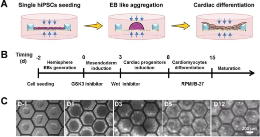
Figure 2. Cardiac differentiation of hiPSCs on patch consisted of a monolayer of electrospun gelatin nanofibers in a honeycomb fabricated PEGDA frame. (A), (B) Schematic and protocol of cardiac differentiation. (C) Bright-field images of the differentiating hiPSCs at different days. Reprinted with permission from [107].
7
Tian et al. [108] fabricated core-shell nanofibers using an emulsion (instead of a solution) for electrospinning, encapsulating a bioactive growth factor for cardiovascular regeneration (VEGF) in dextran or bovine serum albumin (BSA). In vivo studies showed that most of the transplanted myocytes prevented negative ventricular remodeling, while expressing the cardiac marker proteins [109]. In another study, introducing VEGF in electrospun nanofibers provoked angiogenesis and cardiomyogenesis in an acute myocardial infarction rat model [110]. Tang et al. fabricated a honeycomb shaped frame with poly(ethylene glycol) diacrylate (PEGDA) and electrospun a monolayer of gelatin nanofibers on top. Induction and differentiation of hiPSC was performed on the same scaffold, which led to a uniform cardiac tissue with homogeneous beating (figure 2) [107]. Li et al. [111] electrospun a very thin layer of PLGA nanofibers on a PDMS frame, and showed that it promoted the maturation of hiPSC-CMs. They evidenced their results by measuring an increased electrical response comparing to the same CMs cultured on a flat surface, and validated this finding with flow cytometry, immunostaining, and qPCR, which showed that the construct upregulated cardiac biomarkers and enhanced cardiac functions. They named the hybrid nanofiber-CM on the PDMS frame as “cardiac tissue-like construct (CTLC)”. The CTLC was used for in vitro experiments where it managed to connect and synchronize the beating of two disconnected CM tissues. In vivo experiments in rat models of myocardial infarction, showed that the CTLCs with a high number of cells and increased thickness could surpass the problem of the high Young modulus of the PLGA fibers.
The group of Javasinghe have managed to directly electrospin living cells for the development of scaffolds with “living” parts [112]. In order to be able to realise the technique they used a coaxial needle where a concentrated biosuspension flows through the internal needle, while high viscus and low electrical conductivity polymeric medium (in this case PDMS) flows in the external part, showing that cells were not affected from the high voltage. In a more recent study they used primary neonatal CMs to fabricate living scaffolds with cell electrospinning, while maintaining their functionalities [113].
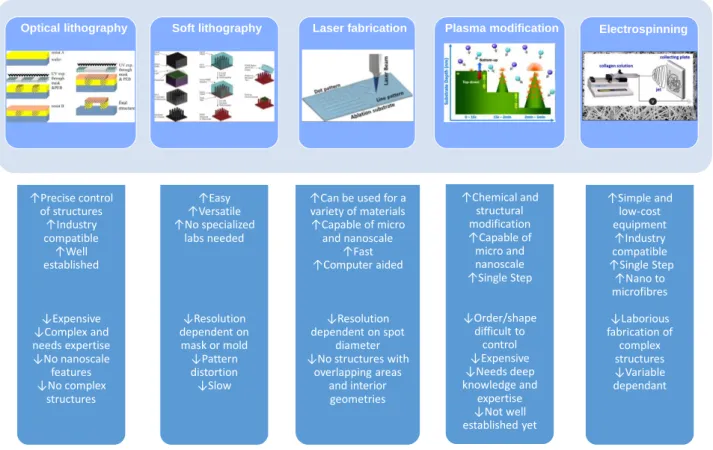
Figure 3. Basic micro and nanoengineering methods used in cardiac tissue engineering. Schematic representations of the method, followed by advantages and disadvantages for their use in cardiac tissue engineering. Figures (reprinted with permission) are modified from the following: optical lithography from [21], soft lithography from [34], laser ablation from [64], plasma modification from [114], and electrospinning from [90].
↑Precise control of structures ↑Industry compatible ↑Well established ↓Expensive ↓Complex and needs expertise ↓No nanoscale features ↓No complex structures ↑Easy ↑Versatile ↑No specialized labs needed ↓Resolution dependent on mask or mold ↓Pattern distortion ↓Slow
↑Can be used for a variety of materials ↑Capable of micro and nanoscale ↑Fast ↑Computer aided ↓Resolution dependent on spot diameter ↓No structures with
overlapping areas and interior geometries ↑Chemical and structural modification ↑Capable of micro and nanoscale ↑Single Step ↓Order/shape difficult to control ↓Expensive ↓Needs deep knowledge and expertise ↓Not well established yet ↑Simple and low-cost equipment ↑Industry compatible ↑Single Step ↑Nano to microfibres ↓Laborious fabrication of complex structures ↓Variable dependant
8 2.6 Additive manufacturing
Additive manufacturing, also known as 3D printing, is the name attributed to a group of techniques. In 1986, Charles Hull invented a process called 3D stereolithography [115]. Using this technique, it became possible to fabricate complex 3D objects that were not possible to be fabricated with any other method. The common basic principle under all additive manufacturing methods, is the modification or addition of consecutive 2D planes in order to achieve a 3D structure [116].
3D printing has been employed by research and industry to create sophisticated instruments and tools. One of the most noticeable examples is its use for fabricating 3D surgical planning models, where a printed organ was fabricated as a guide for surgeons to identify and resolve problems and malfunctions before surgery. This has been used in heart surgery creating a replica of a patient’s heart for the precise understanding of the anatomy, hands-on simulation of surgical and interventional procedures, and morphology teaching of the medical professionals (figure 4) [117].
Figure 4. 3D printed cardiac models for surgical practice or training. Reprinted with permission from [117].
In the last few years, tissue engineering and regenerative medicine are showing great interest in the use of 3D printing, and have started to investigate its potential to directly print scaffolds, tissue, and subsequently organs. This specialised additive manufacturing has been named as “bioprinting”, which includes three steps; pre-bioprinting, bioprinting itself, post-bioprinting. Pre-bioprinting is the step where scanning and imaging, computer aided design and material selection take place. Scanning usually is done either by magnetic resonance imaging or by computer tomography, while material selection is dependent on the type of the printing technique as well as the final aimed construct. Bioprinting in principle works as a typical desktop printer. It uses the (3D) printer, (bio)ink and a biomaterial instead of paper. Post-bioprinting involves all the processes that are needed to acquire a 3D biological structure with the appropriate properties and biological functionality. This for example includes the maturation time of the printed construct in order to fuse different planes into one structure [118].
Bioprinting can be realised by using several approaches, but the ones that are most widely used for depositing and patterning biological materials are laser-assisted printing, multiphoton excitation (MPE)–based fabrication (stereolithography), inkjet printing, and microextrusion. Schematics of these approaches as well as advantages and disadvantages are shown in figure 5 [119,120,129–133,121–128].
9
Figure 5. Schematic representations of the four most widely used techniques of 3D printing for cardiac tissue engineering (reprinted and modified from [121]). (A) Laser-assisted bioprinting (LAB) which is based on laser-induced forward transfer (LIFT). (B) Multiphoton excitation (MPE) which is also called stereolithography. (C) Inkjet printing. (D) Microextrusion ink deposition. The basic advantages and disadvantages for each method for 3D bioprinting are cited below each schematic.
Parker group 3D-printed various different cardiac microphysiological devices with integrated sensors, which were inside their customised inks (described in the section 3.2, figure 6c). This approach showed to improve the ability of tracking temporal development of the mechanics of the tissue and promises to enable new insights for tissue morphogenesis, pathogenesis, as well as drug induced remodeling [134]. Gaetani et al. [135] bioprinted alginate with human CM progenitor cells, fabricating a construct which possessed high cell viability (92% and 89% at 1 and 7 days of culturing). The constructs retained the cardiac lineage, showing an enhanced expression of genes and early cardiac transcription factor sarcomeric protein. Cells were reportedly migrating from the construct matrix to form a tubular structure and colonize on a matrigel layer. The same group [136] also demonstrated tissue printing of an hyaluronic acid/gelatin (HA/gel) scaffold combined with human fetal CM progenitor cells. This was proposed as a cardiogenic patch with defined pore size that could promote cell viability.
Gaebel et al. [137] fabricated a cardiac patch, with LIFT cell printing using human umbilical vein endothelial cells (HUVECs) and human mesenchymal stem cells (hMSCs) on a defined pattern. Performing an in vivo study they showed that the transplantation of the tissue engineered cardiac patch had an increased vessel formation, and a significant improvement in density of the infarcted heart. Cells were integrated into the vessels of the murine vascular system, showing that the patch is a promising approach towards the treatment of myocardial infarction. Izadifar et al. used an alginate laden with human coronary artery endothelial cells as ink for the layer by layer bioprinting of cardiac constructs with varying architectures performing in vivo studies as well as implantation on rat hearts. For the in vivo studies the constructs offered a high cell viability and distribution, while the implanted constructs showed a good agreement with the in vitro measurements in terms of impedance or elastic modulus [138]. Adams et al. [139] printed scaffolds from PCL using a commercial microextrusion 3D printer, and incorporated wirelessly controlled electrical stimulators.
↑High resolution ↑No clogging ↑Variety of materials
used ↓Ink gelation kinetics
lower speed ↓Time consuming
↓Costly ↓Cannot print multiple
cell types simultaneously ↑Complex structures ↑High resolution ↓Limited macroscale size ↓Required to merge fields of view to achieve
larger scales ↓Slow
↑Cost effective ↑High bioink
deposition ↑High cell gradient
capability ↓Cells and biological
species undergo thermal and mechanical
stresses ↓Non-uniform droplet creation ↓Clogging of nozzle ↓Cell encapsulation unreliable ↓Low cell densities ↓Bioink must be liquid
↑Simple ↑High cell concentration can be
achieved ↓High viscosity might
affect cell viability ↓Specialized bioink
needed to ensure viability ↓Cells and biological
species undergo mechanical stresses
10
Electrical stimulation showed signs of improving the attachment and provoking differentiated actin cytoskeletal structures. Ho et al. [140] also used 3D printing with a PCL - carbon nanotube composite, while comparing the viability with a PCL scaffold in the case of rat myoblasts (H9C2). Gao et al. [141] used MPE for fabricating a scaffold, based on photoactive gelatin polymer, with submicron resolution which they seeded with CMs, smooth muscle cells and endothelial cells. All cells were differentiated from hiPSCs and for this reason they named their construct as “human-induced pluripotent stem cell– derived cardiac muscle patch (hCMP)”. The patch was evaluated in a murine model of myocardial infarction. In vivo results showed that hCMP improves cardiac function and reduces infarct size after myocardial infarction. The rate of cell engraftment in hCMP-treated animals was 24.5% at week 1 and 11.2% at week 4. Ciocci et al. [142] fabricated a multitextured 3D PEGDA-based scaffold with microstereolithography, to induce a favorable microenvironment which promotes human cardiac progenitor cell differentiation and orientation. While cultured, cells adopted and showed a 3D spatial ordered orientation, while the expression of sarcomeric α-actinin and connexion-43 was activated.
3D bioprinting shows also potential for fabricating heart valves. The common strategy for creating replacement heart valves is the surgical implantation of a mechanical or chemically crosslinked tissue heart valve [143], which is robust and show extended life-times [144]. Patients with a prosthetic valve, though, are required to permanently take anticoagulants, while when biological valves are implanted, they do not require their use [145]. Neither synthetic nor biological transplants grow and remodel with the patient. A 3D bioprinted valve could possibly address these limitations. Most of the bioprinted heart valves have been fabricated using an extrusion type 3D bioprinter, using photo-crosslinkable hydrogels. Usually a combination of a “soft” and a “rigid” hydrogel is being employed to fabricate the leaflets and the root respectively [146]. This ability to print two different biomaterials allowed for the better biomimicry of the stiffness of different parts of the valve. Interstitial cells from porcine aortic valve survived for up to 3 weeks inside 3D bioprinted heart valves [147]. In another study from the same group, aortic root sinus SMCs and aortic valve leaflet interstitial cells were encapsulated into the root and leaflet portions of an alginate/gelatin 3D bioprinted aortic valve conduit. The study showed that valve leaflet interstitial cells remodeled hydrogels, by depositing collagen and glycosaminoglycan-based ECM [148]. Despite progress shown, heart valve 3D bioprinting has not managed to report results on functional testing of any of the printed valves up to date.
Despite the great advancements of bioprinting techniques and despite the aforementioned 3D printed organ models (for educational and surgical guide purposes), bioprinting technology has not successfully printed yet any tissue constructs which were therapeutically relevant. The basic reason behind this fact is that besides the complexity of such an attempt, for each individual bioprinting technique there are several disadvantages which cannot be surpassed. Potentially a combination of multiple bioprinting techniques, as well as the combination of 3D bioprinting with other methods that are tested and used for tissue engineering, can provide a better strategy towards overcoming the constraints to reach the final aim and 3D print a functional heart.
3. Heart-on-chip systems
This part focuses on surveying characteristic LoC systems that have been reported as cardiac models. Using microfluidic technologies, it is possible to better decipher human heart function and create more-relevant disease models and drug screening systems, which simulate tissue structure and function at a micron level and bridge the gap between the in vivo and in vitro worlds. Bioinspired microfluidic environments allow spatiotemporal control of various chemical and physical culture conditions that are unavailable with standard static cell cultures. Perfusion bioreactor systems with controlled oxygen supply closed to the in vivo environment, can overcome the limitations of diffusional transport in conventional culture systems as reported by Radisic et al. [149]. In the same context, Wang et al. studied the effect of shear stress within PDMS-based microfluidic chips with channels of various widths on the function of aortic valvular interstitial cells [150]. It was revealed the intensity of shear stress regulates cells morphology, transformation as well as the formation of focal adhesions.
11
The importance of LoC platforms is indicated by the fact that drug testing in animal models cannot accurately predict the effects of drugs on humans. Characteristically only a small percentage manages to pass to the clinical phase [151]. Therefore, the development of human models in vitro is of paramount importance for effective drug development and the advances in microfabrication and microfluidics can make this possibility feasible. Herein the presented “heart-on-chip” systems are categorized according to their main functionalities as electrical and mechanical stimulation based systems. Thereafter more complex systems supporting co-culture of cardiomyocytes with other cells are presented, as well as some distinctive cardiac-applied microfluidic approaches.
3.1 Electrical stimulation systems
Devices incorporating micro-electrodes for the stimulation of cardiomyocytes have been utilized for achieving better cell orientation and maturation but also for performing electrophysiology experiments. For the latter, MEAs have been applied for the extracellular recording of field potentials from the contracting cardiomyocytes, a crucial parameter for identifying arrhythmias caused by drugs which is in alliance with the FDA initiative “Comprehensive in-vitro Proarrhythmia Assay” (CiPA) [152]. Herein we present the main microfluidic devices used for inducing electrical stimulation to cardiac cells. It must be noted that our search was focused solely in LoC systems containing microfabricated electrodes and not macro-scaled ones. One of the first microfluidic devices for achieving CMs anisotropy was reported by Yang et al., based on the combination of dielectrophoresis and electro-orientation [153]. They fabricated interdigitated–castellated microelectrodes by standard photolithography and lift-off on glass slides, which were sealed with silicone in order to create a perfusion chamber (figure 6(i)). By applying an appropriate frequency in a low conductive medium, adult rat CMs were trapped and oriented along the ac electric field direction and formed a tissue-like structure. Cheng et al., studied the metabolic monitoring of adult rabbit CMs in a microfluidic device with integrated lactate sensor and stimulating microelectrodes [154]. The microfluidic chambers were designed in order to enable single cell manipulation and measure its contraction, intracellular calcium transients, pH and the released intracellular lactate after stimulation. Using this microfabricated PDMS-on-glass chip, they demonstrated the effect of the restricted extracellular volume on the metabolic activity during continuous stimulation in an ischaemia cellular model.
Tandon et al. developed a bioreactor system containing indium tin oxide (ITO) MEAs which were microfabricated using excimer laser ablation for the electrical stimulation of neonatal rat CMs and human adipose tissue-derived stem cells (hASCs) [155]. ITO advantages are the excellent electrical conductivity along with optical transparency. It was demonstrated that both cell types exhibited enhanced elongation and alignment after 6 days of electrical stimulation. Ma et al. combined laser-patterning and soft lithography to develop multiple muscle fibers (from neonatal rat CMs) with cell bridges on MEA chips, which allowed on chip analyses of electrical conductivities between different cell types in the myocardium [156]. The cell number for bridge formation was controlled by laser-patterning, and their results indicated that stem-cell (rat mesenchymal stem cells from bone marrow, rMSCs-bm) bridges conducted stronger electrical signals in comparison to fibroblast ones.
Qian et al., demonstrated a cardiac platform that combined a MEA for field potential readouts and an interdigitated electrode (IDE) array for impedance readouts [157]. Both the MEA and IDEs were patterned on glass using standard photolithography and electron beam evaporation and were designed to be “interpenetrating” so that they measure the same area of the tissue (figure 6(ii)). This platform was used for performing electrophysiology and contractility measurements of hiPSC-CMs. under physiological conditions and under drug stimuli. In specific the effect of norepinephrine, a clinical drug used to treat low blood pressure and heart failure, indicating that this platform can be used for cardiac toxicity studies.
Another approach included the micromolding of gelatin on top of a commercial MEA [158]. This approach enabled the realization of electrophysiological measurements on laminar cardiac tissue resembling the native myocardium, in contrast to the previous studies that cells were cultured on the high-stiffness glass slide-MEAs. The system was validated by measuring the response of hiPSC-CMs in the presence of cardiotoxic pro-drug (terfenadine) and its non-cardiotoxic metabolite (fexofenadine), and proved to be capable in detecting
12
small molecule-mediated differences in the cardiac tissue electrophysiological properties. The gelatin-micromolded MEA was further integrated in a microfluidic chip and tested by recording the response of this engineered tissue to isoproterenol (figure 6(iii)). The reusability of this system and its designing to support open-well cell culture for more precise cell seeding makes it a valuable platform for drug assessment. Concluding, electrical stimulation using microfabricated electrodes, mainly MEAs, clearly enhanced the structure and alignment of cardiac cells. The LoCs systems used are mainly based on PDMS bonded on thin glass where the latter contains the electrodes part. This scheme proved to be practical for living imaging studies, such the acquisition of the contracted cardiomyocytes and extracellular recording of field potentials.
(i) (ii)
(iii)
Figure 6. Main electrical stimulation heart-on-chip systems: (i) Dielectrophoresis and electro-orientation on chip, reprinted with permission from [153]. (ii) hiPSC-CM tissue growth on a combined MEA-IDE chip. Scale bar: a. 1 cm, b.1 mm, c. d. is 100 μm. Blue: nuclei, green: cardiomyocyte troponin-T, reprinted with permission from [157]. (iii) Micromolded gelatin on a commercial MEA chip, reprinted with permission from [158].
3.2 Mechanical stimulation systems
Except from the electrical stimulation, myocardium withstand mechanical loads due to the contraction/relaxation phases during each heartbeat. Various systems have been developed trying to reproduce this cyclic uniaxial strain that cardiomyocytes are subjected along the locally oriented ECM fibrous network.
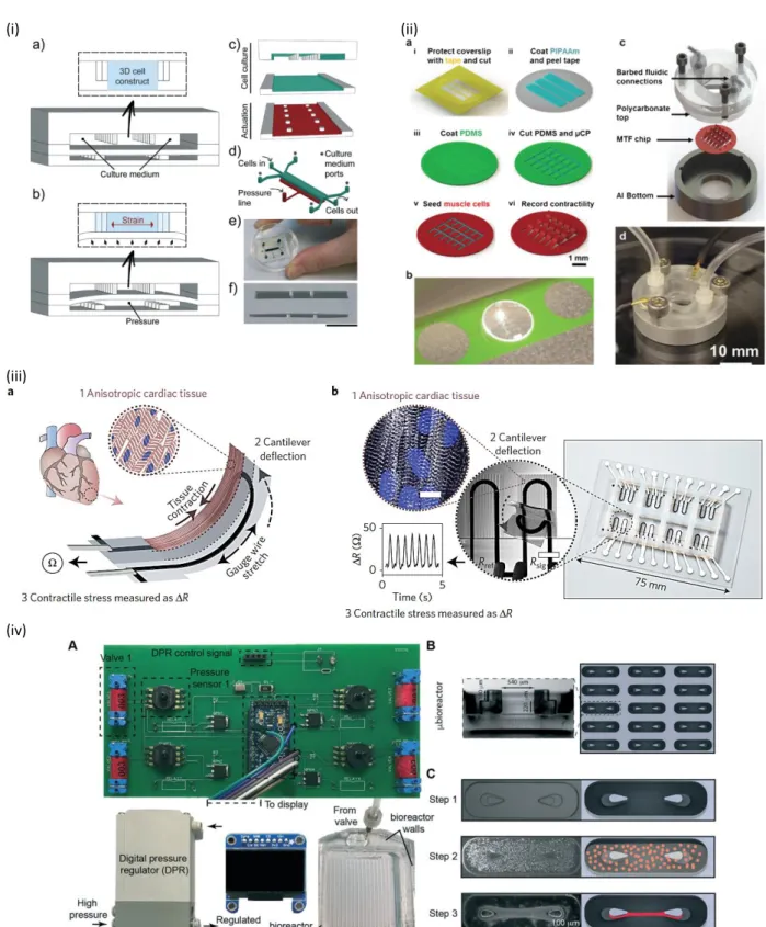
Marsano e al. presented a PDMS-based microfluidic device which mimics the in vivo mechanical stimulation of myocardium and applied to the 3D culture of neonatal and hiPSC-CMs [159]. The device contains an array of hanging posts to confine cell-laden fibrin gels, and a pneumatic actuation system to induce homogeneous uniaxial cyclic strains to the 3D cell constructs during culture (figure 7(i)). The mechanically stimulated microtissues showed early spontaneous synchronous beating and better contractile capability in response to electric pacing. A custom made electrical stimulator was utilized via connecting carbon rod electrodes to the constructs. The microsystem was further evaluated by testing different concentrations of isoproterenol, a commonly used drug for bradycardia, and the results indicated that it can be a promising tool for drug testing in the future.
Giridharan et al. developed a microfluidic cardiac cell culture model that replicates the physiological in vivo biomechanical cues by mimicking the hemodynamic stresses during pressure−volume changes in the left
13
ventricle [160]. The system consisted of a cell culture chamber sandwiched between two rigid plates and integrated within a flow loop. The cells were cultured on a thin PDMS membrane placed at the bottom of the culture chamber, and the device was fabricated in such a way to permit the membrane to deflect and stretch. Both membrane and chamber were fabricated by soft lithography. This microfluidic system has shown that improved the structural and functional maturation of chick embryonic ventricular CMs in comparison to static cultures. It was also demonstrated the importance of mechanical stimulation on maintaining intracellular calcium regulation and enhancing protein synthesis of embryonic chick CMs during embryogenesis [161]. The same group further improved the system by creating a more physiological environment by depositing matrigel/collagen on the chamber, and their results indicated that gradual mechanical loading in a nutrient-rich environment is required for the survival of hiPSC-CMs under physiologically relevant hemodynamic stress [162].
A 3D cardiac μtissue within a microfluidic device based on a micropatterning technology was presented by Aung et al. for the real-time contractile stress assessment [163]. Neonatal mouse CMs were encapsulated within a GelMA hydrogel, which was sandwiched between two polyacrylamide gels, where the latter worked as “stress sensors” for acquiring the contractile stresses that generated by the CMs contraction. This trilayer gel was surrounded by microfluidic channels made by soft lithography, thus creating perfused cardiac cell-laden structures. Drug test using epinephrine, an adrenergic neurotransmitter known to increase the magnitude and frequency of cardiac contractions, demonstrated that the cardiac tissue construct exhibited an increased beating frequency and stress magnitude. Nevertheless, further improvement of the system is required in order to achieve better structural properties of the encapsulated CMs.
Hansen et al. presented one of the first engineered heart tissues (EHT) constructed on PDMS post racks [164]. They simply added a mixture of neonatal rat heart unpurified cells, fibrinogen/Matrigel and thrombin into rectangular casting molds in which two PDMS posts were positioned from above. This method enabled the creation of EHT in a strip format that generates force under auxotonic stretch conditions. This technique enabled the automatic evaluation of the contractility and the evaluation of proarrhythmic (quinidine and erythromycin) and cardiotoxic (doxorubicin) effects of drugs. This EHT was further improved and tested with hiPSC-CMs and commercialized by the company EHT Technologies GmbH [165] [166].
Parker group pioneered in developing the technology of muscular thin films (MTF) which consist of 2D bilaminate constructs with an anisotropic tissue composed of muscle cells cultured on a deformable PDMS thin film coated (or microcontact printed) with fibronectin [167] [168]. The deformation of the MTF was associated to the contractility of the tissue, while its extent was depended on film geometry and tissue organization. They further advanced the throughput of the MTF technology and integrated it in a fluidic microdevice with a heated metallic base for maintaining physiological temperatures, transparent top for optically recording MTF deformation and embedded electrodes for electrical field stimulation of the tissue (figure 7(ii)) [169]. This platform was used to test the effect of the β-adrenergic receptor agonist isoproterenol on the contractility of neonatal rat ventricular CMs. Moreover, Wang et al. used the technology of MTF for modeling the mitochondrial cardiomyopathy of Barth syndrome, a mitochondrial disorder caused by mutation of the gene encoding tafazzin (TAZ) [170]. Thin PDMS films with patterned lines of fibronectin led to self-organized into anisotropic myocardial tissues upon culture of iPSC-CMs onto them. By using Barth Syndrome iPSC-CMs, they manage to elucidate the metabolic, structural and functional abnormalities associated with TAZ mutation, providing new insights into this cardiac disease pathogenesis. The same group recently demonstrated a device made by direct ink writing multimaterial 3D printing which was utilized for the continuous monitoring of cardiac micro-tissue contractile stress (figure 7(iii)) [134]. The microphysiological device included multilayer cantilevers, readout electrical interconnects and independent wells. The cantilevers consisted of a base layer, an embedded strain sensor and a tissue-guiding layer which contained grooved microstructures. Neonatal rat ventricular myocytes and hiPSC-CMs culture and drug testing with the L-type calcium channel blocker verapamil and isoproterenol demonstrated the functionality of this approach even at long-term cultures and thicker microtissues, rendering it a really promising platform for drug screening research.
14
(i) (ii)
(iii)
(iv)
Figure 7. Characteristic mechanical stimulation heart-on-a-chip systems: (i) Design of the 3D heart-on-a-chip microdevice with a pneumatic actuation system for inducing uniaxial cyclic strains to the 3D cell constructs, reprinted with permission from [159]. (ii) Higher throughput heart on a chip and fluidic microdevice based on MTFs consisted of 2D bilaminate constructs with an anisotropic tissue composed of CMs cultured on a deformable PDMS thin film, reprinted with permission from [169]. (iii) Direct ink writing multimaterial 3D printing on a MTF device: a) sketch of the device principle, b) fully printed final device, reprinted with permission from [134]. (iv) Pneumatic microfluidic platform for modeling cardiac hypertrophy; (A) electronic circuitry to control 4 bioreactors independently, (B) Schematic of the bioreactor platform with an array of μbioreactors, (C) Schematic and images of the cardiac μtissue formation, reprinted with permission from [171].
15
The Vunjak-Novakovic group developed a pneumatic microfluidic platform serving as model of cardiac hyperthrophy induced by volume overload (figure 7(iv)) [171]. Soft lithography techniques were employed for the fabrication of μbioreactors arrays containing μpillars, which facilitated the quick formation of large numbers of structurally organized cardiac μtissues in a desired geometry as cells were induced to fuse into μtissues around the elastic μpillars. In this study a mixture of neonatal rat CMs and cardiac fibroblasts was encapsulated in collagen gel. By adding a control layer, pneumatic channels underneath the μwells were pressurized, deflecting the pillars and subjecting the tissues to mechanical stress. With this design each individual μwell was subjected to a specific predetermined strain and enabled to study the effects of mechanical stress on cardiac hypertrophy, by real-time on-chip analysis of the tissues phenotype. The reusability, high throughput studies (it accommodates 900 cardiac μtissues using a small number of cells) and non-contact mechanical stimulation are the main advantages of this device. The same group very recently reported that achieved advanced maturation of human cardiac tissue, upon using early-stage hiPSC-CMs in a platform containing PDMS pillars for the formation of cardiac tissues incorporated in fibrin gel [172]. As it is depicted in figure 7, in most of the works the concept for achieving mechanical stimulation is based on the mechanical deformation of elastomeric thin membranes or posts. Soft lithography is the main technique and, depending the approach, either PDMS was used as substrate for the formation of the heart tissue or as mechanical support for the formation of hydrogel-based tissues. The microfluidic systems were designed to achieve stretching of the fabricated tissues which not only mimics the native stress applied in the myocardium but also aided in the structural organization of the cardiomyocytes. These mechanical stimulation approaches are continuing to advance (see Table I) and the use of hiPSC-CMs have given a further boost to their successful applications in the modelling of cardiac diseases.
3.3 Cardiac models of combined co-culture on chip
Even though the vast majority of the heart-on-a-chip devices focused on mimicking the myocardium, studies on cardiovascular models as well as systems combining heart and other organ tissues have been reported the last years. Ellis et al. developed a microfluidic device to mimic the 3D human cardiac muscle and surrounding microvasculature by spatially controlled co-culture of CMs and endothelial cells, both derived from the same hiPSC line [173]. The microfluidic device was fabricated using PDMS bonded on glass and it consists of three channels separated by micro-posts (figure 8(i)). The CMs were encapsulated in a UV-crosslinkable GelMA hydrogel and seeded on the central channel in order to create a 3D cardiac muscle model, while the endothelial cells were cultured in the side channels for mimicking the microvasculature by providing nutrients and oxygen through media flow. Cell culture indicated that CMs were viable and functional within the device up to 7 days, and were integrated with the endothelial cells, rendering it a promising model for cardiovascular disease studies.
Menon et al. introduced an ECM patterning method based on the principle of capillary burst valve (CBV) for creating perfusable vascularized microchannels of various vessel geometries [174]. The ECM hydrogel loaded into the chamber stopped at the boundaries of a PDMS microchannel design due to the CBV principle, and formed a patterned cell culture channel with ECM sidewalls (figure 8(ii)). They studied the endothelial cell barrier permeability and neutrophil transendothelial migration using collagen-patterned endothelialized microchannels and they further developed a 3D endothelial–smooth muscle cell (EC–SMC) vascular model. Their results indicated the importance of perivascular cells in ECM remodeling and the efficacy of the method for the study of hemodynamics and cellular interactions in cardiovascular diseases.
16
(i) (iii)
(ii)
Figure 8. Cardiomyocyte-endothelial co-culture systems: (i) Schematic of a microfluidic Myocardium-on-a-Chip designed to support controlled co-culture of CMs and endothelial cells, reprinted with permission from [173]. (ii) Flexible 3D ECM patterning using microfluidics for modeling the endothelial–smooth muscle cell vascular system, reprinted with permission from [174]. (iii) Schematics of fabrication of an endothelialized myocardium by combining GelMa hydrogel technology and 3D bioprinting, reprinted with permission from [175].
Khademhosseini group combined the GelMA hydrogel technology with 3D bioprinting towards creating an endothelialized myocardium which was integrated in a microfluidic bioreactor [175]. Endothelial cells (HUVECs) were bioprinted within alginate-GelMA microfibrous hydrogel scaffolds of controlled anisotropy. After their migration to the peripheries of the microfibers, CMs were seeded into the interstitial space of the endothelialized scaffold (figure 8(iii)). In this study both neonatal rat and hiPSC-CMs were utilized. The incorporation of the scaffolds to a microfluidic device demonstrated the positive effect of perfusion at low flow rate in cells viability, where dose-dependent responses of both CMs and endothelial cells were observed when treated with doxorubicin. This technology seems promising, nevertheless it should be optimized for obtaining hollow endothelialized microfibers.
Maoz et al. demonstrated a microfluidic device with integrated MEAs and electrodes for transepithelial electrical resistance (TEER) measurements for the simultaneous measurements of cellular electrical activity and tissue barrier function, which are useful in understanding heart pharmacodynamics [176]. TEER measurements represent the ionic conductance of the paracellular junctions of a cellular monolayer, applied to all epithelial tissues including the endothelium [177]. The device included a MEA, first fluidic layer made of PDMS, followed by a PET membrane, top chip channel and the TEER electrodes. The channel-separating PET membrane was covered with a monolayer of HUVEC, where underneath hiPSC-CMs were cultured on the MEA surface (figure 9(i)). Proof-of-concept tests with the inflammatory stimulus tumor necrosis factor alpha (TNF-α) or isoproterenol indicated its efficacy in detecting simultaneously dynamic alterations of vascular permeability and cardiac function on chip.
17 (i)
(ii)
Figure 9. Cardiac-other tissues co-culture systems: (i) Integrated TEER–MEA microfluidic chip for the simultaneous recording of cellular electrical activity and tissue barrier function, reprinted with permission from [176]. (ii) Integrated Heart/Cancer on a Chip: left-fabrication scheme, right-microfluidic device, reprinted with permission from [178].
A soft lithography-based microfluidic device was fabricated with on-chip integration of pneumatic valves and a peristaltic micropump for establishing precision fluid flow by Kamei et al. in order to emulate the side effects of cancer drugs [178]. The casting mold of the perfusion layer was fabricated by a combination of the negative resist patterning and grayscale lithography using positive resist coating as shown in figure 9(ii). The system contained a set of cell culture chambers with a medium circulation system that mimics the blood circulatory system that interconnects various tissue cells. In the integrated heart/cancer on a chip device, human healthy heart cells (hCMs) and liver cancer cells (HepG2) were cultured in separate chambers. The designing of the device allowed the evaluation of the side effects of doxorubicin on heart cells caused by the production of toxic metabolites (doxorubicinol) by the HepG2 cells through their delivery to heart cells via the circulation loop.
3.4 Other cardiac-applied microfluidic systems
Another approach of drug screening is the use of clusters of hiPSC-CMs which are called cardiac bodies (CBs). Bergström et al. combined CBs in a microfluidic chip and tested the cardiotoxicity of doxorubicin, verapamil and quinidine on these 3D-clustered CMs [179]. The chip was fabricated with standard soft lithography techniques and designed in order to capture the CBs along a perfusion channel (figure 10(i)). By video imaging it was possible to automatically record the beating frequency for each captured CB and assess the effect of drugs on it in a label-free, non-invasive fashion.
18
Moreover, LoC systems have been reported for CMs sorting, taking advantage of microfluidics functionalities. For example, Li et al. achieved to purify CMs derived from hiPSCs, which is critical for avoiding teratomas in clinical applications [180]. The device consisted of integrated microcolumns and ridge-like flow derivation structures, designed such a way that cells were forced to cross these structures, resulting in high efficiency and selective retention of a chosen cell population on the functionalized surfaces (figure 10(ii)). Like that undifferentiated hiPSCs were efficiently captured in this PDMS-based microfluidic device, while the recovered CMs viability was not affected.
(i)
(ii) (iii)
Figure 10. Microfluidic devices for cardiac cells capture/separation: (i) capture of hiPSC-CMs clusters on chip which was used for drug testing, reprinted with permission from [179]. (ii) hiPSC-CMs purification on chip via capturing the undifferentiated cells, reprinted from [180]. (iii) CMs sorting from rest cardiac cells of a neonatal rat heart based on deterministic lateral displacement, reprinted with permission from [181].
Zhang et al. developed a PDMS-based device for the separation of functional CMs from the rest cell populations that exist in the cardiac tissue isolated from neonatal rat hearts [181]. The separation was based on size using the principles of deterministic lateral displacement (figure 10(iii)) [182]. The functionality of the enriched CMs after sorting was demonstrated upon culture on commercial 3D collagen porous scaffolds, where they formed contractile cardiac patches.
19 Table I. Characteristics of “Heart on a Chip” systems.
Cells type Cells direct
substrate/matrix
Microfluidic building block materials
Stimulation In vivo disease
model Tested drugs
Publication
year Ref.
Electrical Mechanical
adult rat CMs glass slide silicone chamber (no
details) glued on glass 2007 [153]
adult rabbit CMs glass slide PDMS bonded on glass ischaemia 2010 [154] neonatal rat CMs, hASCs collagen PDMS bonded on glass-MEA 2010 [155] neonatal rat CMs, fibroblasts, rMSCs-bm fibronectin PDMS bonded on glass-MEA 2012 [156]
hiPSC-CMs fibronectin polystyrene chamber
glued on glass-MEA norepinephrine 2017 [157] hiPSC-CMs micromolded gelatin PEEK well on commercial MEA isoproterenol, terfenadine-fexofenadine 2016 [158] neonatal rat CMs,
hiPSC-CMs fibrin gel PDMS isoproterenol 2016 [159]
chick embryonic CMs collagen PDMS 2013; 2015 [160]; [161] hiPSC-CMs matrigel-collagen PDMS post myocardial
infarction 2016 [162]
neonatal mouse
CMs GelMa PDMS epinephrine 2016 [163]
neonatal rat heart unpurified cells; hiPSC-CMs fibrin-matrigel PDMS quinidine, erythromycin, doxorubicin; rolipram, isoproterenol, verapamil 2010; 2016 [164]; [165] neonatal rat CMs, hiPSC-CMs fibronectin Polycardonate top, Al bottom isoproterenol 2013 [169] neonatal rat CMs, hiPSC-CMs
ABS or PLA printed wells and covers, PDMS gasket verapamil, isoproterenol 2017 [134] neonatal rat CMs & cardiac fibroblasts
collagen gel, fibrin
gel PDMS cardiac hypertrophy isoproterenol, angiotensin II, endothelin-1 2017; 2018 [171]; [172] hiPSC-CMs & endothelial cells GelMA hydrogel,
fibronectin PDMS bonded on glass 2017 [173]
HUVECs & SMC collagen, fibrin,
matrigel PDMS vascular inflammation, atherosclerosis 2017 [174] neonatal rat, hiPSC-CMs & HUVECs
alginate-GelMA PMMA supports,
PDMS gaskets doxorubicin 2016 [175] hiPSC-CMs & HUVECs fbronectin PDMS bonded on glass-MEA vascular inflammation isoproterenol 2017 [176] hCMs, HepG2 fibronectin, gelatin
or matrigel PDMS bonded on glass
doxorubicinol-cardiac toxicity doxorubicin 2017 [178] CBs of hiPSC-CMs - PDMS bonded on glass
doxorubicin, verapamil, quinidine
20
Concluding, there is a high diversity of “Heart on a Chip” systems that have been developed over the last 10 years. Table I summarizes their main characteristics; type of cells used and their direct seeding substrate/scaffold, materials used for the microfluidic block, stimulation applied, disease model and tested drugs. The year of publication gives an overview of the evolution of the approaches used over the last decade. It must be noted that in the table I we included the material that the cells were seeded on (or encapsulated) as separate column from the microfluidic materials, as we wanted to correlate it with the type of materials reported in the section 2. Nevertheless, in the vast majority of the published studies the microfluidic chips were fabricated by standard soft lithography indicating its efficacy in lab-scale applications.
Conclusions
An overview of the micro and nanofabrication and microfluidics approaches that have utilized for developing systems for cardiac tissue engineering has been reported in this review. Firstly, the main fabrication methods applied in cardiac tissue engineering are presented and compared. Advantages and disadvantages of their use as fabrication methods are described, as well as great advancements and typical approaches used. Each method provides for specific functionalities to be achieved. Optical lithography, soft lithography and plasmas have provided tools for structural and chemical modification in planar two dimensional fashion. Lasers, electrospinning and 3D bioprinting have provided the capability to advance the fabrication even further, by providing the tools to fabricate structures into three dimensions. In several cases the combination of these techniques is employed, where additional functionalities or capabilities are needed from the construct. These approaches focused on culturing cardiac cells in a biomimetic environment in order to create a functional cardiac tissue. The main objective was to recapitulate the ECM that support cardiac cells and for this reason the substrates or scaffolds are based on polymeric materials. Depending on the capabilities of method, cardiac cells were seeded on top of micro-nanoengineered materials in 2D or 3D structures or encapsulated within the polymeric matrix. The 2D structures have provided with useful information on the effect of geometries on cardiac cells structural properties. The 3D scaffolds have been applied, although in a limited number, for cardiac cell therapy by means of implantation in animal models carrying a specific cardiac disease. The idea of this therapeutic direction is on restoring some functionality in heart scarred regions by providing a new pool of functional cardiac cells. This application falls under the category of cardiac regenerative medicine with final target the clinical trials by implanting the cellularized scaffolds to patients. The other main category of applications comprises the personalized therapy and drug discovery through the development of in vitro systems that mimic the natural heart microenvironment. The idea is to build a system with cardiac cells derived from patients and then study the cardiac pathophysiology and test drugs in vitro. Advancements in iPSCs engineering and DNA editing have enabled the feasibility of this strategy, which can lead to the achievement of individual patient-targeted therapy. In addition, the in vitro “Heart on a Chip” platforms can provide useful information for effective drug designing through cardiac toxicity studies, avoiding for example drugs for cancer therapy that cause heart dysfunction. Therefore, depending on the application, different “Heart on a Chip” systems have been developed. For example, systems providing electrical stimulation were reported for electrophysiology studies, while microfluidic chips designed to apply mechanical stress were utilized for contractility and structural assessment studies. Moreover, LoCs incorporating both electrical and mechanical stimulation to cardiac cells have been reported, thus providing a higher degree of integration towards the development of more realistic “Heart on a Chip” systems. Thereafter, taking advantage of the capabilities that microfluidics offer, systems for mimicking specific diseases such as pathological cardiac hypertrophy and ischaemia have started to be developed. Actually, some of the described micro/nanofabrication technologies have been incorporated to the microfluidic systems, such as microcontact printed molded structures, as well as bio-printed 3D scaffolds, even though these cases pose the minority. We envisage that the prior knowledge and the correct translation of the information extracted from the use of micro/nanoengineering in cardiac tissue engineering can lead to the development of multi-diverse “Heart on Chip” systems tackling a wide spectrum of cardiac diseases.

![Figure 4. 3D printed cardiac models for surgical practice or training. Reprinted with permission from [117]](https://thumb-eu.123doks.com/thumbv2/123doknet/14462569.520768/9.892.108.789.421.621/figure-printed-cardiac-surgical-practice-training-reprinted-permission.webp)
![Figure 5. Schematic representations of the four most widely used techniques of 3D printing for cardiac tissue engineering (reprinted and modified from [121])](https://thumb-eu.123doks.com/thumbv2/123doknet/14462569.520768/10.892.99.796.98.572/figure-schematic-representations-techniques-printing-engineering-reprinted-modified.webp)
![Figure 6. Main electrical stimulation heart-on-chip systems: (i) Dielectrophoresis and electro-orientation on chip, reprinted with permission from [153]](https://thumb-eu.123doks.com/thumbv2/123doknet/14462569.520768/13.892.126.768.282.675/figure-electrical-stimulation-systems-dielectrophoresis-orientation-reprinted-permission.webp)
![Figure 8. Cardiomyocyte-endothelial co-culture systems: (i) Schematic of a microfluidic Myocardium-on-a-Chip designed to support controlled co-culture of CMs and endothelial cells, reprinted with permission from [173]](https://thumb-eu.123doks.com/thumbv2/123doknet/14462569.520768/17.892.85.784.96.532/cardiomyocyte-endothelial-schematic-microfluidic-myocardium-controlled-endothelial-permission.webp)
![Figure 9. Cardiac-other tissues co-culture systems: (i) Integrated TEER–MEA microfluidic chip for the simultaneous recording of cellular electrical activity and tissue barrier function, reprinted with permission from [176]](https://thumb-eu.123doks.com/thumbv2/123doknet/14462569.520768/18.892.90.805.107.627/cardiac-integrated-microfluidic-simultaneous-recording-electrical-reprinted-permission.webp)
![Figure 10. Microfluidic devices for cardiac cells capture/separation: (i) capture of hiPSC-CMs clusters on chip which was used for drug testing, reprinted with permission from [179]](https://thumb-eu.123doks.com/thumbv2/123doknet/14462569.520768/19.892.163.729.272.776/figure-microfluidic-devices-cardiac-separation-clusters-reprinted-permission.webp)
