SSIEM SYMPOSIUM 2007
AGAT, GAMT and SLC6A8 distribution
in the central nervous system, in relation to creatine
deficiency syndromes: A review
O. Braissant&H. Henry
Received: 30 November 2007 / Submitted in revised form: 1 February 2008 / Accepted: 5 February 2008 / Published online: 4 April 2008
#SSIEM and Springer 2008
Summary Creatine deficiency syndromes, either due to AGAT, GAMT or SLC6A8 deficiencies, lead to a complete absence, or a very strong decrease, of creatine within the brain, as measured by magnetic resonance spectroscopy. While the mammalian central nervous system (CNS) expresses AGAT, GAMT and SLC6A8, the lack of SLC6A8 in astrocytes around the blood–brain barrier limits the brain capacity to import creatine from the periphery, and suggests that the CNS has to rely mainly on endogenous creatine synthesis through AGAT and GAMT expression. This seems contradictory with SLC6A8 deficiency, which, despite AGAT and GAMT expression, also leads to creatine deficiency in the CNS. We present novel data showing that in cortical grey matter, AGAT and GAMT are expressed in a dissociated way: e.g. only a few cells co-express both genes. This suggests that to allow synthesis of creatine within the CNS, at least for a
significant part of it, guanidinoacetate must be trans-ported from AGAT- to GAMT-expressing cells, possibly through SLC6A8. This would explain the creatine deficiency observed in SLC6A8-deficient patients. By bringing together creatine deficiency syn-dromes, AGAT, GAMT and SLC6A8 distribution in CNS, as well as a synthetic view on creatine and guanidinoacetate levels in the brain, this review presents a comprehensive framework, including new hypotheses, on brain creatine metabolism and transport, both in normal conditions and in case of creatine deficiency. Abbreviations
AGAT L-arginine:glycine amidinotransferase BBB blood–brain barrier
CAT cationic amino acid transporter (system y+) CK creatine kinase
CNS central nervous system
Cr creatine
CSF cerebrospinal fluid GAA guanidinoacetate
GAMT guanidinoacetate methyltransferase MCEC microcapillary endothelial cell MRS magnetic resonance spectroscopy SLC6A8 creatine transporter
tCr total creatine (creatine + phosphocreatine)
Introduction
In mammals, creatine (Cr) is taken up from the diet, or can be synthesized endogenously by a two-step mech-anism involving (i) L-arginine:glycine amidinotransfer-ase (AGAT), which, from arginine and glycine as substrates, yields the intermediate guanidinoacetate
DOI 10.1007/s10545-008-0826-9
Communicating editor: Cornelis Jakobs Competing interests: None declared
References to electronic databases:L-Arginine:glycine
amidinotransferase (AGAT) deficiency: OMIM 602360. Guanidinoacetate N-methyltransferase (GAMT) deficiency: OMIM 601240. Creatine transporter (SLC6A8; Slc6a8) deficiency: OMIM 300352. AGAT: EC 2.1.4.1. GAMT: EC 2.1.1.2.
Presented at the Annual Symposium of the SSIEM, Hamburg, 4–7 September 2007.
O. Braissant (*)
:
H. HenryInborn Errors of Metabolism, Clinical Chemistry Laboratory, Centre Hospitalier Universitaire Vaudois and University of Lausanne,
CI 02/33, Avenue Pierre-Decker 2, CH-1011 Lausanne, Switzerland e-mail: [email protected]
(GAA), and (ii) guanidinoacetate methyltransferase (GAMT), which converts GAA to creatine. Creatine is distributed through the blood and is taken up by cells with high energy demands through a specific creatine transporter, SLC6A8, also abbreviated as CT1, CRT1, CRTR, CTR or CreaT (for a review, see Wyss and Kaddurah-Daouk 2000). With the discovery of crea-tine deficiency syndromes due to deficiency in AGAT, GAMT or SLC6A8 (Item et al 2001; Salomons et al
2001; Sto¨ckler et al 1994; for a review, see Sto¨ckler et al2007), the last 15 years have seen a boost in the creatine research field, particularly in the central nervous system (CNS). In this review, we aim at bringing together what is known on creatine deficiency syndromes with the latest research on AGAT, GAMT and SLC6A8 distribution within the brain, in order to delineate a comprehensive framework on creatine metabolism and transport in CNS, both in normal conditions and in case of creatine deficiency. New hypotheses will also be presented.
Functions of creatine within the brain
The creatine/phosphocreatine/creatine kinase (CK) system is essential for the buffering and transport of high-energy phosphates. In the CNS, creatine has been shown to be essential in growth cone migration as well as dendritic and axonal elongation, in Na+/K+-ATPase activity, neurotransmitter release, maintenance of mem-brane potential, Ca2+ homeostasis and the restoration of ion gradients (Wallimann et al 1992; Wyss and Kaddurah-Daouk 2000). Creatine was also recently hypothesized to act as a central neuromodulator, and particularly as co-transmitter on GABA postsynaptic receptors (Almeida et al 2006). Finally, creatine has been proposed to regulate appetite and weight by acting on specific hypothalamic nuclei (Galbraith et al2006).
Creatine deficiency syndromes
The CNS is the main organ affected in patients suffering from creatine deficiency syndromes caused by deficiency of AGAT, GAMT or SLC6A8 (Item et al 2001; Salomons et al 2001; Sto¨ckler et al 1994). These patients present neurological symptoms in infancy (Battini et al 2002; DeGrauw et al 2002; Schulze et al 1997). In particular, mental retardation and delays in speech acquisition can be observed (AGAT, GAMT and SLC6A8 deficiencies), as well as epilepsy (GAMT and SLC6A8 deficiencies), autism, automutilating behaviour, extrapyramidal syndrome
and hypotonia (GAMT deficiency) (for a review, see Sto¨ckler et al2007).
AGAT, GAMT and SLC6A8 present a wide pattern of expression in the mammalian brain, which has been demonstrated in rat (AGAT, GAMT and SLC6A8), mouse (GAMT and SLC6A8) and human (GAMT) (see below; and Braissant et al 2001a,2005; Galbraith et al 2006; Schmidt et al 2004; Tachikawa et al2004). This may, at least in part, contribute to the diverse phenotypic spectrum of neurological symptoms observed in AGAT-, GAMT- and SLC6A8-deficient patients (Anselm et al 2006; Battini et al 2006; Mercimek-Mahmutoglu et al 2006; Schulze 2003). The recently proposed roles of creatine as co-trans-mitter on GABA postsynaptic receptors (Almeida et al 2006) and as regulator of appetite and weight on specific hypothalamic nuclei (Galbraith et al 2006) might also contribute to this phenotypic diversity. Specific features of GAMT deficiency are probably due to the epileptogenic effect of the accumulated GAA (Schulze et al 2001), the activation of GABAA
receptors by GAA (Neu et al 2002) and its inhibitory effect on Na+/K+-ATPase and CK (Zugno et al2006). All three deficiencies are characterized by an absence, or a severe decrease, of creatine in the CNS, as measured by magnetic resonance spectroscopy (MRS) (Stromberger et al 2003; Sykut-Cegielska et al
2004). AGAT- and GAMT-deficient patients can be treated with oral creatine supplementation. Although very high doses of creatine are being used, the replenishment of cerebral creatine takes months and results only in the partial restoration of the cerebral creatine pool (Battini et al 2002; Ganesan et al 1997; Item et al 2001; Schulze et al 1998; Sto¨ckler et al
1996b). The pre-symptomatic treatment of
AGAT-and GAMT-deficient patients has been reported, AGAT-and appears to ameliorate the outcome for these patients (Battini et al 2006; Schulze et al 2006; Schulze and Battini 2007). For GAMT deficiency, lowering GAA by arginine-restricted diet with low-dose ornithine sup-plementation (Schulze et al 2001) or solely by high-dose supplementation of ornithine (Schulze et al2005) has been shown effective. Creatine supplementation of SLC6A8-deficient patients is inefficient in restoring cerebral creatine levels (Bizzi et al 2002; Cecil et al
2001; DeGrauw et al 2002; Po´o-Argu¨elles et al2006).
Expression of AGAT, GAMT and SLC6A8 within the CNS
It has long been thought that most, if not all, of the creatine necessary for the brain is of peripheral origin,
be it taken from the diet or synthesized endogenously through AGAT and GAMT activities in kidney, pancreas and liver (Wyss and Kaddurah-Daouk
2000). It has long been known, however, that the mammalian brain is able to synthesize creatine (Pisano et al1963; Van Pilsum et al1972), which is also true of primary cultures of brain cells and nerve cell lines (Braissant et al2002,2008; Cagnon and Braissant2007; Daly1985; Dringen et al1998). It has now been clearly established that both AGAT and GAMT are expressed within the brain, both during development and in adulthood (Braissant et al 2001b, 2005, 2007; Lee et al 1998; Nakashima et al 2005; Schmidt et al
2004; Tachikawa et al2004,2007). AGAT is expressed throughout the adult rat CNS, including the retina, and can be found in all the main types of brain cells, namely neurons, astrocytes and oligodendrocytes (Braissant et al 2001b; Nakashima et al 2005). In the structures regulating exchanges between periphery and CNS, as well as between brain parenchyma and cerebrospinal fluid (CSF), AGAT is expressed in microcapillary endothelial cells (MCEC) and the astrocytes contacting them at the blood–brain barrier (BBB), as well as in the choroid plexus and ependymal epithelia (Braissant et al 2001b). GAMT is also expressed throughout the main structures of the adult mammalian brain, as shown in rat, mouse and human; furthermore, GAMT is expressed by neurons, astro-cytes and oligodendroastro-cytes, with higher levels found in both glial cell types (Braissant et al2001b; Nakashima et al2005; Schmidt et al2004; Tachikawa et al2004). GAMT is not expressed in MCEC but is present in the astrocytes contacting them (at the BBB), as well as in the choroid plexus and ependymal epithelia (Braissant et al2001b; Tachikawa et al2004).
Organotypic rat cortical cultures, primary brain cell cultures—neuronal, glial or mixed—and neuroblasto-ma cell cultures have creatine uptake activity (Almeida et al 2006; Braissant et al 2002, 2008; Daly
1985; Mo¨ller and Hamprecht1989). In vivo, mouse and rat CNS are able to take up creatine from the blood against its concentration gradient, but this uptake of creatine through the BBB seems relatively inefficient (Ohtsuki et al 2002; Perasso et al 2003). SLC6A8 is expressed throughout the adult mammalian brain (Braissant et al 2001b; Galbraith et al 2006; Guimbal and Kilimann1993; Happe and Murrin1995; Saltarelli et al 1996; Schloss et al 1994). In rat and mouse, SLC6A8 is found in neurons and oligodendrocytes, but, in contrast to AGAT and GAMT, cannot be detected in astrocytes (Braissant et al2001b; Ohtsuki et al2002; Tachikawa et al2004). This holds true also for the retina, where SLC6A8 is expressed in retinal
neurons but not in astrocytes (Acosta et al 2005; Nakashima et al 2004). In contrast to the absence of SLC6A8 in astrocytes lining microcapillaries, MCEC which form the BBB and the blood–retina barrier do express SLC6A8 (Acosta et al 2005; Braissant et al
2001b; Nakashima et al 2004; Ohtsuki et al 2002;
Tachikawa et al2004) and are able to take up creatine (Ohtsuki et al2002). SLC6A8 is also expressed by the choroid plexus and the ependymal epithelia (Braissant et al2001b).
Creatine and guanidinoacetate within the normal versus creatine-deficient CNS
In normal conditions, creatine within human CSF is maintained in the 17–90 2mol/L range (Table 1 and references therein). By MRS, total creatine (tCr) is measured between 5.5 mmol/L and 6.4 mmol/L in the cortical grey matter, and between 4.8 mmol/L and 5.1 mmol/L in the cortical white matter (Table1). GAA is maintained in human CSF at a 1000-fold lower level than creatine, with a 0.015–0.1142mol/L range, while its levels in grey and white matter were estimated as 1.6 mmol/L and 0.9 mmol/L respectively (Table1).
With the exception of SLC6A8-deficient heterozy-gous females, where brain creatine deficiency appears partial (Cecil et al2003), all three creatine deficiencies present the virtual absence (or a very strong decrease) of the creatine peak measured by MRS in the cortical grey and white matter or in basal ganglia (Sto¨ckler et al 2007). However, despite the lack of detection or decrease under MRS measure, creatine remains pres-ent within the brain of creatine-deficipres-ent patipres-ents (Table1).
In SLC6A8 deficiency, creatine levels in CSF do not seem different from those in age-matched controls (Cecil et al 2001; DeGrauw et al2002; Salomons et al
2001) (Table 1). In AGAT deficiency, tCr levels in cortical grey and white matter are decreased to 12% and 10% respectively of age-matched controls (Battini et al2002) (Table1), which suggests that tCr levels in these regions are in the 500 2mol/L range. In GAMT deficiency, CSF levels of creatine are strongly decreased (<2 2mol/L) compared with controls (Ensenauer et al
2004; Schulze et al 1997, 2003), while in cortical grey and white matter, tCr levels were found to be in the 0.2–1.5 mmol/L and 0.3–1.9 mmol/L ranges, respectively (Mancini et al2005; Sto¨ckler et al1994) (Table1).
GAA accumulation in body fluids is characteristic of GAMT deficiency, and the CSF of GAMT-deficient patients presents levels of GAA 60–1000-fold higher than those in age-matched controls (Table 1), while
they are estimated to be 3.6 mmol/L and 3.4 mmol/L within cortical grey and white matter, respectively. No precise data are available on GAA levels within the AGAT- and SLC6A8-deficient CNS, but it was shown recently by MRS that GAA can also accumulate in the brain of SLC6A8-deficient patients (Sijens et al2005) (see also below).
In the rodent brain, creatine concentrations were 8.5 mmol/L (rats) and 8.2 mmol/L (mice) (Renema et al
2003), or 10–112mol/g of tissue (mice) (Schmidt et al
2004; Torremans et al2005) (Table2). In mice, GAA is maintained at a 1000-fold lower level than creatine within CNS (0.012 2mol/g of tissue). As expected, GAMTj/jknockout (KO) mice show decreased levels of creatine within their brain, which however still
reach 1.4 mmol/L or 0.4–0.5 2mol/g of tissue, and a very significant increase in GAA (1.9 2mol/g tissue; Table 2). As for GAMT-deficient patients, GAMTj/j KO mice slowly replenish their brain creatine upon creatine treatment (Kan et al 2007).
Synthesis or uptake of creatine by the brain?
The in vivo expression of AGAT and GAMT within the mammalian brain and the in vitro endogenous synthesis of creatine by various types of cultured brain cells suggest that the CNS synthesizes creatine (for a review, see Braissant et al 2007). However, it was thought for a long time that most, if not all, of the
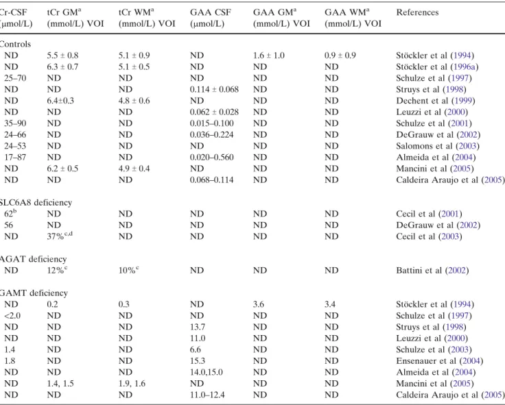
Table 1 Creatine and guanidinoacetate in the human brain of controls and SLC6A8, AGAT or GAMT deficient patients Cr-CSF (2mol/L) tCr GMa (mmol/L) VOI tCr WMa (mmol/L) VOI GAA CSF (2mol/L) GAA GMa (mmol/L) VOI GAA WMa (mmol/L) VOI References Controls ND 5.5T 0.8 5.1T 0.9 ND 1.6T 1.0 0.9T 0.9 Sto¨ckler et al (1994) ND 6.3T 0.7 5.1T 0.5 ND ND ND Sto¨ckler et al (1996a) 25–70 ND ND ND ND ND Schulze et al (1997) ND ND ND 0.114T 0.068 ND ND Struys et al (1998) ND 6.4T0.3 4.8T 0.6 ND ND ND Dechent et al (1999) ND ND ND 0.062T 0.028 ND ND Leuzzi et al (2000) 35–90 ND ND 0.015–0.100 ND ND Schulze et al (2001) 24–66 ND ND 0.036–0.224 ND ND DeGrauw et al (2002) 24–53 ND ND ND ND ND Salomons et al (2003) 17–87 ND ND 0.020–0.560 ND ND Almeida et al (2004) ND 6.2T 0.5 4.9T 0.4 ND ND ND Mancini et al (2005) ND ND ND 0.068–0.114 ND ND Caldeira Araujo et al (2005) SLC6A8 deficiency 62b ND ND ND ND ND Cecil et al (2001) 56 ND ND ND ND ND DeGrauw et al (2002) ND 37%c,d ND ND ND ND Cecil et al (2003) AGAT deficiency ND 12%c 10%c ND ND ND Battini et al (2002) GAMT deficiency ND 0.2 0.3 ND 3.6 3.4 Sto¨ckler et al (1994) <2.0 ND ND ND ND ND Schulze et al (1997) ND ND ND 13.7 ND ND Struys et al (1998) ND ND ND 11.0 ND ND Leuzzi et al (2000) 1.4 ND ND 6.6 ND ND Schulze et al (2003) 1.8 ND ND 15.3 ND ND Ensenauer et al (2004) ND ND ND 14.0,15.0 ND ND Almeida et al (2004) ND 1.4, 1.5 1.9, 1.6 ND ND ND Mancini et al (2005) ND ND ND 11.0–12.4 ND ND Caldeira Araujo et al (2005)
aCortical grey matter (GM) and white (WM) matter.
bWhile on Cr treatment.
c% as compared to age-matched controls.
d
creatine needed by the brain comes from the periphery through the BBB (for a review, see Wyss and Kaddurah-Daouk2000).
The discovery that SLC6A8 cannot be detected in astrocytes, particularly in their feet sheathing micro-capillaries at the BBB suggested, however, that in the mature brain the BBB has a limited permeability for creatine, despite the expression of SLC6A8 by MCEC and their capacity to import creatine (Acosta et al
2005; Braissant et al 2001b; Nakashima et al 2004; Ohtsuki et al 2002; Tachikawa et al 2004). This is further confirmed in vivo, both in rodents and humans. The blood-to-brain transport of creatine through the BBB has been demonstrated in rats and mice, but is relatively inefficient (Ohtsuki et al2002; Perasso et al
2003). Moreover, the long-term treatment of AGAT-and GAMT-deficient patients with high doses of creatine allows the replenishment of their brain creatine pools, but is very slow and only partial (Stromberger et al 2003; Sykut-Cegielska et al 2004). Similarly, GAMTj/jKO mice treated with high doses of creatine replenish their brain creatine, but only slowly (Kan et al2007). One possibility for the limited entry of creatine into the brain parenchyma, without going through astrocytes, could be the use of the limited surface of CNS capillary endothelium that is free of astrocytic endings (Ohtsuki 2004; Virgintino et al 1997). This would explain that the AGAT- or GAMT-deficient CNS, despite its very significant decrease in creatine, still presents measurable levels of creatine (Tables1 and2).
SLC6A8-deficient patients have normal levels of creatine in CSF (Table 1) but are unable to import creatine from the blood (Bizzi et al 2002; Cecil et al
2001; DeGrauw et al2002; Po´o-Argu¨elles et al2006). In contrast, GAMT-deficient patients have strongly decreased levels of creatine in CSF (Table1) but are able to import creatine from the blood (Schulze et al
1997; Sto¨ckler et al 1994). These observations are in favour of endogenous synthesis of creatine within CNS, which would still be operational, at least in some brain cells, under SLC6A8 deficiency while being
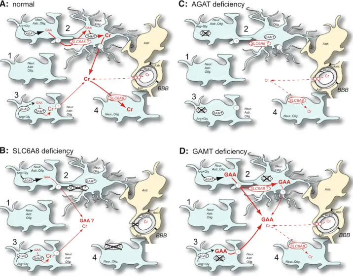
completely blocked in AGAT and GAMT deficiencies (see Fig.1).
Thus, under normal physiological conditions, the adult mammalian brain might depend more on its own creatine synthesis, through the expression of AGAT and GAMT, than on creatine supply from the blood (Braissant et al 2001b, 2007). The brain capacity for creatine synthesis would thus depend on the efficient supply of arginine, the limiting substrate for creatine synthesis, from blood to CNS, and then also on the local trafficking of arginine between brain cells. We and others have shown that the cationic amino acid transporters (CATs) might fulfil these roles in the adult rat brain, as CAT1 is expressed at the BBB as well as ubiquitously in neuronal and glial cells, as CAT2(B) is present in neurons and oligodendrocytes, and as CAT3 is restricted to neurons (Braissant et al
1999,2001a; Hosokawa et al 1999).
However, the hypothesis of endogenous creatine synthesis in the brain might seem contradictory with the in vivo characteristics of SLC6A8 deficiency, which, despite expression of AGAT and GAMT within CNS, shows an absence or a very low level of brain creatine by MRS, as in AGAT and GAMT deficiencies (Salomons et al 2003). This apparent contradiction might be explained by the AGAT, GAMT and SLC6A8 expression patterns in CNS: AGAT and GAMT can be found in every cell type of the brain (Braissant et al 2001b), while they rarely seem to be co-expressed within the same cell.
Dissociated expression of AGAT, GAMT and SLC6A8 within the brain
To elucidate this, we hypothesized that, within the different cell types of the brain, AGAT, GAMT and SLC6A8 might be expressed in a dissociated way, and that GAA, which is known to compete for creatine uptake through SLC6A8 (Ohtsuki et al2002; Saltarelli et al 1996), has to be transported from AGAT- to GAMT-expressing cells, possibly through SLC6A8, for
Table 2 Creatine and guanidinoacetate in the rodent brain, including in GAMTj/jKO mice
tCr brain (mmol/L) VOI Cr brain (2mol/g tissue) GAA brain (2mol/g tissue) References
Control rats 8.5 Renema et al (2003)
Control mice 8.2T 1.2 Renema et al (2003)
10.2T 1.1 0.012T 0.002 Schmidt et al (2004)
11.3T 0.4 0.012T 0.001 Torremans et al (2005)
GAMTj/jKO mice 1.4T 0.4 Renema et al (2003)
0.43T 0.09 1.87T 0.07 Schmidt et al (2004)
creatine to be synthesized within the CNS (Braissant et al2007). This could explain the absence of creatine synthesis in the brain of SLC6A8-deficient patients. Our aim was thus first to dissect the cell-to-cell (co-)expression of AGAT, GAMT and SLC6A8 within the adult rat brain.
To achieve this, in situ hybridization coupled to immunohistochemistry was applied to cryosections of rat brain (Braissant 2004), where the expression pattern of AGAT, GAMT and SLC6A8 was analysed within the grey matter of cortex. Specific RNA probes and polyclonal antibodies were used (Braissant et al
2001b,2005) to unravel, on adjacent sections, the three
different F2 by 2_ combinations of the three genes
(AGAT+GAMT; AGAT+SLC6A8; GAMT+SLC6A8). For each combination, in situ hybridization for gene 1 was coupled to immunohistochemistry for gene 2, followed on adjacent sections by in situ hybridization for gene 2 coupled to immunohistochemistry for gene 1. All combinations were repeated twice, allowing a total of four F2 by 2_ labellings of each combination of the three genes. With each combination, the proportions of cells were obtained with (i) no expression of either genes 1 or 2, (ii) expression of gene 1 only, (iii) expression of gene 2 only, or (iv) co-expression of genes 1 and 2, which finally allowed the calculation of the expression pattern of AGAT, GAMT and SLC6A8 takenF3 by 3_ (Table3). MCEC
1
3
Cr Cr GAA Arg+Gly GAA Arg+Gly GAA4
Cr Cr ? AGAT ? SLC6A8 GAMTA:
normal2
SLC6A8 ? ? BBB Astr. Astr. Astr. Neur. Olig. Neur. Astr.,Olig. Neur.,Olig. Neur. Astr. Olig. Neur. Astr. Olig.B:
SLC6A8 deficiency MCEC1
3
Cr GAA Arg+Gly Arg+Gly GAA4
Cr ? AGAT2
GAMT BBB ? SLC6A8 SLC6A8 ? Astr. Astr. Astr. Neur. Olig. Neur. Astr.,Olig. Neur.,Olig. Neur. Astr. Olig. Neur. Astr. Olig. AGAT GAMT AGAT GAMT Cr GAA ? CrC:
AGAT deficiency MCEC1
3
Cr Arg+Gly GAMT Arg+Gly4
? ? GAMT2
? BBB Cr Cr AGAT AGAT SLC6A8 SLC6A8 ? Astr. Astr. Astr. Neur. Olig. Neur. Astr.,Olig. Neur.,Olig. Neur. Astr. Olig. Neur. Astr. Olig. GAAD:
GAMT deficiency MCEC1
3
Cr Arg+Gly AGAT GAMT Arg+Gly4
? AGAT2
GAMT BBB GAA Cr Cr GAA SLC6A8 GAA SLC6A8 ? ? ? Astr. Astr. Astr. Neur. Olig. Neur. Astr.,Olig. Neur.,Olig. Neur. Astr. Olig. Neur. Astr. Olig.Fig. 1 A proposed model for creatine synthesis and transport within the central nervous system. (A) normal conditions. A high
proportion of cells do not express AGAT, GAMT and SLC6A8 (1). Endogenous synthesis of creatine within CNS can be achieved between AGAT- and GAMT-expressing cells and the concomitant trafficking of GAA between them (2), or in cells co-expressing AGAT + GAMT (3). A low proportion of brain cells express only SLC6A8 (4; i.e. creatine users only). (B) Creatine transporter
(SLC6A8) deficiency. (C)L-arginine:glycine amidinotransferase (AGAT) deficiency. (D) Guanidinoacetate methyltransferase (GAMT)
deficiency. Other abbreviations: Arg, arginine; Astr., astrocytes; BBB, blood–brain barrier; Cr, creatine; GAA, guanidinoacetate; Gly, glycine; MCEC, microcapillary endothelial cells; Neur., neurons; Olig., oligodendrocytes
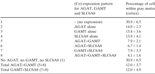
These experiments revealed that within grey matter of the rat cortex, significant proportions of cells do no express either AGAT, GAMT or SLC6A8 (30.9%), or express AGAT only (14.8%), GAMT only (13.4%) or SLC6A8 only (13.9%). Cortical cells co-expressing AGAT+GAMT but not SLC6A8 were 7.9%, those expressing AGAT+SLC6A8 but not GAMT were 6.7%, and those expressing GAMT+SLC6A8 but not AGAT were 7.9%. Finally, cells co-expressing AGAT+GAMT+SLC6A8 were 4.1%.
Altogether, we show that in the rat cortex, a low proportion of cells (12%) appear capable of their own creatine synthesis (i.e. co-expressing AGAT+GAMT), in agreement with the creatine deficiency observed by MRS in SLC6A8-deficient patients. Cells co-expressing GAMT+SLC6A8, and thus equipped for creatine syn-thesis if GAA is taken up by SLC6A8, were also 12%.
Future work will aim at deciphering whether the proportions in the cortical expression pattern of AGAT, GAMT and SLC6A8 are respected within the other regions of the brain, or whether differential expression patterns for AGAT, GAMT and SLC6A8 occur between these structures.
Models and hypotheses to understand creatine synthesis and transport within the brain
Taking together, (i) the expression pattern of AGAT, GAMT and SLC6A8 within CNS, (ii) the absence (or strong decrease) of creatine within CNS of creatine-deficient patients, (iii) the low permeability of the BBB for creatine, and (iv) the creatine and GAA concentrations within the brain, both in normal and in creatine-deficient conditions, leads us to propose the following model to understand creatine synthesis and trafficking within CNS (Fig.1).
In normal conditions (Fig. 1A), SLC6A8 is expressed by CEMC, but not by the feet of surround-ing astrocytes, implysurround-ing that very limited amounts of creatine can enter the brain through the BBB, possibly through the limited surface of CNS capillary endothe-lium that is free of astrocytic endings (Ohtsuki 2004; Virgintino et al1997). Within the cortical grey matter, the high proportion of cells without expression of AGAT, GAMT and SLC6A8 and the low proportion of cells expressing only SLC6A8 suggest that brain cells express AGAT, GAMT and SLC6A8 on demand to adapt their creatine needs in timely manner. Cells equipped with the full creatine synthesis pathway (i.e. co-expressing AGAT and GAMT) are only 12%. Finally, the dissociated expression of AGAT and GAMT among different cells suggests that to allow synthesis of creatine within CNS, at least for a significant part of it, GAA must be transported from AGAT- to GAMT-expressing cells, possibly through SLC6A8 as 12% of cortical cells co-express SLC6A8 and GAMT.
Creatine supplementation of SLC6A8-deficient patients (Fig. 1B) does not restore creatine levels in their brain, as MCEC of these patients lack functional SLC6A8. Moreover, if SLC6A8 also allows GAA uptake, SLC6A8-deficient patients should lack the creatine synthesis pathway from AGAT-expressing to GAMT+SLC6A8 co-expressing cells. This would ex-plain why SLC6A8-deficient patients lack (or present a significant decrease in) creatine in CNS as measured by MRS, having only a small proportion of their brain cells equipped to self-synthesize creatine.
In AGAT deficiency (Fig. 1C), no creatine can be synthesized within the brain, but the expression of SLC6A8 in MCEC allows the very limited entry of creatine within the CNS. Because of the SLC6A8 expression in MCEC, the brain of AGAT-deficient
(Co)-expression pattern for AGAT, GAMT and SLC6A8
Percentage of cells within gray matter (cortex) 1 – (no expression) 30.9T 6.5 2 AGAT alone 14.8T 2.3 3 GAMT alone 13.4T 3.6 4 SLC6A8 alone 13.9T 4.1 5 AGAT+GAMT 7.9T 2.1 6 AGAT+SLC6A8 6.7T 1.4 7 GAMT+SLC6A8 7.9T 3.3 8 AGAT+GAMT+SLC6A8 4.1T 1.6
No AGAT, no GAMT, no SLC6A8 (1) 30.9T 6.5
Total AGAT+GAMT (5+8) 12.0T 3.7
Total GAMT+SLC6A8 (7+8) 12.0T 4.9
Table 3 Dissociated expression of AGAT, GAMT and SLC6A8 in the telencephalic cortex of the rat (grey matter). The proportions (%) of cells with the respective (co-)expression patterns for AGAT, GAMT and SLC6A8 are indicated.
patients can be replenished in creatine by oral creatine treatment.
Finally in GAMT deficiency (Fig.1D), no creatine can be synthesized within the brain, and GAA accu-mulates. As for AGAT deficiency, the expression of SLC6A8 in MCEC allows the very limited entry of creatine within CNS, as well as the replenishment of the GAMT-deficient CNS through oral creatine treatment. To clarify these models and hypotheses, important questions remain to be solved. Future research in the field of brain creatine should aim at analysing the capacity of brain cells to take up GAA and, if this is shown, to demonstrate whether this uptake occurs through SLC6A8. Another important point is to identify how creatine (and GAA) can leave the cells, and whether SLC6A8 or another mechanism is in-volved. Finally, does the brain of SLC6A8-deficient patients accumulate GAA as suggested in our model? So far, data are poor on the GAA level in the brain of SLC6A8-deficient patients. However, a recent work does indeed demonstrate that GAA does not accumu-late in the CNS only in the case of GAMT deficiency, but can also be augmented in the brain of SLC6A8-deficient patients (Sijens et al 2005). The fact that SLC6A8-deficient patients can also develop epilepsy (Hahn et al2002; Mancardi et al 2007; Po´o-Argu¨elles et al2006) is also suggestive of GAA accumulation in the SLC6A8-deficient CNS.
Acknowledgements Our work is supported by the Swiss
National Science Foundation, grants nos 3100A0-100778 and 3100A0-116859. Theo Wallimann is acknowledged for the anti-AGAT antibody, and Marc Loup for excellent technical work.
References
Acosta ML, Kalloniatis M, Christie DL (2005) Creatine trans-porter localization in developing and adult retina: impor-tance of creatine to retinal function. Am J Physiol Cell Physiol 289: C1015–C1023.
Almeida LS, Verhoeven NM, Roos B, et al (2004) Creatine and guanidinoacetate: diagnostic markers for inborn errors in creatine biosynthesis and transport. Mol Genet Metab 82: 214–219.
Almeida LS, Salomons GS, Hogenboom F, Jakobs C, Schoffelmeer AN (2006) Exocytotic release of creatine in rat brain. Synapse 60: 118–123.
Anselm IM, Alkuraya FS, Salomons GS, et al (2006) X-linked creatine transporter defect: a report on two unrelated boys with a severe clinical phenotype. J Inherit Metab Dis 29: 214–219.
Battini R, Leuzzi V, Carducci C, et al (2002) Creatine depletion in a new case with AGAT deficiency: clinical and genetic study in a large pedigree. Mol Genet Metab 77: 326–331. Battini R, Alessandri MG, Leuzzi V, et al (2006)
Arginine:gly-cine amidinotransferase (AGAT) deficiency in a newborn:
early treatment can prevent phenotypic expression of the disease. J Pediatr 148: 828–830.
Bizzi A, Bugiani M, Salomons GS, et al (2002) X-linked creatine deficiency syndrome: a novel mutation in creatine trans-porter gene SLC6A8. Ann Neurol 52: 227–231.
Braissant O (2004) Measurement of nitric oxide-related enzymes in the brain by in situ hybridization. Methods Mol Biol 279: 113–124.
Braissant O, Gotoh T, Loup M, Mori M, Bachmann C (1999)
L-arginine uptake, the citrulline-NO cycle and arginase II
in the rat brain: an in situ hybridization study. Mol Brain Res 70: 231–241.
Braissant O, Gotoh T, Loup M, Mori M, Bachmann C (2001a) Differential expression of the cationic amino acid transporter 2(B) in the adult rat brain. Mol Brain Res 91: 189–195. Braissant O, Henry H, Loup M, Eilers B, Bachmann C (2001b)
Endogenous synthesis and transport of creatine in the rat brain: an in situ hybridization study. Mol Brain Res 86: 193–201. Braissant O, Henry H, Villard AM, et al (2002)
Ammonium-induced impairment of axonal growth is prevented through glial creatine. J Neurosci 22: 9810–9820.
Braissant O, Henry H, Villard AM, Speer O, Wallimann T, Bachmann C (2005) Creatine synthesis and transport during rat embryogenesis: Spatiotemporal expression of AGAT, GAMT and CT1. BMC Dev Biol 5: 9.
Braissant O, Bachmann C, Henry H (2007) Expression and function of AGAT, GAMT and CT1 in the mammalian brain. Subcell Biochem 46: 67–81.
Braissant O, Cagnon L, Monnet-Tschudi F, et al (2008). Ammonium alters creatine transport and synthesis in a 3D-culture of developing brain cells, resulting in secondary
cerebral creatine deficiency. Eur J Neurosci doi:10.1111/
j.1460-9568.2008.06126.x.
Cagnon L, Braissant O (2007) Hyperammonemia-induced tox-icity for the developing central nervous system. Brain Res Rev 56: 183–197.
Caldeira Araujo H, Smit W, et al (2005) Guanidinoacetate methyltransferase deficiency identified in adults and a child with mental retardation. Am J Med Genet A 133: 122–127. Cecil KM, Salomons GS, Ball WS Jr, et al (2001) Irreversible brain
creatine deficiency with elevated serum and urine creatine: a creatine transporter defect? Ann Neurol 49: 401–404. Cecil KM, DeGrauw TJ, Salomons GS, Jakobs C, Egelhoff JC,
Clark JF (2003) Magnetic resonance spectroscopy in a 9-day-old heterozygous female child with creatine transporter deficiency. J Comput Assist Tomogr 27: 44–47.
Daly MM (1985) Guanidinoacetate methyltransferase activity in tissues and cultured cells. Arch Biochem Biophys 236: 576–584. Dechent P, Pouwels PJ, Wilken B, Hanefeld F, Frahm J (1999) In-crease of total creatine in human brain after oral supplementa-tion of creatine-monohydrate. Am J Physiol 277: R698–R704. DeGrauw TJ, Salomons GS, Cecil KM, et al (2002) Congenital creatine transporter deficiency. Neuropediatrics 33: 232–238. Dringen R, Verleysdonk S, Hamprecht B, Willker W, Leibfritz D, Brand A (1998) Metabolism of glycine in primary astroglial cells: synthesis of creatine, serine, and glutathione. J Neurochem 70: 835–840.
Ensenauer R, Thiel T, Schwab KO, et al (2004) Guanidinoacetate methyltransferase deficiency: differences of creatine uptake in human brain and muscle. Mol Genet Metab 82: 208–213. Galbraith RA, Furukawa M, Li M (2006) Possible role of
creatine concentrations in the brain in regulating appetite and weight. Brain Res 1101: 85–91.
Ganesan V, Johnson A, Connelly A, Eckhardt S, Surtees RA (1997) Guanidinoacetate methyltransferase deficiency: new clinical features. Pediatr Neurol 17: 155–157.
Guimbal C, Kilimann MW (1993) A Na(+)-dependent creatine transporter in rabbit brain, muscle, heart, and kidney. cDNA cloning and functional expression. J Biol Chem 268: 8418–8421.
Hahn KA, Salomons GS, Tackels-Horne D, et al (2002) X-linked mental retardation with seizures and carrier manifestations is caused by a mutation in the creatine-transporter gene (SLC6A8) located in Xq28. Am J Hum Genet 70: 1349–1356. Happe HK, Murrin LC (1995) In situ hybridization analysis of CHOT1, a creatine transporter, in the rat central nervous system. J Comp Neurol 351: 94–103.
Hosokawa H, Ninomiya H, Sawamura T, et al (1999) Neuron-specific expression of cationic amino acid transporter 3 in the adult rat brain. Brain Res 838: 158–165.
Item CB, Sto¨ckler-Ipsiroglu S, Stromberger C, et al (2001) Arginine:glycine amidinotransferase deficiency: the third inborn error of creatine metabolism in humans. Am J Hum Genet 69: 1127–1133.
Kan HE, Meeuwissen E, van Asten JJ, Veltien A, Isbrandt D, Heerschap A (2007) Creatine uptake in brain and skeletal muscle of mice lacking guanidinoacetate methyltransferase assessed by magnetic resonance spectroscopy. J Appl Physiol 102: 2121–2127.
Lee H, Kim JH, Chae YJ, Ogawa H, Lee MH, Gerton GL (1998) Creatine synthesis and transport systems in the male rat reproductive tract. Biol Reprod 58: 1437–1444.
Leuzzi V, Bianchi MC, Tosetti M, et al (2000) Brain creatine depletion: guanidinoacetate methyltransferase deficiency (improving with creatine supplementation). Neurology 55: 1407–1409.
Mancardi MM, Caruso U, Schiaffino MC, et al (2007) Severe epilepsy in X-linked creatine transporter defect (CRTR-D). Epilepsia 48: 1211–1213.
Mancini GM, Catsman-Berrevoets CE, de Coo IF, et al (2005) Two novel mutations in SLC6A8 cause creatine transporter defect and distinctive X-linked mental retardation in two unrelated Dutch families. Am J Med Genet A 132: 288–295. Mercimek-Mahmutoglu S, Stoeckler-Ipsiroglu S, Adami A, et al (2006) GAMT deficiency: features, treatment, and outcome in an inborn error of creatine synthesis. Neurology 67: 480– 484.
Mo¨ller A, Hamprecht B (1989) Creatine transport in cultured cells of rat and mouse brain. J Neurochem 52: 544–550. Nakashima T, Tomi M, Katayama K, et al (2004)
Blood-to-retina transport of creatine via creatine transporter (CRT) at the rat inner blood-retinal barrier. J Neurochem 89: 1454–1461. Nakashima T, Tomi M, Tachikawa M, Watanabe M, Terasaki T, Hosoya K (2005) Evidence for creatine biosynthesis in Mu¨ller glia. GLIA 52: 47–52.
Neu A, Neuhoff H, Trube G, et al (2002) Activation of GABA(A) receptors by guanidinoacetate: a novel patho-physiological mechanism. Neurobiol Dis 11: 298–307. Ohtsuki S (2004) New aspects of the blood–brain barrier
transporters; its physiological roles in the central nervous system. Biol Pharm Bull 27: 1489–1496.
Ohtsuki S, Tachikawa M, Takanaga H, et al (2002) The blood-brain barrier creatine transporter is a major pathway for supplying creatine to the brain. J Cereb Blood Flow Metab 22: 1327–1335.
Perasso L, Cupello A, Lunardi GL, Principato C, Gandolfo C, Balestrino M (2003) Kinetics of creatine in blood and brain after intraperitoneal injection in the rat. Brain Res 974: 37–42.
Pisano JJ, Abraham D, Udenfriend S (1963) Biosynthesis and
disposition of+-guanidinobutyric acid in mammalian tissues.
Arch Biochem Biophys 100: 323–329.
Po´o-Argu¨elles P, Arias A, Vilaseca MA, et al (2006) X-Linked creatine transporter deficiency in two patients with severe mental retardation and autism. J Inherit Metab Dis 29: 220–223.
Renema WK, Schmidt A, van Asten JJ, et al (2003) MR spectroscopy of muscle and brain in guanidinoacetate methyltransferase (GAMT)-deficient mice: validation of an animal model to study creatine deficiency. Magn Reson Med 50: 936–943.
Salomons GS, van Dooren SJ, Verhoeven NM, et al (2001) X-linked creatine-transporter gene (SLC6A8) defect: a new creatine-deficiency syndrome. Am J Hum Genet 68: 1497–1500.
Salomons GS, van Dooren SJ, Verhoeven NM, et al (2003) X-linked creatine transporter defect: an overview. J Inherit Metab Dis 26: 309–318.
Saltarelli MD, Bauman AL, Moore KR, Bradley CC, Blakely RD (1996) Expression of the rat brain creatine transporter in situ and in transfected HeLa cells. Dev Neurosci 18: 524–534. Schloss P, Mayser W, Betz H (1994) The putative rat choline
transporter CHOT1 transports creatine and is highly expressed in neural and muscle-rich tissues. Biochem Biophys Res Commun 198: 637–645.
Schmidt A, Marescau B, Boehm EA, et al (2004) Severely altered guanidino compound levels, disturbed body weight homeostasis and impaired fertility in a mouse model of guanidinoacetate N-methyltransferase (GAMT) deficiency. Hum Mol Genet 13: 905–921.
Schulze A (2003) Creatine deficiency syndromes. Mol Cell Biochem 244: 143–150.
Schulze A, Battini R (2007) Pre-symptomatic treatment of creatine biosynthesis defects. Subcell Biochem 46: 167–181. Schulze A, Hess T, Wevers R, et al (1997) Creatine deficiency syndrome caused by guanidinoacetate methyltransferase deficiency: diagnostic tools for a new inborn error of metabolism. J Pediatr 131: 626–631.
Schulze A, Mayatepek E, Bachert P, Marescau B, De Deyn PP, Rating D (1998) Therapeutic trial of arginine restriction in creatine deficiency syndrome. Eur J Pediatr 157: 606– 607.
Schulze A, Ebinger F, Rating D, Mayatepek E (2001) Improving treatment of guanidinoacetate methyltransferase deficiency: reduction of guanidinoacetic acid in body fluids by arginine restriction and ornithine supplementation. Mol Genet Metab 74: 413–419.
Schulze A, Bachert P, Schlemmer H, et al (2003) Lack of creatine in muscle and brain in an adult with GAMT deficiency. Ann Neurol 53: 248–251.
Schulze A, Anninos A, Hoffmann GF, et al (2005) AGAT enzyme inhibition by high-dose ornithine: a new approach in treatment of GAMT deficiency. J Inherit Metab Dis 28(Supplement 1): 227.
Schulze A, Hoffmann GF, Bachert P, et al (2006) Presymptom-atic treatment of neonatal guanidinoacetate methyltransfer-ase deficiency. Neurology 67: 719–721.
Sijens PE, Verbruggen KT, Oudkerk M, van Spronsen FJ,
Soorani-Lunsing RJ (2005)1H MR spectroscopy of the brain
in Cr transporter defect. Mol Genet Metab 86: 421–422. Sto¨ckler S, Holzbach U, Hanefeld F, et al (1994) Creatine
deficiency in the brain: a new, treatable inborn error of metabolism. Pediatr Res 36: 409–413.
Sto¨ckler S, Hanefeld F, Frahm J (1996a) Creatine replacement therapy in guanidinoacetate methyltransferase deficiency, a novel inborn error of metabolism. Lancet 348: 789–790. Sto¨ckler S, Isbrandt D, Hanefeld F, Schmidt B, Von Figura K
first inborn error of creatine metabolism in man. Am J Hum Genet 58: 914–922.
Sto¨ckler S, Schutz PW, Salomons GS (2007) Cerebral creatine deficiency syndromes: clinical aspects, treatment and path-ophysiology. Subcell Biochem 46: 149–166.
Stromberger C, Bodamer OA, Sto¨ckler-Ipsiroglu S (2003) Clinical characteristics and diagnostic clues in inborn errors of creatine metabolism. J Inherit Metab Dis 26: 299–308. Struys EA, Jansen EE, Ten Brink HJ, Verhoeven NM, van der
Knaap MS, Jakobs C (1998) An accurate stable isotope dilution gas chromatographic-mass spectrometric approach to the diagnosis of guanidinoacetate methyltransferase deficiency. J Pharm Biomed Anal 18: 659–665.
Sykut-Cegielska J, Gradowska W, Mercimek-Mahmutoglu S, Sto¨ckler-Ipsiroglu S (2004) Biochemical and clinical char-acteristics of creatine deficiencysyndromes. Acta Biochim Pol 51: 875–882.
Tachikawa M, Fukaya M, Terasaki T, Ohtsuki S, Watanabe M (2004) Distinct cellular expressions of creatine synthetic enzyme GAMT and creatine kinases uCK-Mi and CK-B suggest a novel neuron-glial relationship for brain energy homeostasis. Eur J Neurosci 20: 144–160.
Tachikawa M, Hosoya KI, Ohtsuki S, Terasaki T (2007) A novel relationship between creatine transport at the blood-brain
and blood-retinal barriers, creatine biosynthesis, and its use for brain and retinal energy homeostasis. Subcell Biochem 46: 83–98.
Torremans A, Marescau B, Possemiers I, et al (2005) Biochem-ical and behavioural phenotyping of a mouse model for GAMT deficiency. J Neurol Sci 231: 49–55.
Van Pilsum JF, Stephens GC, Taylor D (1972) Distribution of creatine, guanidinoacetate and enzymes for their biosynthe-sis in the animal kingdom. Implications for phylogeny. Biochem J 126: 325–345.
Virgintino D, Monaghan P, Robertson D, et al (1997) An immunohistochemical and morphometric study on astro-cytes and microvasculature in the human cerebral cortex. Histochem J 29: 655–660.
Wallimann T, Wyss M, Brdiczka D, Nicolay K, Eppenberger HM (1992) Intracellular compartmentation, structure and func-tion of creatine kinase isoenzymes in tissues with high and
fluctuating energy demands: the Fphosphocreatine circuit_
for cellular energy homeostasis. Biochem J 281: 21–40. Wyss M, Kaddurah-Daouk R (2000) Creatine and creatinine
metabolism. Physiol Rev 80: 1107–1213.
Zugno AI, Scherer EB, Schuck PF, et al (2006) Intrastriatal
adminis-tration of guanidinoacetate inhibits Na+,K+-ATPase and