By Eunha Kim
B.S. & M.A. Molecular, Cell, and Developmental Biology University of California, Los Angeles, 2006
Submitted to the Department of Biology
in Partial Fulfillment of the Requirements for the Degree of
Doctor of Philosophy
at the
MASSACHUSETTS INSTUTUTE OF TECHNOLOGY
February 2015
MASSACHUSETTS INSTfTUTE
OF rECHNOLOLGY
FEB 112015
LIBRARIES
@ 2015 Massachusetts Institute of Technology. All rights reserved.
Signature redacted
Signature of Author Eunha Kim Department of Biology February 2, 2015 Signature redacted Certified by Robert nger, Sc.D. In ute Pr essor ' lesis S eplisor Signature redacted)aniel
GAnderson, Ph.D.Associate Profes of Chemical Engineering
Thesis Supervisor
Signature redacted
Accepted by/
(
Amy E. Keating, Ph.D. Associate Professor of Biology Co-Chairman, Graduate CommitteeThis thesis has been examined by the following Thesis Committee:
Thesis Supervisor
Robert S. Langer, Sc.D
Institute Professor
Massachusetts Institute of Technology
Daniel G. Anderson, Ph.D.
Associate Professor of Chemical Engineering Massachusetts Institute of Technology
Thesis Committee Amy E. Keating, Ph.D. Associate Professor of Biology Massachusetts Institute of Technology
Jianzhu Chen, Ph.D.
Professor of Biology
Massachusetts Institute of Technology
Michael Hemann, Ph.D.
Associate Professor of Biology Massachusetts Institute of Technology
External Thesis Committee
Angela Koehler, Ph.D.
Assistant Professor Biological Engineering Massachusetts Institute of Technology
By
Eunha Kim
Submitted to the Department of Biology on February 2, 2015
in Partial Fulfillment of the Requirement for the Degree of Doctor of Philosophy in Biology
Abstract
Islet transplantation has significant potential for the treatment of type I diabetes, but an immunoprotective barrier is necessary to protect the donor tissue from host rejection and to eliminate the need for systemic immunosuppressive therapy. Cell encapsulation is an attractive technology to enable donor cell transplantation, but clinical success has
remained elusive due to immunological responses to the encapsulated materials. Alginate is the leading material for the microencapsulation of islet cells, successfully creating a barrier between the host immune system and implanted islet cells. However, inflammatory monocytes and macrophages initiate a cascade of immunological responses to the
implanted materials, leading to a chronic inflammation that results in fibrosis of the implants and hypoxic death of the islet cells. These macrophages may sense alginate via pattern recognition receptors (PRRs), such as toll-like receptors (TLRs) and NOD-like receptors (NLRs). However, which PRRs are involved, how they recognize alginate, and whether alginate material characteristics and compositions can elicit different responses are not very well understood. To better understand the PRR mediated immune response to alginate, we devised an in vitro system to study the activation of PRRs against several commercially available alginates. Here, we report that alginate compositions and material characteristics can influence which PRRs activate and how strongly they can provoke PRR mediated immune response, and that direct cell-to-material contact is a crucial step in initiating such response.
Thesis Supervisor: Robert S Langer, Sc.D. Title: Institute Professor
Thesis Supervisor: Daniel G. Anderson, Ph.D. Title: Associate Professor of Chemical Engineering
First and foremost, I am extremely grateful to my thesis advisors, professors Robert Langer and Daniel Anderson for their unwavering support, guidance, generosity, and patience, and for giving me freedom and flexibility to pursue the projects that befit my needs and
interests. I had just joined the lab about a month ago when I was diagnosed with cancer. During my cancer journey, many had recommended that I give up pursuing a Ph.D. Without my advisers' understanding, patience, and support, my persistence alone would not have been enough to finish this dissertation. At times, I faced seemingly impossible obstacles to continue pursuing a Ph.D. But, my advisors supported my determination. Numerous times, I sat with Dan and crafted a doable plan for me to get my Ph.D. Many times, I had no idea whether I could really finish it or not. Many times I hit the rock bottom, and was tempted to give it all up to focus on my health. But, I just knew that I could not give up because I could not have the regret of not finishing the Ph.D. program. After all those struggles, I am here with the final product of the Ph.D. program. Graduate school was extremely difficult for me, and I don't think I could have finished it without my advisers' support. No words can describe my gratitude to my advisers who fostered my
development as a graduate student. Also, to the members of my committee, Amy Keating, Jianzhu Chen, and Mike Hemann, for their thoughtful advice, encouragement, at times harsh challenges, and for always making sure that I remain on the track to finish this Ph.D. My life as a graduate student would not have been the same without the enormous help and
support from everyone.
To my lab mates, too many to name, Rose Kanasty, Christina Cortez, Nikita Malavia and Fan Yang, just to name a few, thank you. Thank you for being there for me when I broke down
in tears from the burdens of fighting cancer, trying to make progress in the lab, and dealing with the death of my cancer buddies. I am incredibly fortunate to have found such an
amazing group of friends in the lab. Also, the work described here would truly not have been possible without the collaboration with Arturo Vegas, Omid Veiseh, and Josh Doloff. No words can adequately describe my gratitude to these three who have fostered my development as a scientist and have supported me academically to finish this dissertation.
I also need to thank my medical team, especially Dr. Michael Kane, Dr. Lydia Schapira, Dr.
Michelle Gadd, Dr. AnnKathryn Goodman, Dr. Amy Colwell, Dr. Haleh Rokni, and many others for keeping me alive and sane.
Finally, to my family and my guardian Robert Zurcher for supporting me through this long journey of pursuing a higher education so that I could be the first one with a doctorate degree in my family. And, I want to thank my fellowship buddies John Cassady and Sacha Prashad, and my labmate Brian Timko for proofreading this dissertation. Last but not least, HHMI for providing me the five-year Gilliam Fellowship for Advanced Studies.
Title Page ... p. 1
Abstract ... p. 3
Acknow ledgem ent ... p. 4
Dedication ... P. 5
Table of Contents ... p. 6
Ch
ap
ter1.
Introduction ... p. 12 Diabetes Overview ... p. 14H istorical Developm ent of Diabetes Treatm ent ... p . 15
Discovery of endocrine role of pancreas ... p . 15
Experim ental usage of insulin ... p. 16
Sequencing, synthesis & characterization of Insulin ... p. 16
Developm ent of insulin analogs ... p. 20
Insulin Biosynthesis and Processing ... p. 21
Glucose H om eostasis ... p. 23
Classification and Treatm ents of Diabetes ... p. 26
Islet Transplantation ... p. 28
Principles & M aterials for Cell Encapsulation ... p. 30
Progress of Alginate Cell Encapsulation ... p. 31
Challenges of Encapsulated Islet Transplantation ... p. 33
Future of Islet Encapsulation ... p. 35
Figures and Tables ... p. 37
Figure 1. W orldw ide diabetes statistics ... p. 37
Figure 2. Islet of Langerhans ... p. 37
Figure 3. Processing of insulin ... p. 38
Figure 4. Insulin secretion pathw ay ... p. 40
Figure 5. Assembly and disassembly of insulin monomer, dimer, and hexamer. p. 41 Figure 6. M echanism of insulin secretion ... p. 41
Figure 10. Schematic diagram of islet encapsulation ... p. 47
Figure 11. Foreign body response to implanted biomaterial ... p. 48
Table 1. Etiologic classification of diabetes mellitus ... p. 43
Table 2. Diabetes medications ... p. 44
Table 3. Chemical composition of alginates from various most commonly used industrial sources ... p. 45
References ... p. 49 Chapter 2. Characterization of Pattern Recognition Receptor Responses against
Materials for Cell Encapsulation ... p. 61
A b stra ct ... p . 6 3
I. Introduction ... p. 64
II. Materials and Methods ... p. 69 A. Establishment of the PRR activation assays
1. C ell cu ltu res ... p. 69
2. A lgin ates ... p. 69 3. Quanti-Blue' assay ... p. 70
4. Toll-like receptor agonists ... p. 70 5. Synthesis of alginate hydrogels ... p. 73 6. Quantification of SEAP ... p. 74 7. Statistical analysis ... p. 75
B. Optimization of the immunostimulation assays with alginates
1. Controlled gelation of alginate ... p. 75
2. Synthesis of alginate microcapsules ... p. 76 3. Kinetics studies of PRR activation with alginates ... p. 77
4. Cell adhesion assay ... p. 77 5. Cell staining and immuno fluorescence imaging ... p. 78
3. Elimination of cell-to-material contact with PEG hydrogels ... p. 80 D. NF-KB activation of specific toll-like receptors
1. HEK-Bluer" TLR Cell lines ... p. 81
2. Screening of specific PRRs with TLR specific HEK-Blue'" cell lines ... p. 83 III. R esu lts ... p. 84
A. Establishment of the PRR activation assays
1. Quanti-Blue'" negative control test ... p. 84
2. Optimal agonist concentration ... p. 84
B. Optimization of the immunostimulation assays with alginates
1. Kinetic studies of PRR activation ... p. 85
2. Controlling gelation kinetics ... p. 86 3. Cell adhesion assay ... p. 87
4. Alginate hydrogel formats: flat vs. microcapsules ... p. 89 C. Immunostimulatory capacity of alginates
1. Alginate selection ... p. 90
2. Adherent cells vs. non-adherent cells ... p. 91 D. Effect of eliminating of direct cell-to-material contact
1. Transwell@ system ... p. 92
2. Poly(ethylene glycol) hydrogels ... p. 93 E. NF-KB activation of specific toll-like receptors ... p. 94
IV . D iscu ssio n ... p. 96
A. Present understanding of alginate-induced inflammation ... p. 96
B. Activation of macrophages ... p. 98 C. Cell-contact dependent alginate-induced inflammation ... p. 100 D. Specific TLRs in alginate-induced inflammation ... p. 101
Figure 3. QUANTI-Blue'" negative control test ... p. 106
Figure 4. Determination of the optimal agonist concentration for the
immunostimulatory assays ... p. 107
Figure 5. Kinetic profiles of PRR activation in RAW-Blue" cells against various
agon ists ... p . 108
Figure 6. Alginate hydrogel surface topology and cell seeding behaviors ... p. 110 Figure 7. PRR stimulation of RAW-Blue" cells seeded on smooth surface alginate
hydrogels ... P. 111
Figure 8. Percentage of cell adhesion to alginate hydrogels ... p. 113
Figure 9. Stimulation of PRRs in RAW-Blue" cells on alginate hydrogels at 4, 8, 12, and 24-hour incubation ... p. 114
Figure 10. Stimulation of PRRs with RAW-Blue'" on alginate microcapsules .... p. 115
Figure 11. Stimulation of PRR in adherent and non-adherent cells ... p. 116 Figure 12. Stimulation of PRR activation with RAW-Blue" cells in the Transwell@
system ... p. 117
Figure 13. Stimulation of PRR activation with RAW-Blue" cells on PEG + alginate
hyd rogels ... p. 118
Figure 14. Stimulation of PRR activation with human HEK-Blue" cells on alginate hydrogels ... p. 119
Figure 15. Mammalian TLR signaling pathways ... p. 121
Figure 16. Two mainstream theories of how macrophages are activated by alginate
... p . 1 2 2
Figure 17. Secreted embryonic alkaline phosphatase (SEAP) reporter system p. 123 R eferen ces ... p. 124
Chapter 3. Closing Remarks: Current Status of Islet Encapsulation and Development of
microcapsules in vivo ... Figure 2. In vitro immunostimulatory capacity and in vivo fibrotic profiling of
UPVLVG, E9, and LF10/60 ... p. 137
Figure 3. In vitro immunostimulatory profiles of modified alginate analogs ... p. 138 R eferen ces ... p. 139 Appendix A. In vivo fibrotic profiling of commercial and modified alginate
microcapsules... p. 142 Appendix B. Bright field microscopy images of RAW-Blue' cells seeded modified
Chapter 1
~~~ CHAPTER OUTLINE
* Diabetes Overview
* Historical Development of Diabetes Treatment
Discovery of endocrine role of pancreas Experimental usage of insulin
Sequencing, synthesis & characterization of Insulin Development of insulin analogs
* Insulin Biosynthesis and Processing
* Glucose Homeostasis
* Classification and Treatments of Diabetes
* Islet Transplantation
,e Principles & Materials
for Cell Encapsulation
* Progress of Alginate Cell Encapsulation
* Challenges of Encapsulated Islet Transplantation
* Future of Islet Encapsulation
need of a powerful remedy,for in kind it is the greatest of all sufferings, and when afluid is drunk, it stimulates the discharge of urine. "
- Aretaeus of Cappadocia, 1st century AD
Diabetes Overview
Diabetes mellitus is a metabolic disease characterized by hyperglycemia stemming from
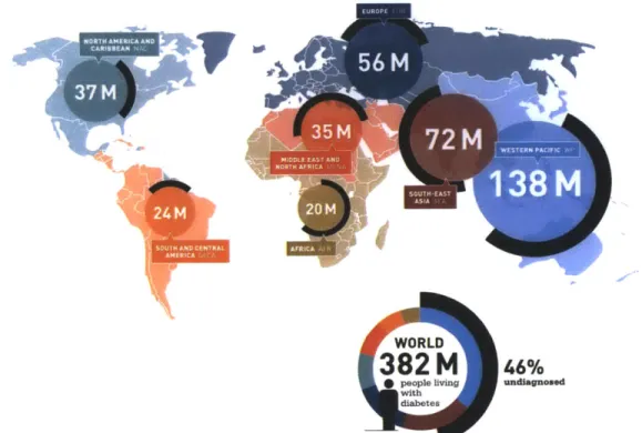
inadequate production and/or utilization of insulin. According to International Diabetes
Federation, there were 382 million people living with diabetes worldwide in 2013;
furthermore, they estimated that by the end of 2013, diabetes could cause 5.1 million
deaths and cost the US $548 billion in healthcare spending, making diabetes one of the
most prevalent, costly, and debilitating disease in the world (Figure 1).
Diabetes generally falls in to one of two categories: type 1 and type 2. Type 1 is an
autoimmune disease caused by cellular-mediated autoimmune destruction of the -cells in
the islets of Langerhans in the pancreas, usually leading to absolute insulin deficiency.
Unlike type 1 diabetes, type 2 diabetes patients usually have intact P-cells, yet they have
insulin resistance with relative insulin deficiency (Drouin et al., 2009).
Discovery of insulin revolutionized the treatment of diabetes. Prior to the discovery of
insulin, most juvenile type 1 diabetes patients, shortly after diagnosis, died of ketoacidosis
(White, 1932). Frederick Banting, a surgeon, and John Macleod, a professor of physiology,
discovered insulin in 1921 and were awarded the Nobel Prize in Medicine in October 1923
(Banting and Best, 1922). Insulin therapy significantly reduced immediate risks of
organs, leading to various complications such as cardiovascular, cerebrovascular,
peripheral vascular diseases, nephropathy, retinopathy, neuropathy, and increased risk of
foot amputation. Occurrence and progression of diabetic complications can be reduced if
hyperglycemia is strictly controlled with precise insulin therapy; however, doing so with
currently available treatments is proven to be difficult (Deedwania and Fonseca, 2005;
Sheetz, 2002).
An alternative to insulin replacement therapy that has shown increasingly promising
results is a cell replacement therapy using beta cells from the islets of Langerhans.
However, there are several technical barriers to be overcome before cell replacement
therapy of diabetes can become a reality. The main barrier is to overcome any immune
responses against transplanted cells. Despite its long history, cell replacement therapy still
has a long way to go before it can become clinically applicable. The effort to make a
cell-based cure for diabetes a success will continue to provide significant milestones, not only
for the cure of diabetes, but also for other regenerative medicine applications.
Historical Development of Diabetes Treatment
Discovery of endocrine role of pancreas
1869 Paul Langerhans, a medical student, discovered "clumps of cells" within pancreas,
which later were named the islet of Langerhans (Figure 2) (Langerhans, 1869;
two dogs and examined their urine for sugar. They demonstrated that pancreas is a
gland, which can prevent the hyperglycemia when implanted under the skin of a
depancreatized dog, and established classic experimental study of diabetes and its
metabolic deviations (Von Mering and Minkowski, 1889; Minkowski, 1989).
1900 E. L. Opie described hyalinization in the islets of Langerhans in diabetic people, and
discovered that the islet of Langerhans produce insulin and that the destruction of
these cells resulted in diabetes (Opie, 1900).
Experimental usage of insulin
1916 Nicolae Paulescu, a Romanian physiologist, developed the first pancreatic extract that
lowered blood sugar in diabetic dogs. He, however, failed to show its application in
human diabetes (Paulesco, 1921).
1921 Frederick Banting, John Macleod, Charles Best, and J.B. Collip produced successful
insulin extract for the treatment of human diabetes. A 14-year-old boy named
Leonard Thompson was the first person to receive the extract to correct the
metabolic acidosis at the Toronto General Hospital in Canada in January of 1922.
Banting and Macleod were awarded the Nobel Prize in Medicine in October 1923
(Banting and Best, 1922; Banting et al., 1922; Best and Scott, 1923).
Sequencing, synthesis & characterization of Insulin
1923 George Walden, a chemical engineer at the Eli Lilly Company, observed that
1982).
1925 First international insulin unit defined (1 unit = 0.125mg of standard material) (Schade et al., 1983)
1926 Crystalline insulin in concentrations of 10, 20, and 40 units per milliliter became
available worldwide
1936 Hans Christian Hagedorn discovered that the action of insulin can be prolonged when
zinc is added to protamine insulin (P.Z.I)(Deckert, 2000).
1939 Reiner, Searle, and Lang developed Globin insulin with shorter duration of action
than P.Z.I. (protamine zinc insulin) (Mosenthal, 1944; Reiner et al., 1939)
1950 Insulin isophane NPH (neutral protamine Hagedorn), an intermediate acting insulin,
with controlled amounts of protamine was developed by Novo Nordisk Company
(Schade et al., 1983)
1951 The amorphous Lente insulin (IZS), an intermediate acting insulin, was developed by
acetate buffering of zinc insulin. The proportion of zinc in the preparation changed
the duration, onset, and peak action of insulin (Hallas-Mo, 1956).
1955 Frederick Sanger and coworkers sequenced Insulin, and it was the first protein to be
fully sequenced. Sanger received the Nobel Prize in Chemistry in 1958 (Stretton,
2002).
1960 Rosalyn Yalow and Solomon Berson developed radioimmunoassasy (RIA), and
demonstrated insulin metabolism in humans with radioactive iodine isotope labeled
laboratories (Katsoyannis et al., 1966; Kung et al., 1966; Meienhofer et al.,
1963).
1967 Donald Steiner discovered that insulin was synthesized as a single polypeptide,
proinsulin precursor, not as two separate A- and B- chains, and a portion of the
proinsulin (C-peptide) was cleaved out after its biosynthesis (Steiner and Oyer,
1967a; Steiner et al., 1967a).
1967 William Kelly and Richard Lillehei performed the first pancreas transplant. A duct
ligated segmental pancreas along with kidney and duodenum, from cadaver donor,
was transplanted into a 28-year-old woman, and insulin independence was achieved;
however, she deceased from pulmonary embolism three months later (Kelly et al.,
1967).
1969 Dorothy Crowfoot Hodgkin, a British biochemist and the Nobel laureate of Chemistry,
deciphered the structure of insulin by x-ray crystallography (Adams et al., 1969).
1971 Insulin receptors discovered and its interaction with insulin defined (Cuatrecasas, 1969, 1971; Freychet et al., 1971).
1973 U100 (100 units per milliliter) insulin became the standardized insulin for human
use in the United States in order to reduce dosage errors and promote better
accuracy in administration (Schade et al., 1983).
1974 Highly purified animal insulin was manufactured with new chromatographic
purification techniques. Eli Lilly Company introduced "single peak" insulin, using
introduced monocomponent (MC) insulin, which was purified ion exchange
chromatography. MC insulin gave a single band in electrophoresis (Root et al., 1972;
Walsh, 2005).
1975 Fully synthetic insulin (CGP12831) was synthesized in the laboratories of Ciba-Geigy
in Basel (Teuscher, 1979).
1976 Serum C-peptide became a clinical tool to access pancreatic beta cell function
(Rubenstein et al., 1977)
1977 The insulin gene was cloned (Cordell et al., 1979; Ullrich et al., 1977).
1978 Open-loop insulin pump delivery system was invented (Pickup et al., 1979). Also,
Genentech used a genetically modified plasmid of E. coli bacteria to synthesize
insulin. Insulin became the first human protein to be manufactured with
recombinant DNA technology (Goeddel et al., 1979).
1980 The human insulin gene was sequenced (Bell et al., 1980). Recombinant DNA human
insulin was first tested on 17 non-diabetic volunteers in England, and the potency
was compared with porcine insulin (Keen et al., 1980).
1981 Insulin receptor kinase activity was described (Kahn et al., 1981).
1982 FDA approved recombinant human insulin, HumulinR (rapid), and HumulinN (NPH),
manufactured by Eli Lilly Company, for the U.S. market.
1989 Islet cells were successfully transplanted into a type I diabetes patient for the first
1996 FDA approved a short-acting insulin analog, lispro (Humalog) developed by Eli Lilly
Company. In lispro, the natural sequence of proline at position B28 and lysine at
position B29 is reversed. This modified amino acid sequence of lispro, decreased the
tendency of insulin to self-associate and increased the rate of absorption after
subcutaneous injection (DiMarchi et al., 1994; Howey et al., 1994).
2000 The "Edmonton Protocol" was devised to improve results of islet transplantation
(Shapiro et al., 2000).
2001 Long acting insulin analog glargine, developed by Aventis Pharma, was approved for
clinical use in the U.S. and Europe. Glargine has two arginine residues added at the
C-terminal end of the B-chain, and asparagine at the position A21 is substituted with
glycine. Glargine has longer duration of action with reduced peak insulin effect
(Jones, 2000; Vajo et al., 2013).
2004 Rapid acting insulin analog glulisine, developed by Aventis Pharma, was approved for
clinical use in the U.S. In glulisine, the natural sequence of asparagine at position B3
is substituted by lysine, and lysine at position B29 is substituted by glutamic acid
(Becker, 2007; Becker and Frick, 2008).
2006 Fatty acid acylated detemir insulin analog (Levemir), developed by Novo Nordisk,
approved for clinical use in the U.S. Insulin detemir is a long-acting analog for
maintaining the basal level of insulin.
2013 Insulin degludec (Tresiba) is an ultra-long acting insulin analog, developed by Novo
Europe and Japan, but not yet in the U.S.
Insulin biosynthesis and processing
Insulin is a small peptide hormone (MW -6kDa) produced by beta cells in the islet of
Langerhans in the pancreas (Figure 2). It consists of two polypeptide chains (A and B
chains) linked together by two disulfide bonds. An additional disulfide bond exist within
the A chain (Figure 3) (Levine and Mahler, 1964; Ryle et al., 1955). Insulin is a highly
conserved protein with a minimal variation among species. The sequences of amino acids
varies between species, but certain segments, especially the positions of three disulfide
bonds, are highly conserved, making insulin from one species likely active in another
species (Steiner et al., 1985). Indeed, pig insulin has been used to treat diabetes in human
(Greene et al., 1983; Richter and Neises, 2003). In most species, the A chain is composed of
21 amino acids, and the B chain of 30 amino acids (Steiner et al., 1985). This two-chain
structure was identified in 1955, but it wasn't until 1967 that the precursor of insulin
(proinsulin) is a single-chain peptide (Ryle et al., 1955; Steiner and Oyer, 1967b; Steiner et
al., 1967b).
In human, there is a precursor of proinsulin - preproinsulin encoded by the INS gene on
chromosome 11 (Owerbach et al., 1980). The first 24 amino acids of preproinsulin form a
hydrophobic signal peptide, which signals the translocation of nascent chain of
preproinsulin into the rough endoplasmic reticulum (RER) (Blobel and Dobberstein, 1975).
trans Golgi network, where endopeptidases cleave off C-peptide. Resulting two peptide
A-and B-chains, linked by two disulfide bonds, are then packaged into immature granules
where it gets further processed by carboxypeptidaseE, which removes two pairs of basic
residues, producing mature insulin. Mature insulin is then packaged into secretory
vesicles, awaiting for metabolic signals to be exocytosed (Figure 3; Figure 4) (Eskridge
and Shields, 1983; Patzelt et al., 1978; Rhodes and Alarc6n, 1994; Walter and Johnson,
1994).
Insulin molecules have a tendency to self-associate and form dimers in solution because
of the hydrogen bonding between the C-termini of B-chains. Moreover, insulin dimers
assemble into hexamers in the presence of zinc ions (Figure 5). This assembly and
disassembly of dimers and hexamers has an important clinical ramification. The active
form of insulin is monomers, but insulin is stored in the pancreas as a hexamer, awaiting
release in response to external stimuli. Hexamers diffuse poorly whereas monomeric and
dimeric insulin diffuse more readily into blood (Brange and Langkjoer, 1993; Derewenda et
al., 1989; Dodson et al., 1979). When the secretory vesicle containing insulin is released
into the bloodstream, the instant dilution causes hexamers to break up into the active form
of monomer quickly (Brange et al., 1990; Sleigh, 1998). However, injected insulin, which at
its storage concentration predominantly exists as hexamers, does not diffuse as readily,
hence delaying absorption and entry into circulation (Brange et al., 1990). A number of
fast-acting insulin analogs were developed by decreasing the tendency to self-associate
This modification decreases the tendency to self-associate and increases the rate of
absorption after subcutaneous injection without affecting receptor binding (DiMarchi et al.,
1994; Howey et al., 1994). Aspart insulin is another example of short acting insulin analog.
In Aspart insulin, proline at the B28 position of the B-chain, the amino acid residue that
participates in self-association, is replaced with negatively charged aspartic acid. This
negative charge eliminates self-association because of charge repulsion (Heinemann et al.,
1993; Kang et al., 1991).
Glucose homeostasis
Insulin is a principal hormonal messenger in fuel homeostasis in human. The basic
circulating units of fuels are glucose and free fatty acids, which are stored intracellularly as
glycogen in skeletal muscles and liver and triglycerides in adipose tissue, respectively
(Cahill, 1976). When food is ingested, insulin levels increases to promote glycogen
synthesis in liver and muscle and lipid formation in adipocytes. In starvation state, insulin
release from the beta cells decreases, and the alpha cells in the islet of Langerhans (Figure
2) start to release glucagon, which stimulates break down of glycogen stored in liver and
muscle (Cryer and Gerich, 1985).
Glucose homeostasis is a complex mechanism that regulates release of insulin from beta
cells in response to changes in blood glucose concentration. The principle objective of
glucose homeostasis is to maintain normoglycemia, and the concentration of blood glucose
continuous need for fuel but does not have fuel storage capacity. It cannot utilize fatty
acids as a fuel source either, though it can use energy derived from fatty acids in a
prolonged starvation state; therefore, it relies solely on blood glucose. Other vital organs,
such as heart, also have continuous need for fuels, but they can utilize fatty acids directly as
needed. Hence, in hypoglycemic state, central nervous function becomes the most
impaired vital organ. Hyperglycemia is detrimental as well because it causes glycosuria
and contributes to the complication of diabetes (Cahill, 1976; Cryer and Gerich, 1985).
Release of insulin from beta cells in the islets of Langerhans is a biphasic process. The
first phase release is rapid, and the amount of initial release, triggered in response to
increased blood glucose level, is dependent on the amounts available in storage. Once
stored insulin is depleted, second phase of slow and sustained release is triggered
independently of glucose. During this latter phase, release of insulin is slow because
insulin has to be synthesized, processed, and packaged into vesicles. Furthermore, beta
cells have to replenish depleted insulin in the initial fast response phase (Curry et al., 1968;
O'Connor et al., 1980; Porte and Pupo, 1969).
Initial release phase is initiated when glucose enters the beta cells through the type 2
glucose transporters (GLUT2). Upon entry, glucose is phosphorylated by the enzyme
glucokinase and is metabolized in glycolysis and the Krebs cycle, producing high-energy
ATP molecules and increasing intracellular ATP/ADP ratio. The increased ATP/ADP ratio
closes ATP sensitive potassium channel, preventing potassium ions from leaving the cells,
activates phospholipase C.
Phospholipase C cleaves the membrane-bound phospholipid phosphatidyl inositol
4,5-biphosphate (PIP2) into inositol 1,4,5-triphosphate (IP3) and diacylglycerol (DAG). DAG
remains within the plasma membrane and activates protein kinase C (PKC), while IP3
diffuses into the cytosol and binds to IP3-gated Ca2+ Channel in the plasmamembrane of the
endoplasmic reticulum (ER). This allows release of Ca2+ from the ER via IP3-gated Ca2
+
channel, further increasing the intracellular Ca2+ concentration. Significantly released
intracellular Ca2+ triggers exocytosis of previously synthesized insulin stored in secretory
vesicles (Figure 6) (Hiriart and Aguilar-Bryan, 2008; Matschinky et al., 1993; Rana and
Hokin, 1990).
Insulin circulates in the blood stream until it binds to transmembrane insulin receptors,
which belong to a family of tyrosine kinase receptors and play an important role in the
regulation of glucose homeostasis. Activated insulin receptors promote uptake of glucose
via type 4 glucose transporters (GLUT4) into various tissues, such as skeletal muscles and
adipose tissues, and increase glycogen, lipid, and protein synthesis. Role of insulin is also
implicated in various gene regulations via control of amino acid uptake and modification of
numerous enzyme activities. (Figure 7) (Bergamini et al., 2007; Dimitriadis et al., 2011;
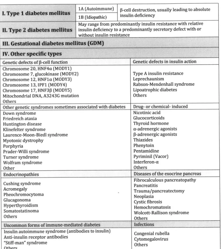
Currently etiological classification of diabetes mellitus falls into four categories - type 1,
type 2, other specific types, and gestational diabetes mellitus (GDM) (Table 1). Most
common forms of diabetes are type 1 and type 2 diabetes. Other specific types of diabetes
encompass a variety of types of diabetes associated with particular diseases or syndromes
with a distinct etiology. Gestational diabetes mellitus is glucose intolerance associated with
varying degrees of hyperglycemia with the onset during pregnancy (Drouin et al., 2009;
Gavin and Alberti, 1997).
Type 1 diabetes is generally caused by destruction of beta cells; therefore, individuals
with this disease require insulin for survival. Idiopathic forms of type I diabetes is further
divided into type 1A and type 1B (Gavin and Alberti, 1997). Type 1A is characterized by
the presence of islet autoantibodies (anti-GAD, anti-islet cell, or anti-insulin antibodies),
which leads to insulitis and selective destruction of islet beta cells, and almost always
progresses to severe insulin deficiency. Type 1A is also strongly associated with human
leukocytes antigen (HLA) alleles (Foulis et al., 1991; Nepom and Kwok, 1998; Noble et al.,
1996; Todd, 1999). Type 1B comprises a minority of type 1 diabetes patients. They are
presented with severe insulin deficiency but without evidence of autoimmune destruction
of beta cells (Sacks et al., 2011).
Type 2 diabetes is the most common form of diabetes and is characterized by defective
insulin action and secretion with no autoimmune destruction of beta cells. Type 2 diabetes
patients usually have insulin resistance and relative insulin deficiency. Their plasma
for survival, although some may require insulin for glycemic control due to progressive
beta cell failure, which can occur with increasing duration of diabetes (Gavin and Alberti,
1997). Some people with type 2 diabetes can regulate blood glucose level with life style
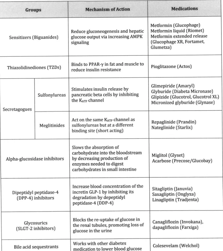
change, diet and exercise alone, but many require diabetes medications, such as metformin
and sulfonylureas (Table 2).
Typical diabetes treatments include oral medications, insulin injections, dietary
restrictions, exercise, and intense self-monitoring of blood glucose; however, these only
provide a short-term relief (Beck et al., 2007). Despite the impressive progress in treating
diabetes, most people with diabetes continue to develop disabling complications, most of
which are directly linked to hyperglycemia. Several advancements have been made to
decrease complications and to treat diabetes more effectively, such as gene therapy and
closed-loop insulin delivery systems. The combination of a glucose sensor and an insulin
pump can mechanically replace beta cell function and provide patients with
normoglycemia (Steil, 2004; Yechoor and Chan, 2005). However, the most promising and
attractive alternative treatment option, especially for type 1 diabetes, remains to be
replacing the missing beta cells with pancreas, islet, or beta cell transplants. This concept
was tested clinically, though unsuccessfully, as early as 1893 in Bristol, England when
pieces of sheep pancreas were transplanted subcutaneously to a 15-year-old boy with
diabetes (Williams, 1894). In 1967, the pancreas transplant from a cadaver donor was
transplanted into a 28-year-old woman for the first time, and the patient achieved insulin
1989 (Scharp et al., 1990). Beta cell replacement with pancreas, islet, or beta cell
transplants can result in long-term relief, providing a glucose homeostasis for an extended
period of time; however, success is limited by the host graft rejection because they can fail
without life long systemic immunosuppression, which can cause many adverse effects such
as renal failure (Gallagher et al., 2011).
Islet transplantation
Pancreas transplantation is an invasive complex surgery; and therefore, regardless of
surgical method, it inevitably comes with a high risk of morbidity and mortality. Despite
the risks and life-long immunosuppression, pancreas transplantation still remains the best
alternative choice for patients, especially those with type 1 diabetes, who do not respond to
conventional treatments. Islet transplantation is an attractive alternative to pancreas
transplantation, since it is a much less invasive procedure (Vardanyan et al., 2010).
Methods to isolation of islet cells were first reported by Lacy and Kostianovsky in 1967,
and since then several studies reported that islet transplantation can successfully reverse
hyperglycemia in both small and large animals (Lacy and Kostianovsky, 1967; Sutherland
et al., 1993). Subsequently, Scharp et al. demonstrated in 1981 that islet allografts
successfully reversed the diabetic state of type 1 diabetes patients, unveiling the promising
future islet transplantation holds for treating diabetes patients (Scharp et al., 1991).
Frustrated with less than optimal outcomes of earlier islet cell transplantation trials, the
spectacularly better than any of the previous islet transplantation trials, patients still have
to receive immunosuppressive therapy.
Despite the conceptual simplicity of the procedure, progress in making islet
transplantation a reliable therapy has been hindered by two major barriers. The first
drawback is the source of the islet cells, which cannot be expanded in vitro -this is beyond
the scope of this chapter; however, there are numerous reports of exploring different
sources of islet cells and even unlocking possibilities of islet cell regeneration from stem
cells. Another major obstacle for islet transplantation is the process of transplant rejection
and autoimmunity from destroying transplanted islet cells. To avoid graft rejection,
patients are again required to take life-long immunosuppressive drugs (Halban et al., 2010;
Weir, 2013). In an attempt to control transplant rejection and autoimmunity, safer and
more effective immunosuppressive drugs are being developed. However, it still does not
eliminate the fact that patients still need to be on life-long immunosuppressive therapy.
Recently, it was found that autoimmune destruction of islet cells can be prevented by
creating a barrier between lymphocytes and transplanted islet cells. This concept of
creating an immunobarrier, if successful, will completely eliminate the use of
immunosuppressive drugs and maintain the long-term islet graft function. For this reason,
transplantation of encapsulated islet cells has created high expectations for treating type 1
Cell encapsulation techniques consist of enclosing therapeutic cells within a
semi-permeable polymeric matrix, which will allow bi-direction diffusions of nutrients, oxygen
and waste, and secretion of therapeutic products while preventing immune cells from
destroying the enclosed cells (Figure 2) (Orive et al., 2003b). Several materials, such as
polysulphone (PS), poly(ethylene glycol) (PEG), dimethylaminoethy methacrylate-methyl
methacrylate copolymer, and poly(vinyl alcohol), have been explored to achieve the
purpose of encapsulation. Successful islet encapsulation was demonstrated with PS by
blending it with poly-vinylpyrrolidone or sodium-dodecyl-sulfate; however, this
encapsulation technique hindered proper islet cell function by limiting insulin diffusion.
Encapsulating with hydroxy-methylated PS showed some promising results (Figliuzzi et al.,
2005; Lembert et al., 2001; Petersen et al., 2002), but, the encapsulated cells had reduced
viability and function due to polymer degradation, as well as fragility and limited
permeability of the capsules (Xie et al., 2005). Additionally, amniotic membranes,
nano-porous microsystems, silica, and synthetic extracellular matrix consisting of
poly(N-isopropyl-acrylamide) and acrylic acid copolymers have also been explored. However, the
properties and manufacturing methods of these materials limit their applicability (Vernon
et al., 2000) (Boninsegna et al., 2003; Desai et al., 2004; Mahgoub et al., 2004).
Alginate, generally extracted from various brown algae (Phaeophyceae) has shown the
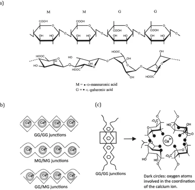
most promising results. Alginate is a family of linear co-polymers of P-D-mannuronic acid
(M) and a-L-guluronic acid (G) with highly variable G and M sequences and compositions
alginates are being commercially manufactured. As an ionic polysaccharide, alginate can
form hydrogels in the presence of divalent cations, such as barium and calcium ions.
Hydrogels are highly hydrated three-dimensional networks of hydrophilic polymers, and
due to their structural similarity to the extracellular matrices in the body, they are often
biocompatible. Only the G-blocks of alginate are known to participate in intermolecular
cross-linking with calcium ions (Figure 9b), while the M-blocks can also participate when
cross-linked with barium ions (Lee and Mooney, 2012). These associations of alginate
chains and divalent cations constitute the junction zone, known as the "egg box model,"
responsible for the gelation (Figure 9c). In the egg box model, oxygen atoms are involved
in the coordination of the divalent cations (Grant et al., 1973; Mackie et al., 1983).
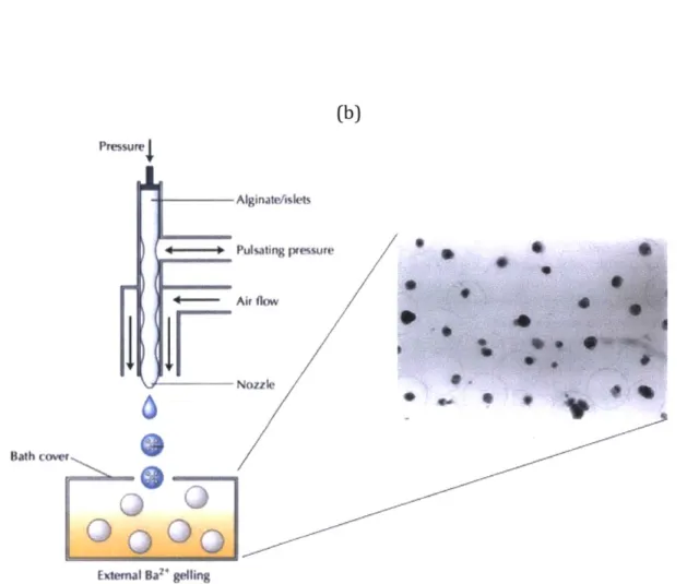
Encapsulating cells with alginate is relatively easy. The cross-linking of alginate happens
almost instantaneously simply by mixing the cells with a solution of sodium alginate and
dripping them into a solution of calcium or barium (Figure 10) (Chaikof, 1999;
Zimmermann et al., 2007). This cross-linking is efficient at near physiological conditions;
and therefore, the cells entrapped inside are highly viable and functional. It is this gelation
property of alginate that has gained high interests for the application of cell encapsulation
(Andersen et al., 2012; Zimmermann et al., 2007).
Progress of Alginate Cell Encapsulation
The first use of a semi-permeable membrane to prevent graft failure was reported in
immune-protection of transplanted cells (Chang, 1964). In 1980, this cell encapsulation
concept was successfully implanted to mobilize xenograft islets cells. Lim and Sun
demonstrated that microencapsulated islets with alginate corrected diabetic state for 2 to 3
weeks and remained functionally viable for over 15 weeks in rats (Lim and Sun, 1980).
Since then, tremendous advancements have been made in using alginate for various
biomedical applications, and alginate remains the leading material for the
microencapsulation of islet cells (Zimmermann et al., 2007).
Since the first demonstration of proof-of-concept with encapsulated cells in humans in
1991 (Scharp et al., 1991), many studies have reported varying degrees of success. In 2003, Omer et al. demonstrated that they could reverse diabetic state in immunocompetent
mice for more than 20 weeks with alginate encapsulated porcine neonatal pancreatic cell
clusters (Omer et al., 2003). In 2007, Calafiore et al. reported two cases where human
diabetic patients received human islet microcapsules in the peritoneal cavity under local
anesthesia. Without immunosuppression, high blood glucose level was reversed for one
year in one of the patients, and for six months in the other patients (Calafiore et al., 2006).
In 2007, Elliott et al. reported a single case of a 9.5-year long-term survival of alginate
encapsulated porcine islets cells in a 41-year-old type 1 diabetes patient. Though
functional, the surviving islet cells were insufficient to reverse the diabetic state after the
first year of transplantation (Elliott et al., 2007). In 2009, Tuch et al. transplanted alginate
encapsulated human islet cells, which were collected from cadaver pancreases, in four type
completely covered with fibrous tissues and contained necrotized islet cells (Tuch et al.,
2009). Most recently, Jacobs-Tulleneers-Thevissen et al. advanced the islet transplantation
field by demonstrating that alginate encapsulated human islets remain functional in the
peritoneal cavity of mice and of a human type 1 diabetes patients
(Jacobs-Tulleneers-Thevissen et al., 2013).
Challenges of Encapsulated Islet Transplantation
Although promising, these studies highlight one of the most serious problems facing
this field: implanted capsules, regardless of whether they contain cells inside or not, elicit a
foreign body response due to the alginate material itself, forming fibrotic structures around
the capsules, which eventually lead the hypoxic death of the islet cells inside (Anderson et
al., 2008; Bridges and Garcia, 2008; Franz et al., 2011; Weir, 2013). How these reactions
occur is not well understood, although it has been suggested that this foreign body
response is a combination of the reactivity to the material itself and/or impurities present
within the material. Genetic makeup of the recipients and possibly the immune reaction to
the biomolecules released by the encapsulated cells can also influence foreign body
responses (Weir, 2013).
The foreign body response is a biomaterial-mediated inflammation, a complex process
initiated immediately upon implantation of the material (Figure 11). When biomaterials
are implanted, various proteins present in the host fluids (blood, lymph, and wound fluids)
adsorbed state by producing various pro-inflammatory cytokines. These neutrophils
eventually recruit tissue resident macrophages and undifferentiated monocytes, and
subsequently exit the implant site. Macrophages respond to the foreign materials by
producing their own set of various inflammatory mediators, which in turn recruit
fibroblasts and fuse into multinucleated foreign body giant cells. Recruited fibroblasts start
infiltrating the site and form thick collagenous fibrous capsules around the implant,
isolating it from the host tissue (Anderson et al., 2008; Bridges and Garcia, 2008; Franz et
al., 2011; Grainger, 2013).
Our current understanding of alginate mediated inflammation is that mannuronic acid
polymers activate Toll-like receptor 2 (TLR2) and TLR4, which are types of pattern
recognition receptors (PRRs), in primary murine macrophages (Flo et al., 2002). A study
by Yang and Jones in 2008 also implicated the involvement of TLRs by demonstrating that
alginate can stimulate innate immune response via macrophage receptors, leading to
NF-KB activation (Yang and Jones, 2009). A number of studies on other biomaterials have
implicated the role of TLRs in the foreign body response as well. Grandjean-Laquerriere et
al. reported that particles of hydroxyapatite, which is widely used biomaterial to fill bone
defects or to coat prosthesis, can induce an inflammatory reaction by activating TLR4
(Grandjean-Laquerriere et al., 2007). Another study by Auquit-Aucbur et al. reported
involvement of TLR4 in inflammation around silicone prosthesis (Auquit-Auckbur et al.,
2011). Pearl et al. investigated involvement of TLRs in polymethylmethacrylate (PMMA)
signaling pathways. Although they were able to demonstrate that TLRs are indeed
involved in PMMA induced inflammatory reaction, they could not identify which TLRs are
involved. Another report on rheumatoid arthritis patients with implants showed
upregulation of TLR2 and TLR4 (Myles and Aggarwal, 2011). All these studies clearly
implicate TLRs are the mediators of the foreign body response. However, which particular
TLRs are involved in macrophage activation in response to alginate microcapsules is
currently not well understood. Additionally, some of these studies are plagued by
questions of whether the materials are contaminated with pathogen-associated molecular
patterns (PAMPs) as alginate is produced not only by brown algae but also by bacteria.
Paredes-Juarez et al. reported that PAMPs, predominantly ligands of TLR2, 5, 8, 9, are
present in some of the commercial alginates (Paredes-Juarez et al., 2013).
Future of Islet Encapsulation
The cell encapsulation technique, based on the principle of immunoisolation, still
remains an attractive therapeutic method with a potential to greatly advance diabetes
treatment, possibly curing type 1 diabetes. It undeniably has the potential to protect
transplanted beta cells from autoimmune destruction. Many challenges remain to be
addressed, and many researchers are collaboratively working on the problems. With
continuous research effort, new and improved materials and formulations are on the
horizon, which may well lead to greater success of not only for the treatment of diabetes,
Insulin is not a curefor diabetes; it is a treatment It enables the diabetic to burn sufficient carbohydrates, so that proteins and fats may be added to the diet in sufficient quantities to provide energyfor the economic burdens of life.
46%
undiagnosed
Figure 1. Worldwide diabetes statistics. Figure adopted from IDF Diabetes Atlas, 6th
ed. Brussels, Belgium: International Diabetes Federation, 2013.
Alpha cell (secretes glucagon) Beta cell (secretes Insulin) Delta cel I (secretes somatostatin) Exocrine pancreas (acinar cells and duct cells)
F cell
(secretes pancreatic polypeptide)
Figure 2. Islet of Langerhans. Located within pancreas, they consist of four distinct cell
types - alpha, beta, delta, and F cells. The beta cells are the most common islet cells, and they produce insulin, which is the major hormone responsible for glucose metabolism. The alpha cells produce glucagon which can trigger the release of stored glucose from the liver and fat tissues (@2011 Pearson Education, Inc).
Preproinsulin
-c S SEndoplasmic
reticulum
8-chain1~
~mrneesmmininw
N %pnal peptideProinsulin
Ce
Endopep C-PePtlde Endopepidas-m
A-6ain Carboxypep daseE CarboxypeptidaseE B-caJSecretory
vesicles
SMature insulin
I.N -;-) S S A-chain r-o-C 8-chain 5Golgi
apparatus
$ $ N..mm
~m
i
Noin S - SFigure 3. Processing of insulin. In human, the initial precursor of insulin is preproinsulin,
which consists of four domains - signal peptide, A-chain, B-chain, C-peptide. The signal peptide, the first 24 amino acids at N-terminus of preproinsulin, translocates the nascent protein into the rough endoplasmic reticulum (RER) where the signal peptide gets cleaved off and the protein gets folded into correct conformation, producing proinsulin. Proinsulin is then transported into the Golgi apparatus where C-peptide gets cleaved off by
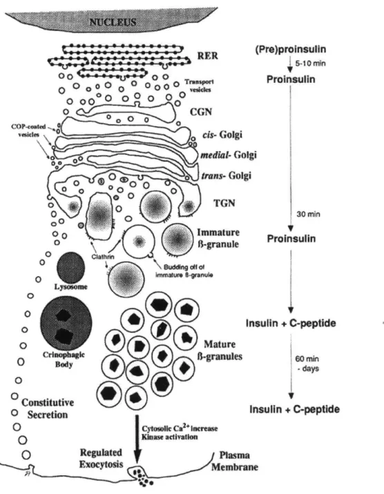
endopeptidases. The resulting two peptide chains (A- and B-chains) are packaged into secretory vesicles where they get further processed by carboxypeptidaseE, which removes two pairs of basic residues, producing mature insulin. Mature insulin in the secretory vesicles then waits for metabolic signals to be exocytosed.
0 000 0RER 0 0 0 00 00000 0 00 0 C(N cop o COP-Cw '4 r cis- Golgi medial- Golgi trans- Golgi 0 0 0(3j, 0 G TGN '0 0 00 Immature 0 >. -granule Budgalt 4at" 0
000
00
Constitutive o S Mature 8-granules In~ ItTocr
MIMI 0 ya~n ca +reso
Regulated Plsma EXOCytOSIS MembraneFigure 4. Insulin secretion pathway. Newly made proinsulin in the rough endoplasmic reticulum (RER) is transferred to the cis-Golgi network (CGN). Proinsulin is packaged into immature
s-granules
in the trans-Golgi network (TGN). Proinsulin is sent to the P-granule compartment, where the C-peptide is cleaved off to produce insulin. Mature p-granules are held in an intracellular storage compartment awaiting a signal for exocytosis ( 2004 Lippincott Williams & Wilkins).Pre)proinsulln Proinsulin 30 min Proinsulin uIn + C-peptide 60 min -days sulin + C-peptide
Monomers 10-'M Dimer 10-SM Hexamer 10M
Figure 5. Assembly and disassembly of insulin monomer, dimer, and hexamer. At
different concentrations, insulin assembles and disassembles into monomers, dimers, and hexamers. Insulin molecules tend to form dimers in solution due to the hydrogen bonds between the C-termini of B-chains. Furthermore, in the presence of zinc ions, insulin
dimers assemble into hexamers.
(a) (b)
ATP-sensitive
91ucos~e . ,GLUT2 potassium
uptake charnel
n ADPa
insulin relea! e calciumn channel ,e
swoage eanules S 1 ci Phospholipase C Exterior Plasm mnib Cytos a DAG ol P PIP2 '3 (pKC} Phosphoylatlon a of substrates Protein ** kinase C 1P0 IP 3-gated Ca2 channel * ec , C 0
Ca* Endoplasmic reticulum
Figure 6. Mechanism of insulin secretion. (a) Insulin secretion in beta cells is triggered by elevated blood glucose. Cells uptake glucose via GLUT2 transporter, and the glycolytic
phosphorylation of glucose causes a rise of ATP:ADP ratio. This increased ATP:ADP ratio deactivates the potassium channel, causing depolarization of the membrane, which then leads to opening of calcium channel, allowing inflow of calcium ions and activates
phospholipase C. This subsequently leads to exocytosis of stored insulin. (b)
Phospholipase C cleaves phospholipid phosphatidyl inositol 4,5-biphosphate (PIP2) to inositol 1,4,5-triphosphate (IP3) and diacylglycerol (DAG). DAG activates protein kinase C (PKC), and IP3 activates IP3-gated calcium channel, which leads to further increase of intracellular calcium ion concentration (@ 2004 Beta Cell Biology Consortium).
ore-operated RpCa2l 0 Ca2 -hannel :0000 **
(: 14
-
It
-
4-o 0 0 o insulin 0 00 glucose 0 0 0 0 0 GLUT insulin .. receptors
m expression & gucose a O
glycogent I pid I protein
Figure 7. Effect of insulin on glucose uptake and metabolism. Activated insulin receptor promotes uptake of glucose via GLUP4 transporter. Binding of insulin to its receptor also activates a cascade of reactions, such as glycogen/lipid/protein synthesis, glycolysis (@ 2004 Beta Cell Biology Consortium).
Table 1. Etiologic classification of diabetes mellitus
1A (Autoimmune) P-cell destruction, usually leading to absolute 1B (Idiopathic) insulin deficiency
May range from predominantly insulin resistance with relative insulin deficiency to a predominantly secretory defect with or without insulin resistance
Genetic defects of $-cell function Genetic defects in insulin action Chromosome 20, HNF4a (MODY1)
Chromosome 7, glucokinase (MODY2) Type A insulin resistance Chromosome 12, HNF1a (MODY3) Leprechaunism
Chromosome 13, IPF1 (MODY4) Rabson-Mendenhall syndrome Chromosome 17, HNF3P (MODY5) Lipoatrophic diabetes
Mitochondrial DNA, A3243G mutation Others Others
Other genetic syndromes sometimes associated with diabetes Drug- or chemical- induced
Down syndrome Nicotinic acid
Friedreich ataxia Glucocorticoids Huntington disease Thyroid hormone Klinefelter syndrome a-adrenergic agonists Laurence-Moon-Biedl syndrome 0-adrenergic agonists
Myotonic dystrophy Thiazides
Porphyria Phenytoin
Prader-Willi syndrome Pentamidine
Turner syndrome Pyriminil (Vacor)
Wolfram syndrome Interferon-a
Other Others
Endocrinopathies Diseases of the exocrine pancreas Fibrocalculous pancreatopathy
Cushing syndrome Pancreatitis
Acromegaly . Trauma/pancreatectomy
Pheochromocytoma Neoplasia
Glucagonoma Cystic fibrosis
Hyperthyroidism Hemochromatosis
Somatostatinoma Wolcott-Rallison syndrome
Others Others
Uncommon forms of immune-mediated diabetes Infections
Insulin autoimmune syndrome (antibodies to insulin) Congenial rubella Anti-insulin receptor antibodies Cytomegalovirus
"Stiff-man" syndrome Others
Others
Adapted from Joslin's Diabetes Mellitus. 14th ed. In: Bennett, PH and Knowler, WC. Definition, Diagnosis and Classification of Diabetes
Table 2. Diabetes medications
Sensitizers (Biguanides)
Reduce gluconeogenesis and hepatic glucose output via increasing AMPK signaling
Metformin (Glucophage) Metformin liquid (Riomet) Metformin extended release
(Glucophage XR, Fortamet, Glumetza)
Thiazolidinediones (TZDs) Binds to PPAR-y in fat and muscle to reduce insulin resistance Pioglitazone (Actos)
Stimulates insulin release by Glimepiride (Amaryl)
Sulfonylureas a ca tels bGlipizide (Glucotrol, Glucotrol XL)pancreatic beta cells by inhibiting thefy a Glyburide (Diabeta Micronase)
the KATP channel Micronized glyburide (Glynase)
Secretagogues
Act on the same KATP channel as Repaglinide (Prandin)
Meglitinides sulfonylureas but at a different Nateglinide (Starlix) binding site (short acting)
Slows the absorption of
carbohydrate into the bloodstream Miglitol (Glyset)
Alpha-glucosidase inhibitors by decreasing production of Acarbose (Precose/Glucobay) enzymes needed to digest
carbohydrates in small intestine
Increase blood concentration of the Sitagliptin (Januvia) Dipeptidyl peptidase-4 incretin GLP-1 by inhibiting its Saxagliptin (Onglyza)
(DPP-4) inhibitors degradation by depeptidyl Linagliptin (Tradjenta) peptidase-4 (DDP-4)
Glycosurics Blocks the re-uptake of glucose in Canagliflozin (Invokana),
(SLGT-2 inhibitors) the renal tubules, promoting loss of dapagliflozin (Farxiga) glucose in the urine
Works with other diabetes
Bile acid sequestrants medication to lower blood glucose olesevelam (Welchol)
Table 3. Chemical composition of alginates from various most commonly used industrial sources. FGM Source FG Fm FGG FMM FMG FGGG FGGm FMGM NG>I Durvillea antarctica 0.32 0.68 0.16 0.51 0.17 0.11 0.05 0.12 4 Laminaria japonica 0.35 0.65 0.18 0.48 0.17 Ascophyllum nodosum 0.39 0.61 0.23 0.46 0.16 0.17 0.07 0.09 5 Lessonia nigrescens 0.41 0.59 0.22 0.40 0.19 0.17 0.05 0.14 6 Laminaria digitata 0.41 0.59 0.25 0.43 0.16 0.20 0.05 0.11 6 Macrocystis pyrifera 0.42 0.58 0.20 0.37 0.21 0.16 0.04 0.02 6
Laminaria hyperborea (leaf) 0.49 0.51 0.31 0.32 0.19 0.25 0.05 0.13 8
Laminaria hyperborea (stipe) 0.63 0.37 0.52 0.26 0.11 0.48 0.05 0.07 15
Adapted from Andersen et al., 2012.
Inward diffusion of )
nutrients and oxygen
is allowed
Inflammatory cells are excluded
0 0' Antibodies are exclude Therapeutic and waste products
can diffuse freely d
Therapeutic
product-secreting cells
Microcapsule
Figure 8. Principles of cell encapsulation. Cells are enclosed within a semi-permeable
polymeric matrix, which can circumvent host immune rejection. The matrix allows in-flow of oxygen and nutrients and out-flow of therapeutic and waste products, while preventing immune cells and antibodies come in direct contact with the enclosed cells. Figure adapted from Orive et al., 2003.
(a) M M G 0 COOH COOH O OOH COOH OH HO OH HO OH HO OH HO HOOC OH HOOC HO OH 0 0 HOOC OH HOOC M = -D-nannuronic acid G = * -L-guIurornic acid (b) (c) GG/GG junctions MG/MG junctions GG/MG junctions Z \ OH O2
o
0 0 0 H GG/GG junctionsDark circles: oxygen atoms involved in the coordination of the calcium ion.
Figure 9. Schematic diagrams of alginate. (a) Chemical structure of alginate MMGG
block; (b) alginate crosslinked with calcium; (c) Schematic drawing and calcium coordination of the "egg box model" as described for the pair of guluronate chains in calcium alginate junction zones. Dark circles represent the oxygen atoms involved in the coordination of the calcium ion. (Andersen et al., 2012; Braccini and Perez, 2001)
(a) (b) Pressurel Alginate/islis ) + - Pulsating pressure + - Air flow A Rath 1COVe External Ba gelling
Figure 10. Schematic diagram of islet encapsulation. (a) An air-jet droplet generator is
used to encapsulate islet cells with alginate; (b) Rat islet cells encapsulated in alginate hydrogel. Figure adapted from Zimmermann et al., 2007 and Chaikof, 1999.
Protein Matrix Fibrous Bare 0-biomnatri -surface 60. J Time
Figure 11. Foreign body response to implanted biomaterial. Diverse adsorbed protein
layers, which happens instantaneously upon implantation of the material, on the implant surface recruit neutrophils. Neutrophils produce various inflammatory mediators and recruit macrophages and monocytes. Within days, neutrophils exit the site, and
macrophages start recruiting fibroblasts. Some macrophages fuse to create foreign body giant cells. Fibroblasts start forming thick collagenous layers on the implant. Figure adapted from Grainger, 2013.