O R I G I N A L P A P E R
Maziar Assadi Æ Markus Mu¨ntener
Utrophin is lacking at the neuromuscular junctions
in the extraocular muscles of normal cat: artefact or true?
Accepted: 13 December 2004 / Published online: 24 February 2005 Ó Springer-Verlag 2005
Abstract Extraocular muscles (EOM) are typically spared in Duchenne muscular dystrophy. We hypothe-sized that this might be due to different patterns of utrophin expression. The expression of utrophin was examined in EOM of normal cats using immunohisto-chemical methods and Western blot. For detecting ace-tylcholine receptors (AChR), we used a-bungarotoxin. Surprisingly, a-bungarotoxin failed to stain the AChR and no expression of utrophin could be detected at the neuromuscular junctions. Our study could indicate that the expression of utrophin is dependent on the structure of the AChR.
Keywords Extraocular muscle Æ Utrophin Æ Acetylcholine receptor Æ Immunohistochemistry Æ Duchenne muscular dystrophy
Abbreviations AChE: Acetylcholinesterase Æ AChR: Acetylcholine receptors Æ DMD: Duchenne muscular dystrophy Æ EOM: Extraocular muscles Æ NMJ: Neuromuscular junctions
Introduction
Duchenne muscular dystrophy (DMD) is one of the most common inherited neuromuscular diseases and occurs in about 1:3,500 live male births (Worton1995). DMD is characterized by the almost complete absence
of dystrophin either due to promoter defects or nonsense mutations and deletions leading to an unstable tran-script or protein. In DMD, extraocular muscles (EOM) remain unaffected during the course of the disease (Kaminski et al. 1992; Karpati et al. 1988; Khurana et al.1995; Ragusa et al.1996). The compensatory fac-tors or mechanisms that allow EOM to escape the consequence of dystrophin deficiency are unknown. One hypothesis is that an alternative expression of a protein that is structurally and functionally homologous to the defective dystrophin could save EOM by assuming the role of dystrophin. This would prevent the loss of other vital components of the transmembrane protein com-plex, thereby stabilizing the sarcolemma and providing myofiber survival. A possible candidate for such a function is utrophin, which has a ubiquitous expression pattern in the brain (Khurana et al. 1990), muscle (Nguyen et al.1991) and other tissues (Love et al.1991). In normal mature striated muscle, utrophin expression is detected at the neuromuscular (Nguyen et al. 1991; Takemitsu et al. 1993) and myotendinous junctions (Khurana et al. 1992), in the wall of endomysial capil-laries and other blood vessels and in the perineurium and Schwann cells of the intramuscular nerves (Karpati et al.1993). During ontogeny (Lin and Burgunder2000) and regeneration (Lin et al. 1998), it is found at the whole sarcolemma. The utrophin and dystrophin genes exhibit a marked degree of homology although the utrophin gene is smaller (Brown1997). Both dystrophin and utrophin proteins have the same four domains: the amino terminus, the rod domain, the cysteine-rich domain and the carboxy terminus. Both proteins have almost the same molecular weight (dystrophin 427 kDa, utrophin 420 kDa) and the same associated proteins (dystroglycans, sarcoglycans, and syntrophins).
There are three animal models of DMD: mdx mouse (Bulfield et al.1984; Cooper1989; Hoffmann2001), dog (Cooper et al.1988a, b; Kornegay et al. 1988) and cat (Carpenter et al.1989; Gaschen et al.1992; Winand et al.
1994). They all lack dystrophin in skeletal muscle tissue because of loss-of-function mutations in the highly
M. Assadi (&) Æ M. Mu¨ntener
Institute of Anatomy, University of Zu¨rich-Irchel, Zu¨rich, Switzerland
M. Mu¨ntener
Department of Chemistry and applied Biosciences, ETH, Zu¨rich, Switzerland
M. Assadi
F. Hoffmann-La Roche, 93/8.08, 4070 Basel, Switzerland E-mail: maziar.assadi@roche.com
conserved dystrophin gene. The clinical phenotype of each animal model differs from the human disease and from one another. The fact that they show differences in phenotype is valuable in that they provide insight into the secondary consequences of primary dystrophinopathy.
Our hypothesis for the sparing of EOM in DMD was that in normal EOM, utrophin is naturally overexpres-sed at the extrajunctional sites and replaces dystrophin. Because cat is one of the animal models of DMD, we studied the expression of utrophin in EOM and leg muscles of normal cats using immunohistochemical methods and Western blot.
Materials and methods Tissue
Three domestic cats and one Wistar rat without appar-ent myopathy were obtained from the departmappar-ent for
veterinary medicine of the University of Zu¨rich. The cats and the rat were killed with a high dose of anesthetics in accordance with the guidelines of the local animal care committee. Samples of EOM and leg muscles (quadri-ceps muscle) were taken immediately after death and were snap-frozen in isopentane cooled in liquid nitrogen. Until the analysis, the biopsies were stored at 70°C.
Histology
Detection of utrophin and acetylcholine receptors (AChR)
Peroxidase immunohistochemistry For the histological procedures, the muscle specimens were sectioned in a cryostat at 7 lm and dried overnight at room tempera-ture. Processing of sections for detecting utrophin with peroxidase immunohistochemistry was performed as described by Lin et al. (1998). A monoclonal antibody against the amino terminus of utrophin was used (NCL-DRP2, Novocastra Laboratories Ltd.). The second antibody was a biotinylated sheep antimouse antibody (RPN1001, Amersham Biosciences) followed by perox-idase-conjugated streptavidin (016-030-084, Jackson ImmunoResearch Laboratories Inc.). Peroxidase activ-ity was visualized using DAB tablets (DO426, Sigma).
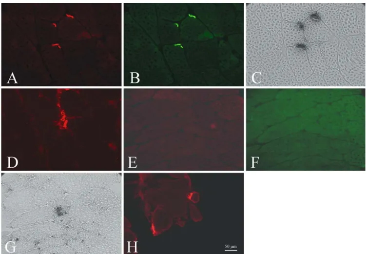
Fig. 1 Neuromuscular junctions (NMJ) were stained in leg muscles (a–d) and extraocular muscles (EOM) (e–h) of cat. Double staining with a-bungarotoxin/utrophin showed a positive staining for both utrophin (a) and a-bungarotoxin in leg muscles (b). In EOM, neither utrophin (e) nor a-bungarotoxin (f) could be detected. By using the method of Karnovsky on consecutive sections, NMJ could be shown in leg muscles (c) and EOM (g). NMJ could also be detected with rapsyn in leg muscles (d) and EOM (h)
Fluorescence immunohistochemistry The same protocol as for peroxidase immunohistochemistry was adapted for fluorescence immunohistochemistry. Sections were double-stained either with utrophin/a-bungarotoxin or with rapsyn/a-bungarotoxin. a-bungarotoxin binds to the extracellular part of the AChR whereas rapsyn interacts with the cytoplasmatic part of the AChR. a-bungarotoxin was conjugated with Alexa Fluor 488 (B-13422, Molecular Probes). Primary antibodies were: (1) monoclonal antibody against the amino terminus of utrophin (NCL-DRP2, Novocastra Laboratories) and (2) rabbit polyclonal antibody against rapsyn (see Acknowledgments). Utrophin was detected with donkey antimouse antibody conjugated with Texas Red (715-075-150, Jackson ImmunoResearch Laboratories). For detection of rapsyn, sections were incubated with a biotinylated goat antirabbit antibody (B-2770, Molecu-lar Probes) followed by Texas Red conjugated strepta-vidin (RPN1233V, Amersham Biosciences).
Staining of acetylcholinesterase (AChE)
Neuromuscular junctions (NMJ) were demonstrated by staining for AChE activity using the method of Kar-novsky (KarKar-novsky and Roots 1964). All the prepara-tions were examined with a Polyvar 2 microscope (Reichert-Jung) equipped with a CCD camera (Visitron Systems). The digital pictures were processed with Adobe Photoshop 7.
Western blot
For the Western blot, we used a protocol established by Anderson and Davison (1999). As primary antibody, a monoclonal antibody against the amino terminus of utrophin (NCL-DRP2, Novocastra Laboratories) was used. Goat antimouse IgG conjugated with peroxidase (A-2554, Sigma) was detected with ECL Western blot-ting detection reagents (RPN2109, Amersham Bio-sciences).
Results Muscle tissue
In leg muscles, the utrophin distribution (Fig. 1a) was restricted to NMJ as shown by double staining with a-bungarotoxin (Fig.1b). In EOM, there was neither any utrophin immunoreactivity (Fig.1e) nor a positive staining of the AChR with a-bungarotoxin (Fig.1f) along the sarcolemma of any muscle fiber.
On consecutive sections, we also used the method of Karnovsky for detecting NMJ. In leg muscles, the staining of AChE (Fig.1c) showed the same pattern as staining with a-bungarotoxin. AChE staining of the EOM showed an occurrence of NMJ (Fig.1g), in con-trast to the staining with a-bungarotoxin. For checking our results about the distribution of NMJ, we did a double staining with a-bungarotoxin and a polyclonal
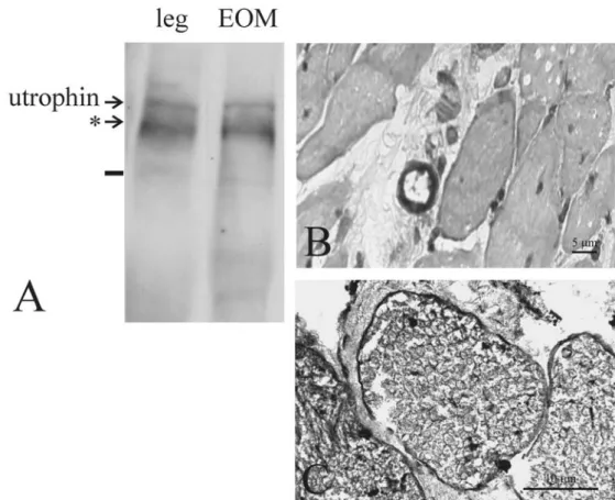
Fig. 2 Utrophin was detected with Western blot in leg muscles and extraocular muscles (EOM) (a). The double bands on both blots were interpreted as degradation products (*). Staining with indirect peroxidase immunohisto-chemistry in EOM showed a positive signal for utrophin in blood vessels (b) and nerves (c)
antibody against rapsyn. Slides from leg muscles (Fig.1d) and the EOM (Fig.1h) showed a positive staining of the AChR with rapsyn. The staining of the AChR with a-bungarotoxin showed again a positive result only in leg muscle but not in the EOM (data not shown). The Western blot analysis of both leg muscles and EOM showed a low level of expression for utrophin (Fig.2a). The double band on both blots could be due to degradation products.
Nerves and blood vessels
Staining of utrophin with indirect peroxidase reaction showed in EOM a positive result in blood vessels (Fig.2b) and nerves (Fig.2c). The same was found in leg muscles (data not shown).
Discussion
Specific skeletal muscles are involved in a selective manner during muscular dystrophy. It is well known that in mdx mouse the small-caliber skeletal muscle fibers are less affected although dystrophin is deficient in all muscle fibers (Karpati et al.1988). In DMD patients, EOM are structurally and functionally spared during the whole course of the disease. In hematoxylin and eosin staining, EOM lack the cardinal pathological manifestation of dystrophin deficiency such as hypertrophy, fiber size variation, fiber splitting, central nucleation, fatty degeneration and fibrotic scarring (Khurana et al.1995). Also, clinical study of the ocular motility of DMD pa-tients using infrared oculography showed the absence of altered function (Kaminski et al. 1992). The sparing of EOM in DMD may be linked to differences in mor-phology, cell and molecular biology of this muscle group compared with other skeletal muscles (Porter et al.1997). In this study, we evaluated the hypothesis that utrophin would also be expressed outside NMJ in normal EOM and thus could replace dystrophin in DMD. We used immunohistochemical methods to investigate distribution of utrophin along the sarco-lemmal membrane in normal EOM and leg muscles of cats. For comparing the expression level of utrophin in these two muscles, we used Western blot. Our results clearly showed that in cat, there is a different expres-sion of utrophin in EOM compared with other skeletal muscles. In leg muscles, the distribution of utrophin was restricted to NMJ while in EOM, we could not detect any immunoreactivity of utrophin. Another interesting finding was that by using a-bungarotoxin, we could detect NMJ in leg muscles but not in EOM while staining of AChE showed NMJ in both muscles. For checking these results, a double staining of AChR with a-bungarotoxin and an antibody against rapsyn was performed. In leg muscles, AChR were detected with both a-bungarotoxin and rapsyn. In EOM, there
was again no signal for a-bungarotoxin although NMJ could be shown with rapsyn.
Our study shows two surprising findings in EOM of normal cat: the lack of utrophin at NMJ and the failure of staining AChR with a-bungarotoxin. To our knowl-edge, there is no study about the distribution of utrophin in EOM of cat. Further, we also could not find any data about staining with a-bungarotoxin NMJ in EOM of cat.
We are aware that our results contradict reports from other groups. In different studies, the expression of utrophin in EOM was examined (Khurana et al. 1995; Porter et al. 1998). In addition, various papers showed a-bungarotoxin staining in EOM of a variety of species (Briggs and Schachat2002; Khanna et al.2003; Kusner et al. 1999; Lukas et al. 2000). None of these treatises were done in EOM of cat. Nevertheless, there are dif-ferent reasons which could let assume that our negative results are not true and were caused by processing ar-tefacts:
1. The NMJ was missing in the serial sections chosen for the double staining of utrophin and a-bungarotoxin. To exclude this possibility, different samples from all three cats were cut along the tendon-to-tendon length of the muscle. The results presented in Fig. 1e–g are representative of our findings in all these sections. 2. Our protocol for the staining of NMJ with
a-bun-garotoxin was not suitable for EOM. To find out if this could be true, we applied the same protocol in EOM of a rat. Like in previous reports, we were able to detect NMJ with a-bungarotoxin in this species. On account of all the controls performed, we believe that it is rather unlikely that our results were caused by a technical failure.
In normal mature striated muscle, utrophin expres-sion is also detected in the wall of blood vessels and in the perineurium and Schwann cells of intramuscular nerves (Karpati et al.1993). In EOM of cat, we did not find utrophin at the NMJ, but we were able to detect utrophin in blood vessels and nerves of this muscle group. This result explains why the Western blot of EOM showed a low expression level for utrophin al-though in the immunohistochemical analysis of this muscle group, no utrophin was found at NMJ.
The AChR are a large complex of four transmem-brane glycoprotein subunits, which form an a2bcd
pen-tameric complex (Raftery et al. 1980). a-bungarotoxin binds to AChR by interacting with the extracellular part of a-subunits (Harel et al. 2001; Moise et al. 2002; Young et al. 2003). Rapsyn interacts with the cytoplas-matic part of all subunits of AChR (Huebsch and Maimone 2003; Maimone and Merlie 1993; Willmann and Fuhrer 2002). Therefore, one possible explanation as to why in EOM of cats NMJ are detectable with rapsyn but not with a-bungarotoxin could be that in this species, a-subunits of AChR do not have the binding site for a-bungarotoxin.
At NMJ, utrophin is found at the crests of junctional folds whereas dystrophin occurs mainly in troughs (Be-wick et al.1992). AChR are also concentrated at the crests (Flucher and Daniels1989). The clear functional role of utrophin and its interaction with AChR remain unclear. There are different explanations as to why we could not detect utrophin expression at NMJ in EOM of cat. One could be that in this species, utrophin is expressed at NMJ of EOM without the epitope that is recognized by the antibody we used in our study. Another interpretation of the outcome of our study would be that utrophin does not occur at NMJ of EOM because its expression depends on the structure of a-subunits of AChR.
We believe that the lack of utrophin at NMJ in EOM of cat is not an artefact but a true result. A possible explanation could be that in EOM of cat, NMJ are ar-ranged differently from other species. Further studies are needed for a better characterization of AChR and their subunits in EOM of cat. Also, a more specific method than Western blot has to be used to better distinguish between expression of utrophin in muscle fibers of EOM and their blood vessels and nerves.
Acknowledgements We thank Dr. Jean-Marc Burgunder for the discussions that led to the idea for the study. We are grateful to Dr. Christian Fuhrer from the Brain Research Institute at the Uni-versity of Zu¨dieresisrich-Irchel for kindly providing antibodies against rapsyn. We also thank Theres Lauterburg, Astrid Rhyner, and Sulamith Gehr for their technical assistance.
References
Anderson LV, Davison K (1999) Multiplex Western blotting sys-tem for the analysis of muscular dystrophy proteins. Am J Pathol 154:1017–1022
Bewick GS, Nicholson LV, Young C, O‘Donnell E, Slater CR (1992) Different distributions of dystrophin and related proteins at nerve–muscle junctions. Neuroreport 3:857–860
Briggs MM, Schachat F (2002) The superfast extraocular myosin (MYH13) is localized to the innervation zone in both the global and orbital layers of rabbit extraocular muscle. J Exp Biol 205:3133–3142
Brown RH, Jr (1997) Dystrophin-associated proteins and the muscular dystrophies. Annu Rev Med 48:457–466
Bulfield G, Siller WG, Wight PA, Moore KJ (1984) X chromo-some-linked muscular dystrophy (mdx) in the mouse. P Natl Acad Sci USA 81:1189–1192
Carpenter JL, Hoffman EP, Romanul FC, Kunkel LM, Rosales RK, Ma NS, Dasbach JJ, Rae JF, Moore FM, McAfee MB et al.(1989) Feline muscular dystrophy with dystrophin defi-ciency. Am J Pathol 135:909–919
Cooper BJ (1989) Animal models of Duchenne and Becker mus-cular dystrophy. Br Med Bull 45:703–718
Cooper BJ, Valentine BA, Wilson S, Patterson DF, Concannon PW (1988a) Canine muscular dystrophy: confirmation of X-linked inheritance. J Hered 79:405–408
Cooper BJ, Winand NJ, Stedman H, Valentine BA, Hoffman EP, Kunkel LM, Scott MO, Fischbeck KH, Kornegay JN, Avery RJ et al. (1988b) The homologue of the Duchenne locus is defective in X-linked muscular dystrophy of dogs. Nature 334:154–156
Flucher BE, Daniels MP (1989) Distribution of Na+ channels and ankyrin in neuromuscular junctions is complementary to that of acetylcholine receptors and the 43 kd protein. Neuron 3:163–175
Gaschen FP, Hoffman EP, Gorospe JR, Uhl EW, Senior DF, Cardinet GH III, Pearce LK (1992) Dystrophin deficiency causes lethal muscle hypertrophy in cats. J Neurol Sci 110:149– 159
Harel M, Kasher R, Nicolas A, Guss JM, Balass M, Fridkin M, Smit AB, Brejc K, Sixma TK, Katchalski-Katzir E, Sussman JL, Fuchs S (2001) The binding site of acetylcholine receptor as visualized in the X-Ray structure of a complex between alpha-bungarotoxin and a mimotope peptide. Neuron 32:265–275 Hoffmann EP (2001) Dystrophinopathies. In: Karpati G,
Hilto-Jones D, Griggs RC (eds.) Disorders of voluntary muscle. Cambridge University Press, Cambridge, pp. 385–432 Huebsch KA, Maimone MM (2003) Rapsyn-mediated clustering of
acetylcholine receptor subunits requires the major cytoplasmic loop of the receptor subunits. J Neurobiol 54:486–501 Kaminski HJ, al-Hakim M, Leigh RJ, Katirji MB, Ruff RL (1992)
Extraocular muscles are spared in advanced Duchenne dystro-phy. Ann Neurol 32:586–588
Karnovsky MJ, Roots L (1964) A direct-coloring thiocholine method for cholinesterases. J Histochem Cytochem 12:219–221 Karpati G, Carpenter S, Prescott S (1988) Small-caliber skeletal muscle fibers do not suffer necrosis in mdx mouse dystrophy. Muscle Nerve 11:795–803
Karpati G, Carpenter S, Morris GE, Davies KE, Guerin C, Hol-land P (1993) Localization and quantitation of the chromosome 6-encoded dystrophin- related protein in normal and patho-logical human muscle. J Neuropathol Exp Neurol 52:119–128 Khanna S, Richmonds CR, Kaminski HJ, Porter JD (2003)
Molecular organization of the extraocular muscle neuromus-cular junction: partial conservation of and divergence from the skeletal muscle prototype. Invest Ophth Vis Sci 44:1918–1926 Khurana TS, Hoffman EP, Kunkel LM (1990) Identification of a
chromosome 6-encoded dystrophin-related protein. J Biol Chem 265:16717–16720
Khurana TS, Watkins SC, Kunkel LM (1992) The subcellular distribution of chromosome 6-encoded dystrophin-related protein in the brain. J Cell Biol 119:357–366
Khurana TS, Prendergast RA, Alameddine HS, Tome FM, Far-deau M, Arahata K, Sugita H, Kunkel LM (1995) Absence of extraocular muscle pathology in Duchenne’s muscular dystro-phy: role for calcium homeostasis in extraocular muscle spar-ing. J Exp Med 182:467–475
Kornegay JN, Tuler SM, Miller DM, Levesque DC (1988) Mus-cular dystrophy in a litter of golden retriever dogs. Muscle Nerve 11:1056–1064
Kusner LL, Kim E, Kaminski HJ (1999) Heme oxygenase-2 expression at rat neuromuscular junctions. Neurosci Lett 273:143–146
Lin S, Burgunder JM (2000) Utrophin may be a precursor of dystrophin during skeletal muscle development. Brain Res Dev Brain Res 119:289–295
Lin S, Gaschen F, Burgunder JM (1998) Utrophin is a regenera-tion-associated protein transiently present at the sarcolemma of regenerating skeletal muscle fibers in dystrophin- deficient hypertrophic feline muscular dystrophy. J Neuropathol Exp Neurol 57:780–790
Love DR, Morris GE, Ellis JM, Fairbrother U, Marsden RF, Bloomfield JF, Edwards YH, Slater CP, Parry DJ, Davies KE (1991) Tissue distribution of the dystrophin-related gene product and expression in the mdx and dy mouse. P Natl Acad Sci USA 88:3243–3247
Lukas JR, Blumer R, Denk M, Baumgartner I, Neuhuber W, Mayr R (2000) Innervated myotendinous cylinders in human extra-ocular muscles. Invest Ophth Vis Sci 41:2422–2431
Maimone MM, Merlie JP (1993) Interaction of the 43 kd post-synaptic protein with all subunits of the muscle nicotinic ace-tylcholine receptor. Neuron 11:53–66
Moise L, Piserchio A, Basus VJ, Hawrot E (2002) NMR structural analysis of alpha-bungarotoxin and its complex with the prin-cipal alpha-neurotoxin-binding sequence on the alpha 7 subunit of a neuronal nicotinic acetylcholine receptor. J Biol Chem 277:12406–12417
Nguyen TM, Ellis JM, Love DR, Davies KE, Gatter KC, Dickson G, Morris GE (1991) Localization of the DMDL gene-encoded dystrophin-related protein using a panel of nineteen monoclo-nal antibodies: presence at neuromuscular junctions, in the sarcolemma of dystrophic skeletal muscle, in vascular and other smooth muscles, and in proliferating brain cell lines. J Cell Biol 115:1695–1700
Porter JD, Karathanasis P, Bonner PH, Brueckner JK (1997) The oculomotor periphery: the clinician’s focus is no longer a basic science stepchild. Curr Opin Neurobiol 7:880–887
Porter JD, Rafael JA, Ragusa RJ, Brueckner JK, Trickett JI, Davies KE (1998) The sparing of extraocular muscle in dys-trophinopathy is lost in mice lacking utrophin and dystrophin. J Cell Sci 111:1801–1811
Raftery MA, Hunkapiller MW, Strader CD, Hood LE (1980) Acetylcholine receptor: complex of homologous subunits. Sci-ence 208:1454–1456
Ragusa RJ, Chow CK, St Clair DK, Porter JD (1996) Extraocular, limb and diaphragm muscle group-specific antioxidant enzyme
activity patterns in control and mdx mice. J Neurol Sci 139: 180–186
Takemitsu M, Nonaka I, Sugita H (1993) Dystrophin-related protein in skeletal muscles in neuromuscular disorders: immu-nohistochemical study. Acta Neuropathol 85:256–259
Willmann R, Fuhrer C (2002) Neuromuscular synaptogenesis: clustering of acetylcholine receptors revisited. Cell Mol Life Sci 59:1296–1316
Winand NJ, Edwards M, Pradhan D, Berian CA, Cooper BJ (1994) Deletion of the dystrophin muscle promoter in feline muscular dystrophy. Neuromuscular Disord 4:433–445
Worton R (1995) Muscular dystrophies: diseases of the dystrophin-glycoprotein complex. Science 270:755–756
Young HS, Herbette LG, Skita V (2003) alpha-bungarotoxin binding to acetylcholine receptor membranes studied by low angle X-ray diffraction. Biophys J 85:943–953