HAL Id: hal-02448295
https://hal.archives-ouvertes.fr/hal-02448295
Submitted on 7 Jun 2021
HAL is a multi-disciplinary open access
archive for the deposit and dissemination of
sci-entific research documents, whether they are
pub-lished or not. The documents may come from
teaching and research institutions in France or
abroad, or from public or private research centers.
L’archive ouverte pluridisciplinaire HAL, est
destinée au dépôt et à la diffusion de documents
scientifiques de niveau recherche, publiés ou non,
émanant des établissements d’enseignement et de
recherche français ou étrangers, des laboratoires
publics ou privés.
Type 4 Metabotropic Glutamate Receptor
Bruno Vilar, Jérôme Busserolles, Bing Ling, Sophie Laffray, Lauriane Ulmann,
Fanny Malhaire, Eric Chapuy, Youssef Aissouni, Monique Etienne, Emmanuel
Bourinet, et al.
To cite this version:
Bruno Vilar, Jérôme Busserolles, Bing Ling, Sophie Laffray, Lauriane Ulmann, et al.. Alleviating
Pain Hypersensitivity through Activation of Type 4 Metabotropic Glutamate Receptor. Journal of
Neuroscience, Society for Neuroscience, 2013, 33 (48), pp.18951-18965.
�10.1523/JNEUROSCI.1221-13.2013�. �hal-02448295�
Neurobiology of Disease
Alleviating Pain Hypersensitivity through Activation of Type
4 Metabotropic Glutamate Receptor
Bruno Vilar,
1,2Je´roˆme Busserolles,
3,4,Bing Ling,
3,4Sophie Laffray,
1,2Lauriane Ulmann,
1,2Fanny Malhaire,
1,2Eric Chapuy,
3,4Youssef Aissouni,
3,4Monique Etienne,
3,4Emmanuel Bourinet,
1,2Francine Acher,
5Jean-Philippe Pin,
1,2Alain Eschalier,
3,4,6and Cyril Goudet
1,21Institut de Ge´nomique Fonctionnelle, Centre National de la Recherche Scientifique Unite´ Mixte de Recherche 5203, Universite´ de Montpellier, F-34094 Montpellier, France,2Institut National de la Sante´ et de la Recherche Me´dicale, U661, F-34094 Montpellier, France,3Clermont Universite´, Universite´ d’Auvergne, Pharmacologie Fondamentale et Clinique de la Douleur, F-63000 Clermont-Ferrand, France,4Inserm, U 1107, Neuro-Dol, F-63001 Clermont-Ferrand, France,5Laboratoire de Chimie et Biochimie Pharmacologiques et Toxicologiques, Centre National de la Recherche Scientifique Unite´ Mixte de Recherche 8601, Universite´ Paris Descartes, Sorbonne Paris Cite´, F-75270 Paris, France, and6CHU Clermont-Ferrand, Service de pharmacologie, F-63003 Clermont-Ferrand, France
Hyperactivity of the glutamatergic system is involved in the development of central sensitization in the pain neuraxis, associated with
allodynia and hyperalgesia observed in patients with chronic pain. Herein we study the ability of type 4 metabotropic glutamate receptors
(mGlu4) to regulate spinal glutamate signaling and alleviate chronic pain. We show that mGlu4 are located both on unmyelinated C-fibers
and spinal neurons terminals in the inner lamina II of the spinal cord where they inhibit glutamatergic transmission through coupling to
Cav2.2 channels. Genetic deletion of mGlu4 in mice alters sensitivity to strong noxious mechanical compression and accelerates the onset
of the nociceptive behavior in the inflammatory phase of the formalin test. However, responses to punctate mechanical stimulation and
nocifensive responses to thermal noxious stimuli are not modified. Accordingly, pharmacological activation of mGlu4 inhibits
mechan-ical hypersensitivity in animal models of inflammatory or neuropathic pain while leaving acute mechanmechan-ical perception unchanged in
naive animals. Together, these results reveal that mGlu4 is a promising new target for the treatment of chronic pain.
Key words: allodynia; analgesia; GPCR; hyperalgesia; mGlu; pain
Introduction
Chronic pain is among the most debilitating and costly afflictions
in North America and Europe, seriously affecting the quality of
life of
⬎19% of adult Europeans (
Verhaak et al., 1998
;
Ospina
and Harstall, 2002
;
Breivik et al., 2006
;
Bouhassira et al., 2008
).
With regard to neuropathic pain, the burden of disease is further
worsened by the large proportion of patients whose treatment
does not offer significant relief (
Mico´ et al., 2006
;
Finnerup et al.,
2010
), highlighting the crucial need of progress in the
manage-ment of pain. Comprehensive understanding of the cellular and
molecular mechanisms involved in chronic pain may in turn lead
to the development of effective targeted therapies.
The blockade of increased glutamatergic activity may
repre-sent a pivotal means of reducing chronic pain but await a clearer
identification of adequate targets. Central sensitization of the
pain neuraxis is associated with hyperexcitability of the
glutama-tergic system and leads to the development of the evoked pain
symptoms, allodynia and hyperalgesia (
Latremoliere and Woolf,
2009
). At the synaptic level, glutamate activity is mediated
through two classes of receptors, ionotropic receptors (iGluRs),
which are ligand-gated ion channels responsible for the fast
syn-aptic response, and metabotropic receptors (mGluRs), which are
G-protein-coupled receptors that modulate synaptic activity.
Both iGluRs and mGluRs are involved in the induction and the
maintenance of central sensitization.
Given that mGluRs are expressed all along the pain neuraxis
where they modulate the perception of pain (
Varney and Gereau,
2002
;
Neugebauer, 2007
;
Goudet et al., 2009
;
Chiechio and
Nico-Received March 21, 2013; revised Oct. 21, 2013; accepted Oct. 24, 2013.
Author contributions: B.V., J.B., S.L., E.B., A.E., and C.G. designed research; B.V., J.B., B.L., S.L., L.U., F.M., E.C., Y.A., M.E., and C.G. performed research; F.A. contributed unpublished reagents/analytic tools; B.V., J.B., B.L., S.L., L.U., F.M., E.C., Y.A., E.B., J.-P.P., A.E., and C.G. analyzed data; B.V., J.B., J.-P.P., A.E., and C.G. wrote the paper.
This work was supported by Eranet Neuron and Agence Nationale de la Recherche Grants ANR-08-NEUR-006, ANR-09-MNPS-037, and 2010 ANR-MALZ105– 01, La Marato de TV3, Fondation pour la Recherche Me´dicale (Equipe FRM DEQ20130326522), Fondation pour la Recherche sur le Cerveau, Institut UPSA de la Douleur, and Socie´te´ Franc¸aise d’Etude et de Traitement de la Douleur. B.V. was supported by a fellowship from the Ministère de la Recherche et de la Technologie. We thank Yves Le Feuvre and Fre´de´ric Nagy from Institut National de la Sante´ et de la Recherche Me´dicale U862 and Federica Bertaso from IGF Centre National de la Recherche Scientifique Unite´ Mixte de Recherche 5203 for their technical help on electrophysiology on slices; Julien Cau from the Montpellier RIO Imaging facility of campus Centre National de la Recherche Scientifique Arnaud de Villeneuve and Delphine Rigault from Centre National de la Recherche Scientifique Unite´ Mixte de Recherche 8601 for the synthesis of LSP4 –2022; David Hampson for kindly providing mGlu4KO mice; and Gregory Stewart and Ebba L. Lagerqvist from IGF Centre National de la Recherche Scientifique Unite´ Mixte de Recherche 5203 for critical reading of the manuscript. The IGF belongs to the Laboratories of Excellence, Ion Channel Science and Therapeutics, and EpiGenMed.
The authors declare no competing financial interests.
Correspondence should be addressed to either of the following: Dr. Cyril Goudet, Institut de Ge´nomique Fonc-tionnelle, Centre National de la Recherche Scientifique Unite´ Mixte de Recherche 5203, Institut National de la Sante´ et de la Recherche Me´dicale U661, Universite´ de Montpellier, 141, Rue de la Cardonille, F34094 Montpellier cedex 5, France, E-mail: cyril.goudet@igf.cnrs.fr; or Prof. Alain Eschalier, Institut National de la Sante´ et de la Recherche Me´dicale Unite´ Mixte de Recherche 1107, Pharmacologie Fondamentale et Clinique de la Douleur, Universite´ d’Auvergne, 28, Place Henri Dunant, F63001 Clermont-Ferrand cedex 1, France, E-mail: alain.eschalier@udamail.fr.
DOI:10.1523/JNEUROSCI.1221-13.2013
letti, 2011
), the selective targeting of mGluRs to modulate
synap-tic activity could constitute a viable alternative to the direct
blockade of synaptic glutamate transmission by iGluRs
antago-nists. Eight mGluRs have been identified so far (mGlu1-mGlu8)
and classified in three groups (
Niswender and Conn, 2010
;
Nico-letti et al., 2011
). Group I mGluRs (mGlu1, mGlu5) are
postsyn-aptic receptors that positively modulate neuronal excitability,
whereas Group II (mGlu2-mGlu3) and Group III (mGlu4,
mGlu6-mGlu8) are mainly presynaptic receptors that exert a
negative retrocontrol on neurotransmission. The role of Group
III mGluRs in pain has been less studied than the other groups,
but their ability to reduce hyperalgesia in animal models of
chronic pain (
Fisher et al., 2002
;
Mills et al., 2002
;
Chen and Pan,
2005
;
Goudet et al., 2008
), and to regulate neurotransmission in
the dorsal horn of spinal cord (
Chen and Pan, 2005
;
Zhang et al.,
2009
;
Carlton et al., 2011
;
Wang et al., 2011
), reveals their
poten-tial for the development of analgesics. However, the precise role
and contribution of each of the Group III subtypes at the spinal
cord level on pain modulation remain to be elucidated.
In the present study, we focused our attention on mGlu4. We
have determined its precise localization in sensory neurons and
spinal interneurons. We have evaluated its modulation of
gluta-matergic neurotransmission on spinal cord slices of naive or
in-flamed animals. Then, combining selective pharmacological and
genetic tools in behavioral studies, we highlighted the key role of
presynaptic mGlu4 receptors in the modulation of mechanical
hypersensitivity in inflammation or nerve injury, thus
exempli-fying the significant potential of mGlu4 receptors as a therapeutic
target for the treatment of chronic pain.
Materials and Methods
Animals
Animal care and experiments were performed in accordance with the Committee for Research and Ethical Issues of the International Associa-tion for the Study of Pain (Zimmermann, 1983).
Male Sprague Dawley rats were purchased from Charles River, and C57BL/6 mice from Elevage Janvier. Homozygous mGlu4KO mice and wild-type littermates were generated from crosses between heterozygous animals. mGlu4KO mice were first described byPekhletski et al. (1996), and their genotyping was performed according to the method described byPitsch et al. (2007). Animals were housed under controlled environ-mental conditions (22°C; 55% humidity) and kept under a 12/12 h light/ dark cycle, with food and water ad libitum for a week before start the experiments to acclimatize.
Immunofluorescence
The mice were anesthetized with pentobarbital (100 mg/kg,i.p.), and tissues were fixed via transcardiac perfusion of 4% paraformaldehyde in PBS and postfixed overnight. Transverse lumbar spinal cords slices of 30 m were cut with a vibratome.
Transverse sections of mouse spinal cords were preincubated for 1 h in PBS containing 0.3% Triton X-100 and 5% donkey serum and then immunostained overnight at 4°C in the same buffer containing primary antibodies. After three washes, sections were incubated in a conjugated secondary donkey antibody for 2 h at room temperature.
The following antibodies were purchased from commercial sources: rab-bit anti-mGlu4a (Invitrogen, 51–3100; 1/100), rabrab-bit anti-PKC␥ (Santa Cruz Biotechnology, sc-166385; 1/500), mouse anti-NF200 (Sigma, clone N52, N0142; 1/400), mouse anti-CGRP (Santa Cruz Biotechnology, sc-8857; 1/50), guinea pig VGLUT3 (Millipore, AB5421; 1/5000), rat anti-Substance P (Medimabs, NC1/34; 1/25), IB4 FITC conjugate (Sigma, L 2895; 1/100), mouse anti-bassoon (Enzo Life Sciences, ADI-VAM-PS003; 1/1000), mouse anti-synaptophysin (Sigma, S5768; 1/1000), donkey anti-rabbit Alexa-546 (Invitrogen), donkey anti-rabbit DyLight 488 (Jackson noResearch Laboratories; 1/500), donkey anti-mouse FITC (Jackson Immu-noResearch Laboratories; 1/500), donkey anti-guinea pig DyLight 488
(Jackson ImmunoResearch Laboratories; 1/500), and donkey anti-rat FITC (Jackson ImmunoResearch Laboratories; 1/500).
Immunostained sections were mounted with ProLong Gold (Invitro-gen), viewed with a Bio-Rad MRC 1024 or a Zeiss LSM510 META con-focal microscope, and images were analyzed with ImageJ.
Rhizotomy
Mice were anesthetized with an intraperitoneal injection of ketamine/ xylazine (100 mg/kg and 10 mg/kg, respectively). A 1-cm-long incision was made in the skin of the back, and the L4 spinal cord segment was exposed by laminectomy. A pool was formed around the exposed spinal segment with the incised skin and filled with artificial CSF (ACSF). A small hole was made in the dura overlying the dorsal root branches that enter the L4 lumbar segment. A fine forceps (Dumont #55) was intro-duced subdurally, and the dorsal root branches were resected with micro-scissors. To minimize scar formation on the spinal tissue, a piece of artificial dura (Gore Preclude MVP Dura Substitute, W.L. Gore and Associates) was used to cover the laminectomy site. Musculature and skin were then closed with sterile 4 – 0 Vicryl sutures. Animals were given subcutaneous injections of 0.9% NaCl sterile solution to prevent dehy-dration and kept on a heating pad until fully awake. Animals were housed in individual cages and killed15 d later for immunohistochemical analysis.
Spinal cord transmission
Spinal cord slice preparation. Young C57BL/6 mice (14 –19 d old) were
anesthetized with isoflurane, and the lumbar segment of spinal cord at L4 to L6 level was rapidly removed and immediately placed in an ice-cold dissection solution presaturated with 95% O2and 5% CO2. This solution
contained (in mM) as follows: glucose, 25; NaCl, 101; KCl, 3.8; MgCl2,
18.7; MgSO4, 1.3; KH2PO4, 1.2; HEPES, 10; CaCl2, 1; pH 7.3. The tissue was then placed in a groove formed in an agarose block and glued onto the metal block of a vibratome (Vibratome 3000). Transverse spinal cord slices were cut to 300m in the ice-cold dissection solution and then preincubated in ACSF oxygenated with 95% O2and 5% CO2at room
temperature for at least 1 h before being transferred to the recording chamber. The ACSF contained (in mM) as follows: glucose, 10; NaCl,
130.5; KCl, 2.4; MgSO4, 1.3; KH2PO4, 1.2; HEPES, 1.25; CaCl2, 2.4;
NaHCO3, 19.5; pH 7.3. In the recording chamber, the slice was
contin-uously perfused with ACSF solution at 31°C. In the experiments per-formed on spinal cord of inflamed animals, inflammation was induced by bilateral injections of 15l of complete Freund’s adjuvant (CFA) 24 h before the slice preparation.
Electrophysiological recording. Recordings of postsynaptic currents
were performed using the whole-cell voltage-clamp method (holding potential⫺70 mV). The spinal lamina II has a distinct translucent ap-pearance and can easily be distinguished under a microscope. Lamina II neurons in the spinal slice were visualized with infrared optics using an X40 0.80 water-immersion objective on an Olympus BX50WI upright microscope equipped with a video camera system (COHU 4912).
Pipettes of typical resistance of 6 –9M⍀, made of borosilicate glass,
were filled with an internal solution containing (in mM) the following: K-gluconate, 135; KCl, 5; MgCl2, 2; CaCl2, 0.5; HEPES, 5; EGTA, 5;
ATP-Mg, 5; Tris-GTP, 0.5; adjusted to pH 7.3. In GDP-S experiments, Tris-GTP was replaced by GDP-S (1 mM), a nonhydrolyzable analog of
GDP (Eckstein et al., 1979), as previously described byHolz et al. (1986). Recordings of EPSCs in whole-cell patch-clamp using an Axopatch 200B amplifier (Molecular Devices) began after the current reached a steady state. The evoked EPSCs (eEPSCs) of lamina II neurons were induced by an electrical stimulation with a fixed intensity (0.4 ms,⬃0.4 mA, and 0.2 Hz) through a bipolar electrode (Pt/Ir) placed on the dorsal root entry zone.
Recordings were filtered at 2 kHz. Data were analyzed using pCLAMP10 (Molecular Devices) and GraphPad Prism (GraphPad) soft-ware. In all experiments, a single neuron per slice was recorded.
All data are expressed as the mean⫾ SEM, and statistical significance was assessed with an unpaired Student’s t test, with p⬍ 0.05 (two-tailed) considered being significant.
Behavioral testing in mice
Mice were acclimatized to handling and testing procedures twice a day for 3 d before behavioral testing.
Tail immersion test. The tail of the mice was immersed in a water bath
maintained at 4, 10, 15, 42, 46, 48, and 50°C. The latency time for tail withdrawal was determined and a cutoff time of 30 s was set to avoid injury.
Paw lift tests. Mechanical allodynia and hyperalgesia were assessed on
mice using the von Frey hair filaments of three different bending forces (0.07 g, corresponding to an innocuous stimulation, 0.6 g intermediate, and 1.4 g noxious). The filaments were pressed perpendicularly to the plantar surface of the hindpaw until they bent. For each filament, five stimuli were applied with an interval of 3–5 s. Data were analyzed by a two-way ANOVA and Dunnett’s post hoc test, for time course studies. One-way ANOVA with a Newman-Keuls post hoc test was used to analyze the effect of the different treatments determined by the AUCs. The level of statistical significance was set at p⬍ 0.05.
Formalin test. After acclimatization for 20 min in the test chamber,
mice received 20l of 5% formalin injected subcutaneously into the plantar surface of the hindpaw. They were then placed in a Plexiglas box. Biting and licking of the injected paw were monitored, and the total duration of these actions in seconds was measured during the two peaks of the typical biphasic pain behavior. The spontaneous aversive response corresponding to the early phase was assessed during the first 10 min. The second peak of aversive behavior was observed from 15 to 50 min after formalin administration. Data were analyzed by a two-way ANOVA and Bonferroni’s post hoc test.
Behavioral testing in rats
Paw pressure test in rats. Rats were submitted to the paw pressure test
previously described byRandall and Selitto (1957). Nociceptive thresh-olds, expressed in grams, were measured with an Ugo Basile analgesim-eter (Apelex, tip diamanalgesim-eter of the probe 1 mm, weight 30 g) by applying an increasing pressure to the right hindpaw of rats until a squeak (vocaliza-tion threshold) was obtained (cutoff was 750 g, except for carrageenan-treated animals for which the cutoff was 500 g). Before treatments, rats were habituated to the test by handling without submitting them to paw pressure. Then, after having obtained two consecutive stable vocalization threshold values, treatment effects were assessed after 15, 30, 45, 60, 90, and 120 min. The results are expressed as vocalization thresholds, in grams. To investigate global effects, areas under the time course curves (AUCs, g.min) of the antihyperalgesic effects were calculated from indi-vidual scores at each time, using the trapezoidal method. Data were analyzed by a two-way ANOVA followed by a Bonferroni’s test, when the time course of the effects was studied. One-way ANOVA followed by a Student-Newman-Keuls’ test was used to analyze the effect of the differ-ent treatmdiffer-ents determined by the AUCs. The level of statistical signifi-cance was set at p⬍ 0.05.
Inflammation or neuropathic pain models
Carrageenan-induced inflammatory mechanical hyperalgesia. Thresholds
to mechanically induced vocalization were assessed with animals pre-senting hyperalgesia elicited by a subcutaneous injection of 2% -carrageenan (200 l for rat and 20 l for mice) into the right hindpaw.
Chronic constriction injury (CCI) model. Unilateral peripheral
monon-europathy was induced according to the method described byBennett and Xie (1988). Briefly, after determining vocalization thresholds, rats were anesthetized with sodium pentobarbital (50 mg/kg i.p.) and four chromic gut (5– 0) ligatures were tied loosely (with⬃1 mm spacing) around the right common sciatic nerve. The nerve was constricted to a barely discernible degree, so that circulation through the epineurial vas-culature was not interrupted. Only animals presenting a decreaseⱖ15% of the presurgery value of vocalization threshold were selected.
Experimental procedure
For all experiments, unless stated otherwise, treatments were injected intrathecally. To avoid uncontrolled environmental influences, a block procedure was used whereby animals were tested in groups, with the number of animals in each group corresponding to the number of con-ditions of the experiment. Therefore, all the concon-ditions were tested at
once on the different individuals of the group in the same short lapse of time. For example, for an experiment with five different conditions (e.g., negative control, positive control, and three different doses of drug), each group contained five animals, each of which received one of the five treatments of the experiment. Treatments were randomized and per-formed blind. Experiments were perper-formed in a quiet room where ani-mal behaviors were observed by a single experimenter.
Intrathecal injections were performed under isoflurane anesthesia (4% induction, 2% maintenance), as previously described (Mestre et al., 1994). Briefly, the anesthetized animal was held in one hand by the pelvic girdle, and a 25-gauge X1-inch needle connected to a 25l Hamilton syringe was inserted into the subarachnoid space between lumbar verte-brae L5 and L6, until a tail flick was elicited. The syringe was held in position for few seconds after the injection of a volume of 10l/rat and of 5l/mouse.
Knockdown of spinal mGlu4 receptor expression by
antisense oligonucleotides
An antisense (AS) oligonucleotide was designed based on rat mGlu4 sequences (GenBank gene ID: 24417) in regions lacking known splice variants (AS: TAAAGGCTGAGGAGTAGG). A scramble oligonucleo-tide control was used (scrambled control: GCCTGCTAGAATGC-CATT). The absence of complementarity of the scrambled control to any registered nucleotide sequences was verified by a blast search in the Gen-Bank. They were synthesized by Eurofins MWG Operon.
The oligonucleotides were reconstituted in saline before administra-tion. Intrathecal injection of oligonucleotides (AS/scrambled control, 12.5g) or saline was performed using a volume of 10 l via direct transcutaneous injection between the dorsal aspects of L5 and L6 under slight anesthesia with volatile isoflurane (3.5%). This treatment was re-peated twice daily for 4 d. The studies were performed in naive, inflamed, and mononeuropathic rats. Pain scores were determined before oligonu-cleotide treatments and then on day 4. In each experiment, 6 –10 animals per group were used.
Sample collection
Four days after intrathecal injections of AS or mismatch (MM) oligonu-cleotide, the spinal cords were collected from CCI and carrageenan-treated rats (n ⫽ 4 for each group). Animals were first deeply anesthetized, and the L4 –L6 spinal cord segments were then extracted and immediately placed on ice-cooled glass dish. The spinal cords were further subdivided into dorsal and ventral halves by cutting straight across from the central canal laterally to a midpoint in the white matter.
Western blot
The ipsilateral and contralateral of each CCI or carrageenan-treated rat spinal cord were separated and subsequently processed for Western blot analysis. The L4 –L6 ipsilateral and contralateral spinal cord dorsal seg-ments were homogenized in lysis buffer containing 50 mMHEPES, pH
7.5, 1% Triton X-100, 10 mMEDTA, 150 mMNaCl, 10 mMNa4P2O7, 0.1
MNaF, 2 mMvanadate, 100 U/ml aprotinin, 20Mleupeptin, and 0.5 mM
PMSF. The total amount of protein in each sample was determined using the BCA Protein Assay Kit (Pierce-Thermo Scientific) before loading on polyacrylamide gels. Spinal cord homogenates (80g protein) were sep-arated using 7.5% SDS-PAGE and transferred to nitrocellulose. After the blots had been washed with TBST (10 mMTris-HCl, pH 7.6, 150 mM
NaCl, 0.05% Tween-20), the membranes were blocked with 5% skim milk for 1.5 h and incubated at 4°C overnight with a primary antibody specific for-actin (1:5000, loading control, Sigma) and mGlu4 (1 g/ ml, catalog #51–3100, Invitrogen). After washing with TBST, membranes were incubated for 1 h with HRP-conjugated rabbit IgG or anti-mouse IgG secondary antibody (1:10,000, Pierce-Thermo Scientific) to detect mGlu4 or-actin, respectively. The bands were visualized with SuperSignal WestPico chemioluminescent substrate (Pierce-Thermo Scientific). The positive pixel area of specific bands was measured with a computer-assisted image analysis system (ChemiDoc XRS, Bio-Rad) and normalized against the corresponding-actin loading control bands. The ratio of knockdown of spinal mGlu4 expression by AS oligonucleo-tide was calculated. The mean value of ipsilateral and contralateral spinal mGlu4 expression in animals treated by MM oligonucleotide was set
at 100%. Thus, the percentage change in the mGlu4 expression in each sample was calculated.
RNA expression
Extraction. Total RNAs were prepared from
pooled DRG (L4 –L6) using the TRIzol method according to the manufacturer’s instructions (Invitrogen). Reverse transcriptions were per-formed using 1g of total RNA with random primers and M-MuLV reverse transcriptase (New England Biolabs).
qPCR. The expression levels of genes were
determined by qPCR using SYBR Green and a Roche LightCycler 480. Primer pairs were val-idated using DNA plasmid of the gene of inter-est as a template. Experiments were performed on 2 ng of the cDNA product using 1⫻ SYBR Green PCR Master (Roche) and 300 nM con-centration of each primer pair.
Data were analyzed using the threshold cycle (Ct) relative quantification method.
Three housekeeping genes (HKGs) among the eight tested were chosen to normalize the results using the geNorm software. The
ex-pression level of each gene was normalized according to the formula 2⫺[Ct (gene) ⫺ mean Ct (HKG)]. Each bar graph represents the average
gene expression levels measured in DRG from at least three animals each being tested in three independent qPCR experiments.
qPCR data were analyzed with unpaired Student’s t test. Data are expressed as mean⫾ SEM, and the levels of significance were set at p ⬍ 0.05, p⬍ 0.01, and p ⬍ 0.001.
Drugs
ACPT-I was purchased from Tocris Bioscience,L-AP4, -conotoxin
GVIA,-agatoxin IVA, NBQX, and Tertiapin Q from Abcam Biochem-icals. LSP4 –2022 was synthesized in the laboratory of F.A. following a procedure analogous to that previously described (Selvam et al., 2010;
Acher et al., 2012). All solutions were prepared just before experiments.
Results
mGlu4 receptors are localized in the lamina II of the mice
spinal cord both in the terminals of unmyelinated afferents
and spinal neurons
The superficial laminae of the dorsal horn of the spinal cord
represent the area of the CNS where the first modulation of
pain-related information occurs. This region receives sensory
infor-mation from primary afferents responding to noxious and
non-noxious stimuli. This incoming information is then
pro-cessed by interneurons, important for maintaining normal
sen-sory function, and then transmitted to projection neurons for
relay to the brain (
Basbaum et al., 2009
;
Todd, 2010
).
Previous immunocytochemistry studies revealed the presence
of mGlu4 receptors in presynaptic elements from afferent fibers
in lamina II of the dorsal horn in the rat spinal cord (
Azkue et al.,
2001
). However, the nature of the neurons expressing this
recep-tor was not determined. We therefore performed
immunofluo-rescence staining in mice spinal cord slices and DRG with an
mGlu4 antibody and various markers of neurons involved in pain
transmission.
Immunoreactivity was absent in mGlu4KO mice both in
im-munofluorescence (
Fig. 1
A) and in Western blot experiments
(
Fig. 1
B), ruling out nonspecific labeling of the mGlu4 antibody.
Although the presence of mRNA encoding mGlu4 receptor has
previously been detected by in situ hybridization in neurons with
small- and medium-size bodies in the DRG (
Ohishi et al., 1995a
),
we could not detect mGlu4 receptor immunostaining in DRGs,
suggesting that their expression level in these neuronal cell bodies
is under the detection limit.
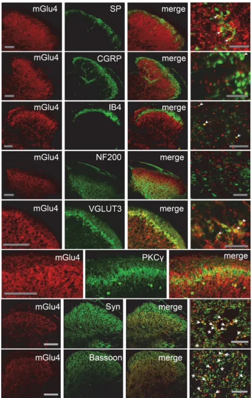
We observed that mGlu4 receptors are expressed mostly in the
inner lamina II of the dorsal horn of the spinal cord in mice (
Fig.
2
). In this lamina, we detected mGlu4 immunoreactivity in
C-tactile low threshold mechanical receptive (C-LTMR)
affer-ents expressing the vesicular glutamate transporter 3 (VGLUT3).
Of note, these C-LTMR neurons convey non-noxious touch
sen-sations in healthy conditions (
Lo¨ken et al., 2009
;
Li et al., 2011
)
and are involved in mechanical hypersensitivity associated with
chronic pain (
Seal et al., 2009
). A small proportion of mGlu4
receptor staining (⬍10%) merged with peptidergic and
nonpep-tidergic C fibers markers, such as substance P (SP), calcitonin
gene-related peptide (CGRP), or isolectin B4 (IB4), respectively.
The labeling of mGlu4 receptors also partially overlaps with
staining of protein kinase C␥ (PKC␥) interneurons in inner
lam-ina II (
Fig. 2
). PKC
␥-expressing interneurons are excitatory
neu-rons, which play an important role in the processing of tactile
inputs both in physiological and pathological conditions and are
notably thought to mediate injury-induced hypersensitivity
(
Malmberg et al., 1997
;
Polga´r et al., 1999
;
Miraucourt et al.,
2007
;
Neumann et al., 2008
;
Lu et al., 2013
). By contrast, mGlu4
receptor labeling does not significantly overlap with NF200
(
⬍1%), a marker of myelinated A or A␦ fibers. In accordance
with previous electronic microscopy data indicating that mGlu4
is commonly observed in presynaptic terminals in the dorsal
spinal cord (
Azkue et al., 2001
), most mGlu4 receptor
immuno-reactivity merges with that of synaptophysin, a membrane
glyco-protein characteristic of presynaptic vesicles (
Wiedenmann and
Franke, 1985
), and bassoon, a protein expressed at the
presynap-tic nerve terminals (
tom Dieck et al., 1998
).
To further examine the nature of neurons expressing mGlu4
receptors, we performed a unilateral dorsal rhizotomy of the
fourth lumbar cord segment on mice. Fifteen days after the
rhi-zotomy, we performed a triple immunostaining of mGlu4,
VGLUT3, and IB4 on cervical to lumbar spinal cord slices (
Fig.
3
). As expected, both IB4 and VGLUT3 labeling in superficial
layers of the dorsal horn of the fourth lumbar cord segment was
markedly reduced (⬎70%) on the ipsilateral, but not the
con-tralateral, side to the dorsal rhizotomy. On the other hand,
Figure 1. Lack of mGlu4 immunoreactivity in mGlu4KO mice. A, Double labeling of a transverse section of the lumbar spinal cord from mGlu4KO or WT mice with a rabbit antiserum against mGlu4 receptor (red) and isolectin B4 conjugated to FITC (IB4, green). Immunofluorescence staining of mGlu4 receptors is detected in inner lamina II and lamina III of dorsal horn in WT mice but not in mGlu4KO mice. Scale bars, 100m.B,WesternblotanalysisofmGlu4proteinlevelexpressioninlumbarspinalcordextractsfrom mGlu4KO or WT mice. Two specific bands at⬃95 and ⬃240 kDa corresponding to monomeric and dimeric mGlu4 receptors were detected in the extract from WT mice and absent in mGlu4KO mice (20g of total proteins). Immunoreactivity was absent in mGlu4KO mice both in immunofluorescence and Western blot experiments, thus ruling out nonspecific labeling of the mGlu4 antibody.
mGlu4 imnuoreactivity was only partially reduced after
rhizot-omy on the ipsilateral side (⬍25%, n ⫽ 4 independent
experi-ments), suggesting both a peripheral and spinal origin of these
processes.
The localization of mGlu4 receptors in
superficial layers of the dorsal horn of the
spinal cord, both in the terminals of
un-myelinated C-fibers and in spinal
in-terneurons, provides neuroanatomical
clues of the putative involvement of this
receptor in the modulation of pain.
Presynaptic mGlu4 receptors reduce
glutamatergic neurotransmission
through inhibition of Ca
V2.2 channels
in spinal cord slices
We then studied the role of mGlu4
recep-tors on glutamatergic neurotransmission
in lamina II dorsal horn neurons. Using
the patch-clamp technique, we measured
eEPSCs on lamina II neurons from
lum-bar (L4 –L6) spinal cord slices of 2- to
3-week-old mice.
Having confirmed that in young
ani-mals, as in adult mice, mGlu4 receptor
im-munostaining could be observed in lamina
II (
Fig. 4
A), we then compared the effects of
two ligands: LSP4 –2022, a recently
identi-fied mGlu4 selective agonist, and
L-AP4, a
nonselective Group III mGluRs agonist.
LSP4 –2022 is 100 and 500 times more
po-tent at mGlu4 than at mGlu7 and mGlu8,
respectively (
Goudet et al., 2012
). Both
li-gands significantly decreased the amplitude
of eEPSCs (
Fig. 4
B,C). Interestingly,
appli-cation of 5
MLSP4 –2022 or 50
M L-AP4
yielded to a similar inhibition of eEPSC
am-plitude by 43.9
⫾ 3.9% (n ⫽ 10) and 47.4 ⫾
4.4% (n
⫽ 9), respectively (
Fig. 4
C). At the
concentrations used, in addition to
activat-ing mGlu4,
L-AP4 will also activate mGlu8,
whereas LSP4 –2022 will not, suggesting
that most, if not all, of the effects are
medi-ated by mGlu4 receptor activation. The full
blockade of the eEPSCs by addition of 50
MNBQX, an iGluR (AMPA receptors)
an-tagonist, at the end of the experiment, proved
their glutamatergic nature.
In the next experiment, we replaced GTP
by GDP-
S, a stable GDP analog that blocks
G-protein-mediated responses, in the patch
pipette. Into the recorded neuron, GDP-
S
competes with GTP on G-proteins, thereby
blocking GTP-dependent activation of the
G-proteins by hormones and
neurotrans-mitters (
Holz et al., 1986
). This method has
been proven to efficiently block
postsynap-tic G-protein-mediated modulation of
glu-tamatergic transmission in various CNS
synapses, including spinal cord (e.g.,
Rebola
et al., 2008
;
Yang et al., 2011
;
Fan et al.,
2013
). However, in our experiments, the
in-hibition of eEPSC amplitude by LSP4 –2022
is not modified, suggesting that the effect of the drug on transmission
occurs through presynaptic receptors (
Fig. 4
D), in accordance with
immunofluorescence data showing colocalization of mGlu4 with
presynaptic markers (
Fig. 2
).
Figure 2. Localization of mGlu4 receptors in inner lamina II of the dorsal horn of mice spinal cord. Double immunolabeling of transverse sections of lumbar spinal cord of C57BL/6 mice with a rabbit antiserum against mGlu4 receptor (red) and various antibodies against main markers of the different sensory fibers or interneurons (green), and markers of presynaptic element (green). Immunoreactivity of mGlu4 receptors is detected in the inner lamina II of dorsal horn. The mGlu4 staining is mostly, but not exclusively, colocalized with VGLUT3 and overlaps to a large extent with interneurons that express PKC␥(green).Ofnote,because mGlu4 and PKC␥ primary antibodies originate from the same species (rabbit), the two labeling were performed in consecutive adjacent slices. A small proportion of mGlu4 receptor staining merges with SP, CGRP, or IB4 (green) staining (arrows), whereas no mGlu4 receptor staining merges with NF200 (green). Labeling of mGlu4 and the presynaptic proteins synaptophysin and bassoon merges to a large extent (arrows), but not exclusively (asterisks). Scale bars: three left columns, 100m;rightcolumncorresponds to a section of the third column at higher magnification. Scale bars, 10m.
Using specific ion channel blockers, we
then demonstrated that the modulation
of spinal glutamatergic transmission by
mGlu4 receptor required Cav2.2, but not
Cav2.1 or GIRK channels. Indeed, neither
Cav2.1 blockade by
-agatoxin IVA
(-Agtx, 100 n
M) nor GIRK blockade by
ter-tiapin Q (200
M) altered the amplitude
of eEPSCs inhibition by 50
MLSP4-2022,
whereas Cav2.2 blockade by
-conotoxin
GIVA (-Cgtx, 1
M) almost completely
abolished the effect of LSP4 –2022 (
Fig. 5
).
Cav2.2 is known to be mostly expressed in
presynaptic terminals of C- and A
␦-fibers
(
Heinke et al., 2004
), further supporting the
presynaptic localization of mGlu4 receptors
and an involvement in the regulation of
no-ciceptive transmission.
Because neurotransmission is known
to be modulated in the spinal cord under
conditions of chronic pain, we compared
the depressant effect of mGlu4 receptor
activation on transmission in spinal cord
slices from naive and inflamed mice.
Twenty-four hours before dissection,
inflammation was induced by bilateral
in-jection of a solution of CFA in the
hind-paws of young mice. Interestingly, the
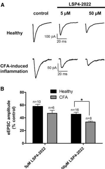
inhibition of eEPSC amplitude by 5 or 50
MLSP4 –2022 was increased in spinal cord slices from inflamed
compared with naive animals (
Fig. 6
). Indeed, whereas 50
MLSP4 –2022 depressed the eEPSC amplitude by 53.1
⫾ 4.7% in
control conditions, this depressant effect is significantly raised to
67.6
⫾ 1.5% in inflammatory conditions. This indicates that the
inhibition of glutamatergic neurotransmission by mGlu4
recep-tor is reinforced in the spinal cord of inflamed mice.
Physiological and pathophysiological role of mGlu4 receptors
in nociception and inflammatory pain in mice
After showing that mGlu4 receptors are present in lamina II and
modulate the neurotransmission, we evaluated the physiological
consequences of the genetic deletion of mGlu4 receptors in mice
on thermal, mechanical, or chemical sensitivity.
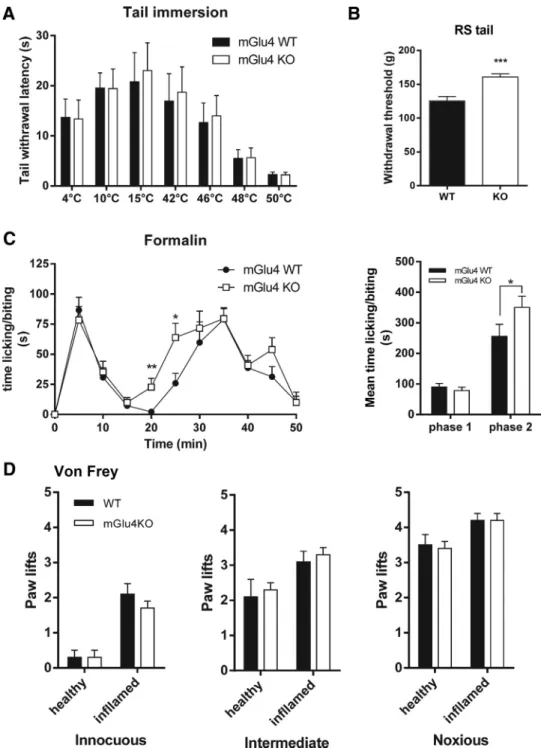
Sensitivity to cold or heat measured by the tail immersion test
at temperatures ranging from 4°C to 50°C did not differ between
mGlu4 knock-out (KO) mice and wild-type (WT) littermates
(
Fig. 7
A). Similarly, mechanical perception in response to
punc-tate innocuous, intermediate, and noxious stimuli elicited by von
Frey filaments of different strength (0.07, 0.6, and 1.4 g,
respec-tively) was not altered in mGlu4KO mice (
Fig. 7
D), although we
noticed a significantly higher threshold for tail withdrawal in
response to noxious mechanical compression using the Randall
& Sellitto apparatus (
Fig. 7
B).
In the formalin test, we observed a weak but significant
in-crease of nociceptive behavior of mGlu4KO compared with WT
mice during the second phase (
Fig. 7
C) but not in the first phase.
This increase seems to be the result of a faster development of the
inflammatory phase of the response to formalin injection, as can
be seen by its kinetics. We further investigated the possible
in-volvement of mGlu4 receptors in pathological inflammatory
conditions using the usual model of carrageenan-induced
in-flammatory pain and mechanical stimuli. Four hours after
induc-tion of inflammainduc-tion by an injecinduc-tion of carrageenan soluinduc-tion, a
comparable mechanical hypersensitivity was observed in
in-flamed mGlu4KO and their WT littermates (
Fig. 7
D). These data
suggest that the lack of mGlu4 receptors does not affect the
in-flammatory hypersensitivity once established.
Exogenous activation of spinal mGlu4 receptors reduced
inflammatory and neuropathic mechanical hypersensitivity
We then examined whether exogenous pharmacological
activa-tion of spinal mGlu4 receptors was able to reduce mechanical
hypersensitivity induced by inflammation or neuropathy.
We first evaluated the effect of spinal mGlu4 receptors activation
using von Frey filaments eliciting innocuous to noxious
mechani-cal stimuli in a carrageenan-induced model of inflammation in
C57BL/6 mice. As can be seen in
Figure 8
, activation of spinal mGlu4
receptors by intrathecal injection (i.t.) of LSP4 –2022
dose-depen-dently reduced mechanical hypersensitivity.
We verified that the mGlu4 receptor-induced correction of
mechanical hypersensitivity remained true across species because
terminals of afferent fibers have been shown to exhibit
differen-tial segregation in PKC␥-positive layer in spinal cord of rodents
(
Neumann et al., 2008
). We tested the effect of spinal mGlu4
receptor activation in naive “healthy” rats and in models of
in-flammation and neuropathic pain using the paw pressure test
(
Randall and Selitto, 1957
).
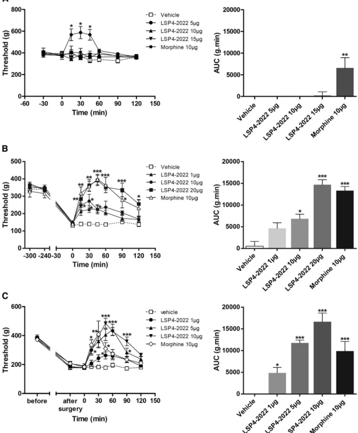
In inflamed or neuropathic rats, mechanical sensitivity was
assessed by measuring the vocalization threshold to paw pressure
on right hindpaw. Inflammation was induced by injection of
car-rageenan in the rat hindpaw and mononeuropathy by CCI of the
sciatic nerve. As previously shown in healthy rats with ACPT-I, a
Group III mGluR agonist (
Goudet et al., 2008
), administration of
LSP4 –2022 (5, 10, or 15
g/rat, i.t.) failed to induce
antinocice-ption, in contrast to morphine (10
g/rat, i.t.) that significantly
increased the vocalization threshold after mechanical stimulation
Figure 3. Partial reduction of mGlu4 receptor staining in the dorsal horns of the spinal cord after rhizotomy. A cervical and two lumbar transverse sections of the spinal cord of a C57BL/6 mouse subjected to unilateral dorsal rhizotomy of the fourth lumbar nerve are displayed. Triple staining was performed against mGlu4 receptors (red), VGLUT3 (blue), and IB4 (green). The solid lines indicate the separation between gray and white matter; the dotted lines indicate lamina I (LI), outer lamina II (LIIo), and the dorsal and ventral inner lamina II (LIIid and LIIiv). Fifteen days after the operation, immunoreactivity of mGlu4 receptors is partially reduced, whereas most IB4 and VGLUT3 labeling disappears in the dorsal horn of the fourth lumbar cord segment on the side ipsilateral to the lesion. Images are representative of four independent experiments. Scale bars, 100m.
(
Fig. 9
A). However, both in the rat model of
carrageenan-induced inflammatory pain (
Fig. 9
B) and in mononeuropathic
rats (CCI model) (
Fig. 9
C), intrathecal injection of LSP4 –2022
(1–20
g/rat) dose-dependently inhibited mechanical
hyperalge-sia. Maximal increase of the vocalization threshold was observed
45 min after the injection of LSP4 –2022 and corresponded to a
complete reversal of hyperalgesia.
Together, these results illustrate that pharmacological
activa-tion of spinal mGlu4 receptors abolishes mechanical hyperalgesia
associated with both inflammation and nerve injury but does not
affect reaction to noxious mechanical stimulus in healthy rats.
Demonstration of the mGlu4 receptor involvement in the
antihyperalgesic effect of LSP4 –2022
A genetic approach was used to obtain specific inactivation of
mGlu4 receptors by testing the impact of both knock-out and a
knockdown strategies on the involvement of mGlu4 receptors in
the effects of the agonist.
We first evaluated the consequences of the genetic deletion of
mGlu4 receptors in mice on the ability of LSP4 –2022 to alleviate
inflammatory mechanical hypersensitivity (
Fig. 10
A). Using the
model of carrageenan-induced inflammatory pain and von Frey
stimuli, we compared the effect of the maximal dose of LSP4 –
2022 (10
g/mice, i.t.) on mGlu4KO mice and their WT
litter-mates. The ability of LSP4 –2022 to reduce carrageenan-induced
hypersensitivity in response to applied stimuli is significantly
re-duced in the mGlu4 KO mice compared with their WT
litter-mates (
Fig. 10
A). The effect of LSP4 –2022 on sensitivity to
noxious mechanical stimulation is reduced by 78% in mice
lack-ing mGlu4, compared with their WT littermates. The reduced
effect of LSP4 –2022 in mGlu4KO mice reveals the major role of
mGlu4 receptors in the observed effects. However, there is still a
slight but significant remaining effect of LSP4 –2022 in mGlu4
KO mice. This suggests that, at this dose, other Group III
sub-types may be also involved in this response, albeit to a much lesser
extent.
Using a knockdown strategy in rats, we further examined the
role of spinal mGlu4 receptors in hyperalgesia. The aim of this
approach was as follows: (1) to compare the consequences of a
local and transient silencing of mGlu4 to the results obtained
with the constitutive knock-out of mGlu4 in mice; and (2) to
specifically block the mGlu4 mediated response in the absence of
selective antagonists. Silencing of spinal mGlu4 was achieved
us-ing AS oligonucleotides injected intrathecally accordus-ing to the
method described by
Mestre et al. (1994
). This method allows the
AS oligonucleotides to reach the DRG neurons (
Bourinet et al.,
2005
). Rats were treated by vehicle, scrambled control
oligonuce-lotides (MM), or mGlu4 receptor targeting AS oligonucleotides
(AS) injected intrathecally twice daily for 4 d. On the fourth day,
mechanical sensitivity was assessed by the paw pressure test
be-Figure 4. Activation of mGlu4 strongly reduces excitatory neurotransmission in spinal cord slices. Electrophysiological recording of eEPSCs in lamina II neurons from spinal cord slices of 2- to 3-week-old mice were performed using the whole-cell patch-clamp technique. Postsynaptic currents were evoked by an electrical stimulation using a bipolar electrode placed on the dorsal root entry zone. A, Immunofluorescence staining of mGlu4 receptors in lamina II of dorsal horn of spinal cord section from young mice. B, Time course of eEPSC amplitude before and after bath application of 50MLSP4 –2022, followed by a wash out and application of 50MNBQX. The amplitude of eEPSCs was normalized to amplitude before drug application. Top, Sample current traces recorded in
these conditions. C, Histogram of mean⫾ SEM of eEPSC amplitude in the absence and in the presence of 5 or 50Mof LSP4 –2022 or in the presence of 50M L-AP4, expressed as the percentage
of amplitude in control conditions, before drug application. D, Histogram of mean⫾ SEM of eEPSC amplitude in the presence of 50Mof LSP4 –2022 with or without GDP-S,astableGDPanalog
fore killing the animals and quantifying the degree of
knock-down. Expression of mGlu4 mRNA in lumbar DRG extracts was
significantly decreased by 38% after chronic treatment with AS,
whereas expression in animals treated with MM was not altered
(
Fig. 10
B). The protein level of mGlu4 in lumbar spinal cord
extracts of animals treated by AS was decreased by 58% compared
with animals treated by MM (
Fig. 10
C).
In the carrageenan-induced inflammation model, the effect of
LSP4 –2022 was significantly reduced (⫺54%) in animals treated
with AS oligonucleotides (
Fig. 10
D). Similarly, in
mononeuro-pathic rats, this AS strategy significantly reduced the amplitude of
antihyperalgesia induced by LSP4 –2022 (
⫺77%), whereas this
effect was unchanged in animals treated with MM (
Fig. 10
E).
These results clearly confirm the antihyperalgesic role of the
ac-tivation of spinal mGlu4 receptors both in inflammatory and
neuropathic pain models and the potential therapeutic interest of
mGlu4 receptor agonists.
Consistent with data obtained from the mGlu4KO mice, the
lack of variation in vocalization thresholds after repeated
treat-Figure 5. mGlu4 receptors regulates excitatory transmission through coupling to Cav2.2 chan-nels. Evoked EPSCs were recorded in lamina II neurons from spinal cord slices of 2- to 3-week-old mice using the whole-cell patch-clamp technique. A, Bottom, Time course of eEPSC amplitude before and after bath application of-Cgtx, a selective blocker of Cav2.2 channels, and 50 MLSP4 –2022, an
mGlu4 agonist. The amplitude of eEPSCs is normalized to amplitude before drug application. Top, Sample current traces recorded before drug application, after-Cgtx,andinthepresenceof-Cgtx andLSP4 –2022.B,Histogramofmean⫾SEMofeEPSCamplitudeintheabsenceandinthepresence ofLSP4 –2022anddifferentchannelblockers:-Cgtx,aselectiveblockerofCav2.2channels;-Agtx, a selective blocker of Cav2.1 channels; and tertiapin Q, a selective blocker of GIRK channels. Current amplitudes are expressed as the percentage of amplitude before drug application. C, Histogram of mean⫾ SEM of inhibition of eEPSC amplitude in the presence of the different channel blockers. CurrentamplitudesareexpressedasthepercentageofinhibitionoftheamplitudebeforeLSP4 –2022 application. **p⬍0.01.***p⬍0.001.ns,Notsignificant.
Figure 6. Inhibition of spinal excitatory neurotransmission by mGlu4 receptor is reinforced in inflammation. Electrophysiological recording of current eEPSCs in lamina II neurons from spinal cord slices of naive or inflamed young mice. Inflammation was induced by administration of CFA solution in hindpaws. Spinal cords were removed and recordings performed 24 h after the injection. A, Sample current traces recorded before and after 5 or 50MLSP4 –2022
appli-cation in naive or CFA-treated mice. B, Histogram of mean⫾ SEM eEPSC amplitude after application of 5 or 50MLSP4 –2022 in naive or inflamed mice, expressed as percentage of
ment with AS compared with saline- or MM-treated groups
sug-gests that, once established, the mechanical hypersensitivity is not
affected by mGlu4 receptor silencing.
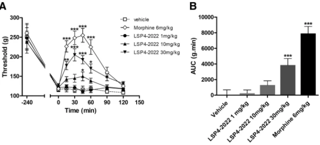
Systemic administration of an mGlu4 receptor agonist
alleviates pain hypersensitivity in inflammation
The ability of spinal mGlu4 receptors to abolish the
mechan-ical hypersensitivity caused either by inflammation or nerve
injury highlights the therapeutic potential of these receptors
for the development of novel analgesics. However, the
ques-tion of whether a reducques-tion of pain can be observed after a
systemic administration of an mGlu4 receptor agonist
re-mained unanswered. To address this point, we injected LSP4 –
2022 intraperitoneally (1–30 mg/kg) in rats subjected to
carrageenan-induced inflammation. This particular ligand
was chosen because of its ability to cross the blood– brain
barrier, which has previously been documented in an animal
model of Parkinson disease (
Goudet et al., 2012
).
Figure 7. Perception of thermal, mechanical, and chemical stimuli in naive mGlu4KO mice. A, mGlu4KO mice and their WT littermates display a similar thermal sensitivity, determined by measuring the latency to withdraw the tail immersed in water at temperatures ranging from 4°C to 50°C (n⫽10animalspergroup).B,mGlu4KOmicedisplayasignificantlyhigherthresholdfortailwithdrawalafternoxious mechanical stimuli evoked by tail compression in the Randall–Selitto (RS) than WT mice (n⫽ 10 animals per group). C, Both mGlu4KO and WT mice displayed the typical biphasic time course of nocifensive behavior (paw licking/biting) after injection of 15lofa5%formalinsolutionintherighthindpaw.ThesecondphaseoftheresponseoccursfasterformGlu4KOmice.Right,Histogramrepresentingthemean time⫾SEMofnocifensivebehaviorduringthefirstphase(0–10min)andthesecondphase(15–50min)oftheformalintest.TheresponseinthesecondphaseissignificantlyhigherformGlu4KOmicethanfor WT mice (n⫽8animalspergroup).D,MechanicalsensitivityassessedbymeasuringthenumberofpawliftsoffivestimulationsusingvonFreyfilamentscorrespondingtoinnocuous(0.07g),intermediate(0.6 g), and noxious (1.4 g) bending forces does not differ between mGlu4KO and WT mice (n⫽10animalspergroup).*p⬍0.05.**p⬍0.01.***p⬍0.001.
The acute intraperitoneal injection of
LSP4 –2022 (1–30 mg/kg) in inflamed rats
led to a significant and dose-dependent
reduction of mechanical hyperalgesia
mea-sured using the paw pressure test (
Fig. 11
).
The time course of LSP4 –2022
anti-hyperalgesic effect was similar to that of
morphine (6 mg/kg, i.p.), with a peak effect
30 min after administration, but the
maxi-mal efficacy of 30 mg/kg LSP4 –2022 was
40% lower than that of morphine.
This result further highlights the interest
of targeting mGlu4 receptors to alleviate
painful hypersensitivity in an inflammatory
context using LSP4 –2022, and possibly
other activators of mGlu4 receptors able to
cross the blood– brain barrier.
Discussion
Because of current limitations in the
thera-peutic arsenal for pain management, there is
a crucial need for the identification and
de-velopment of alternative targets and
strate-gies for the treatment of chronic pain.
Targeting the modulatory role of mGluRs to
prevent the glutamatergic overactivity
asso-ciated with central sensitization of the pain
neuraxis may be one of the hitherto
unappreciated modalities to reduce chronic
pain. In the present study, we present
neuroanatomical, electrophysiological,
pathophysiological, and pharmacological
data that, together, highlight the effect of
spinal mGlu4 receptor activation to alleviate
the mechanical hypersensitivity observed in
inflammatory or neuropathic pain.
One of the most striking findings of our
study is that mGlu4 receptors are only
influ-ential in painful and pathological conditions
after exogenous activation. Physiologically,
the mGlu4 receptor seems to be devoid of a
tonic role in pain perception as observed
when using brief stimuli, such as von Frey
hair application or tail immersion. Indeed,
thermal sensitivity of mGlu4KO mice is not
affected, and they do not differ from WT in
their response to innocuous or noxious
me-chanical stimuli elicited by von Frey hairs on
their paw. However, the role of the mGlu4
receptor is revealed by the altered responses
of mGlu4KO mice to more noxious stimuli,
such as tail pressure, or to an inflammatory
agent, such as formalin. Indeed, the
nocice-ptive behavior of mGlu4KO mice is
in-creased in the second phase of the formalin test, suggesting that the
lack of mGlu4 receptor may accelerate the development of
inflam-matory pain. Exogenous mGlu4 activation reduces mechanical
allo-dynia and hyperalgesia in an inflammatory state or in a neuropathic
situation but does not modulate acute pain perception after brief
stimuli. These results are consistent with the antihyperalgesia
ob-served after activation of Group III mGluRs with Group III selective
agonists (
Goudet et al., 2008
). Antihyperalgesic effects of Group III
agonists could be mediated by mGlu4, mGlu7, and/or mGlu8
recep-tors, which are expressed either in the spinal cord (
Ohishi et al.,
1995b
;
Azkue et al., 2001
) or in cell bodies of sensory neurons (
Car-lton and Hargett, 2007
). This notwithstanding, the use of selective
mGlu4 agonists in this study or allosteric enhancers (
Goudet et al.,
2008
;
Wang et al., 2011
) suggests that this particular subtype plays a
pivotal role in pain modulation. This is further supported by the
strong reduction of the antihyperalgesic effect only by exogenous
activation in mGlu4KO mice but also after a local and transient
knockdown of spinal mGlu4 by AS oligonucleotides in rats.
How-Figure 8. Pharmacological activation of spinal mGlu4 receptors reduces mechanical hypersensitivity induced by inflammation on mice. Time course and area under the curve of mean⫾ SEM of the number of paw lifts of five stimulations using von Frey filaments corresponding to innocuous (A), intermediate (B), and noxious (C) bending forces (0.07, 0.6, and 1.4 g, respectively) on inflamed C57BL/6 mice treated by various doses of LSP4 –2022 (as indicated) 240 min after carrageenan injection (n⫽ 6–8 animals per group). *p⬍ 0.05, vehicle versus treated conditions. **p ⬍ 0.01, vehicle versus treated conditions.
Figure 9. Pharmacological activation of spinal mGlu4 receptors does not modify mechanical sensitivity of naive rats but reduces mechanical hypersensitivity induced by inflammation or neuropathy. Time course of mean⫾ SEM of vocalization threshold to paw pressure (in grams) and area under the curve (AUC, g.min) of inflamed rats treated with vehicle, LSP4 –2022 (as indicated in micrograms per rat, i.t.) or morphine (10g/rat, i.t.) administered on naive rats (A), on inflamed rats 240 min after carrageenan injection (B), and on mononeuropathic rats 2 weeks after ligature of the sciatic nerve (C) (n⫽ 6 – 8 animals per group). *p ⬍ 0.05, vehicle versus treated conditions. **p ⬍ 0.01, vehicle versus treated conditions. ***p ⬍ 0.001, vehicle versus treated conditions.
Figure 10. Antihyperalgesia induced by pharmacological activation of spinal mGlu4 is markedly reduced in mGlu4KO mice and rats treated by selective mGlu4 AS oligonucleotides. Area under the time course curves of paw lifts after application of von Frey filaments (five stimulations) corresponding to innocuous (0.07 g), intermediate (0.6 g), and noxious (1.4 g) (Figure legend continues.)
ever, there is still a small but significant antihyperalgesic effect of
LSP4 –2022 in mGlu4KO mice, suggesting that other Group III
mGluRs subtypes may also be involved. Interestingly, we show here
that a systemic injection of mGlu4 agonists induces a robust
me-chanical antihyperalgesia. Together, these results illustrate the
ther-apeutic potential of mGlu4 receptors in a pathological context.
Activation of Group III mGluRs has been shown to decrease
firing of spinal cord dorsal horn projection neurons (
Chen and
Pan, 2005
) and reduce spontaneous and evoked spinal synaptic
transmission (
Zhang et al., 2009
;
Cui et al., 2011
). Our results
underline the important role of mGlu4 in this regulatory process
because the selective activation of mGlu4 receptors by LSP4 –
2022 replicates the effect obtained by the nonselective Group III
agonist
L-AP4. Our results further demonstrate that mGlu4 is
functionally coupled to Cav2.2 channels in sensory neurons.
These channels are mostly expressed in presynaptic terminals of
C- and A␦-fibers and are key targets against chronic pain, as
illustrated by the clinical efficacy of ziconotide or the
anticonvul-sants gabapentin and pregabalin (
Zamponi et al., 2009
). Indeed,
gabapentin and pregabalin, blockers of the regulatory subunit
␣2␦ of Cav2.2 channels, are effective analgesics for neuropathic
pain and are also able to reduce pain and opioid consumption
after surgery (
Dauri et al., 2009
). Furthermore, the Cav2.2
chan-nel blocker, ziconotide, is used to manage severe chronic pain
(cancer or neuropathic pain). The identification of a new way to
inhibit Cav2.2 at the spinal cord level by activating mGlu4
recep-tors may lead to improved therapeutic strategies, as illustrated
by
Brittain et al. (2011
). Interestingly, we also show here that
mGlu4-induced inhibition of glutamatergic neurotransmission
is reinforced in spinal cord slices from inflamed mice. This is
consistent with the observation that the ability of Group III
mGluRs to control the excess of excitatory transmission is
rein-forced in spinal cord of neuropathic pain animal models (
Zhang
et al., 2009
). This may reflect both the synaptic plasticity
associ-ated with central sensitization observed in chronic pain and the
use-dependent activity of Group III mGluRs, mGlu4 receptors in
particular. The latter point will be further discussed below. The
molecular remodeling that takes place under chronic pain
con-ditions notably includes overexpression of Cav2.2 channels and
its regulatory subunits (
Lu et al., 2010
), could explain the
rein-forcement of mGlu4 inhibitory effect.
How is it that mGlu4 receptors are only modulating painful or
pathological pain states and not pain perception when using brief
stimuli in naive animals? First, this could be due to the
localiza-tion of the mGlu4 receptor in a particular subset of sensory
neu-rons and in PKC
␥-expressing interneurons, as suggested by our
results obtained after rhizotomy and data from of imunostaining
studies. We localized some mGlu4 receptors to the sensory
ter-minals of unmyelinated fibers in the inner lamina II of the dorsal
horn of spinal cord. Although there is some mGlu4 receptor
staining in peptidergic and nonpeptidergic C fibers in which the
role in nociception is well known (
Basbaum et al., 2009
;
Todd,
2010
), most of the labeling is located in a subset of C-LTMR
expressing VGLUT3. While normally devoted to the sensing of
pleasant touch in humans, these fibers are involved in mechanical
hypersensitivity in animal models of inflammation or
neuropa-thy (
Lo¨ken et al., 2009
;
Seal et al., 2009
;
Li et al., 2011
). Moreover,
mGlu4 immunoreactivity overlaps in great part with PKC␥
im-munoreactivity. PKC␥-expressing interneurons are excitatory
interneurons located where low-threshold mechanoreceptive
and nociceptive inputs terminate and play a key role in the
pro-cessing of tactile inputs both in physiological and pathological
conditions (
Malmberg et al., 1997
;
Polga´r et al., 1999
;
Neumann
et al., 2008
) These interneurons are activated by innocuous
stim-uli in physiological conditions (
Neumann et al., 2008
;
Li et al.,
2011
) but are thought to mediate pain hypersensitivity induced
by injury (
Malmberg et al., 1997
;
Polga´r et al., 1999
), probably
4
(Figure legend continued.) bending forces on inflamed mGlu4KO or WT mice treated by LSP4 –2022 240 min after injection of carrageenan (A) (n⫽ 7 animals per group). Top, Illus-tration of a time course of paw lifts for a noxious bending force. Bottom, Mean⫾SEMofrelative antihyperalgesia, expressed as the area under curve of mGlu4KO or WT mice treated by vehicle and 10gLSP4-2022.QuantificationofselectiveknockdownofspinalGlu4inratsweretreated during 4 d by vehicle, scrambled control, or mGlu4 receptor targeting AS oligonucleotides by measuring DRG mGlu4 mRNA level by qRT-PCR (B) or spinal protein level by Western blot (C). Effect of mGlu4 receptor knockdown on LSP4 –2022-mediated antihyperalgesia in inflamed rats (D) (n⫽ 7 animals per group) and neuropathic rats (E) (n ⫽ 8 animals per group). Left, Time course curves of mean⫾ SEM of vocalization threshold to paw pressure (expressed in grams). Right, Mean⫾ SEM of relative antihyperalgesia, expressed as percentage of the area under the curve of rats treated by vehicle and 10g LSP4–2022 (control). ns, Not significant. *p⬍ 0.05 versus vehicle-treated group. **p ⬍ 0.01 versus vehicle-treated group. ***p ⬍ 0.001 versus vehicle-treated group. #p⬍ 0.05 versus corresponding LSP4–2022 treated WT groups. ##p⬍0.01versuscorrespondingLSP4–2022treatedWTgroups.###p⬍0.001versus corresponding LSP4 –2022 treated WT groups.
Figure 11. Systemic injection of mGlu4 agonist alleviates mechanical hypersensitivity in inflammation. Results are expressed by the time course curves of mean⫾SEMofvocalizationthreshold to paw pressure (expressed in grams) in the left panel and by the area under the curve (AUC, g.min) in the right panel. Rats were treated with vehicle, LSP4 –2022 (various doses as indicated in milligrams per kilogram, i.p.), or morphine (6 mg/kg, i.p.) administered 240 min after intraplantar injection of carrageenin (n⫽8animalspergroup).*p⬍0.05versusvehicle-treatedgroup.**p⬍ 0.01 versus vehicle-treated group. ***p⬍ 0.001 versus vehicle-treated group.