HAL Id: hal-02940233
https://hal.archives-ouvertes.fr/hal-02940233
Submitted on 22 Oct 2020
HAL is a multi-disciplinary open access
archive for the deposit and dissemination of
sci-entific research documents, whether they are
pub-lished or not. The documents may come from
teaching and research institutions in France or
abroad, or from public or private research centers.
L’archive ouverte pluridisciplinaire HAL, est
destinée au dépôt et à la diffusion de documents
scientifiques de niveau recherche, publiés ou non,
émanant des établissements d’enseignement et de
recherche français ou étrangers, des laboratoires
publics ou privés.
Bonafos, Christine Roques, K Makasheva
To cite this version:
Marvine Soumbo, Adriana Scarangella, Christina Villeneuve-Faure, Caroline Bonafos, Christine
Roques, et al.. Combined effect of proteins and AgNPs on the adhesion of yeast Candida albicans
on solid silica surfaces. 2020 IEEE 20th International Conference on Nanotechnology (IEEE-NANO),
Jul 2020, Montreal, Canada. pp.242-245, �10.1109/NANO47656.2020.9183494�. �hal-02940233�
Abstract— Research and development of new biomaterials
has considerably increased in the last ten years. Description of their properties involves among others their interaction with proteins. Due to the exposure of proteins to non-biological solid surfaces the relationship between protein structure and function might be modified. The conformation and arrangement of the adsorbed proteins on the surface further control the subsequent biological processes and thus determine the biological response to the material. In this work we present the impact of silver nanoparticles (AgNPs) embedded in the near-surface of thin silica layers on the adhesion forces of Candida albicans IP48.72 in conditioned by different proteins environment. Depending on the nature of proteins the effect can be opposite: The Bovin Serum Albumine (BSA) decreases the yeast adhesion on solid silica surfaces while the Fibronectin (Fn) favors it. It is also found that the AgNPs, when embedded in the near-surface of thin silica layers, impair the adhesion forces of C. albicans through the release of silver ions and lead the cell dead.
I. INTRODUCTION
The emergence and selection of antibiotic-resistant bacteria is an ever increasing Public Health problem. Microbial adhesion and subsequent biofilm formation are at the origin of hospital-acquired infections (HAIs), known also as healthcare-associated infections (HCAIs), often leading to septic complications and even lethal issues, and entailing large economical losses for the health-care systems [1 – 3]. This threat is of particular concern when compared with the very limited number of new antimicrobial agents in the pipeline of the pharmaceutical industry and the ability of microorganisms to be less sensitive under biofilm organization. Biofilms are constituted by a community of microorganisms adhered to solid surfaces of materials and protected by a self-produced extracellular polymeric matrix. They are complex and dynamic ecosystems, with well developed resistance to external agents. Surface cleaning procedures traditionally involve the use of detergents or/and biocides [4]. Unfortunately, these methods may turn out to be ineffective against mature biofilms [5 – 7].
In this context, new strategies oriented to the prevention of environmental contamination of medical devices, catheters, implants, etc., are under scrutiny. One possible approach is related to the elaboration of antimicrobial
*Resrach supported by the program IDEX Transversalité of University of Toulouse (ANR-11-IDEX-0002-02), under project ADAGIO. M. S. acknowledges PhD-grant from Université de Toulouse and Région Occitanie, under APR-project ADAGIO.
M. Soumbo, A. Scarangella, C. Villeneuve-Faure, and K. Makasheva are with LAPLACE laboratory, Université de Toulouse ; CNRS, UPS,
surface coatings. The antimicrobial agent is dispersed in the coating and progressively released on the surface providing thus continuous inhibition of the microbial adhesion, so surface colonization over long term.
Silver, and particularly silver nanoparticles (AgNPs), exhibit inherent antimicrobial properties. Recently, the AgNPs proved the largest antimicrobial activity against bacteria, viruses and eukaryotic microorganisms. The antimicrobial activity of AgNPs leaded to the increased and wide use of silver-containing materials, especially in the biomedical domain. The biological activity of AgNPs is closely related to ionic Ag (Ag+) release or to direct contact
of the microorganisms with AgNPs [8 – 13], resulting in protein denaturation at different cell locations; especially sensible are those enzymes of the respiratory chain and transport channels [14]. On the other hand, the uncontrolled use of AgNPs hides environmental risks due to the AgNPs-toxicity [15]. Modulation of the silver ion release from AgNPs would allow delivery of the appropriate dose of Ag+
for biomedical uses and for environmental protection. The successful and sustainable use of nanocomposite materials containing AgNPs is mostly supported by the knowledge of how to use them safely prior to their large distribution. To that end a better understanding of the molecular mechanisms of interaction of AgNPs with microorganisms, considering also the environment, is highly demanded in order to localize the AgNPs antimicrobial activity.
In many systems the microorganisms are dispersed in a flowing carrier fluid and adhere on immersed surfaces. It defines the importance to evaluate their adhesion forces. The relationship between microbial adhesion and shear flow, as well as the fate of the microorganisms after interaction with the solid surface, defines the efficiency of antimicrobial properties of the surface coating [9]. Among other constituents, the different kinds of proteins and other macromolecules, present in the environment, further condition the adhesion of microorganisms. The present study focuses on the evaluation of shear-induced detachment of the yeast Candida albicans IP48.72 in contact with plasma mediated thin silica (SiO2) layers
containing AgNPs at very low shear-stresses. To that end a shear-stress flow chamber is used. The experiments are performed in presence of two different proteins, Bovin
INPT; 118 route de Narbonne, F-31062 Toulouse, France (e-mail: kremena.makasheva@laplace.univ-tlse.fr).
M. Soumbo, and C. Roques are with LGC laboratory, Université de Toulouse ; CNRS, UPS, INPT; 35 chemin des maraîchers, F-31062 Toulouse, France.
A. Scarangella and C. Bonafos, are with CEMES-CNRS, Université de Toulouse, 29 Jeanne Marvig, BP 94347, F-31055 Toulouse, France.
Combined effect of proteins and AgNPs on the adhesion
of yeast Candida albicans on solid silica surfaces*
M. Soumbo, A. Scarangella, C. Villeneuve-Faure, Member IEEE,
C. Bonafos, C. Roques, and K. Makasheva, Member IEEE
Serum Albumine (BSA) and Fibronectin (Fn), to assess their respective contributions.
II. EXPERIMENTAL PART
The substrates used in this work were thin thermal silica (SiO2) layers grown on Si-intrinsic wafers at 1100°C under
slightly oxidizing atmosphere using a N2–O2 gas mixture
containing 1.0% of O2. The plasma processed AgNPs-based
nanocomposite coatings, consisted of a single plan of AgNPs embedded at a few nanometers beneath the surface of thin SiO2-layers. The surface of each sample was of
1 cm2. The plasma deposition was performed in an
axially-asymmetric capacitively-coupled RF discharge sustained at low pressure that successfully combines sputtering of a metal target and plasma polymerization. More details about the plasma process and its monitoring during deposition of the nanocomposites are given elsewhere [16].
The AgNPs-based nanocomposite coatings were characterized by Transmission Electron Microscopy (TEM), Spectroscopic Ellipsometry (SE) and Fourier Transform InfraRed (FTIR) spectroscopy. The specimens for TEM observations were prepared according to a standard procedure, i.e. mechanical polishing and Ar+ ion
milling for both cross-section and plane-view configurations.
The BSA and Fn proteins were purchased from Sigma Aldrich in a lyophilized powder form. Protein stock solutions were prepared with a concentration of 5.0 g/Lof BSA and 1.0 g/Lof Fn in water for injectable preparations (WIP, European Pharmacopoeia, COOPER). The BSA and Fn thin protein monolayers were deposited on the samples, from solutions with protein concentration of 0.05 g/L, by dip coating [17]. The SiO2 samples and the AgNPs-based
nanocomposites were immersed for 1h in 1mL of protein solution. After the immersion, the samples were rinsed in WIP to remove all the non-adhered proteins and were left dehydrating at room temperature and atmospheric pressure before starting the physico–chemical characterization.
Thickness and organization of the protein layers were characterized by spectroscopic ellipsometry and Atomic Force Microscopy (AFM) in Peak-Force mode. To probe soft material as the resulting protein layers a SNL tip with spring constant of 0.24 N/m and curvature radius of around 5 nm was used. The peak force was set to 0.5 nN. More details can be found elsewhere [17].
Adhesion of C. albicans on the SiO2 substrates and on
the AgNPs-based nanocomposites, and their detachment profiles were studies in vitro by a hydrodynamic method using a shear-stress flow chamber. The selected yeast strain was C. albicans IP48.72 (Institut Pasteur, Paris, France). The test suspensions were made from frozen aliquots, from the second and third subcultures of C. albicans on Sabouraud agar (30°C, 48-72h). The culture was carried out in Sabouraud liquid medium (Sabouraud Dextrose liquid Medium, OXOID, CM0147) (30 g/L, pH = 5.6 at 23°C). After 72 h of incubation at 30°C without shaking, the cells were recovered by filtration. The cells were collected by centrifugation at 13000 rpm for 3 min at room temperature, washed twice and then suspended in WIP in order to avoid any possible interaction with extracell molecules. For all
assays, the cell suspension was adjusted in WIP to DO600nm,
corresponding to 1.107 CFU/mL.
The shear-stress flow chamber was a commercially available one from BioSurface Technologies Corporation (BST Model FC 71 Coupon Evaluation Flow Cell, USA). The coupon compartment was in-house adapted to receive
the above described samples. The shear-stress flow
chamber was connected to a flow system designed to address a large range of shear stresses, up to 80 Pa. Specific attention was paid to the very low shear-stress domain, between 0.01 Pa and 0.5 Pa. The idea behind was to be able to reproduce also flow stresses comparable to those in the human body.
III. RESULTS AND DISCUSSION
According to the SE measurements the thickness of the thermal silica layers is 80 nm. Their structural properties studied by FTIR show a very well organized silica layers. Only the three typical transversally optic (TO) modes of molecular vibrations of amorphous silica are present in the infrared spectrum (results not shown here) [16]: the Si-O-Si rocking vibration at 457 cm−1, the symmetric stretching mode at 810 cm−1, and the asymmetric stretching mode at 1062 cm−1. The shoulder centered around 1250 cm−1, characteristic of thermal SiO2 is also clearly observable on
the spectrum.
The plasma processed AgNPs-based nanocomposite layers are shown in Figure 1. The AgNPs are bimodal in size with larger AgNPs of 19.0 ± 10.0 nm and density of 1.7 × 10¹¹ NPs/cm2, covering 42% surface area as obtained
after processing of the TEM image in plan-view shown in Figure 1(a). The small AgNPs are rather spherical in shape while the shape of large AgNPs evolves to prolate spheroid, with highest eccentricity found of c = 0.52. The AgNPs are
well aligned in a single plane. They are polycrystalline and/or twinned. The AgNPs single plane is covered with a very thin plasma deposited SiO2pl layer, conformal to the
AgNPs shape as can be seen in the TEM cross-section image in Figure 1(b). The SiO2pl layers are very close in
structure to the thermal silica layers. Their FTIR study shows spectra containing the same peaks as for the thermal SiO2. The total thickness of the structures is 100 nm.
The ellipsometric spectrum of the SiO2pl cover layer was
used to determine its refractive index (n), extinction coefficient (k) and layer thickness by applying the Forouhi-Bloomer dispersion law. The SiO2pl layer is found
Figure 1. Bright field TEM images of the samples containing AgNPs (a) in plan-view and (b) in cross-section,. The cross-section
image also shows the single plane of AgNPs with their conformal 5.5 nm thin silica cover layer.
transparent (k = 0) with n = 1.45 (at = 632.8 nm), which testify for the high-quality of the plasma deposited silica cover layer. Its thickness is found of 5.5 nm. It means that the AgNPs are embedded at 5.5 nm beneath the surface [16]. The value of the SiO2pl cover layer thickness extracted from
the ellipsometry spectrum corresponds exactly to the one obtained from TEM observations (Figure 1(a)).
Whenever a foreign material is brought to contact with blood or physiological fluids one of the first process to occur is protein adsorption on the material surface. The conformation and arrangement of the adsorbed proteins further control the subsequent biological processes and thus determine the biological response to the material. The “protein-adsorption problem” remains fundamental and comes to the fore especially when protein interaction with silicon oxide-related materials are under study. The SiO2 is
an inorganic material with strongly pronounced hydrophilic properties, thus not favoring protein adsorption. In general, protein adsorption on solid surfaces depends on different factors related to both the protein itself and the surface as the final outcome is shared between the two sides. In terms of microbial adhesion, the adsorbed proteins on the surface impact it. The proteins create a conditioning film on the solid surface, thus modulating the interactions of microorganisms with surfaces expressing modified properties. Therefore, before evaluating the adhesion of
C. albicans we have studied the organization and
conformational changes of thin protein layers of BSA and Fn after adsorption and dehydration on the surface of the elaborated samples. The choice to work with dehydrated thin protein layers is in link with natural conditions during drying. The selected low concentration of the two proteins in solution (0.05 g/L) is comparable to protein concentrations in human body fluids.
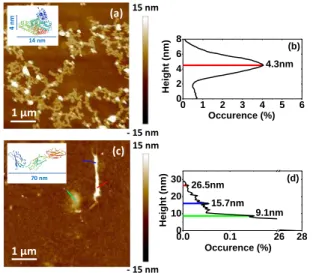
Figure 2 shows the organization of BSA and Fn proteins on the surface of the thermal SiO2 layer. It is worth to
remind here that a silica layer is also the top layer in the AgNPs-based nanocomposits. The resulting protein layers are very thin and non-continuous monolayers due to the
very small protein concentration in the solutions. The BSA proteins organizes in a lace-like network on the surface (Figure 2(a)). The height histogram created after the AFM investigation in PF-QNM mode (Figure 2(b)) shows a single peak with value of 4.3 nm between the substrate surface and the protein layer. This value is consistent with the BSA smaller dimension (minor axis of the spheroid, insert of Figure 2(a)) suggesting that the proteins are ‘side-on’ adsorbed on the surface [18, 19]. It also confirms the monolayer organization of BSA on the surface.
Due to their structural differences and much larger size (2 × 70 nm) compared to the BSA larger dimension (14 nm), the Fn proteins organize on the silica surface in a very different way. They either fold up to achieve a globular shape or adopt more complex branch-type structures as shown in Figure 2(c). This is represented on the height histogram in Figure 2(d), where three characteristic heights can be observed. They suggest different Fn conformations on the surface. The most frequently occurring one is with height of 9.1 nm. This value corresponds well to the hydrodynamic radius of Fn in aqueous solution, which is 8.7 nm [20], and suggest globular compact conformation. The two other specific heights are of 15.7 nm and 26.5 nm. They can be assigned to partially unfolded and elongated protein conformations [20]. It appears that after adsorption and dehydration on the silica surfaces the three conformational models for protein: globular (folded compact), branched, or linear (extended), can be assumed for the Fn. Moreover, other complex random or looped protein models may exist. The results also demonstrate the trend of unfolding of Fn under harsher and denaturing conditions, represented in this study by interaction with the solid surfaces of SiO2 layers.
Since the two proteins are optically non-absorbing, the Cauchy dispersion law was used for interpretation of the recorded ellipsometry spectra of the protein monolayers, as detailed in our earlier work [17]. The SE measurements of the adsorbed proteins corroborate with the results from the AFM study for both proteins. The effective thickness of the BSA layer is found 4.1 ± 0.1 nm and the one for the Fn 8.6 ± 0.2 nm. These results confirm that the BSA proteins are ‘side-on’ adsorbed on the SiO2 surface and that the Fn
proteins are mainly under globular compact conformation. The extracted refractive indexes of BSA and Fn protein layers are np-BSA = 1.61 and np-Fn = 1.62, respectively.
Based on the obtained refractive indexes and applying the de Feijter expression [21], the protein surface concentrations, i.e. the adsorbed protein mass per unit area, are found BSA = 0.58 g/cm2 for BSA and
Fn = 1.32 g/cm2 for Fn. These results show that even
though the concentration of proteins in solution is the same (0.05 g/L), the amount of adsorbed on the SiO2 surface
proteins differs significantly. The mass per unit area of Fn is more than twice larger compared to the one of BSA. Without any doubt, one can expect that the SiO2 surfaces
will be conditioned differently for the C. albicans adhesion according to the nature of adsorbed proteins, BSA or Fn.
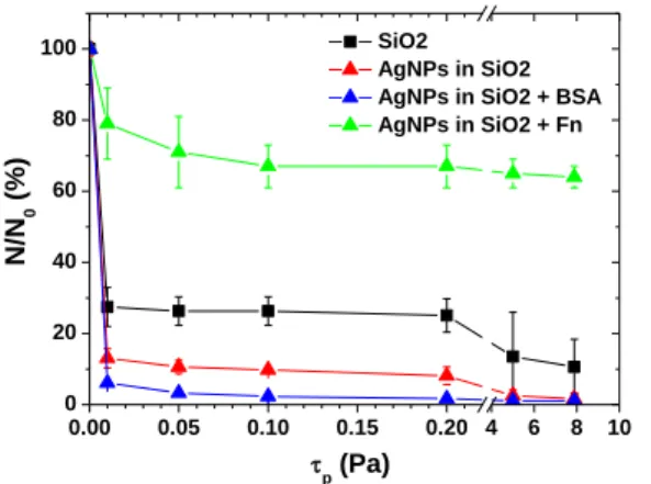
Figure 3 shows the shear-flow induced profiles of
C. albicans at very low shear-flows. The results are
averaged over three independent measurements per sample.
Figure 2. Organization of thin protein layers on silica surface after adsorption and dehydration (solution concentration of 0.05 g/L): (a) BSA layer with the corresponding height hystogram on (b) and
(c) Fn layer, with the characteristic height peaks given in (d).
0.0 0.1 26 28 0 10 20 30 9.1nm 15.7nm 26.5nm (d) Occurence (%) He ight ( nm) 0 1 2 3 4 5 6 0 2 4 6 8 Occurence (%) He ight ( nm) 4.3nm (b) 1 µm - 15 nm 15 nm (c) 70 nm 1 µm - 15 nm 15 nm (a) 14 nm 4 n m
As reported by other authors for SiO2, the obtained results
confirm that the SiO2 surfaces do not favor cell adhesion.
More than 70% of the cells are detached even for the lowest applied shear-flow (0.01 Pa). One can immediately notice that the adhesion forces of C. albicans are even more reduced on the surface of AgNPs-based nanocomposites. It is due to the combined effect of silica surfaces and progressive release of Ag+ from the AgNPs; the latter was
evaluated to 0.48 µM over 20 h, representing 1.7% of the total silver amount in the layer [15]. Besides, the released from the nanocomposites Ag+ lead the dead of 92% of the
remained adhered cells in the assay conditions.
As expected, the nature of proteins forming the conditioning layer strongly modulates the adhesion of yeast cell. C. albicans adhesion is additionally reduced when BSA proteins are adsorbed on the SiO2 surface. In opposite,
when Fn-protein layer conditions the surface, a dramatic increase in C. albicans adhesion forces is observed. Moreover, the C. albicans adhesion is so strong in presence of Fn on the SiO2 surface that the majority of cells cannot
be detached even for very high shear-flow stresses (80 Pa). IV. CONCLUSION
The presence of protein conditioning layer alters the adhesion of C. albicans on solid surfaces depending on the protein nature. The effect of AgNPs is cumulative. Through the release of Ag+ ions, the AgNPs embedded close to SiO
2
surface lead also the dead of adhered cells. Surprisingly, cell death does not impair the major impact of protein nature on cell adhesion. Further work will be directed to deeper our study towards quantification of the C. albicans adhesion forces and to explore more complex protein organizations, such as intermixed proteins/proteins and proteins/microorganisms systems.
REFERENCES
[1] M. Habash and G. Reid, “Microbial biofilms: their development and significance for medical device-related infections,” J. Clin.
Pharmacol, vol. 39, p. 887-898, 1999.
[2] C. Roques, H. Al Mousa, A. Duse, R. Gallagher, T. Koburger, E. Lingaas, N. Petrosillo, and J. Škrlin, “Consensus statement: patient
safety, healthcare-accociated infections and hospital environmental surfaces,” Future Microbiol., vol. 10, pp. 1615-1620, 2015. [3] C. Feuillolay, L. Haddioui, M. Verelst, A. Furiga, L. Marchin, and
C. Roques, “Antimicrobial activity of metal oxide microspheres: an innovative process for homogeneous incorporation into materials,’
Appl. Microbiol., vol. 125, p. 45-55, 2018.
[4] S. G. Parkar, S. H. Flint, and J. D. Brooks, “Evaluation of the effect of cleaning regimes on biofilms of thermophilic bacilli on stainless steel,” Appl. Microbiol., vol. 96, pp. 110-116, 2004.
[5] B. Joseph, S.K. Otta, and I. Karunasagar, “Biofilm formation by
Salmonella spp. On food contact surfaces and their sensitivity to
sanitizers,” Int. J. Food Microbiol., vol. 64, pp. 367-372, 2001. [6] H. Gibson, J. H. Taylor, K. E. Hall, and J.F. Hollah, “Effectiveness of
cleaning techniques used in the food industry in terms of the removal of bacterial biofilms,” Appl. Microbiol., vol. 87, pp. 41-48, 1999. [7] P. J. Taormina and L. R. Beuchat, “Survival of Listeria
monocytogenes in commercial food-processing equipment cleaning
solutions and subsequent sensitivity to sanitizers and heat,” Appl.
Microbiol., vol. 92, pp. 71-80, 2002.
[8] M. Rai, A. Yadav, and A. Gade, “Silver nanoparticles as a new generation of antimicrobials,” Biotechnol. Adv., vol. 27, p. 76-83, 2009.
[9] C. Saulou, B. Despax, P. Raynaud, S. Zanna, P. Marcus, and M. Mercier-Bonin, “Plasma deposition of organosilicon polymer thin films with embedded nanosilver for prevention of microbial adhesion,” Appl. Surf. Sci., vol. 256S, p. S35-S39, 2009.
[10] B. Despax, C. Saulou, P. Raynaud, L. Datas, and M. Mercier-Bonin, “Transmission electron microscopy for elucidating the impact of silver-based treatments (ionic silver versus nanosilver-containing coating) on the model yeast Saccharomyces cerevisiae,”
Nanotechnology, vol. 22, p. 175101, 2011.
[11] B. Reidy, A. Haas, A. Luch, K. A. Dawson, and I. Lynch,
“Mechanisms of Silver Nanoparticle Release, Transformation and
Toxicity: A Critical Review of Current Knowledge and Recommendations for Future Studies and Applications,” Materials,
vol. 6, pp. 2295-2350, 2013.
[12] S. Eckhardt, P. S. Brunetto, J. Gagnon, M. Priebe, B. Giese, and K. M. Fromm, “Nanobio Silver: Its Interactions with Peptides and Bacteria, and Its Uses in Medicine,” Chemical Reviews, vol. 113, pp. 4708-4754, 2013.
[13] B. Le Ouay and F. Stellacci, “Antibacterial activity of silver nanoparticles: A surface science insight,” Nano Today, vol. 10, pp. 339-354, 2015.
[14] K. B. Holt and A. J. Bard, “Interaction of silver(I) ions with the respiratory chain of Escherichia coli: an electrochemical and scanning electrochemical microscopy study of the antimicrobial mechanism of micromolar Ag+,” Biochemistry, vol. 44, pp.13214 -13223, 2005.
[15] A. Pugliara, K. Makasheva, B. Despax, M. Bayle, R. Carles, P. Benzo, G. BenAssayag, B. Pécassou, M.-C. Sancho, E. Navarro, Y. Echegoyen, and C. Bonafos, “Assessing bio-available silver released from silver nanoparticles embedded in silica layers using the green algae Chlamydomonas reinhardtii as bio-sensors,” J. Science of the
Total Environment, vol. 565, pp. 863-871, 2016.
[16] A. Pugliara, C. Bonafos, R. Carles, B. Despax, and K. Makasheva, “Controlled elaboration of large-area plasmonic substrates by plasma process,” Mat. Res. Express, vol. 2, p. 065005, 2015. [17] A. Scarangella, M. Soumbo, C. Villeneuve-Faure, A. Mlayah, C.
Bonafos, M.-C. Monje, C. Roques, and K. Makasheva, “Adsorption properties of BSA and DsRed proteins deposited on thin SiO2 layers: optically non-absorbing versus absorbing proteins,”
Nanotechnology, vol. 29, p. 115101, 2018.
[18] P. Silva-Bermudez, S. E. Rodila and S. Muhla, “Albumin adsorption on oxide thin films studied by spectroscopic ellipsometry,” Appl.
Surf. Sci., vol. 258, pp. 1711-1718, 2011.
[19] D. Whitford, Proteins: Structure and Function. Chichester: Wiley, 2005.
[20] V. Nelea, Y. Nakano, and M. T. Kaartinen, “Size Distribution and Molecular Associations of Plasma Fibronectin and Fibronectin Crosslinked by Transglutaminase 2,” Protein J, vol. 27, pp. 223-233, 2008.
[21] J. A. de Feijter, J. A. Benjamins, and F. A. Veer, “Ellipsometry as a tool to study the adsorption behaviour of synthetic and biopolymers at the air– water interface,” Biopolymers, vol. 17, pp. 1759-1772, 1978. Figure 3. Normalized (to the number of initially adhered cells)
shear-flow induced detachment profiles of C. albicans adhered on the surface of silica layer (■), AgNPs-based nanocomposite (▲) and
AgNPs-based nanocomposite covered with BSA (▲) and Fn (▲).
0.00 0.05 0.10 0.15 0.20 4 6 8 10 0 20 40 60 80 100 SiO2 AgNPs in SiO2 AgNPs in SiO2 + BSA AgNPs in SiO2 + Fn N/N 0 ( % ) p (Pa)