HAL Id: cea-02495105
https://hal-cea.archives-ouvertes.fr/cea-02495105
Submitted on 18 Mar 2020
HAL is a multi-disciplinary open access
archive for the deposit and dissemination of
sci-entific research documents, whether they are
pub-lished or not. The documents may come from
teaching and research institutions in France or
abroad, or from public or private research centers.
L’archive ouverte pluridisciplinaire HAL, est
destinée au dépôt et à la diffusion de documents
scientifiques de niveau recherche, publiés ou non,
émanant des établissements d’enseignement et de
recherche français ou étrangers, des laboratoires
publics ou privés.
Synthesis, Electronic Properties and OLED Devices of
Chromophores Based on λ
5
-Phosphinines
Gregor Pfeifer, Faouzi Chahdoura, Martin Papke, Manuela Weber, Rozsa
Szücs, Bernard Geffroy, Denis Tondelier, László Nyulászi, Muriel Hissler,
Christian Müller
To cite this version:
Gregor Pfeifer, Faouzi Chahdoura, Martin Papke, Manuela Weber, Rozsa Szücs, et al.. Synthesis,
Electronic Properties and OLED Devices of Chromophores Based on λ
5Phosphinines. Chemistry
-A European Journal, Wiley-VCH Verlag, 2020, 26 (46), pp.10534-10543. �10.1002/chem.202000932�.
�cea-02495105�
Synthesis, Electronic Properties and OLED Devices of
Chromophores Based on
λ
5
-Phosphinines
Gregor Pfeifer,
[a]Faouzi Chahdoura,
[b]Martin Papke,
[a]Manuela Weber,
[a]Rózsa Szűcs,
[c], Bernard
Geffroy,
[d]Denis Tondelier,
[d]László Nyulászi,*
[c]Muriel Hissler,*
[b]and Christian Müller*
[a]In memory of Prof. Dr. Pascal Le Floch
Abstract: We have synthesized a new series of 2,4,6-triaryl-λ5
-phosphinines containing different substituents both at the carbon-backbone and the phosphorus atom of the six-membered heterocycle. Their optical and redox properties were studied in detail, supported by in-depth theoretical calculations. The modularity of the synthetic strategy allowed us to establish structure-property relationships for this class of compounds and an OLED based on a blue phosphinine emitter could be developed for the first time.
Introduction
During the last decades, phosphorus(III) heterocycles have evolved to important key structures in modern chemical research, especially concerning applications. In the field of homogeneous catalysis, for instance, transition metal complexes based on saturated and unsaturated 5- and 6-membered phosphorus heterocycles show excellent performance.[1] Chiral 5-membered
phospholanes are among the most efficient ligands for asymmetric homogeneous catalysis.[2] In molecular material
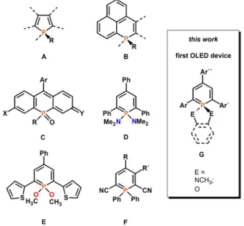
science, the most widely used P-based building block is the phosphole ring (A, Figure 1) embedded in π-conjugated systems, which can be used as emitter in Organic-Light-Emitting Diodes (OLEDs).[3] The flexibility in fine-tuning the optical and
electrochemical properties of such compounds through manipulation of their chemical structure has been exploited for the preparation of tailored white-light-emitting devices.[4]
Apart from the 5-membered rings, polycyclic phosphaphenalenes (B) as well as phospha-fluoresceins, phospha-rhodols and phospha-rhodamines (C) with incorporated 6-membered phosphorus heterocycles have recently been used
for the development of highly fluorescent molecular materials, also for applications in biological imaging.[5,6] In contrast, the
6-membered aromatic phosphinines have received only little attention as building blocks for the construction of emissive π-conjugated systems as it has been shown that 2,4,6-triaryl-λ3
-phosphinines are mostly non-emissive at room temperature.[7] In
view of the strong relationship between a λ3-P=C and a C=C
bond,[8] it seems somewhat surprising that conjugated systems
with P=C double bonds, such as phosphinines, are often non-fluorescent, while most fluorescent molecules are based on conjugated C=C bonds. However, some notable exceptions show that not the P=C bond itself is responsible for the non-emissive behavior.[9,10] Nevertheless, it is possible to restore the
emission of phosphinine-based π-systems by introducing additional substituents at the P-atom, or by coordination of the heterocycle to a transition metal center, respectively.[11,12] First
quantitative photophysical measurements by one of us on λ5
-phosphinines, such as D and E, revealed a rather strong fluorescence emission at λmax = 503 nm and a quantum yield of
20% for E.[7]
Figure 1. Fluorescent organophosphorus compounds A-F and schematic structure of a λ5-phosphinine G. Ar, Ar´and Ar´´: substituted aryl-groups.
More recently, 2,4,6-triaryl- λ5-phosphinines have been
implemented in π‐conjugated, covalent phosphinine‐based frameworks.[13] In 2018, Hayashi and coworker described the
synthesis and optical properties of several λ5-phosphinines of
type F and fairly high quantum yields and tunable fluorophore
[a] Dr. G. Pfeifer, Dr. M. Papke, M. Weber, Prof. Dr. C. Müller Freie Universität Berlin
Institut für Chemie und Biochemie Fabeckstr. 34/36, 14195 Berlin, Germany E-mail: c.mueller@fu-berlin.de [b] Prof. Dr. M. Hissler, Dr. F. Chahdoura
Univ Rennes, CNRS, ISCR - UMR 6226, 35000 Rennes, France E-mail: muriel.hissler@univ-rennes1.fr
[c] Prof. Dr. L. Nyulászi, Dr. R. Szűcs
Department of Inorganic and Analytical Chemistry Budapest University of Technology and Economics
and MTA-BME Computation Driven Chemistry Research Group Szt. Gellért tér 4
H-1111 Budapest, Hungary E-mail: nyulaszi@mail.bme.hu [d] Dr. D. Tondelier, Dr. B. Geffroy
LICSEN, NIMBE, CEA, CNRS, Université Paris-Saclay, CEA Saclay, Gif-sur-Yvette CEDEX 91191, France
Supporting information for this article is given via a link at the end of the document.
Accepted
FULL PAPER
properties were reported.[14] These observations prompted us to
envisage for the first time strategic structural variations on λ5
-phosphinines in order to fine-tune their electronic properties and to elucidate structure-property relationships. Indeed, the classical synthetic route to λ3- and λ5-phosphinines via pyrylium
salts allows the introduction of different substituents in the 2,4,6-positions of the phosphinine ring, while the substituents at the phosphorus atom can also be varied to a great extent by using amines or alcohols in combination with Hg(OAc)2 as an oxidation
reagent. Here, we report on a detailed study on the chemistry of compounds of type G, including crystallographic characterizations, UV/Vis absorption, and fluorescence data, electrochemical behavior as well as theoretical calculations. Most importantly, we report also for the first time on the development of an OLED, based on a blue λ5-phosphinine
emitter of type G.
Results and Discussion
The modular synthesis of 2,4,6-triaryl-λ3-phosphinines allows the
preparation of 2,6-diphenyl-4-tolyl-phosphinine 1, pyridyl-functionalized phosphinine 2 as well as ortho-fluoro-phenyl substituted phosphinine 3 (Figure 2). The pyridyl substituent was chosen, as the steric demand of a nitrogen lone pair is smaller than a CH group of a phenyl moiety, which permits a planar ground state of the conjugated ring system.[9] In contrast, the
increased steric bulk of the ortho-fluoro-phenyl group most likely destabilizes the planar structure. Consequently, the rotational barrier should increase considerably. λ3-Phosphinines 1-3 were
synthesized according to known literature procedures from the corresponding pyrylium-salts and P(SiMe3)3.[7,15,16]
Figure 2. 2,4,6-Triaryl-λ3
-phosphinines 1-3.
Crystals of 2,6-di(2’-fluorophenyl)-4-phenyl-phosphinine, suitable for X-ray diffraction, were obtained by slow crystallization from acetonitrile. The molecular structure of 3 in the crystal (Figure 3) shows the expected planar phosphorus heterocycle, while the three aryl groups are not in plane with the central hexagon. It should be noted that crystallographically characterized 2,4,6-triaryl-substituted λ3-phosphinines are rare. This is in fact the
first observation of the “statistical average” arrangement of the aryl groups attached to C(1) and C(5) for this class of compounds.[15,17] The rotational barrier of the ortho-fluoro-phenyl
groups in 3 amounts to 4.9 kcal/mol (the rotational maximum corresponds to the planar structure, with the fluorine atom pointing toward the phosphorus atom at ω = 0°, see Table S1). This value is higher than the typical 3 kcal/mol for
2,4,6-triphenyl-substituted phosphinines,[18] which is apparently due to
the presence of the sterically more demanding F-substituent.
Figure 3. Molecular structure of 3 in the crystal. Displacement ellipsoids are shown at the 50% probability level. Selected bond lengths (Å) and angles (°): P(1)-C(1): 1.756(6); P(1)-C(5): 1.753(6); C(1)-C(2): 1.386(7); C(2)-C(3): 1.375(8); C(3)-C(4): 1.422(7); C(4)-C(5): 1.401(7). C(5)-P(1)-C(1): 99.4(3); C(2)-C(1)-C(12)-C(13): -134.0(5); C(4)-C(5)-C(6)-C(7): 135.4(5); C(2)-C(3)-C(18)-C(19): -142.6(5).
The λ3-phosphinines 1-3 were further converted quantitatively
into a series of λ5-phosphinines by reaction with Hg(OAc) 2 as
oxidation reagent in the presence of either 1,2-ethanediol, catechol, or N,N´-dimethyl-ethylenediamine, according to a modified procedure described by Dimroth et al. (Scheme 1).[19]
Scheme 1. Synthesis of λ5-phosphinines starting from λ3-phosphinines.
After column chromatography, the λ5-phosphinines 4-10 (Figure
4) were obtained as fairly air- and moisture stable orange and yellow solids in high isolated yields and were characterized by means of 1H-, 13C- and 31P{1H} NMR spectroscopy. Compounds
4-10 show single resonances at around δ(ppm) = +60 in the
31P{1H} NMR spectrum. The shielding by approximately 130 ppm
compared to the values of the corresponding λ3-phosphinines is
characteristic for λ5-phosphinines.[19]
Crystals of 8 suitable for X-ray diffraction were obtained by slow recrystallization from THF/Et2O. Compound 8 crystallizes with
two independent molecules in the asymmetric unit and the molecular structure along with selected bond lengths and angles of one molecule is depicted in Figure 5. The crystallographic characterization of 8 shows the expected tetrahedral arrangement around the phosphorus atom, while the catechol-unit is located perfectly perpendicular to the plane of the phosphorus heterocycle. Since the three aryl substituents in both the λ5-phosphinine (8) and the λ3-phosphinine (3) are
Accepted
FULL PAPER
attached at the 2-,4- and 6-position of the central core, a direct comparison of the crystallographic data is possible.
Figure 4. 2,4,6-Triaryl-λ5-phosphinines 4-10.
Figure 5. Molecular structure of 8 in the crystal. Displacement ellipsoids are shown at the 50% probability level. Only one independent molecule is shown. Selected bond lengths (Å) and angles (°): P(1)-C(1): 1.714(3); P(1)-C(5): 1.715(3); C(1)-C(2): 1.387(4); C(2)-C(3):1.397(4); C(3)-C(4): 1.382(5); C(4)-C(5): 1.403(4); C(5)-C(6): 1.477(4); P(1)-O(1): 1.642(2); P(1)-O(2): 1.646(2); C(2)-C(1)-C(11)-C(12): 138.3(3); C(4)-C(5)-C(6)-C(7): 11.7(4); C(4)-C(3)-C(17)-C(18): 140.3(3).
Upon oxidation of the P-atom, the P(1)-C(1) and P(1)-C(5)-distances become with 1.714(3) and 1.715(3) Å somewhat shorter than in a λ3-phosphinine (see Figure 3), while the C-C
distances are with 1.382-1.403 Å well equalized (benzene: 1.396 Å).[20] These structural characteristics are in accordance with our
earlier theoretical predictions for the parent λ5-phosphinine. Its
electronic structure can be explained by a specific ylidic cyclic delocalization in which the orbitals composed of the phosphorus atom and its two additional substituents are also involved (hyperconjugative effect).[20a] This non-classical cyclic
delocalization turns into a classical aromaticity when electronegative substituents (F, O, N) attached to the phosphorus atom, as shown by Schleyer and subsequently by Rzepa and co-worker, who have attributed this observation to the dominance of negative hyperconjugation.[21,22] The modest
aromaticity of the λ5-phosphinines is also in agreement with our
calculated NICS(1) values of -5 to -8 ppm for the investigated phosphinine rings (Table S2), which demonstrates that the aryl substituents have a minor effect on the electronic structure of the λ5-phosphinines.
For compound 8, we further found a C(2)-C(1)-C(11)-C(12) torsion angle of 138.3(3)° for the phenyl group in 2-position with the central heterocycle, while the pyridyl-group is essentially coplanar with the phosphinine-ring (C(7)-C(6)-C(5)-C(4) = 11.7(4)°). This is in accordance with our expectations for the steric demand of the nitrogen lone pair as discussed above.[9]
In contrast to 2,4,6-triphenyl-λ3-phosphinine, for which a low
barrier for the rotation of the phenyl groups was determined computationally (vide infra),[9] we anticipated that the situation
should be significantly different in 2,4,6-triaryl-λ5-phosphinines.
In these heterocycles, the two additional substituents at the phosphorus atom should have a substantial impact on the rotational barriers of the ortho-aryl groups and their physical properties.
Consequently, to establish structure-property relationships, the optical and electrochemical features of compounds 4-10 were examined. First, we investigated the redox properties of compounds 4-10 by means of cyclic voltammetry (CH2Cl2, 0.2 M,
TBAPF6, v = 200 mV.s-1, see Table 2). All compounds show two
oxidation waves, while no reduction processes were observed in the electrochemical window. The ionization energy of ylides in general,[23] and particularly of λ5-phosphinines,[24] is low. This is
in accordance with the partial negative charge at the carbon-based C5-fragment of the molecule (ylide-character).[20a,24] The
ylide-character is a consequence of the electron distribution in the b1 type HOMO, which is influenced by the hyperconjugative
interaction with the σ-orbitals of the two additional phosphorus-substituents, leading to its destabilization, as has been discussed before.[20a] Furthermore, the nature of the
P-substituents has a significant impact on the HOMO energy. An electronegative O-substituent results in a less destabilized HOMO compared to an N-substituent (Table 1). Thus, 9 and 10 (containing an N,N´-dimethyl-ethylenediamine moiety) have more destabilized HOMOs and show lower Eox1 oxidation
potentials than 4-8, in which oxygen is attached to the phosphorus atom. Interestingly, compounds 7 and 8, containing a catechol moiety at the phosphorus atom, are the most difficult
Accepted
FULL PAPER
to oxidize. Since the aryl rings in 2- and 6-position of the P-heterocycle do not contribute significantly to the HOMO (Figure 6 and Figure S23), the substitution of these rings has only a minor effect on the HOMO energies and the oxidation potentials. Only the aryl ring in 4-position contributes weakly to the HOMO. The good correlation between the measured Eox1 values and the
HOMO energies is noteworthy.
Table 1. B3LYP/6-31+G* HOMO energies [eV], first oxidation potentials (Eox1
[V]) and decomposition temperatures (Td5 [°C]). λ5 HOMO [eV] [a] Eox1 [V] [b] Td5 [°C] [d] 4 -5.18 +0.79 264 5 -5.17 +0.83 271 6 -5.27 +0.93 249 7 -5.44 +1.09 240 8 -5.41 +1.04[c] 253 9 -4.83 +0.47 240 10 -4.91 +0.59 244
[a] All potentials were obtained during cyclic voltametric investigations in 0.1 M Bu4NPF6 in CH2Cl2. Platinum electrode diameter 1 mm, sweep rate: 200 mV·
s-1. All reported potentials are referenced to the reversible formal potential of
the decamethyl-ferrocene/decamethylferrocenium couple. [b] Irreversible process. [c] Decomposition temperature at 5% weight loss, measured by thermogravimetric analysis (TGA) under nitrogen.
Figure 6. B3LYP/6-31+G* HOMO and LUMO of 3, 7 and 10.
The LUMO of a λ5-phosphinine has a nodal plane through the
heteroatom (a2 symmetry) and is similar to the LUMO+1 orbital
of a λ3-phosphinine (Figure 6 and 7), as we noted also
before.[20a] This can be rationalized by the fact that the LUMO of
a λ3-phosphinine (b
1 symmetry, likewise the HOMO) is
destabilized by the hyperconjugative interactions with the σ-orbitals at the P-substituents being present in the λ5-phosphinine.
Consequently, the energy of the corresponding orbital in the λ5
-phosphinine is pushed above the orbital with a2 symmetry, which
then becomes the LUMO, however, at significantly higher energies compared to the b1 type orbital of the λ3-counterpart
(Figure 7). All of this is in line with our observations, that no reduction wave was detected for λ5-phosphinines 4-10.
Figure 7. Frontier orbitals of a λ3- and a λ5-phosphinine and formation of the
3c-4e bond.
It is further noteworthy, that the aryl substituents in 2- and 6-position of the heterocycle are involved in the LUMO to some extent. This is particularly obvious from the bonding interactions between the connecting carbon atoms, even in those cases, where the substituent in 2- or 6-position is rotated somewhat out of the plane of the heterocycle (Figure 6, LUMO of 7, 10). Next, the optical properties (UV-vis absorption and fluorescence) of compounds 4-10 were studied both in dichloromethane and in the solid-state (Table 2). Compounds 4-10 show similar absorptions with two broad bands around λ = 380 nm and λ = 280 nm respectively (Figure 8 and Figures S8-S22). The TD-DFT calculated band maxima (vertical transition energies) are essentially HOMO-LUMO (see Figure S23) transitions and were obtained for the most stable rotamers. These values are systematically located by approximately 30 nm lower wavelengths than the experimentally observed ones (slightly different TD-DFT calculated excitation energies for other rotamers are given in Tables S3a-S9a). The oscillator strength values are in most cases larger than 0.2 (see Table S3a-S9a) in accordance with the different (quasi)symmetry of the HOMO and LUMO. These spectra differ significantly from the spectrum recorded for λ3-phosphinine 3, which presents an intense band
centered at λ = 279 nm along with a red-shifted shoulder tailing down to λ = 345 nm. The lowest energy band for the λ3
-phosphinine was assigned to a HOMO→LUMO (π-π*) transition as was shown by our TD-DFT calculations and corresponds to an intra-phosphinine charge transfer with a strong contribution of the phosphorus atom in both orbitals. Since this transition is weakly allowed due to the small dipole moment change (note that in the parent λ3-phosphinine both HOMO and LUMO have
Accepted
FULL PAPER
the same b1 symmetry), only a low fluorescence intensity is
observed (quantum yields << 1%) as we noted already before.[7]
Figure 8. Absorption spectrum of compound 3 (top) and absorption and emission spectra of compound 10 recorded in CH2Cl2 (c = 10-5 M) at room
temperature.
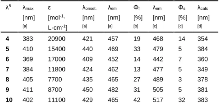
The λ5-phosphinines 4-10 exhibit moderate to high fluorescence
in solution (Table 2), which is in accordance with the rather large oscillator strength discussed above. Since the Stokes shift is different for each compound, the reorganization of the molecules between the ground state and the excited state should contribute to the different quantum efficiencies (vide infra).
Table 2. Optical properties of λ5
-phosphinines 4-10. λ5 λ max [nm] [a] ε [mol-1· L·cm-1 ] λonset. [nm] [a] λem [nm] [a] Φf [%] [b] λem [nm] [c] Φs [%] [c] λcalc [nm] [d] 4 383 20900 421 457 19 468 14 354 5 410 15400 440 469 33 479 5 384 6 369 17000 409 452 14 442 7 360 7 384 11800 424 462 13 477 5 349 8 405 7700 435 465 27 489 3 378 9 411 8700 450 482 31 505 5 381 10 402 11100 429 465 42 517 32 383 [a] Measured in CH2Cl2. [b] Fluorescence quantum yields determined using
quinine sulfate as standard, ± 15%. [c] Measured in an integrated sphere. [d] TD-DFT vertical absorption energy.
The emission band maxima were also calculated for the most stable isomers by optimizing the excited state geometries by TD-DFT (Table S3a-S9a). The calculated and the measured Stokes shifts are in reasonable agreement, while the largest deviation is seen in the case of compound 6. The pyridyl-functionalized
systems 5 and 8, in which the pyridyl-substituent and the P-heterocycle are coplanar both in the ground and excited states, show a small Stokes shift and, accordingly, a high quantum efficiency.
To further understand the photophysical properties of the different λ5-phosphinines, we investigated the rotational barriers
for all the connecting aryl groups, since this is the most likely pathway for a radiationless energy relaxation of the excited state. As a function of the angles ω, θ and φ, the highest rotational barriers for the λ3-phosphinine 3 and the λ5-phosphinines 4-10
are illustrated in Table 3 (for the more detailed rotational analysis see Tables S1b and S3b-S9b). The rotational barriers for the aryl group in 4-position of the heterocycle (characterized by θ) in 4-10 are low and rather independent from the additional substituents at the phosphorus atom, including the case of λ3
-phosphinine 3. As expected, the rotational barriers for the aryl groups in 2- and 6-position (ω and φ) are higher for λ5
-phosphinines 6 and 10 than for the corresponding λ3
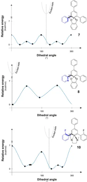
-phosphinine 3. This hindrance of the rotation decreases the efficiency of the vibronic deactivation of the excited state and, consequently, contributes to the observed increase of the quantum yield. Also compounds containing an α-pyridyl group (5 and 8) have rather high rotational barriers. For the three representative examples 7, 8 and 10 with rather different barriers, the full 360° relaxed rotation scan of one aryl group in 2-position is depicted in Figure 9.
Table 3. Rotational barriers in kcal/mol for λ3-phosphinine 3 and λ5
-phosphinines 4-10. ω θ φ 3 4.9[a] 3.1 4.9[a] 4 4.3[a,b] 2.6 4.3[a,b] 5 6.6[c] 2.7 3.9[b] 6 5.8[b] 2.8 5.8[b] 7 2.9[a,b] 2.7 2.9[a,b] 8 5.7[c] 2.8 3.2[a,b] 9 3.4[a,b] 2.4 3.4[a,b] 10 6.8[a] 2.5 6.8[a]
[a] Rotational maximum at about ω=0°. [b] Rotational maximum at about ω=180°. [c] Rotation of the pyridyl group, rotational maximum at about ω=90°.
In case of 8, the two rotational minima of the pyridine ring are located at the coplanar positions of the pyridyl- and phosphinine-rings due to the small steric need of the nitrogen lone pair.[9] This allows for an efficient π-conjugation. The
rotational maxima are at the perpendicular positions of the substituent, where the overlap between the π-systems is minimal. For 7 and 10, the rotational potential energy surface is completely different. Repulsion of the hydrogen, or the α-fluoro atoms, with the substituents both at the 6-membered ring and the P-atom, hinders a coplanar arrangement. A planar form in case of 7 and 10 represents indeed a rotational maximum (with additional, but smaller rotational maxima at the perpendicular positions). For the 2-fluoro-phenyl substituted phosphinine 10, the rotational maxima at the coplanar positions are even higher than for 7.
The TD-DFT calculated excited states exhibit somewhat
Accepted
FULL PAPER
shortened C-C distances (by ca. 0.02-0.03 Å) between the phosphinine ring and the aromatic rings in 2- and 6-position, in accordance with the π-bonding nature of the LUMO between the two respective carbon atoms (Figure 6, 7 and Figure S23). As a consequence of the population of the LUMO in the excited state, the central P-heterocycle and the aryl-rings in 2- and 6-position tend to reach a coplanar arrangement (see the insert showing the position of the excited state on the rotational scan in Figure 9).
Figure 9. Rotational analysis for the groups at the 2-position of 7, 8 and 10.
The flattening is, however, less pronounced for the fluoro-phenyl substituted λ5-phosphinines, which are more rigid due to the
higher rotational barriers. In case of 8 (and 5) having already a (nearly) coplanar arrangement of the pyridyl-group and the core in the ground state (see above), the geometry change upon excitation is only minimal. Since this change is related to the Franck-Condon factor, which influences the transition probability,
the fluorescent quantum yields are beneficially influenced by the small geometry change upon excitation, as it was also shown before.[8,9] As a consequence, the increased quantum yields for
certain λ5-phosphinines can be attributed to the high oscillator
strength of the electronic transition, and also to the increase of the rotational barrier of the α-substituent. No increase of the quantum yields is observed in the solid-state indicating the presence of aggregate quenched emissions in this series of compounds (Table 2).
Taking into account the thermal stabilities and the optical and redox properties of compounds 4-10, only compound 10 was used as emitting material (EM), either pure or doped in a DPVBi (4,4’-bis(2,2’-diphenylvinyl)-1,1’-biphenyl) matrix. In the first attempt, compound 10 was used as pure emitter in an Organic Light Emitting Diode (OLED) having the following configuration: Glass / ITO / CuPc (10nm) / α-NPB (50nm) / 10 (40nm) / BCP (10nm) / Alq3 (10nm) / LiF (1.2nm) / Al (100nm) (ITO =
indium-tin-oxide; CuPC = Cu(II)phtalocyanine; α-NPB = N,N'-Bis-(1-naphthalenyl)-N,N'-bis-phenyl-(1,1′-biphenyl)-4,4′-diamine; BCP = bathocuproine; Alq3 = tris-(8-hydroxyquinolinato)aluminum).
The electroluminescence (EL) performance of the resulting devices are reported in Table 4 and the EL spectra are shown in Figure 10.
For the pure emitter, the EL emission peaked at λ = 485 nm with a red-shifted shoulder in the range of λ = 550-600 nm. This indicates that compound 10 may form aggregates after vacuum evaporation. The EL performance is moderate (Table 4) since the charge transport in the emitting layer, containing compound
10, maybe low and the emission is quenched by the formation of
the proposed aggregates.
Table 4. Electroluminescent performance of devices A-C.
Device Emitter Doping rate (%) Von[a] (V) EQE[b] (%) CE[b] (cd/A) PE[b] (lm/W) A 10 pure 5.3 0.04 0.09 0.04 B 10 3.2 5.0 0.96 1.87 0.58 C 10 7.9 4.9 0.67 1.40 0.46
[a] Threshold voltage recorded at luminance of 1 cd/m2
. [b] EQE (external quantum efficiency), CE (current efficiency) and PE (power efficiency) recorded at 10 mA/cm2.
Figure 10. Normalized EL spectrum of doped and non-doped OLEDs devices.
Accepted
FULL PAPER
Nevertheless, an increase of the performance could be observed when compound 10 is used as dopant (3.2-7.9 %wt, Table 4) in a DPVBi matrix. As a result of diluting compound 10 in the DPVBi matrix, efficient charge transport properties are generated. Moreover, doping the blue matrix with 3.2% of compound 10 leads to an OLED, which exhibits a turn-on voltage of 5.0 V with current and power efficiencies of 1.87 cd A -1 and 0.58 lm/W, respectively. Interestingly, the external
quantum efficiency (EQE) is dramatically improved compared to the pure emitter (0.9% for device B and 0.04% for device A). However, an increase of the doping ratio of up to 7.9 %wt led to a decrease of the performance (device C) and a shift of the EL peak from λ = 474 nm for device B to λ = 485 nm for device C (Figure 10). It is noteworthy for the higher doping rate, that the EL peak appears at the same value as for the pure emitter (device A).
Conclusions
In conclusion, we have synthesized a series of 2,4,6-triaryl-λ5
-phosphinines by combining the highly modular pyrylium salt route for the preparation of λ3-phosphinines with an efficient
oxidation process to introduce additionally different R2N- or
RO-substituents at the phosphorus atom. We found that the optical and redox properties of these compounds can be varied to some extend by the nature of the substituents. Theoretical calculations helped to rationalize our observations and we could clearly demonstrate that λ5-phosphinines can be efficient emitters in
contrast to their λ3-counterparts. In addition, the thermal stability
of 10 prompted us to use this compound as a blue fluorescent emitting material for the construction of an OLED with current and power efficiencies of 1.87 cd A-1 and 0.58 lm/W. These
preliminary results demonstrate that λ5-phosphinine-based
emitters can indeed be used to fabricate optoelectronic devices. Further structural variations in λ5-phosphinines, supported by
DFT calculations and improvements of their performance in OLED devices are currently performed in our laboratories. Main Text Paragraph.
Experimental Section
General. Unless otherwise stated, all syntheses were performed under
an inert argon atmosphere using modified Schlenk techniques or in a MBraun glovebox. All common chemicals were commercially available and were used as received. Dry or deoxygenated solvents were prepared using standard techniques or used from a MBraun solvent purification system. The NMR spectra were recorded on a JEOL ECX400 (400 MHz) spectrometer and chemical shifts are reported relative to the residual resonance in the deuterated solvents. Phosphinines 1[15], 2[7] and 3[16]
were prepared according to the literature.
UV/Vis spectra were recorded at RT with a VARIAN Cary 5000 spectrophotometer. UV/Vis/NIR emission and excitation spectra measurements were recorded with an FL 920 Edinburgh Instrument equipped with a Hamamatsu R5509-73 photomultiplier for the NIR domain (300-1700 nm) and corrected for the response of the photomultiplier. Quantum yields were calculated relative to quinine
sulfate (H2SO4, 0.1 m, φref = 0.55). The electrochemical studies were
carried out under argon with an Eco Chemie Autolab PGSTAT 30 potentiostat for cyclic voltammetry with the three-electrode configuration: the working electrode was a platinum disk, the reference electrode was a saturated calomel electrode, and the counter-electrode was a platinum wire. All potentials were internally referenced to the ferrocene/ferrocenium couple. For the measurements, concentrations of 10-3 m of the electroactive species were used in freshly distilled and
degassed dichloromethane and 0.2 m tetrabutylammonium hexafluorophosphate. Thermogravimetric analyses were performed with a Mettler-Toledo TGA-DSC-1 apparatus under dry nitrogen flow at a heating rate of 10°C min-1. All measurements were performed with quartz
cuvettes with a path length of 1.0 cm.
For NMR, absorption, emission and excitation spectra see supporting information.
Synthesis of 1,1-Ethylenglycolyl-λ5-2,6-diphenyl-4-(p-tolyl)phosphinine
(4): Phosphinine 1 (200 mg, 0.59 mmol) and mercury acetate (207 mg,
0.65 mmol) were dissolved in 5 mL toluene in an argon atmosphere and then mixed with ethylene glycol (0.04 mL, 0.60 mmol) at room temperature. After stirring for overnight, the solution was filtered over silica (3 cm) to remove the mercury residues. The solvent of the neon yellow filtrate was then removed under vacuum and the residue was washed with pentane. After drying under high vacuum, the product is obtained as a neon yellow solid (157 mg, 0.39 mmol, 66%). 1H NMR (400
MHz, CDCl3): δ = 7.69 (d, 3JH,P = 39.5 Hz, 2H, C5H2P), 7.63-7.54 (m, 4H, Har), 7.46-7.36 (m, 4H, Har), 7.36 (d, J = 8.1 Hz, 2H, Har), 7.31 (t, J = 7.2 Hz, 2H, Har), 7.15 (d, J = 7.8 Hz, 2H), 4.19 (d, 3JH,P = 10.3 Hz, 4H, CH2 -CH2), 2.35 (s, 3H, CH3) ppm; 13C{1H} NMR (101 MHz, CDCl3): δ = 140.3 (d, J = 2.6 Hz), 139.5 (d, J = 4.1 Hz), 138.9 (d, J = 10.7 Hz), 134.5, 129.4, 129.3 (d, J = 1.7 Hz), 128.8 (d, J = 1.1 Hz), 126.6 (d, J = 1.5 Hz), 126.1 (d, J = 0.7 Hz), 116.4 (d, J = 19.5 Hz), 97.2 (d, 1J P,C = 145.4 Hz, C1,5(C 5H2P)), 66.6 (d, 2JP,C = 1.8 Hz, CH2-CH2), 21.1 (CH3) ppm; 31P{1H} NMR (162 MHz, CDCl3): δ = 69.2 ppm. EI (m/z): 398.1580 g/mol (calculated: 398.1435 g/mol) [M]+.
Synthesis of 1,1-Ethylenglycolyl-λ5-2-(2‘-pyridyl)-4,6-diphenylphosphinine
(5): Phosphinine 2 (100 mg, 0.31 mmol) and mercury acetate (103 mg,
0.33 mmol) were dissolved in 5 mL toluene in an argon atmosphere and then mixed with ethylene glycol (0.02 mL, 0.33 mmol) at room temperature. After stirring for overnight, the solution was filtered over silica (3 cm) to remove the mercury residues. The solvent of the fluorescent yellow-green filtrate was then removed under vacuum and the residue was washed with pentane. After drying in high vacuum, the product was obtained as a neon yellow solid (70.4 mg, 0.18 mmol, 59%).
1H NMR (400 MHz, CDCl 3): δ = 8.46 (d, J = 5.8 Hz, 1H, Har), 8.01 (dd, 3J H,P = 40.4 Hz, 4JH,H = 2.7 Hz, 1H, Har), 7.76 (dd, J = 38.2, 2.7 Hz, 1H, Har), 7.69-7.59 (m, 2H, Har), 7.64-7.44 (m, 4H, Har), 7.47-7.26 (m, 5H, Har), 7.26-7.15 (m, 1H, Har), 7.05-6.96 (m, 1H, Har), 4.76-4.66 (m, 2H, CH2-CH2), 4.36-4.25 (m, 2H, CH2-CH2) ppm; 13C{1H} NMR (101 MHz, CDCl3): δ = 148.4, 143.2, 141.6 (d, J = 10.2 Hz), 136.9, 133.6 (d, J = 9.2 Hz), 129.2 (d, J = 5.6 Hz), 128.7 (d, J = 9.6 Hz), 126.7, 126.3, 125.1, 119.4, 117.6 (d, J = 9.2 Hz), 116.6 (d, J = 18.9 Hz), 67.4 (d, J = 1.1 Hz) ppm; 31P{1H} NMR (162 MHz, CDCl 3): δ = 72.9 ppm. EI (m/z): 385.1201
g/mol (calculated: 385.1232 g/mol) [M]+.
Synthesis of 1,1-Ethylenglycolyl-λ5
-2,6-bis(2-fluorophenyl)-4-phenyl-phosphinine (6): Phosphinine 3 (100 mg, 0.28 mmol) and mercury
acetate (95.0 mg, 0.30 mmol) were dissolved in 5 mL toluene in an argon atmosphere and subsequently mixed with ethylene glycol (0.02 mL, 0.30 mmol) at room temperature. After stirring for overnight, the solution was filtered over silica (3 cm) to remove the mercury residues. The solvent of the neon yellow filtrate was then removed under vacuum and the residue
Accepted
FULL PAPER
was washed with pentane. After drying under high vacuum, the product was obtained as a neon yellow solid (75.6 mg, 0.21 mmol, 74%). 1H NMR
(400 MHz, CDCl3): δ = 7.68 (d, 3JH,P = 39.6 Hz, 2H), 7.57-7.52 (m, 2H, Har), 7.45-7.41 (m, 2H, Har), 7.37-7.25 (m, 4H, Har), 7.23-7.11 (m, 5H, Har), 4.04 (d, 3J H,P = 10.4 Hz, 4H, CH2-CH2) ppm. 19F NMR (376 MHz, CDCl3): δ = -116.9 (m) ppm; 31P{1H} NMR (162 MHz, CDCl 3): δ = 67.6 ppm. EI
(m/z): 420.1138 g/mol (calculated: 420.1091 g/mol) [M]+.
Synthesis of 1,1-Brenzcatechinyl-λ5-2,6-diphenyl-4-(p-tolyl)phosphinine
(7): Phosphinine 1 (200 mg, 0.59 mmol) and mercury acetate (207 mg,
0.65 mmol) were dissolved in 5 mL toluene in an argon atmosphere and then catechol (71.0 mg, 0.65 mmol) was added at room temperature. After stirring for overnight, the solution was filtered over silica (3 cm) to remove the mercury residues. The solvent of the neon yellow filtrate was then removed under vacuum and the residue was washed with pentane. After drying under high vacuum, the product was obtained as a neon yellow solid (230 mg, 0.52 mmol, 87%). 1H NMR (400 MHz, CD
2Cl2): δ = 7.82 (d, 3J P,H = 42.5 Hz, 2H, C5H2P), 7.51-7.43 (m, 4H, Har), 7.46-7.36 (m, 2H, Har), 7.32-7.22 (m, 4H, Har), 7.24-7.16 (m, 4H, Har), 7.01-6.93 (m, 4H, Har), 2.36 (s, 3H, CH3) ppm; 13C{1H} NMR (101 MHz, CD2Cl2): δ = 145.5 (d, J = 1.0 Hz), 140.0 (d, J = 3.1 Hz), 138.6 (d, J = 10.5 Hz), 138.2 (d, J = 4.0 Hz), 135.8, 129.9, 129.4 (d, J = 0.9 Hz), 128.9 (d, J = 7.1 Hz), 127.3 (d, J = 1.5 Hz), 126.7 (d, J = 0.9 Hz), 124.2, 119.4 (d, J = 21.3 Hz), 111.8 (d, J = 11.0 Hz), 98.2 (d, J = 144.5 Hz, C5H2P), 21.2 (d, J = 1.5 Hz, CH3) ppm; 31P{1H} NMR (162 MHz, CD 2Cl2): δ = 70.6 ppm. 31P NMR (162 MHz, CD2Cl2): δ = 70.6 (t, 3JP,H = 42.5 Hz) ppm. EI (m/z): 446.1528 g/mol (calculated: 446.1436 g/mol) [M]+.
Synthesis of 1,1-Brenzcatechinyl-λ5
-2-(2‘-pyridyl)-4,6-diphenyl-phosphinine (8): Phosphinine 2 (100 mg, 0.31 mmol) and mercury
acetate (103 mg, 0.33 mmol) were dissolved in 5 mL toluene in an argon atmosphere and then catechol (35.0 mg, 0.33 mmol) was added at room temperature. After stirring for overnight, the solution was filtered over silica (3 cm) to remove the mercury residues. The solvent of the fluorescent yellow-green filtrate was then removed under vacuum and the residue was washed with pentane. After drying under high vacuum, the product was obtained as a neon yellow solid (115 mg, 0.27 mmol, 86%). 1H NMR (400 MHz, CDCl 3): δ = 8.08 (dd, 3JH,P = 43.9 Hz, 4JH,H = 2.7 Hz, 1H, C5H2P), 7.89 (dd, 3JH,P = 41.2 Hz, 4JH,H = 2.7 Hz, 1H, C5H2P), 7.76 (d, J = 4.8 Hz, 1H), 7.63-7.49 (m, 6H, Har), 7.41 (t, J = 7.6 Hz, 2H, Har), 7.32-7.18 (m, 4H, Har), 7.04-6.92 (m, 4H, Har), 6.88-6.83 (m, 1H, Har) ppm; 13C{1H} NMR (101 MHz, CDCl 3): δ/ppm = 157.6 (d, J = 1.9 Hz), 148.5, 146.2, 142.7 (d, J = 2.7 Hz), 141.1 (d, J = 10.5 Hz), 138.1 (d, J = 3.8 Hz), 136.8, 132.7 (d, J = 8.6 Hz), 128.9 (d, J = 0.9 Hz), 128.8, 128.7, 128.6, 126.9 (d, J = 1.4 Hz), 126.5 (d, J = 1.1 Hz), 125.7, 123.2, 119.9 (d, J = 0.9 Hz), 118.7 (d, J = 21.1 Hz), 116.8 (d, J = 10.1 Hz), 110.9 (d, J = 11.3 Hz), 101.6 (d, 1J P,C = 144.9 Hz, C1,5(C5H2P)) ppm; 31P{1H} NMR (162 MHz, CDCl3): δ = 75.2 ppm. EI (m/z): 433.1298 g/mol (calculated: 433.1232 g/mol) [M]+.
Synthesis of 1,1-N,N‘-Dimethylethylendiaminyl-λ5
-2,6-diphenyl-4-(p-tolyl)-phosphinine (9): Phosphinine 1 (200 mg, 0.59 mmol) and mercury
acetate (207 mg, 0.65 mmol) were dissolved in 5 mL toluene in an argon atmosphere and subsequently mixed with N,N' dimethylethylenediamine (0.07 mL, 0.65 mmol) at room temperature. After stirring for overnight, the solution was filtered over silica (3 cm) to remove the mercury residues. The solvent of the neon yellow filtrate was then removed in a vacuum and the residue washed with pentane. After drying under high vacuum, the product was obtained as a neon yellow solid (117 mg, 0.28 mmol, 47%). 1H NMR (400 MHz, CDCl 3): δ = 7.74 (d, 3JH,P = 33.5 Hz, 2H, C5H2P), 7.50-7.41 (m, 4H, Har), 7.38 (d, J = 8.1 Hz, 2H, Har), 7.33 (t, J = 7.6 Hz, 4H, Har), 7.23-7.17 (m, 2H, Har), 7.13 (d, J = 7.9 Hz, 2H, Har), 3.11 (d, 3J H,P = 7.5 Hz, 4H, CH2-CH2), 2.36 (d, 3JH,P = 10.4 Hz, 6H, N-CH3), 2.34 (s, 3H, Ph-CH3) ppm; 13C{1H} NMR (101 MHz, CDCl3): δ = 142.2 (d, J = 6.0 Hz), 140.8, 138.1 (d, J = 9.6 Hz), 133.4, 129.3, 128.4, 128.3 (d, J = 5.7 Hz), 125.4-125.3 (d, J = 1.2 Hz), 125.2, 113.5 (d, J = 15.6 Hz), 95.8 (d, 1J P,C = 126.2 Hz, C1,5(C5H2P)), 48.3 (d, 2JP,C = 8.7 Hz, CH2-CH2), 31.3 (d, 2J P,C = 8.2 Hz, N-CH3), 21.0 (Ph-CH3) ppm; 31P{1H} NMR (162 MHz, CDCl3): δ = 36.2 ppm. EI (m/z): 424.2075 g/mol (calculated: 424.2063 g/mol) [M]+.
Synthesis of 1,1-N,N‘-Dimethylethylendiaminyl-λ5
-2,6-bis(2-fluorophenyl)-4-phenylphosphinine (10): Phosphinine 3 (500 mg, 1.39 mmol) and
mercury acetate (486 mg, 1.53 mmol) are dissolved in 15 mL toluene in an argon atmosphere and subsequently mixed with N,N' dimethylethylenediamine (0.16 mL, 1.53 mmol) at room temperature. After stirring for overnight, the solution was filtered over silica (3 cm) to remove the mercury residues. The solvent of the neon yellow filtrate was then removed under vacuum and the residue was washed with pentane. After drying under high vacuum, the product was obtained as a neon yellow solid (141 mg, 0.32 mmol, 23%). 1H NMR (400 MHz, CDCl
3): δ = 7.68 (d, 3J H,P = 33.8 Hz, 2H, C5H2P), 7.45 (d, J = 8.0 Hz, 2H, Har), 7.35-7.18 (m, 6H, Har), 7.16-7.02 (m, 5H, Har), 2.89 (d, 3JH,P = 8.0 Hz, 4H, CH2-CH2), 2.48 (d, 3JH,P = 10.4 Hz, 6H, N-CH3) ppm; 13C{1H} NMR (101 MHz, CDCl3): δ = 140.4, 132.4, 128.5, 128.2, 127.7 (d, J = 8.0 Hz), 125.1, 123.8, 123.6 (d, J = 3.7 Hz), 123.4, 121.8, 121.5, 115.8 (d, J = 23.5 Hz), 98.2 (d, 1J P,C = 131.4 Hz, C1,5(C5H2P)), 47.4 (d, 2JP,C = 8.8 Hz, CH2-CH2), 31.3 (d, 2J P,C = 8.4 Hz, N-CH3) ppm; 19F NMR (376 MHz, CDCl3): δ = -114.7 ppm; 31P{1H} NMR (162 MHz, CDCl 3): δ = 34.6 ppm. EI (m/z):
446.1903 g/mol (calculated: 446.1718 g/mol) [M]+.
X-ray crystal structure determination of 3: C23H15F2P, Fw = 360.32,
colourless stick, 0.01 × 0.03 × 0.17 mm3, orthorhombic, Pna2
1, a =
7.7176(3), b = 19.6751(7), c = 11.5400(5) Å, V = 1752.29(12) Å3, Z = 4,
Dx= 1.366 g/cm3, µ = 1.587 mm-1. 11690 reflections were measured by a
Bruker D8-Venture diffractometer with a Photon area detector (CuKα radiation; λ = 1.54178 Å) at a temperature of T = 100(2) K up to a resolution of θmax = 79.29. The reflections were corrected for absorption
and scaled on the basis of multiple measured reflections by using the SADABS program (0.77–0.98 correction range).[25] 2572 reflections were
unique (Rint = 0.045). The structures were solved with SHELXS-1997 by
using direct methods and refined with SHELXL-2017 on F2 for all
reflections.[26] Non-hydrogen atoms were refined with anisotropic
displacement parameters. 235 parameters were refined without restraints.
R1 = 0.057 for 2572 reflections with I>2s(I) eÅ3, and wR2 = 0.154 for 2878
reflections, S = 1.081. Geometry calculations and checks for higher symmetry were performed with the PLATON program.[27] CCDC-1968707
contains the supplementary crystallographic data for this paper. These data can be obtained free of charge from The Cambridge Crystallographic Data Centre via www.ccdc.cam.ac.uk/data_request/cif.
X-ray crystal structure determination of 8: C28H20NO2P, Fw = 433.42,
orange block, 0.16 × 0.31 × 0.32 mm3, monoclinic, P2
1, a = 10.6265(2), b
= 7.5595(4), c = 12.1521(3) Å, V = 2137.37(8) Å3, Z = 4, D
x= 1.347 g/cm3,
µ = 0.155 mm-1. 78641 reflections were measured by a Bruker
D8-Venture diffractometer with a Photon area detector (MoKα radiation; λ = 0.71073 Å) at a temperature of T = 100(2) K up to a resolution of θmax =
27.16. The reflections were corrected for absorption and scaled on the basis of multiple measured reflections by using the SADABS program (0.92–1.00 correction range).[25] 28752 reflections were unique (R
int =
0.056). The structures were solved with SHELXS-2013 by using direct methods and refined with SHELXL-2013on F2 for all reflections.[26]
Non-hydrogen atoms were refined with anisotropic displacement parameters. 578 parameters were refined without restraints. R1 = 0.035 for 8752
reflections with I>2s(I) eÅ3, and wR
2 = 0.096 for 9479 reflections, S =
1.119. Geometry calculations and checks for higher symmetry were performed with the PLATON program.[27] CCDC-1968706 contains the
supplementary crystallographic data for this paper. These data can be
Accepted
FULL PAPER
obtained free of charge from The Cambridge Crystallographic Data Centre via www.ccdc.cam.ac.uk/data_request/cif.
Computational details: Density functional calculations were carried out
with the Gaussian 09 program package.[28] All structures were optimized
using the B3LYP functional,[29] combined with the 6-31+G* basis set, and
these results were discussed throughout. For the conformational search of 3 further calculations were carried out using the M06-2X and the ωB97XD functionals and the cc-pVTZ basis, which gave similar results to B3LYP/6-31+G* (See Table S1a in the Supporting Information). At each of the optimized structures vibrational analysis was carried out to check that the stationary point located is a minimum of the potential energy hypersurface (no imaginary frequencies were obtained). Relaxed scans were calculated to describe the rotational behavior of the α-aryl groups. To obtain vertical excitation energies and optimized excited state structures TD DFT B3LYP/6-31+G* calculations were carried out. The optimized excited state geometries were used for the calculation of the position of the emission spectral maxima. For the visualization of the molecular orbitals the VMD program[30] was used.
OLEDs device fabrication: The OLED devices were fabricated onto
indium tin oxide (ITO) glass substrates purchased from Xin Yang Technology (90 nm thick, sheet resistance of 15 Ω/m). Prior to organic layer deposition, the ITO substrates were cleaned by sonication in a detergent solution, rinsed twice in de-ionized water and then in isopropanol solution and finally treated with UV-ozone during 15 minutes. The OLEDs stack is the following: Glass / ITO / CuPc (10 nm) / α-NPB (40 nm) / EML 40 nm / BCP (10 nm) / Alq3 (40 nm) / LiF (1.2 nm) / Al
(100 nm). Cu(II)phthalocyanine (CuPc) is used as hole injection layer (HIL), N,N'-Bis-(1-naphthalenyl)-N,N'-bis-phenyl- (1,1′-biphenyl)-4,4′-diamine (α-NPB) as hole transport layer (HTL), bathocuproine (BCP) as hole blocking layer (HBL), tris-(8-hydroxyquinoline)aluminum (Alq3) as
electron transport layer (ETL), lithium fluoride as electron injection layer (EIL) and 100 nm of aluminum as the cathode, respectively. The emitting layer (EML) is compound 10 either as a neat film (device A) or a host-guest system (devices B and C). The host material is 4,4′-bis(2,2-diphenylvinyl)-1,1′-biphenyl, (DPVBi). The doping ratio were 3.2 and 7.9 %wt respectively. All the organic materials were purchased from commercial companies except molecule 10. Organic layers were sequentially deposited onto the ITO substrate at a rate of 0.2 nm/s under high vacuum (10−7 mbar). The doping rate was controlled by simultaneous co-evaporation of the host and the dopant. An in-situ quartz crystal was used to monitor the thickness of the layer depositions with an accuracy of 5%. The active area of the devices defined by the Al cathode was 0.3 cm2. The organic layers and the LiF/Al cathode were deposited
in a one-step process without breaking the vacuum.
Device characterization: After deposition, all the measurements were
performed at room temperature and under ambient atmosphere with no further encapsulation of devices. The current–voltage–luminance (I–V–L) characteristics of the devices were measured with a regulated power supply (ACT100 Fontaine) combined with a multimeter (Keithley) and a 1 cm2 area silicon calibrated photodiode (Hamamatsu).
Electroluminescence (EL) spectra and chromaticity coordinates of the devices were recorded with a PR650 SpectraScan spectrophotometer, with a spectral resolution of 4 nm.
Acknowledgements
OTKA NN 113772 and BME-Nanotechnology FIKP grant of EMMI (BME FIKP-NAT) and the Alexander von Humboldt Foundation for a reinvitation grant for (LN), PICS SmartPAH
(08062) and MTA – NKM 44/2019 (LN and MH) is acknowledged for financial support. Funding by the Deutsche Forschungsgemeinschaft (MU1657/5-1) is gratefully acknowledged.
Keywords: Phosphinine • heterocycles • π-system • OLED •
DFT calculations
[1] a) L. Kollár, G. Keglevich, Chem. Rev. 2010, 110, 4257; b) C. Müller, D. Vogt in Phosphorus Chemistry: Catalysis and Material Science Applications, Vol. 36 (Ed.: M. Peruzzini, L. Gonsalvi), Chapter 6, Springer, 2011; c) C. Müller in Phosphorus Ligand Effects in Homogeneous Catalysis: Design and Synthesis (Ed.: P.C.J. Kamer, P.W.N.M van Leeuwen), Wiley-VCH, 2012.
[2] See for example: a) M. J. Burk, J. E. Feaster, R. L. Harlow, Tetrahedron: Asymmetry 1991, 2, 569-592; b) T. Clark, C. R. Landis, Tetrahedron: Asymmetry 2004, 15, 2123-2137; c) A. C. Brezny, C. R. Landis, Acc. Chem. Res. 2018, 51, 2344-2354.
[3] For recent reviews see : a) D. Joly, P.-A. Bouit, M. Hissler, J. Mater. Chem. C 2016, 4, 3686-3698; b) M. P. Duffy, W. Delaunay, P.-A. Bouit, M. Hissler, Chem. Soc. Rev. 2016, 45, 5296-5310; c) T. Baumgartner, Acc. Chem. Res. 2014, 47, 1613-1622; d) M. A. Shameem, A. Orthaber, Chem. Eur. J. 2016, 22, 10718-10735.
[4] O. Fadhel, M. Gras, N. Lemaitre V. Deborde, M. Hissler, B. Geffroy, R. Réau, Adv. Mater. 2009, 20, 1-5.
[5] a) P. Hindenberg, C. Romero-Nieto, Synlett, 2016, 27, 2293-2300; b) P. Hindenberg, A. López-Andarias, F. Rominger, A. de Cózar, C. Romero-Nieto, Chem. Eur. J. 2017, 23, 13919-13928; c) E. Regulska, P. Hindenberg, C. Romero-Nieto, Eur. J. Inorg. Chem. 2019, 1519-1528; d) P. Hindenberg, J. Zimmermann, G. Hernandez-Sosa, C. Romero-Nieto, Dalton Trans. 2019, 48, 7503-7508; e) E. Regulska, S. Christ, J. Zimmermann, F. Rominger, G. Hernandez-Sosa, C. Romero-Nieto, Dalton Trans. 2019, 48, 12803.
[6] M. Grzybowski, M. Taki, K. Senda, Y. Sato, T. Ariyoshi, Y. Okada, R. Kawakami, T. Imamura, S. Yamaguchi, Angew. Chem. Int. Ed. 2018, 57, 10137-10141.
[7] C. Müller, D. Wasserberg, J. J. M. Weemers, E. A. Pidko, S. Hoffmann, M. Lutz, A. L. Spek, S. C. J. Meskers, R. A. J. Janssen, R. A. van Santen, D. Vogt, Chem. Eur. J. 2007, 13, 4548-4559.
[8] All the measured π-ionization energies (thus π-orbital energies) are matching: L. Nyulászi, T. Veszprémi, J. Réffy. J. Phys. Chem.1993, 97, 4011-4015.
[9] J. A. W. Sklorz, S. Hoof, N. Rades, N. De Rycke, L. Könczöl, D. Szieberth, M. Weber, J. Wiecko, L. Nyulászi, M. Hissler, C. Müller, Chem. Eur J. 2015, 21, 11096-11109.
[10] S. Sarkar, J. D. Protasiewicz, B. D. Dunietz, J. Phys. Chem. Lett. 2018, 9, 3567−3572.
[11] K. Dimroth, Top. Curr. Chem. 1973, 38, 1-147.
[12] a) P. Roesch, J. Nitsch, M. Lutz, J. Wiecko, A. Steffen, C. Müller, Inorg. Chem. 2014, 53, 9855-9859; b) J. Moussa, T. Cheminel, G. R. Freeman, L.-M. Chamoreau, J. A. G. Williams, H. Amouri, Dalton Trans. 2014, 43, 8162; c) X. Chen, Z. Li, F. Yanan, H. Grützmacher, Eur. J. Inorg. Chem. 2016, 633-638; d) Y. Li, Y. Hou, Y.-N. Fan, C.-Y. Su, Inorg. Chem. 2018, 57, 13235-13245.
[13] J. Huang, J. Tarábek, R. Kulkarni, C. Wang, M. Dračínský, G. J. Smales, Y. Tian, S. Ren, B. R. Pauw, U. Resch-Genger, M. J. Bojdys, Chem. Eur. J. 2019, 25, 12342-12348.
[14] N. Hashimoto, R. Umano, Y. Ochi, K. Shimahara, J. Nakamura, S. Mori, H. Ohta, Y. Watanabe, M. Hayashi, J. Am. Chem. Soc. 2018, 140, 2046-2049.
[15] M. Bruce, G. Meissner, M. Weber, J. Wiecko, C. Müller, Eur. J. Inorg. Chem. 2014, 1719–1726.
Accepted
FULL PAPER
[16] L. E.E. Broeckx, S. Güven, F. J. L. Heutz, M. Lutz, D. Vogt, C. Müller, Chem. Eur. J. 2013, 19, 13087-13098.
[17] Z. Varga, C. Müller, L. Nyulászi, Struct. Chem. 2017, 28, 1243-1253. [18] As in the case of the parent 2,4,6-triphenylphosphinine, this
arrangement is calculated at several levels of theory to be higher in energy (although by less than 0.5 kcal/mol) than the “propeller” arrangement of the three aryl-groups (see 3a vs. 3b in Table S1a/b). Thus the observed preference for the statistical average structure is likely due to crystal forces.
[19] K. Dimroth, W. Städe, Angew. Chem. 1968, 80, 966.
[20] a) L. Nyulaszi, T. Veszpremi J. Phys. Chem. 1996, 100, 6456-6462; b) L. Nyulaszi, Chem. Rev. 2001, 101, 1229-1246; c) Z. Benkő, L. Nyulászi, Top. Heterocyc. Chem. 2009, 19, 27-83.
[21] Z.-X. Wang, P. v. R. Schleyer Helv. Chim. Acta. 2001, 84, 1578-1600 [22] H. S. Rzepa, K. Y. Taylor J. Chem. Soc., Perkin Trans. 2, 2002, 1499–
1501.
[23] K. A. Ostoja Starzewski, H. Bock, J. Am. Chem. Soc. 1976, 98, 8486-8494.
[24] a) A. Schweig, W. Schäfer, K. Dimroth, Angew. Chem. Int. Ed. Engl. 1972, 11, 631-632; b) A. Schweig, W. Schäfer, G. Märkl, Tetrahedron Lett. 1973, 39, 3743-3746
[25] Bruker (2013). APEX2, SAINT, XPREP and SADABS. Bruker AXS Inc., Madison, Wisconsin USA.
[26] G. M. Sheldrick, Acta Cryst. 2015, C71, 3-8. [27] PLATON. A. L. Spek, Acta Cryst. 2009, D65, 148–155.
[28] Gaussian 09, Revision E.01, M. J. Frisch, G. W. Trucks, H. B. Schlegel, G. E. Scuseria, M. A. Robb, J. R. Cheeseman, G. Scalmani, V. Barone, B. Mennucci, G. A. Petersson, H. Nakatsuji, M. Caricato, X. Li, H. P. Hratchian, A. F. Izmaylov, J. Bloino, G. Zheng, J. L. Sonnenberg, M. Hada, M. Ehara, K. Toyota, R. Fukuda, J. Hasegawa, M. Ishida, T. Nakajima, Y. Honda, O. Kitao, H. Nakai, T. Vreven, J. A. Montgomery, Jr., J. E. Peralta, F. Ogliaro, M. Bearpark, J. J. Heyd, E. Brothers, K. N. Kudin, V. N. Staroverov, T. Keith, R. Kobayashi, J. Normand, K. Raghavachari, A. Rendell, J. C. Burant, S. S. Iyengar, J. Tomasi, M. Cossi, N. Rega, J. M. Millam, M. Klene, J. E. Knox, J. B. Cross, V. Bakken, C. Adamo, J. Jaramillo, R. Gomperts, R. E. Stratmann, O. Yazyev, A. J. Austin, R. Cammi, C. Pomelli, J. W. Ochterski, R. L. Martin, K. Morokuma, V. G. Zakrzewski, G. A. Voth, P. Salvador, J. J. Dannenberg, S. Dapprich, A. D. Daniels, O. Farkas, J. B. Foresman, J. V. Ortiz, J. Cioslowski, and D. J. Fox, Gaussian, Inc., Wallingford CT, 2013.
[29] (a) A. D. Becke J. Chem. Phys. 1993, 98, 5648-5652; (b) C. Lee, W. Yang, R. G. Parr. Phys. Rev. B, 1988, 37, 785-789.
[30] W. Humphrey, A. Dalke, K. Schulten, "VMD - Visual Molecular Dynamics", J. Molec. Graphics, 1996, 14, 33-38.
Accepted
FULL PAPER
Entry for the Table of Contents
FULL PAPER
A new series of 2,4,6-triaryl-λ5
-phosphinines were synthesized and characterized. Supported by DFT calculations, the structure-property relationships of these unsaturated phosphorus heterocycles were investigated in detail. We could develop for the first time an Organic Light Emitting Device based on a blue phosphinine emitter.
G. Pfeifer, F. Chahdoura, M. Papke, M. Weber, R. Szűcs, B. Geffroy, D. Tondelier, L. Nyulászi, M. Hissler, C. Müller
Page No. – Page No.
Synthesis, Electronic Properties and OLED Devices of Chromophores
Based on λ5-Phosphinines


