HAL Id: hal-02989009
https://hal.archives-ouvertes.fr/hal-02989009v2
Submitted on 4 Jan 2021
HAL is a multi-disciplinary open access
archive for the deposit and dissemination of
sci-entific research documents, whether they are
pub-lished or not. The documents may come from
teaching and research institutions in France or
abroad, or from public or private research centers.
L’archive ouverte pluridisciplinaire HAL, est
destinée au dépôt et à la diffusion de documents
scientifiques de niveau recherche, publiés ou non,
émanant des établissements d’enseignement et de
recherche français ou étrangers, des laboratoires
publics ou privés.
Impairment of Glycolysis-Derived l-Serine Production in
Astrocytes Contributes to Cognitive Deficits in
Alzheimer’s Disease
Juliette Le Douce, Marianne Maugard, Julien Veran, Marco Matos, Pierrick
Jégo, Pierre-Antoine Vigneron, Emilie Faivre, Xavier Toussay, Michel
Vandenberghe, Yaël Balbastre, et al.
To cite this version:
Juliette Le Douce, Marianne Maugard, Julien Veran, Marco Matos, Pierrick Jégo, et al.. Impairment of
Glycolysis-Derived l-Serine Production in Astrocytes Contributes to Cognitive Deficits in Alzheimer’s
Disease. Cell Metabolism, Elsevier, 2020, 31 (3), pp.503-517.e8. �10.1016/j.cmet.2020.02.004�.
�hal-02989009v2�
Cell Metabolism
Impairment of glycolysis-derived L-serine production in astrocytes contributes to
cognitive deficits in Alzheimer’s disease
--Manuscript
Draft--Manuscript Number: CELL-METABOLISM-D-19-00854R4
Full Title: Impairment of glycolysis-derived L-serine production in astrocytes contributes to cognitive deficits in Alzheimer’s disease
Article Type: Research Article
Keywords: Astrocytes; Alzheimer's Disease; L-serine; D-serine; NMDA; synaptic plasticity; spatial behavior
Corresponding Author: Gilles Bonvento, Ph.D. CNRS CEA URA 2210 Orsay, FRANCE
First Author: Juliette Le Douce
Order of Authors: Juliette Le Douce
Marianne Maugard Julien Veran Marco Matos Pierrick Jego Pierre-Antoine Vigneron Emilie Faivre Xavier Toussay Michel Vandenberghe Yael Balbastre Juliette Piquet Elvire Guiot Nguyet Thuy Tran Myriam Taverna Stephane Marinesco Ayumi Koyanagi Shigeki Furuya Mylene Gaudin-Guerif Sebastien Goutal Aurélie Ghettas Alain Pruvost Alexis-Pierre Bemelmans Marie-Claude Gaillard Karine Cambon Lev Stimmer
Anne-Sophie Herard Thierry Delzescaux Philippe Hantraye Emmanuel Brouillet Bruno Cauli Stephane Oliet Aude Panatier Gilles Bonvento, Ph.D. Pierrick Jégo Yaël Balbastre Nguyet Thuy Tran Stéphane Marinesco Mylène Gaudin-Guérif Sébastien Goutal Véronique Sazdovitch Anne-Sophie Hérard Stéphane H.R. Oliet
Abstract: Alteration of brain aerobic glycolysis is often observed early in the course of
Alzheimer’s disease (AD). Whether and how such metabolic dysregulation contributes to both synaptic plasticity and behavioral deficits in AD is not known. Here, we show that the astrocytic L-serine biosynthesis pathway, which branches from glycolysis, is impaired in young AD mice and in AD patients. L-serine is the precursor of D-serine, a co-agonist of synaptic NMDA receptors (NMDARs) required for synaptic plasticity. Accordingly, AD mice display a lower occupancy of the NMDAR co-agonist site as well as synaptic and behavioral deficits. Similar deficits are observed following inactivation of the L-serine synthetic pathway in hippocampal astrocytes, supporting the key role of astrocytic L-serine. Supplementation with L-serine in the diet prevents both synaptic and behavioral deficits in 3xTg-AD mice. Our findings reveal that astrocytic glycolysis controls cognitive functions and suggest oral L-serine as a ready-to-use therapy for AD.
Suggested Reviewers: Pierre Magistretti
pierre.magistretti@kaust.edu.sa
Strong expertise in neuron-glia metabolic interactions Giles Hardingham
Giles.H@ed.ac.uk
Strong expertise in energy metabolism in Alzheimer Giovanni Marsicano
giovanni.marsicano@inserm.fr
Expertise in glia metabolism and behavior Philip Haydon
philip.haydon@tufts.edu Expert in glia physiology Andrea Volterra
andrea.volterra@unil.ch
Expert in glia physio and pathology Christian Henneberger
brian.macvicar@ubc.ca
Expert in neuron-glia metabolic interactions Cristina Alberini
ca60@nyu.edu
Expert in neuron-glia metabolism and behavior
Opposed Reviewers: Herman Wolosker
hwolosker@tx.technion.ac.il conflict of interest Gerry Dienel gadienel@uams.edu conflict of interest Joseph Coyle joseph_coyle@hms.harvard.edu conflict of interest
Graphical Abstract Click here to access/download;Graphical Abstract;graphical abstract square1200.tif
Impairment of glycolysis-derived L-serine production in astrocytes contributes to cognitive deficits in Alzheimer’s disease
Juliette Le Douce1*, Marianne Maugard1*, Julien Veran2,3*, Marco Matos2,3*, Pierrick Jégo1+,
Pierre-Antoine Vigneron1+, Emilie Faivre1+, Xavier Toussay4, Michel Vandenberghe1, Yaël Balbastre1, Juliette
Piquet4, Elvire Guiot4, Nguyet Thuy Tran5, Myriam Taverna5,6, Stéphane Marinesco7, Ayumi Koyanagi8,
Shigeki Furuya8, Mylène Gaudin-Guérif1, Sébastien Goutal1, Aurélie Ghettas9, Alain Pruvost9,
Alexis-Pierre Bemelmans1, Marie-Claude Gaillard1, Karine Cambon1, Lev Stimmer9, Véronique Sazdovitch11,
Charles Duyckaerts11, Graham Knott12, Anne-Sophie Hérard1, Thierry Delzescaux1,Philippe Hantraye1,
Emmanuel Brouillet1, Bruno Cauli4, Stéphane H.R. Oliet2,3, Aude Panatier2,3,#, and Gilles Bonvento1,#,&
1 Commissariat à l’Energie Atomique et aux Energies Alternatives (CEA), Direction de la Recherche
Fondamentale, Institut François Jacob, Molecular Imaging Center (MIRCen), CNRS UMR 9199, Université Paris-Sud, Université Paris-Saclay, Fontenay-aux-Roses, France
2 Neurocentre Magendie, INSERM U1215, Bordeaux, France
3 Université de Bordeaux, Bordeaux, France
4 Sorbonne Universités, UPMC Université Paris 06, Institut de Biologie Paris-Seine (IBPS), UM 119,
Neuroscience Paris Seine, Paris, France, CNRS, UMR 8246, Neuroscience Paris Seine, Paris, France, INSERM, UMR-S 1130, Neuroscience Paris Seine, Paris, France
5 Université Paris-Saclay, CNRS, Institut Galien Paris Sud, Châtenay-Malabry, France
6 Institut Universitaire de France (IUF)
7 AniRA-NeuroChem Technological Platform, Lyon Neuroscience Research Center, CNRS, UMR 5292,
INSERM U1028, University Lyon 1, Lyon, France
8 Laboratory of Functional Genomics and Metabolism, Department of Bioscience and Biotechnology,
Graduate School of Bioresource and Bioenvironmental Sciences, Kyushu University, Fukuoka, Japan
9 Université Paris-Saclay, CEA, INRAE, Département Médicaments et technologies pour la
santé,Gif-sur-Yvette, France.
10 INSERM US27, Platform for Experimental Pathology, Molecular Imaging Research Center (MIRCen),
Fontenay-aux-Roses, France
11 Laboratoire de Neuropathologie Raymond Escourolle, Hôpital de la Pitié-Salpêtrière, AP-HP, Paris,
France
12 BioEM Facility, School of Life Sciences, Swiss Federal Institute of Technology Lausanne (EPFL),
Lausanne, Switzerland
* These authors contributed equally
+ These authors contributed equally
# Senior authors
& Lead contact
CORRESPONDING AUTHORS:
Gilles Bonvento, PhD
MIRCen and CNRS UMR 9199, Fontenay-aux-Roses, France Tel.: +33 1 4654 8330 Fax: +33 1 4654 9116
Email: gilles.bonvento@cea.fr
Aude Panatier, PhD
Neurocentre Magendie, INSERM U1215 146 rue Léo Saignat
Bordeaux, France Tel: +33 5 5757 3730
ABSTRACT
Alteration of brain aerobic glycolysis is often observed early in the course of Alzheimer’s disease (AD). Whether and how such metabolic dysregulation contributes to both synaptic plasticity and behavioral deficits in AD is not known. Here, we show that the astrocytic L-serine biosynthesis pathway, which branches from glycolysis, is impaired in young AD mice and in AD patients. L-serine is the precursor of D -serine, a co-agonist of synaptic NMDA receptors (NMDARs) required for synaptic plasticity. Accordingly, AD mice display a lower occupancy of the NMDAR co-agonist site as well as synaptic and behavioral deficits. Similar deficits are observed following inactivation of the L-serine synthetic pathway in hippocampal astrocytes, supporting the key role of astrocytic L-serine. Supplementation with L-serine in the diet prevents
both synaptic and behavioral deficits in AD mice. Our findings reveal that astrocytic glycolysis controls cognitive functions and suggest oral L-serine as a ready-to-use therapy for AD.
INTRODUCTION
Alzheimer’s disease (AD) is a neurological disorder characterized by progressive memory decline and cognitive impairment. Functional and physical disruption of synapses is central to the disease process (Forner et al., 2017; Spires-Jones and Hyman, 2014). Dementia due to AD is also characterized by early and progressive metabolic disturbances as observed using [18F]-fluorodeoxyglucose positron emission
tomography (FDG-PET). FDG-PET changes even precede brain atrophy and neuronal dysfunction (Gordon et al., 2018; Mosconi et al., 2006; Protas et al., 2013). As such, FDG-PET is included among the core biomarkers for clinical and prodromal AD (Garibotto et al., 2017). Although both amyloid-β (A) and tau are central to AD pathogenesis, it remains enigmatic whether, and how, glucose dysregulation contributes to synaptic defects in AD. Recent observations suggest that changes in aerobic glycolysis prevail in the early phase of AD (An et al., 2018; Vlassenko and Raichle, 2015). One major function of aerobic glycolysis is to maintain high levels of glycolytic intermediates to support anabolic reactions in cells (Lunt and Vander Heiden, 2011). L-serine, a non-essential amino acid, is generated through diversion of the glycolytic intermediate 3-phosphoglycerate (3PG) into the phosphorylated pathway. This L-serine biosynthesis pathway is a short metabolic sequence consisting of three enzymatic reactions starting with the oxidation of 3PG to 3-phosphohydroxypyruvate by 3-Phosphoglycerate Dehydrogenase (PHGDH). L-serine is a carbon donor for the biosynthesis of many macromolecules and is racemized into D-serine in the brain (Ehmsen et al., 2013). We and others have previously shown that the induction of long-term synaptic potentiation in the adult hippocampus requires the presence of D-serine, the endogenous co-agonist of synaptic NMDARs (Henneberger et al., 2010; Panatier et al., 2006; Papouin et al., 2012; Yang et al., 2003). Therefore, a shortage of glucose may not only directly reduce the availability of ATP required to sustain synaptic transmission, but also decrease the production of both L- and D-serine. In this study, we investigated the contribution of the phosphorylated pathway to early synaptic and behavioral deficits in AD. Our findings highlight oral L-serine as a potential therapy for AD as well as other neurodegenerative diseases.
RESULTS
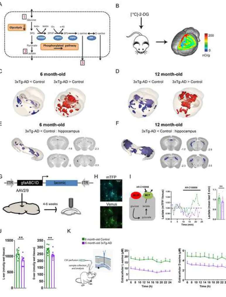
The serine biosynthesis pathway directly branches from glycolysis (Figure 1A). Alteration of aerobic glycolysis is an early feature of AD. In the brain, a substantial fraction of glucose is thought to be processed via aerobic glycolysis in astrocytes (Bak and Walls, 2018; Barros and Weber, 2018; Magistretti and Allaman, 2015, 2018). We used young 3xTg-AD mice to measure brain glucose metabolism in vivo, aerobic glycolysis flux in astrocytes and serine levels in vivo.
Alteration of glucose metabolism in 3xTg-AD mice
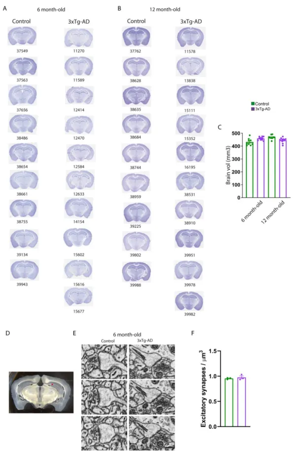
We measured energy metabolism in two groups of animals at two stages of progression of the pathology. We used six to seven months-old 3xTg-AD female mice to study the early phase of AD, as only intraneuronal A is present in the brain, and 11 to 12 months-old animals, that display senile plaques (Figures S1A-S1C for the anatomopathological evaluation of our colony of 3xTg-AD mice). In vivo 2-deoxyglucose autoradiography (Figure 1B) was followed by 3D voxel-wise statistical analysis to map the differences in cerebral glucose metabolism between control and 3xTg-AD mice (Barros et al., 2018). This approach allows statistically significant unbiased identification of metabolic differences between groups at the voxel level, without operator-dependent delineation of regions of interest (Dubois et al., 2010). We found that six to seven months-old 3xTg-AD mice displayed localized decreases in glucose uptake in the amygdala, entorhinal cortex, and hippocampus including CA1, dentate gyrus and CA2 (Figures 1C and 1E). We also observed increases in glucose uptake in the striatum, auditory cortex, and retrospenial cortex. Older 3xTg-AD mice (11 to 12 months-old) displayed the same hypermetabolic clusters, but the size of the hypometabolic clusters was bigger, in particular in the hippocampus (Figures 1D and 1F). The magnitude of the changes in glucose uptake was similar at both ages. Very little neurodegeneration has been detected in 3xTg-AD mice, at least up to the age of 24 months (Oh et al., 2010; Rohn et al., 2008). Our current observation of an unchanged brain volume between the four groups of mice as measured on Nissl stained brain sections for all animals used for in vivo metabolism (Figures S2A-S2C) corroborates the previous experimental evidence. It is therefore unlikely that cell death could contribute to hypometabolism in the 2 cohorts of six to seven months-old and 11 to 12 months-old 3xTg-AD female mice. Since most of energy is used at the level of excitatory synapses (Harris et al., 2012), we also probed whether the density of excitatory synapses were altered in 3xTg-AD mice. We performed serial block-face scanning electron microscopy at the level of the CA1 region of the hippocampus. We found that the density of excitatory
0.956 ± 0.012; Figures S2D-S2F). This result strongly suggests that the hypometabolism we observed in 3xTg-AD mice is not due to anatomical/structural changes and that 3xTg-AD mice display localized metabolic changes which start at an early stage of the disease. We next investigated whether the metabolic status of astrocytes was altered in 3xTg-AD mice.
Alteration of glucose metabolism in astrocytes of 3xTg-AD mice
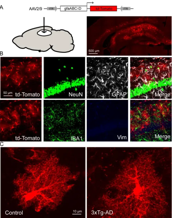
Resting astrocytes produce lactate in a tonic manner via aerobic glycolysis (Sotelo-Hitschfeld et al., 2015). We monitored astrocyte lactate dynamics in real time in hippocampal slices using Laconic, a FRET lactate nanosensor that was expressed in the hippocampus using an AAV2/9 (Figure 1G). We first confirmed the specific astrocytic tropism of the AAV2/9 expressing td-Tomato under the control of the short gfaABC1D promoter (Figures S3A-S3C). Accordingly, expression of Laconic was restricted to astrocytes (Figure 1H). Lactate transport across the astrocytic plasma membrane is mediated by monocarboxylate transporters (MCTs). We therefore used AR-C155858, a MCT blocker (Ovens et al., 2010), to measure the accumulation of lactate in single astrocytes (Figure 1I) at physiological extracellular glucose concentrations (Silver and Erecinska, 1994). The accumulation of intracellular lactate under MCT blockade observed in astrocytes of control mice was absent in 3xTg-AD astrocytes (Figure 1I). This result indicates that resting lactate production via the aerobic glycolytic pathway is altered in 3xTg-AD astrocytes.As L-serine is generated from glucose through diversion of the glycolytic intermediate 3-phosphoglycerate into the phosphorylated pathway (Figure 1A), we next investigated whether the defective glycolytic flux in 3xTg-AD astrocytes translated into reduced L-serine production/release.
Lower levels of L-serine in 3xTg-AD mice
We first measured total L-serine level in the hippocampus and found that it was significantly lower in 3xTg-AD (880 ± 48) than in control mice (1089 ± 42 nmol/g wet tissue; Figure 1J). We then measured extracellular
L-serine in the hippocampus by in vivo microdialysis and found that its concentration was also significantly lower in 3xTg-AD (7.64 ± 1.10) than in control mice (16.61 ± 1.73 µM; Figure 1K). These results strongly suggest that the production and release of L-serine is reduced in young 3xTg-AD mice. We observed these
reductions in six-month-old 3xTg-AD mice that do not exhibit A plaques, overt astrocyte reactivity (GFAP staining), or activated microglia (IBA1 staining) in the CA1 region (Figure S1A and Figures S4A-S4D). L -serine is the obligatory precursor of D-serine, the main co-agonist of synaptic NMDARs in the adult hippocampus. We next monitored the levels of D-serine in the hippocampus of 3xTg-AD mice.
Lower levels of D-serine in 3xTg-AD mice
We measured total D-serine level in the hippocampus and found that it was significantly lower in 6 month-old control 3xTg-AD (244 ± 8) than in control mice (283 ± 8 nmol/g wet tissue; Figure 1J). We then measured extracellular D-serine in the hippocampus by in vivo microdialysis and found that the concentration of extracellular D-serine was also significantly lower in 6 month-old control 3xTg-AD (2.90 ± 0.38) than in control mice (4.53 ± 0.72 µM; Figure 1K). These results strongly suggest that the production and release of D-serine is reduced in young 3xTg-AD mice. We did not find any difference in the level of both total and extracellular glutamate between control and 3xTg-AD mice (Figure S6D and S6E) suggesting no major impairment in glutamate synthesis and release/uptake in young 3xTg-AD mice. We next performed electrophysiological recordings to assess the functional impact of a lower extracellular D-serine on the activity of NMDARs in 3xTg-AD mice.
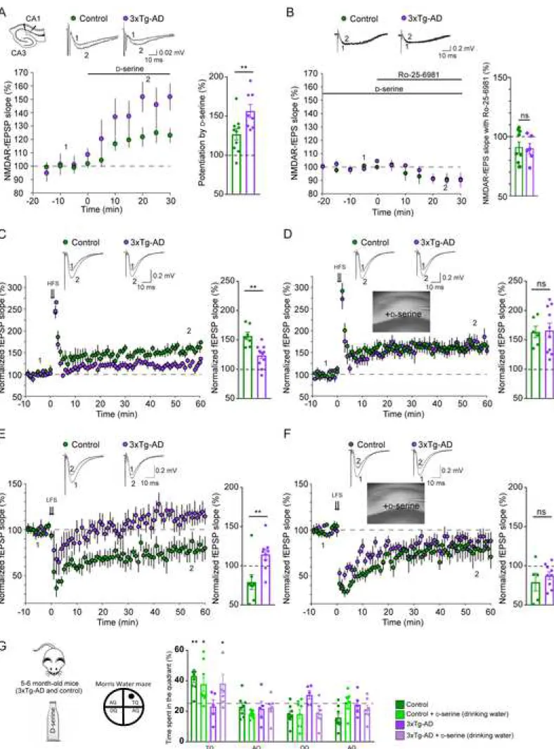
Lower level of occupancy of the co-agonist-binding site of NMDARs in 3xTg-AD mice
We thus recorded the NMDAR component of field excitatory post-synaptic potentials (NMDAR-fEPSPs) in acute hippocampal slices. In control mice, bath application of D-serine (50 µM) induced an increase of NMDAR-fEPSPs (126.8% ± 7.3, Figure 2A). This is in accordance with previous work (Henneberger et al., 2010; Papouin et al., 2012), indicating that the co-agonist binding sites of synaptic NMDARs in this region are not fully saturated. Importantly, the D-serine-mediated increase in synaptic NMDAR-fEPSPs was
significantly larger in 6 month-old control 3xTg-AD mice (156.6% ± 8.3, Figure 2A). This is consistent with a reduced level of occupancy of the NMDAR co-agonist-binding site in 3xTg-AD mice because of a reduced level of D-serine within the cleft. Another possibility would be that the NMDAR subunit composition was switched from GluN2B to GluN2A in 3xTg-AD mice, since GluN2A-containing NMDARs have a lower affinity for D-serine than GluN2B receptors (Ferreira et al., 2017). In that case, the amount of NMDARs recruited during synaptic stimulation could be reduced even if endogenous D-serine levels were unchanged. We tested this possibility by investigating the inhibitory action of a GluN2B antagonist (Ro-25-6981, 2 µM) on NMDAR-EPSPs in both control and 3xTg-AD mice. Experiments were performed in the presence of D-serine (50 µM) to make sure that all synaptic NMDARs were available for activation and to have a better quantification of the NMDAR population present at the synapse. In WT animals, NMDAR-fEPSPs were slightly decreased by Ro-25-6981 (90.8 ± 4.7% of baseline, Figure 2B). Interestingly, a similar effect was observed in 3xTg mice (89.8 ± 4.4% of baseline, Figure 2B). Taken together, these data indicate, that the
fraction is unchanged in 3xTg-AD mice. This result argues against the possibility of a switch of NMDARs subunits in these mice. We next monitored hippocampal excitatory synaptic transmission and plasticity in 3xTg-AD female mice, given the role played by D-serine in NMDAR activity.
Impaired synaptic plasticity in 3xTg-AD mice
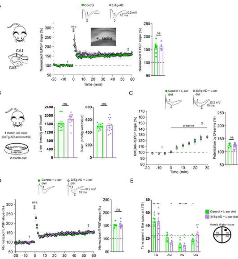
As originally reported (Oddo et al., 2003), high-frequency stimulation (HFS)-induced LTP was impaired in 6 month-old control 3xTg-AD (156.7% ± 6.7 in control vs. 123.5% ± 5.4 in 3xTg-AD, Figure 2C). We found that low-frequency stimulation (LFS)-induced LTD was also reduced in these animals (79.9% ± 9.5 in control vs. 114.5% ± 6.6 in 3xTg-AD, Figure 2E).
Impaired synaptic plasticity in 3xTg-AD mice is rescued by D-serine
All these results support the hypothesis that the deficits in synaptic plasticity observed in 3xTg-AD mice are due to a lower concentration of D-serine in the synaptic cleft and thus to fewer NMDARs available for
activation during HFS and LFS. In agreement with this hypothesis, LTP was rescued by supplying exogenous D-serine (50 µM) to slices obtained from 3xTg-AD mice (163.6% ± 9.7 in control vs. 166.1% ± 12.4 in 3xTg-AD; Figure 2D). Similarly, exogenous D-serine (50 µM) completely rescued LTD in 3xTg-AD mice (79.1 ± 11.3% in control vs. 87.5% ± 4.9 in 3xTg-AD, Figure 2F). Acute application of D-serine did not affect LTP and LTD in slices obtained from control mice (Figures 2C-2F). We next assessed whether D -serine could rescue the spatial memory deficits of 3xTg-AD mice.
Impaired spatial memory in 3xTg-AD mice is rescued by D-serine
We evaluated learning and memory using the spatial reference version of the Morris water maze that is hippocampal-dependent (Morris, 1984). 3xTg-AD mice displayed a similar spatial learning performance than age-matched control mice but did not show any preference for the target quadrant during the probe trial (Figure 2G). We chronically treated mice with D-serine in the drinking water (100mg/kg body weight), a regimen that has been previously shown to increase levels of D-serine in the brain (Otte et al., 2013). Oral supplementation of D-serine for two weeks rescued the cognitive deficits of six-month-old female 3xTg-AD mice but did not modify the performance of control mice (Figure 2G). These results all point to defective
D-serine availability at the hippocampal synapses of 3xTg-AD mice. We then asked whether the reduced production of L-serine could be responsible for the D-serine-mediated impairment of synaptic plasticity and spatial behavior. If this is the case, inactivation of the L-serine biosynthetic pathway should recapitulate the
recently reported to be expressed mostly in astrocytes and oligodendrocytes and not in neurons (see
http://www.brainrnaseq.org/ and http://mousebrain.org/genesearch.html), the cellular localization of PHGDH has not been thoroughly studied in the brain. We therefore performed a histological study focusing at the hippocampus.
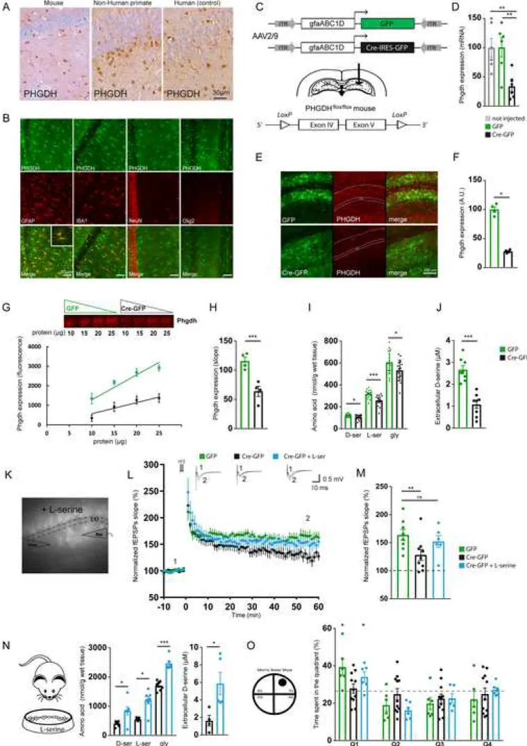
The serine synthetic pathway is primarily located in astrocytes
The first committed step of the L-serine biosynthetic pathway is the oxidation of 3PG to 3-phosphohydroxypyruvate (3PHP) by PHGDH (Figure 1A). A specific antibody directed against PHGDH showed prominent staining of astrocytes not only in mouse but also in non-human primate and human brain (Figure 3A). We performed co-localization studies with confocal microscopy in a control mouse brain and confirmed that PHGDH was mainly expressed by GFAP-expressing astrocytes and not microglial cells (IBA1-positive), neurons (NeuN-positive), or oligodendrocytes (Olig2-positive) in the CA1 region of the hippocampus (Figure 3B). We next developed a conditional mouse model to interrogate the role of the astrocytic hippocampal PHGDH in synaptic plasticity and spatial behavior.
Conditional inactivation of Phgdh leads to impaired synaptic plasticity and spatial memory
Mice conditionally lacking Phgdh in astrocytes have been previously generated by crossing Phgdhflox/flox
mice with hGFAP-Cre transgenic mice (Yang et al., 2010). Since hGFAP-cre transgene mediates gene deletions in a much wider spectrum of neural cells than just astrocytes (Zhuo et al., 2001), we locally and conditionally inactivated Phgdh by injecting an AAV expressing Cre recombinase under the control of the astrocytic gfa-ABC1D promoter in the hippocampus of adult Phgdhflox/flox mice (Figure 3C). At the
transcriptional level, Phgdh mRNA was 25% of its initial value found in both non-injected and AAV-GFP-injected mice (Figure 3D). At the protein level, PHGDH expression was significantly reduced in Phgdhflox/flox
mice injected with AAV-Cre in comparison to mice injected with AAV-GFP (Figures 3E-3H). Astrocytes lose some of their normal functions when they become reactive (Liddelow et al., 2017). The levels of expression of mRNA for vimentin and gfap and of GFAP were not significantly different between Phgdhflox/flox mice
injected with AAV-Cre or AAV-GFP (Figure S5A-S5D). We next performed a Sholl analysis on GFAP immunostained astrocytes and showed that the complexity of astrocyte processes was not altered following inactivation of Phgdh (Figure S5E and S5F). These results strongly suggest that astrocytes did not adopt a reactive phenotype following inactivation of Phgdh. The levels of expression of selected neuronal and
microglial mRNAs (Neun, eaat3, cd68, iba1) and of D-amino acid oxidase (daao), the enzyme that degrades
D-serine, were not significantly different between Phgdhflox/flox mice injected with AAV-Cre or AAV-GFP
(Figures S5G-S5K), indicating no marked neuronal death, no microglial activation as well as no compensatory mechanisms targeting D-serine levels following local inactivation of Phgdh.
We then confirmed that the levels of L- and D-serine in the brain are determined by their de novo biosynthesis through the phosphorylated pathway in astrocytes. Indeed, the total L-serine, D-serine and glycine content in the whole hippocampus, measured by HPLC (Figure 3I), as well as the level of extracellular D-serine, measured in vivo via a biosensor (Figure 3J), were significantly lower in Phgdhflox/flox
mice injected with AAV-Cre in comparison to mice injected with AAV-GFP. We subsequently investigated whether long-term synaptic plasticity was impaired in those mice. HFS-induced LTP was significantly impaired in these animals (166.8% ± 5.8 in AAV-GFP-injected, and 133.9% ± 13.5 in AAV-Cre-injected mice, Figures 3L and 3M). Bath application of L-serine (50 µM) rescued LTP (152.2% ± 12.0, Figures 3L and 3M), indicating that L-serine produced by the phosphorylated pathway is required for activity-dependent synaptic plasticity. We performed several behavioral tests on these mice to assess their anxiety and monitor their motor and cognitive performance. The mice with inactivated Phgdh in the hippocampus did not show any alterations in anxiety or spontaneous motor behavior, as assessed using the elevated plus-maze and the Phenotyper® (Figures S5L and S5M). Those mice were tested using the Morris water maze and displayed a similar spatial learning performance than control mice. However, they did show significant impairment of spatial memory when they were tested 72 h after learning the location of the platform (Figure 3O), as we observed with 3xTg-AD mice. Finally, to determine whether L-serine is necessary and sufficient to promote spatial memory in the hippocampus, we fed Phgdhflox/flox mice injected with AAV-Cre with a diet
enriched in L-serine for one month. We found that the diet was sufficient to restore total levels of L-serine,
D-serine and glycine in the whole hippocampus as well as the extracellular levels of D-serine (Figure 3N).
Accordingly, the spatial memory was restored in those animals (Figure 3O). Overall, these results show that the availability of L-serine synthesized via the phosphorylated pathway in astrocytes is a key
determinant for normal NMDAR function and support our hypothesis that its dysregulation could contribute to memory deficits in AD. To gain further insight into the potential contribution of the astrocytic L-serine synthesis pathway in AD, we investigated whether the expression of PHGDH was altered both in young 3xTg-AD mice and in post-mortem brain tissue of AD patients.
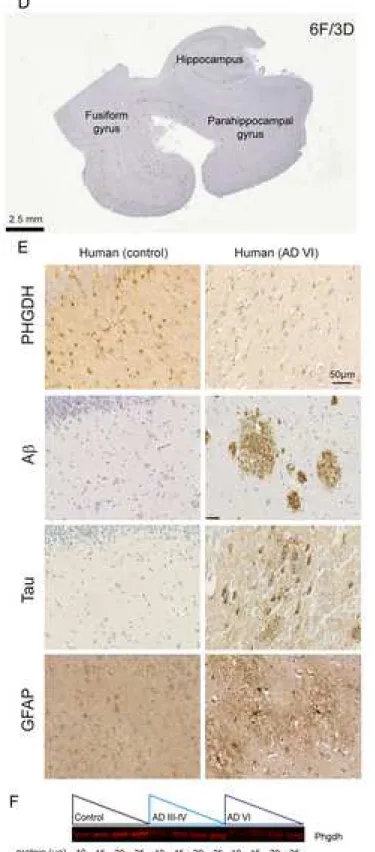
Expression of PHGDH is reduced in the AD brain
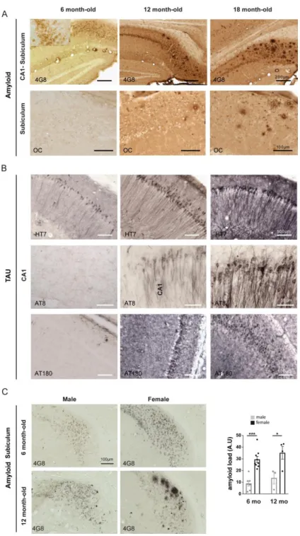
We analyzed the expression of PHGDH in 6 month-old female 3xTg-AD mice that do not display Aβ plaques (Figure 4A) and found no difference as compared to control age-matched animals using immunofluorescent confocal analysis (Figure 4B) or immunoblotting (Figure 4C). This result suggests that the lower L-serine levels in 3xTg-AD mice is more likely resulting from reduced glucose uptake and less substrate available for the L-serine synthesis pathway rather than from a direct effect of Aβ on PHGDH expression, at least
during the early phase of AD (i.e. astrocytes are not reactive and Aβ plaques are not formed). We then performed immunohistochemistry on human formalin-fixed paraffin-embedded sections (Figure 4D) from control individuals, intermediate AD (Braak III-IV), and advanced AD (Braak VI) patients (Table 1). Contrarily to 6 month-old female 3xTG-AD mice, advanced AD patients presented numerous Aβ plaques, hyperphosphorylated Tau and reactive GFAP-positive astrocytes (Figure 4E). We observed a reduction of PHGDH staining (Figure 4E), which was confirmed by fluorescent immunoblotting (Figure 4F) of frozen sections from the same patients. The reduction was statistically significant in intermediate AD (-62%) and even more pronounced in advanced AD patients (-82%). These results indicate that a progressive alteration of the astrocytic serine synthetic pathway occurs in the brain of AD patients. Altogether, these data support the contribution of defective production/release of L-serine in astrocytes to the pathogenesis of AD and point to L-serine supplementation as a potential therapy. We finally asked whether supplying exogenous L
-serine could also rescue both synaptic plasticity and spatial behavior deficits in 3xTg-AD mice.
Oral L-serine as a potential therapy for AD
We first assessed the effects of acute L-serine (50 µM) on LTP. Bath application of L-serine (50 µM) fully rescued LTP in slices obtained from 3xTg-AD mice (156.8 % ± 7.4 in control vs. 160.5 % ± 6.4 in 3xTg-AD; Figure 5A). Since long-term treatment with exogenous L-serine can compensate for its deficient production
in the context of inherited metabolic disorders of L-serine biosynthesis (see discussion), we next assessed the effects of chronic oral L-serine supplementation upon both the long-term synaptic plasticity and the
spatial memory performance of 3xTg-AD and control mice. Mice aged four to five months were given a 10%
L-serine-enriched diet for two months (Figure 5B). Total L-serine content in the whole hippocampus was significantly higher in the two groups of animals that received the diet (Figure 5B) as compared to untreated animals (Figure 1J). Two months after chronic L-serine diet, total L-serine content was no longer significantly different between control and 3xTg-AD mice (1590 ± 87 vs. 1844 ± 73 nmol/g wet tissue,
respectively, Figure 5B). The diet also boosted de novo production of D-serine since its content in the whole hippocampus was significantly higher in the two groups of animals that received the diet (see Figure 1J for comparison with untreated animals) up to a level that was not different between control and 3xTg-AD mice (496 ± 11 vs. 512 ± 28 nmol/g wet tissue for control and 3xTg-AD mice, respectively, Figure 5B). Using in vivo microdialysis, we found that the levels of extracellular L- and D-serine were higher in 3xTg-AD mice receiving the diet vs. untreated (24.91 ± 6.29 vs. 8.66 ± 1.83 µM and 8.22 ± 1.79 vs. 2.28 ± 0.31 µM for L- and D-serine, respectively). The diet did not significantly alter the total levels of other amino acids measured in the hippocampus; only a modest increase in phenylalanine level was observed (Figures S6A-S6I). We then observed that in 3xTg-AD mice fed with L-serine, bath application of D-serine (50 µM) induced a similar
increase of NMDAR-fEPSPs to the one measured in control animals (126.3% ± 3.5 in control vs. 124.4% ± 2.3 in 3xTg-AD, Figure 5C). This result indicates that the chronic supplementation of L-serine was able to rescue the deficit of D-serine availability observed at hippocampal synapses in 3xTg-AD mice, thereby restoring the level of occupancy of the NMDAR co-agonist binding site to its control values. Accordingly, LTP was fully rescued in those animals (152.0% ± 3.2 in control vs. 154.5% ± 5.0 in 3xTg-AD, Figure 5D). We finally probed the spatial memory performance in 3xTg-AD mice fed with L-serine and found that it reached a level similar to the one measured in control mice fed with L-serine (Figure 5E). Altogether, these data support the use of oral L-serine as a ready-to-use and safe therapy for of AD.
DISCUSSION
Glial cells have been recently implicated in AD with astrocytes and microglia now recognized as potential cellular mediators of synapse dysfunction and loss (Hong et al., 2016; Jo et al., 2014). Here, we have identified an astrocytic metabolic pathway that directly contributes to early synaptic deficits underlying cognitive decline in AD, well before microglia and astrocytes become reactive (Liddelow et al., 2017). We showed that the production of L-serine, a non-essential amino acid that is generated from glucose, is impaired in AD astrocytes.
The L-serine biosynthetic pathway has been well studied in cancer cells that exhibit increased glucose metabolism to lactate (the so-called Warburg effect) and in which multiple copies of Phgdh divert glycolytic flux and provide L-serine and glycine for rapid cell growth and proliferation (Locasale et al., 2011; Possemato et al., 2011). L-serine is a central node for the biosynthesis of many molecules such as sphingolipids and phospholipids. It also supplies carbon to the one-carbon pool, which is involved in folate
metabolism, contributing to purine synthesis (Mattaini et al., 2016). This metabolic pathway is particularly essential for the nervous system as homozygous mutations of each of the three enzymes of the serine biosynthesis pathway cause a syndrome called Neu-Laxova, a rare autosomal-recessive disorder characterized by severe peripheral and central nervous system malformations (microcephaly), leading to prenatal or early postnatal death (Acuna-Hidalgo et al., 2014). This clinical phenotype of L-serine biosynthesis is reminiscent of defects in brain serine transport induced by mutations in SLC1A4, the gene coding for the neutral amino acid transporter ASCT1 (Damseh et al., 2015), underlying the critical role of L -serine biosynthesis and shuttling. L-serine treatment during pregnancy and after birth improves fetal brain growth and prevents neurological symptoms (de Koning et al., 2004), suggesting that exogenous L-serine
can compensate for its lack of de novo production in the brain.
In the present work, we focused our attention on glycolysis as a possible regulator of L-serine production in
the brain as L-serine is generated from glucose. In AD, early alterations of cerebral glucose metabolism, as measured using fluoro-2-deoxy-D-glucose (FDG) and positron emission tomography (PET), are well established. Several studies have shown that abnormalities in FDG-PET can even predict progression from mild cognitive impairment (MCI) to AD (for references see (Cohen and Klunk, 2014)). We used 3D analysis of in vivo brain glucose metabolism to show that 3xTg-AD mice display metabolic impairment in specific brain regions, including the hippocampal formation. Vascular dysregulation (Iturria-Medina et al., 2016) as well as endothelial glucose transporter 1 (GLUT1) reductions are mechanisms by which A could likely exert its deleterious metabolic effects (Winkler et al., 2015). Glucose, the almost exclusive energy substrate of the brain, is glycolytically processed to produce pyruvate and lactate in astrocytes, whereas neurons predominantly process glucose through the pentose phosphate pathway to produce NADPH and utilize pyruvate and lactate from astrocytes to feed the TCA cycle and the associated oxidative phosphorylation to produce ATP (Herrero-Mendez et al., 2009). Our observation that PHGDH, whose substrate is a glycolytic intermediate, is exclusively expressed by astrocytes is consistent with the notion that astrocytes are glycolytic cells. Even if we do not provide a causal link between the defective hippocampal glycolysis and the alteration of the phosphorylated pathway, our findings suggest that astrocytic energy metabolism may control L-serine production, thereby influencing glutamatergic neurotransmission. In addition to lactate (Suzuki et al., 2011; Yang et al., 2014), we propose that another byproduct of aerobic glycolysis (L-serine) may play an important role in plasticity and learning.
In the brain, L-serine is the obligatory precursor of D-serine (Ehmsen et al., 2013), a physiological ligand of the co-agonist site of synaptic NMDARs in the hippocampus, that is required to sustain synaptic transmission (Panatier et al., 2006; Papouin et al., 2012). Our electrophysiological recordings show a reduced level of occupancy of the NMDAR glycine-binding site in seven-month-old 3xTg-AD mice. Accordingly, D-serine completely rescued both forms of synaptic plasticity (LTP and LTD) as well as spatial memory. Chronic D-serine supplementation has long been considered to be a therapeutic option for pathological conditions involving NMDARs malfunction such as schizophrenia (Kantrowitz et al., 2016) but its slow and weak diffusion through the blood–brain barrier and potential nephrotoxicity have limited its clinical use. L-serine represents a more favorable therapeutic option because it is considered to be safe by
the Food and Drug Administration; it has been approved as a normal food additive, it is widely sold as a dietary supplement and is well-tolerated, even at high doses. L-serine is currently prescribed for the
treatment of Neu-Laxova syndrome that results in L-serine deficiency (van der Crabben et al., 2013) and is currently in a phase I clinical trial as a treatment for hereditary sensory autonomic neuropathy type 1 (Garofalo et al., 2011) and amyotrophic lateral sclerosis (Levine et al., 2017). L-serine supplementation was recently shown to ameliorate motor and cognitive performance in rare NMDA-related severe encephalopathy leading to glutamatergic signaling deficiency (Soto et al., 2019). Glutamate excitotoxicity has long been implicated in AD pathophysiology, as supported by the use of the uncompetitive NMDAR partial antagonist memantine to treat moderate-severe AD patients. Although it is possible that increasing
D-serine concentrations in AD patients may exacerbate glutamate excitotoxicity, this would probably not
occur at an early stage of the pathology, when memantine is not yet effective (Schneider et al., 2011). It is noteworthy that people living in Okinawa, a Japanese island known for the overall longevity of its population, have a diet characterized by an L-serine content that is four times higher than that of the average American (Cox and Metcalf, 2017).
This study finally points to direct impairment of the phosphorylated pathway in AD, as shown by a marked reduction of PHGDH expression in AD patients. This is the first demonstration of the selective expression of PHGDH in human hippocampal astrocytes, as well as its decrease during the progression of AD. Since we did not observe any change in PHGDH expression in six-month old 3xTg-AD mice that do not display Aβ plaques and whose astrocytes are not reactive, it is therefore highly conceivable that the primary event leading to less serine production would be related to altered glycolysis flux in early AD and that PHGDH expression would be affected in later AD stages. The fact that brain glucose metabolism is altered in the
early phase of AD patients is compatible with such hypothesis. Unfortunately, there is no consensus on the evolution of L- and D-serine levels in AD (Biemans et al., 2016; Lin et al., 2017; Madeira et al., 2015). Additional studies are necessary to determine whether, where (brain region, extracellular vs. tissue vs. CSF) and when the levels of L- and D-serine change during disease progression. Altogether, these data raise the prospect of L-serine supplementation as a treatment option for AD and possibly other neurodegenerative diseases.
Limitations of Study
There are some limitations to our study, which should be acknowledged. First, we only performed this study in a single model of AD (3xTg-AD mice). It would be important to investigate whether the reduced production of L-serine by astrocytes we uncovered is a general feature that can be found in another transgenic AD mice models that do not present the Tau mutations. Second, we analyzed a rather limited number of human hippocampal samples and it may be worth to complete the analysis with more samples coming from other brain regions as well. Finally, an intriguing question that will be important to investigate in the future is the impact of the supplementation of L-serine in the diet on the amyloid load and the Tau hyperphosphorylation.
Funding
This work was supported by grants from the Agence Nationale de la Recherche (ANR 2011 MALZ-0003 to G.B. and B.C.), Association France Alzheimer and Fondation de France (Prix Spécial 2012 to G.B., B.C. and S.H.R.O.), Fondation Plan Alzheimer (G.B.), Infrastructure de Recherche translationnelle pour les Biothérapies en Neurosciences-NeurATRIS ANR-11-INBS-0011 (G.B.), Fondation pour la Recherche Médicale and EU Joint Programme – Neurodegenerative Disease Research (JPND ; Horizon 2020 Framework Programme, grant agreement 643417/DACAPO‐AD to S.H.R.O.). J.L.D. is a recipient of a PhD fellowship from the CEA (IRTELIS program). M.Mau. is a recipient of a PhD fellowship from the Ecole Normale Supérieure (ENS). P.A.V is a recipient of a PhD fellowship from the Fondation pour la Recherche Médicale (FRM). P.J. fellowship was provided by Enhanced Eurotalents, a Marie Sklodowska-Curie Actions Programme, co-funded by the European Commission and managed by the French Alternative Energies and Atomic Energy Commission (CEA). M.M is a recipient of a post-doctoral fellowship (DACAPO-AD / JPND / EU H20H20 grant agreement N°643417). This work benefited from the support of various facilities granted by INSERM and LabEX BRAIN ANR-10-LABX-43. The team of G.B. is part of the Ecole des Neurosciences de Paris (ENP) training network and that of S.H.R.O is part of the LaBex BRAIN.
Author contributions
J.L.D. performed the confocal analysis, carried out the biochemical, RT-qPCR and the metabolic studies and developed the AAVs with the help of M.Mau, A.P.B. and M.C.G.
J.V. and M.M. performed electrophysiology on 3xTg-AD mice with the help of A.P.
M.Mau. performed all experiments on Phgdhflox/flox mice, immunoblotting and all behavioral tests with the
help of K.C.
P.J carried out electrophysiology on Phgdhflox/flox mice
P.A.V, P.J. and M.G.G. performed microdialysis
T.T., M.T., S.G., A.G. and A.P. measured extracellular amino acid concentrations using CE-LIF or LC-LC/MS
E.F. performed the histological analysis of the 3xTg-AD colony and performed the in vivo metabolic study with the help of G.B. and M.G.G.
X.T. carried out the FRET imaging experiments with the help of J.P., E.G and B.C.
M.V., Y. B., A-S. H. and T.D. performed the 3D image processing and the analysis of the in vivo metabolic data
S.M. and M.Mau. measured extracellular D-serine using biosensors
A.K. and S.F. provided the Phgdhflox/flox mice and measured amino acid concentrations using HPLC
L.S., V.S and C.D performed and analyzed immunohistochemical stainings on human samples with the help of P.A.V.
G.N. performed EM studies
P.H. and E.B. provided materials and discussed the data B.C., S.H.R.O. and G.B. obtained funding for the research A.P. and G.B. coordinated the study and wrote the manuscript
Acknowledgments
The authors thank Martine Guillermier and Sueva Bernier for useful technical help and Felipe Barros, Juan Bolanos and Igor Allaman for helpful discussion. We thank Noëlle Dufour and Charlène Joséphine for viral production, the Neuro-CEB biobank at the Hôpital de la Pitié-Salpétrière for providing human samples, Anaelle Dubois for electron microscopy at the BioEM Facility of the EPFL (Switzerland) and Alain Chédotal
and Stéphane Fouquet at the Institut de la Vision for high resolution scans. Authors thank also Delphine Gonzales, Sara Laumond and all the people of the Animal Facility and the Genotyping Facility of the NeuroCentre Magendie for mouse care and genotyping.
Figures and Legends
Figure 1. 3xTg-AD mice display energy metabolism alterations and decreased L- and D-serine levels
(A) The phosphorylated pathway of L-serine biosynthesis is a short metabolic sequence consisting of three enzymatic reactions. The glycolytic intermediate 3-phosphoglycerate (3PG) is converted into phosphohydroxypyruvate (3PHP), in a reaction catalyzed by 3-phosphoglycerate dehydrogenase (PHGDH). 3PHP is metabolized to phosphoserine (3PS) by phosphohydroxypyruvate aminotransferase (PSAT1) and, finally, 3PS is converted into L-serine by phosphoserine phosphatase (PSPH). In the brain,
L-serine is converted into D-serine by serine racemase (SRR). 1,2 and 3 indicates the functional parameters
we have measured in 3xTg-AD mice. (B) In vivo glucose uptake was measured using 3D-reconstructed autoradiography and voxel-wise statistical analysis to map the cerebral metabolic differences between (C) 6 month-old control (n=10) and 3xTg-AD mice (n=10) and (D) between 12 month-old control (n=9) and 3xTg-AD mice (n=10). Brains of 3xTg-AD mice display hypometabolic (blue) and hypermetabolic (red) clusters at both ages. (E) Only hypometabolic clusters were observed in the hippocampus in 6 month-old 3xTG-AD and (F) their size were larger in 12 month-old 3xTg-AD mice (Student’s t test p<0.01). Numbers indicate the anatomical position relative to Bregma. (G) Design of the AAV2/9 expressing the FRET sensor Laconic that was injected in the CA1 region of the hippocampus in 6 month-old control (n = 3) and 3xTg-AD (n = 3) mice. (H) CA1 astrocytes expressing the FRET lactate sensor Laconic in an acute hippocampal slice showing mTFP (blue) and Venus (green). (I) Typical response of intracellular lactate to bath application of 1µM AR-C155858, a monocarboxylate transporter blocker. Astrocytes of 6 month-old 3xTg-AD mice (n=3, 7 slices, 26 cells) do not accumulate lactate as compared to control astrocytes (n = 3 mice, 6 slices, 33 cells; Mann-Whitney U Test, **p<0.01). (J) Total levels of L-serine and D-serine measured by HPLC are significantly lower in 6 month-old 3xTg-AD mice (Mann-Whitney U test, ** p=0.005). (K) Extracellular levels of L-serine and D-serine measured by microdialysis and LC-MS/MS are significantly lower in 6 month-old 3xTg-AD mice as compared to control animals (Repeated measures ANOVA, F(1, 10)=15.691, p=0.0027 for L-serine and F(1, 11)=5.4482, p=0.0396 for D-serine).
Figure 2. Deficits of synaptic plasticity at CA3-CA1 synapses and of spatial memory in 3xTg-AD mice are rescued by D-serine
(A) Electrophysiological recordings were performed on acute brain slices of 6-7 month-old 3xTg-AD and control mice with the stimulating and recording electrodes located in CA1. Effect of D-serine application (50 µM) on NMDAR-fEPSPs in slices obtained from control (green) and 3xTg-AD mice (purple). Traces of representative NMDAR-fEPSPs recorded before (1) and during (2) D-serine application. Bar graph summarizing results presented in (A) indicates that NMDAR co-agonist binding site occupancy was decreased in 3xTg-AD mice (Mann-Whitney U test, p = 0.008; control: n=10; 3xTg-AD: n=8). (B) The application of Ro-25-6981 (2µM) only slightly reduces NMDAR-fEPSPs in slices obtained from control and 3xTg-AD mice. D-serine (50 µM) was bath applied to make sure that all synaptic NMDARs were available for activation. Bar graph summarizing results presented in B indicates that GluN2B receptors contribute to a small fraction of the synaptic NMDARs in both control and 3xTg-AD mice (Mann-Whitney U test, p = 0.75; control: n=9; 3xTg-AD: n=6). (C) Traces represent typical fEPSPs evoked before (1) and after (2) high frequency stimulation (HFS). LTP was significantly impaired in 3xTg-AD mice (Mann-Whitney U test, p=0.012). Bar graph summarizing experiments presented in C (control: n=7; 3xTg-AD: n=11). (D) D-serine application (50 µM) rescued LTP in 3xTg-AD mice (Mann-Whitney U test,p=0.958). Bar graph summarizing experiments presented in D (control: n=6; 3xTg-AD: n=10). (E) Representative traces of characteristic fEPSPs evoked before (1) and after (2) low frequency stimulation (LFS). LTD was significantly impaired in 3xTg-AD mice (Mann-Whitney U test, p=0.008). Bar graph summarizing experiments presented in E (control: n=8; 3xTg-AD: n=9). (F) D-serine application (50 µM) rescued LTD in 3xTg-AD mice (Mann-Whitney U test,p=0.699). Bar graph summarizing experiments presented in F (control: n=5; 3xTg-AD: n=9). (G) 3xTg-AD mice present spatial memory deficits (one tailed one sample t-test against chance level (25%), p=0.19) when probed in the Morris Water maze 72h after training as compared to control mice (p=0.0037). Chronic oral D-serine supplementation (drinking water for 2 weeks) restored the spatial memory deficits of 3xTg-AD mice (p=0.035) and was without any effect in control mice (p=0.014). AQ, adjacent quadrant, OQ, opposite quadrant, TQ, target quadrant. n = number of mice, 1 slice/animal.
Figure 3. Conditional inactivation of Phgdh in astrocytes impairs synaptic plasticity and spatial memory
(A) Immunostaining of PHGDH in the hippocampus of a mouse, a non-human primate and a human brain. PHGDH is mainly expressed in astrocytes. (B) Double immunofluorescent staining of brain sections from
6-month old control mice using PHGDH (green) and either an astrocyte marker (GFAP, red), a microglial marker (IBA1, red), a neuronal marker (NeuN, red) or an oligodendrocyte marker (Olig2, red). (C) Design of the AAV2/9 expressing Cre-IRES-GFP that was injected in the CA1 region of the hippocampus in
Phgdhflox/flox mice (D) Level of phgdh mRNA was 25% of its initial value as compared to both non-injected
and GFP-injected mice (Kruskal-Wallis followed by Dunn test, p=0.004 vs. not-injected and p=0.005 vs. GFP)). (E) Three weeks after injection of the AAV-Cre, expression of PHGDH (red) was markedly decreased compared to the AAV-GFP. (F) Quantification of the PHGDH fluorescence (Nested model p=0.011, n=2 mice, 10-12 confocal images/mouse). (G) Fluorescent immunoblotting of PHGDH performed using 4 different protein concentrations showed a linear relationship and (H) a significant reduction of PHGDH expression (linear regression model, p<0.001). (I) Total levels of D-serine, L-serine and glycine measured by HPLC were significantly reduced in the hippocampus (Mann-Whitney U test, p=0.012; p<0.001, p=0.027, respectively) as well as (J) extracellular levels of D-serine measured using biosensors (Mann-Whitney U test, p<0.001). (K) Electrophysiological recordings were performed in CA1 region expressing fluorescent astrocytes. (L) LTP was significantly impaired in Phgdhflox/flox mice injected with the
AAV-Cre. Traces represent typical fEPSPs evoked before (1) and after (2) LTP induction. LTP was rescued by bath application of 50µM L-serine. (M) Bar graphs summarizing results presented in L (Kruskal-Wallis followed by Dunn test, p=0.031). (N) Chronic supplementation with L-serine restored both the total levels of D-serine, L-serine and glycine (Mann-Whitney U test, p=0.026; p=0.017, p<0.001, respectively) as well as the extracellular levels of D-serine (Mann-Whitney U test, p=0.016). (O) Phgdhflox/flox mice injected with the
AAV-Cre displayed a significant impairment of spatial memory during the probe test of the Morris water maze 72h after learning the location of the platform (one tailed one sample t-test against chance level (25%), p=0.14), while Phgdhflox/flox mice injected with the AAV-GFP significantly spent more time in the TQ
(p=0.02). Chronic supplementation with L-serine restored spatial memory in Phgdhflox/flox mice injected with
Figure 4. Expression of PHGDH in 3xTg-AD mice and in AD Human samples
(A) Six-month old female 3xTg-AD mice that do not present Aβ plaques (4G8 immunostaining) (B) did not display significant reduction of PHGDH expression as observed using immunofluorescent staining or immunoblotting in the hippocampus (C). Four different concentrations of proteins were loaded per sample and the expression of PHGDH was derived from the slopes of the regression lines (linear regression model, p=0.79). (D) Human post-mortem brain samples included the hippocampus, parahippocampal gyrus and fusiform gyrus and presented numerous Aβ plaques (6F/3D immunostaining). (E) Expression of PHGDH was reduced in an advanced AD brain (Braak VI) that present amyloid plaques (A staining using 6F/3D antibody), hyperphosphorylated Tau (AT8) and GFAP reactive astrocytes as observed on formalin-fixed paraffin-embedded sections. (F) Fluorescent immunoblots of fresh brain tissues from Controls (n = 5), AD Braak III-IV (n = 4) and AD Braak VI (n = 5). Four different concentrations of proteins were loaded per sample and the expression of PHGDH was derived from the slopes of the regression lines. Expression of PHGDH was significantly lower in intermediates AD cases (-62%) and in advanced AD patients (-82%) as compared to controls (linear regression model, p<0.001).
Figure 5. Deficits of synaptic plasticity at CA3-CA1 synapses and of spatial memory in 3xTg-AD mice are rescued by L-serine
(A) Bath L-serine (50 µM) application in hippocampal acute slices rescued LTP in 3xTg-AD mice to the same level than in control mice (Mann-Whitney U test, p=0.97; control: n=8; 3xTg-AD: n=5). Traces represent typical fEPSPs evoked before (1) and after (2) LTP induction. Bar graph summarizing experiments presented in A. (B) 3xTg-AD and control 4 month-old mice received chronic oral L-serine supplementation (food enriched with 10% L-serine for 2 months). The total concentration of L- and D-serine in the hippocampus was similar in both groups of mice after L-serine treatment. (C) Chronic L-serine diet restored NMDAR co-agonist binding site occupancy measured in 3xTg-AD mice to the same value measured in control mice (Mann-Whitney U test, p=0.63; control: n=5; 3xTg-AD: n=13). Traces of representative NMDAR-fEPSPs recorded before (1) and during (2) D-serine application (50 µM). Bar graph summarizing results presented in C. (D) Chronic oral L-serine supplementation fully restored the LTP
deficits of 3xTg-AD mice (Mann-Whitney U test, p=0.8; control: n=7; 3xTg-AD: n=7). Representative traces of characteristic fEPSPs evoked before (1) and after (2) LTP induction. Bar graph summarizing results presented in d. (E) 3xTg-AD mice fed with a food enriched in 10% L-serine for 2 months spent more time in the target quadrant (one tailed one sample t-test against chance level (25%), p=0.007), similarly to control mice fed with the same diet (p=0.0011), during the probe test of the Morris water maze. AQ, adjacent quadrant, OQ, opposite quadrant, TQ, target quadrant.
STARMethods
Lead Contact and Materials Availability
Further information and requests for resources and reagents should be directed to and will be made available upon reasonable request by the Lead Contact, Gilles Bonvento (gilles.bonvento@cea.fr). This study did not generate new unique reagents.
Experimental Model and Subject Details
Mouse models
All experimental procedures using animals were performed in strict accordance with French regulations (Code Rural R214/87 to R214/130). They conform to the ethical guidelines of both the European Directive (2010/63/EU) and the French National Charter on the ethics of animal experimentation. They were approved by a local ethics committee and submitted to the French Ministry of Education and Research (Approvals 12-084, 14-015, APAFIS#617 and APAFIS#617-2015050516068503 v2). The animal facility is accredited by the French authorities (B9-032-02). All mice were group-housed and maintained in individually ventilated cages with standard enrichment and conventional health status. Mice were housed in a temperature (22 ± 1°C) and humidity-controlled room (50%) on a 12 h light/dark cycle (lights on 7:00) with ad libitum access to water and food (Altromin 1310). We used homozygous female 3xTg-AD mice (age range 6-12 months) generated and maintained on a mixed J29/C57BL6 background (RRID:MMRRC_034830-JAX). 3xTg-AD mice express the mutated gene PS1M146V (knock-in) and the mutated human genes APPSwe and tauP301L in the same locus, both under the control of the mouse Thy1.2 regulatory element (Oddo et al., 2003). We also used age-matched NonTg control mice on the same background strain (B6129SF1/J; RRID:IMSR_JAX:101043). Mice with homozygous conditional Phgdh alleles (Phgdhflox/flox) have been previously described (Yang et al., 2010). These mice were produced by
introducing two loxP sites flanking exons 4 and 5 and were backcrossed with the C57BL/6J strain for more than 10 generations. We used Phgdhflox/flox male mice (age range 3-6 months).
Human samples
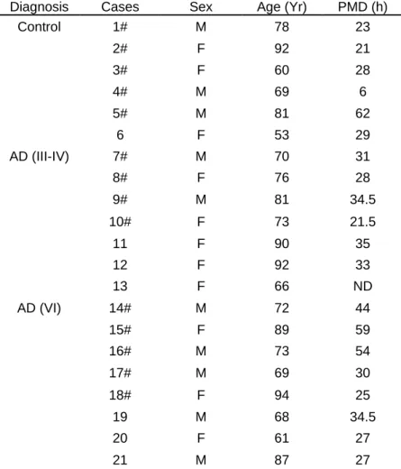
Frozen brain tissue (hippocampus, parahippocampal gyrus and fusiform gyrus) from Controls (n = 5), AD Braak III-IV (n = 4), and AD Braak VI (n = 5) were provided by the GIE NeuroCeb biobank in Paris. Paraffin
Braak III-IV (n = 7; age average 79.4yr, female and 75.5yr, male) and AD Braak VI (n = 8; age average 81.3yr, female and 73.8yr, male) were provided by the Laboratory of Neuropathology at the Hôpital Pitié-Salpêtrière. We did not analyze the influence of sex on our results because of the low number of samples. Previous immunostaining of Tau (AT8, Innogenetics, 1/500) and Aβ (6F/3D, Dako, 1/200) had been performed on all cases. Detailed information on pathology and demographic data are summarized in Table 1. The NeuroCEB brain bank meets the criteria of the French law on biological resources, including consent and data protection (Article L1243-4 of the Public Health Code, August 2007). It has received approval from the Ministry of Higher Education, Research and Innovation to retain human samples and provide them to research teams (CODECOH approval number: AC-2018-3290).
Method details
In Vivo [14C]-2-Deoxyglucose Uptake
We measured [14C]-2-Deoxyglucose (2-DG) uptake in four experimental groups: 6-month-old female 3xTg-AD mice (n = 10), 6-month-old female control mice (n = 9), 12-month-old female 3xTg-3xTg-AD mice (n = 10), and 12-month-old female control mice (n = 10). Experiments were performed in conscious, lightly restrained animals that were previously habituated to the constraint. Animals fasted for 12 h before the experiment but had free access to water. On the day of the experiment, the mice were anesthetized with isoflurane (2% in O2) and two catheters were inserted into the femoral artery and vein, respectively. Body temperature was
maintained at 37°C. The mice were allowed to recover from anesthesia for 1 h and intravenously injected with [14C]-2DG (16.5 µCi/100 g body weight; Perkin Elmer, Boston, MA, USA). Glycemia was measured
using a DIGL-100 QuantiChrom™ Glucose Assay Kit. The mice were euthanized by injection of a lethal dose of sodium pentobarbital 45 min after injection. The brains were rapidly removed and immediately frozen at -40 °C in isopentane. They were cut into 20 µm-thick coronal sections with a CM3050S cryostat (Leica, Rueil-Malmaison, France). Every other section was mounted on a coverslip, rapidly heat-dried, and placed against autoradiographic film (Kodak Biomax MR) for 10 days together with radioactive [14C] standards (146C, American Radiochemical Company, St Louis, MO, USA). The sections not used for the autoradiography study were mounted on Superfrost glass slides that were then processed for Nissl staining, to obtain additional information about brain anatomy.
Autoradiographic and Nissl-stained sections were digitized using a high-resolution flatbed scanner (ImageScanner III; GE Healthcare Europe, Orsay, France) at 600 dpi (pixel size 42 x 42 µm2) and 1,200
dpi (pixel size 21 x 21 µm2) in-plane resolution, respectively. Image processing was performed using our
in-house software platform BrainVISA (http://brainvisa.info/). Each anatomic volume was first reconstructed in 3D, and each autoradiographic volume was reconstructed in 3D using the corresponding anatomic volume as a geometrical reference for co-registration (Dubois et al., 2008). The non-rigid registration approach is based on FFD (Free Form Deformation, polynomial model) and has been previously found to be very robust (Lebenberg et al., 2010; Rueckert et al., 1999). Autoradiographic images were converted from gray scale to activity (nCi/g) using the [14C] standards and normalized at 200 nCi/g. A brain template was created using all the anatomic brain volumes at a final resolution of 0.1 x 0.1 x 0.1 mm3. Regional
differences in cerebral glucose uptake between 3xTg-AD and control mice were then assessed using a voxel-wise two-tailed unpaired t-test implemented in BrainVISA (Vandenberghe et al., 2016).
Generation of AAVs and stereotaxic injection in mice
The plasmid encoding the sensor Laconic (San Martin et al., 2013) was obtained from Addgene (www.addgene.org). The plasmid encoding Cre-IRES-GFP was kindly provided by Martine Cohen-Salmon. We generated adeno-associated viral vectors (AAV2/9) expressing Laconic, td-Tomato, GFP or Cre-IRES-GFP under the control of the short gfaABC1D promoter. The design, production, and titration of AAV2/9
vectors have been described previously (Berger et al., 2015). Mice were anesthetized with a mixture of ketamine (150 mg/kg) and xylazine (10 mg/kg). Lidocaine (5 mg/kg) was injected subcutaneously under the scalp 5 min before the beginning of surgery. The mice received bilateral stereotaxic injections of AAVs administered by a 10-µl Hamilton syringe via a 34-gauge blunt needle. The stereotaxic coordinates used were as follows: AP - 2 mm; L +/- 2 mm; V -1.2 mm with the tooth bar set at 0. Mice received a dose of 1010
viral genomes (VG) in a total volume of 2 µl per injection site at a rate of 0.2 µl/min. At the end of the injection, the needle was left in place for 5 min before being slowly removed. The skin was sutured, and the mice were allowed to recover for three to six weeks.
FRET imaging of brain slices
Injections of AAVs in mice
Five-month old female 3xTg-AD and control mice were injected in the dorsal hippocampus with an AAV2/9 expressing Laconic under the control of the short gfaABC1D promoter and used four to six weeks later.
Slice Preparation
The mice were deeply anesthetized with Ketamine/Xylazine. Heparin (1000 U) was directly injected into the left heart ventricle to prevent blood coagulation, and the mice were perfused transcardially with oxygenated ice-cold sucrose artificial cerebrospinal fluid (ACSF) containing: 126 mM NaCl, 2.5 mM KCl, 1.25 mM NaH2PO4, 2 mM CaCl2, 1 mM MgCl2, 26 mM NaHCO3, 2.5 mM glucose, 25 mM sucrose, and 3 mM
kynurenic acid (Sigma) as previously described (Hay et al., 2015). The brains were quickly removed and sagittal slices (300 µm thick) containing the hippocampus were cut with a vibratome (VT1000S; Leica). Slices were allowed to recover 10 min at 37°C in oxygenated sucrose ACSF and then placed at room temperature for 1 h in ACSF saturated with O2/CO2 (95%/5%), containing 2.5 mM glucose and 22.5 mM
sucrose. The slices were transferred to a submerged recording chamber and perfused with oxygenated ACSF lacking kynurenic acid.
Fluorescence imaging
Two-photon images were obtained with a custom-built 2-photon laser scanning microscope based on an Olympus BX51WI upright microscope (Olympus) with a 40X (0.8 NA) water-immersion objective and a titanium:sapphire laser (MaiTai HP; SpectraPhysics) tuned at 850 nm for mTFP excitation. Galvanometric scanners (model 6210; Cambridge Technology) were used for scanning, and a piezo-driven objective scanner (P-721 PIFOC; Physik Instrumente GmbH) was used for z-stack image acquisition. A two-photon emission filter was used to reject residual excitation light (E700 SP; Chroma Technology). A fluorescence cube containing 479/40 and 542/50 emission filters and a 506-nm dichroic beam splitter (FF01-479/40, FF01-542/50 and FF506-Di02-25x36 Brightline Filters; Semrock) was used for the separation of the mTFP and Venus signals. Two imaging channels (H9305 photomultipliers; Hamamatsu) were used for simultaneous detection of the two types of fluorescence emission. An image stack of 10 to 20 images with a 1-µm interval was acquired every 30 s. Occasional x, y, and z drifts were corrected using custom macros developed from ImageJ plugins TurboReg,StackReg, MultiStackReg and Image CorrelationJ. mTFP and Venus fluorescence intensities were measured at the soma of astrocytes. Fluorescence ratios (R) were calculated by dividing the mean mTFP intensity of a given ROI by the mean Venus intensity of the same ROI. The ratios were normalized to the baseline ratio (R0) calculated in the same ROI during the 10-min prior to the application of AR-C155858 (1µM). Cells exhibiting a baseline with a slope smaller than 0.5%.min-1 were considered to be stable and further analyzed. Effect of monocarboxylate transporter
blockade was quantified by averaging the normalized ratio (R/R0) during the last five minutes of drug application.
Electron microscopy
Six-month-old female mice (3 control and 3xTg-AD) were euthanized with an overdose of pentobarbital and immediately perfused, via the heart, with a buffered mix of 2.5 % glutaraldehyde and 2.0 % paraformaldehyde in 0.1M phosphate buffer (pH 7.4). Brains were then removed and stored overnight in PBS at 4°C. Coronal vibratome sections were cut at 80 microns thickness, post-fixed in potassium ferrocyanide (1.5%) and osmium (2%), then stained with thiocarbohydrazide (1%) followed by osmium tetroxide (2%). They were then stained overnight in uranyl acetate (1%), washed in distilled water at 50°C, before being stained with lead aspartate at the same temperature. They were finally dehydrated in increasing concentrations of alcohol and then embedded in Spurs resin and hardened at 65 degrees for 24 h between glass slides. The regions containing the hippocampal region CA1 were trimmed from the rest of the section using a razor blade and glued to an aluminium stub and mounted inside scanning electron microscope (Zeiss Merlin, Zeiss NTS). Serial sections were cut from the face using an ultramicrotome mounted in the microscope (Gatan, 3View) and the block face imaged after every cut using a beam voltage of 1.7 kV and pixel size of 6 nm with a dwell time of 1 microsecond. Series of 200 – 300 sections were collected, aligned in the FIJI imaging software (www.fiji.sc) and synapses counted in the TrakEM2 plugin. Synapses were counted through discrete volumes through the series of images and classified according to their morphology. Asymmetric synapses were presumed excitatory (glutamatergic) and symmetric, inhibitory (GABAergic). A total of 1067 synapses were counted in the 3xTg-AD mice and 1094 in the controls.
Immunohistochemistry of human brain samples
Immunohistochemistry was performed on a VENTANA Discovery XT automated staining instrument according to the manufacturer's instructions. All solutions were from Roche Diagnostics (Meylan, France). Briefly, slides were de-paraffinized using EZprep solution for 30 min at 75°C, followed by epitope retrieval with RiboCC solution for 20 min at 95°C. Primary rabbit polyclonal PHGDH antibody (Frontier Institute, Hokkaido, Japan) was diluted with the provided Discovery Ab Diluent at 1:300 and applied manually. Slides were developed using the DABMap detection kit, counterstained with hematoxylin II for 8 min, washed,