HAL Id: hal-02006448
https://hal-amu.archives-ouvertes.fr/hal-02006448
Submitted on 31 Aug 2020HAL is a multi-disciplinary open access
archive for the deposit and dissemination of sci-entific research documents, whether they are pub-lished or not. The documents may come from teaching and research institutions in France or abroad, or from public or private research centers.
L’archive ouverte pluridisciplinaire HAL, est destinée au dépôt et à la diffusion de documents scientifiques de niveau recherche, publiés ou non, émanant des établissements d’enseignement et de recherche français ou étrangers, des laboratoires publics ou privés.
Alain Boussuges, Fabienne Bregeon, Philippe Blanc, Jean-Marie Gil, Laurent
Poirette
To cite this version:
Alain Boussuges, Fabienne Bregeon, Philippe Blanc, Jean-Marie Gil, Laurent Poirette. Characteristics of the paralysed diaphragm studied by M-mode ultrasonography. Clinical Physiology and Functional Imaging, Wiley, 2019, 39 (2), pp.143-149. �10.1111/cpf.12549�. �hal-02006448�
Characteristics of the paralyzed diaphragm studied by M-Mode
Ultrasonography
Authors: Alain Boussuges1,2, Fabienne Brégeon3, Philippe Blanc4, Jean-Marie Gil5, Laurent
Poirette6
1. Institut de Recherche Biomédicale des Armées (IRBA), Brétigny sur Orge
2. Center for Cardiovascular and Nutrition Research (C2VN), Aix Marseille Université, INSERM, INRA, Marseille, France
3. Service d’Explorations Fonctionnelles Respiratoires, CHU Nord, Assistance Publique des Hôpitaux de Marseille et Aix Marseille Univ, IRD, APHM, MEPHI, IHU-Méditerranée Infection, Marseille, France
4. Sainte Clotilde Rehabilitation Center, Reunion Island, France 5. Service de Cardiologie, HIA Laveran, Marseille
6. Leon Berard Hospital, Unité de Réhabilitation, Hyères, France
Running Head : Ultrasonography of paralyzed diaphragm Conflicts of Interest: None to report
Abstract word count: 221 words, Text-only word count: 2916 words, 4 figures and 2 tables Address for correspondence: Dr. Alain Boussuges, C2VN, Center for Cardiovascular and Nutrition Research, Aix-Marseille Université et Institut de Recherche Biomédicale des Armées (IRBA), Faculté de Médecine de Marseille, France Tel: 33 4 91 38 56 50; Fax: 33 4 91 38 56 12
Characteristics of the paralyzed diaphragm studied by M-Mode Ultrasonography Abstract
Background: M-Mode ultrasonography might be useful for detecting hemidiaphragm
paralysis. The objective of the present study was to describe the motion recorded by M-mode ultrasonography of both diaphragmatic leaves in patients with a pre-established diagnosis of hemidiaphragm paralysis.
Methods: A study was conducted in 26 patients (18 men, 8 women) with unilateral diaphragmatic paralysis. They were referred at two different rehabilitation centers after thoracic surgery in 23 cases and cardiac interventional procedures in 3 cases. The pulmonary function tests and the study of the diaphragmatic motion using M-mode ultrasonography were recorded.
Results: The pulmonary function tests showed a restrictive pattern. The M-mode ultrasonography reported either the absence of motion or a weak paradoxical (cranial) displacement (less than 0.5cm) of the paralyzed hemidiaphragm during quiet breathing. A paradoxical motion was recorded in all patients during voluntary sniffing, reaching around -1cm. During deep breathing, a paradoxical motion at the beginning of the inspiration was observed. Thereafter, a re-establishment of the motion in the cranio-caudal direction was recorded. The excursions measured on the healthy side, during quiet breathing and voluntary sniffing, were increased in patients suffering from contralateral hemidiaphragm paralysis, when compared with 170 healthy volunteers.
Conclusions: To detect diaphragmatic dysfunction in patients at risk it would be useful to study diaphragmatic motion by M-mode ultrasonography during quiet breathing, voluntary sniffing and deep breathing.
Key words: chest ultrasonography, diaphragmatic motion, phrenic nerve, respiratory maneuvers
Characteristics of the paralyzed diaphragm studied by M-Mode Ultrasonography
Ultrasonography using B-mode or M-mode has demonstrated its advantage in the assessment of diaphragmatic function. Motions of both hemidiaphragms can be recorded by M-mode ultrasonography using the sub-costal approach(Targhetta et al., 1995; Kantarci et al., 2004; Epelmann et al., 2005). Normal values for the excursions of the hemidiaphragms, during quiet breathing, voluntary sniffing and deep breathing, have been previously reported (Boussuges et al., 2009). M-mode US might be useful for detecting diaphragmatic paralysis, but its use has only been reported in case reports (Patel et al., 2007; Boussuges et al., 2015) or in small series of adult patients referred for suspected diaphragmatic paralysis(Lloyd et al., 2006). In patients suffering from unilateral diaphragmatic paralysis, the hemidiaphragm mobility has been reported to be absent or paradoxical during breathing at rest (Patel et al., 2007; Lloyd et al., 2006). During voluntary sniffing, a paradoxical (i.e. cranial) movement of the hemidiaphragm has been reported using fluoroscopy (Nason et al., 2012) or M-mode ultrasonography (Lloyd et al., 2006; Boussuges et al., 2015). Nevertheless, a paradoxical movement can be observed in healthy subjects. Using fluoroscopy, Alexander demonstrated that a unilateral paradoxical movement on sniffing occurred in 6% of normal subjects (Alexander, 1966). Therefore, the unique presence of a paradoxical movement recorded during voluntary sniffing cannot be definitely attributed to hemidiaphragm paralysis. Kinetics of the paralyzed hemidiaphragm during deep breathing can be informative. Scillia et al. (2004) have studied the parameters involved in the displacement of a paralyzed
hemidiaphragm. They reported that in dogs affected by unilateral diaphragmatic paralysis, the course of the inactive hemidiaphragm during inspiration is determined by the balance between
the decrease in pleural pressure, which leads to a cranial displacement, and the force
generated by the intact hemidiaphragm, which induces a caudal displacement. Furthermore, it has also been reported that the relaxation of the abdominal wall could induce a caudal
displacement of the paralyzed diaphragm during inspiration(McCool & Mead, 1989).
Consequently, a full description of the diaphragmatic excursions in patients suffering from unilateral diaphragmatic paralysis can be useful for investigators. This study was designed to describe the motion recorded by M-mode ultrasonography of the two hemidiaphragms in patients with a diagnosis of hemidiaphragm paralysis established on the basis of paraclinical data and medical history.
Methods
Population
The patients were recruited from two rehabilitation centers: the Leon Berard Hospital, Hyères, France (annual number of admissions: 1100) and the Sainte Clotilde Rehabilitation Center, Reunion Island, France (annual number of admissions: 350). Data were collected from patients admitted to both centers between 2013 and 2017 and presenting with a suspected diagnosis of diaphragmatic dysfunction. The diagnosis of diaphragmatic dysfunction relied on clinical and functional assessments including pulmonary function testing, fluoroscopy and diaphragm electromyography (Jammes et al., 2010). Only subjects with a diagnosis of
diaphragmatic paralysis, as confirmed by the physician in charge of the patient, were included in the study. The Ethics Committee of the Aix Marseille University (CPPRB 1, No
During the predetermined period, 26 patients were diagnosed with paralyzed hemidiaphragm by their physician. These patients (18 men, 8 women) were referred to the rehabilitation centers after heart valve surgery in 9 cases, coronary artery bypass graft surgery in 8 cases, heart transplantation in 4 cases, atrial fibrillation ablation in 2 cases, surgery of the thoracic aorta in 2 cases and percutaneous coronary intervention in one case. Before this last procedure, their past medical history included a previous thoracic surgery in 11 patients, and a chest trauma due to a motorcycle accident in one patient. Consequently, in some patients, diaphragmatic dysfunction could not definitely be attributed to the procedure immediately performed before admission in the rehabilitation center.
They were admitted as a mean 30+/-14 days (from 9 to 61 days) after the surgical or interventional procedures. Sixteen patients were classified as NYHA Functional Class II, 8 patients were classified as NYHA III and 2 patients NYHA I.
A pleural effusion was detected by chest radiography in 13 cases corresponding to 10 patients. The effusion was recorded on the left side in 7 cases and on both sides in 3 cases. Pleural fluid volume was considered to be small in 8 cases and moderate in 5 cases.
Results of the investigations, including pulmonary function tests and the study of the diaphragmatic motion using M-mode ultrasonography, were recorded at admission to the rehabilitation centers.
Pulmonary function tests were performed in the sitting position using a commercially available spirometer (Ilmeter 1304; Masterlab Jaeger, Wurzberg, Germany) according to American Thoracic Society Standards (Miller et al. 2005). The best forced expiratory volume in 1sec (FEV1.0), Forced Vital Capacity (FVC) and Slow Vital Capacity (SVC) of at least
normal (LLN) and to the mean values predicted by the CECA 93 equations (Quanjer et al. 1993).
Diaphragmatic function was studied by M-mode ultrasonography using a Philips iE33 ultrasound system (Andover, MA, USA) equipped with a 1-5MHz transducer (S5-1 probe). Ultrasonographic examinations were performed in a seated position. The excursions of the diaphragm were studied on both sides during quiet breathing, voluntary sniffing and deep breathing according to a previously published method (Boussuges et al., 2009). Briefly,the measurement of the diaphragmatic motion included the diaphragmatic excursion (in cm), i.e. the vertical distance between the point corresponding to the beginning of the inspiration and the point corresponding to the maximal diaphragm displacement, and the duration of the diaphragmatic contraction (in sec). The velocity of contraction (in cm/sec) was calculated as the ratio excursion/duration. Several respiratory cycles were recorded on both sides.
Measurements were averaged from at least three different cycles.
Statistical analysis
Results of the measurements were reported as Mean+/-Standard Deviation. Statistical tests were run on Sigma Stat software (SPSS Inc, Chicago, USA). The excursions recorded on the non-paralyzed side were compared with measurements performed in a group of 170 healthy volunteers (121 men, 49 women) with normal lung function tests. Analysis of the differences between patients and control subjects was performed using unpaired Student’s t-test. In the event of cohorts of variables not having a normal distribution, comparisons were performed with the Mann-Whitney U test.
Furthermore, it was determined the number of patients reporting a diaphragmatic excursion upper than normal values on the healthy side during quiet breathing, according to reference values previously published by Boussuges et al. (2009). A chi-square test was used to compare the number of subjects with diaphragmatic excursion upper than normal values in patients and healthy controls. The significance level was p<0.05.
Results
A total of 26 patients were sent to the ultrasonographic laboratory with a pre-established diagnosis of paralyzed hemidiaphragm (right side=13 and left side=13).
In the studied population, the pulmonary function tests were compatible with a restrictive pattern with a mean SVC= 2±0.7L (57±15% of predicted), mean FVC=1.8±0.7L (52±15% of predicted), mean FEV1.0=1.45±0.5L (52±12% of predicted), and mean FEV1.0
/FVC= 82±9% (106±11% of predicted). 24 patients out of 26 had both SVC and FVC below the LLN.
One patient with a pre-established right-sided paralysis of the hemidiaphragm had a ultrasonographic aspect of hypokinesis of the left hemidiaphragm. This patient was excluded from the statistical analysis comparing the excursions recorded on the non-paralyzed side with the measurements performed in a group of healthy volunteers.
Motion of the paralyzed hemidiaphragm (Table 1)
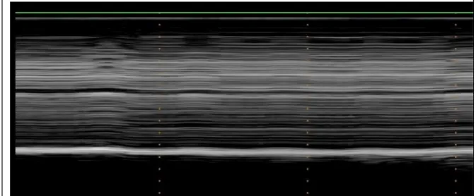
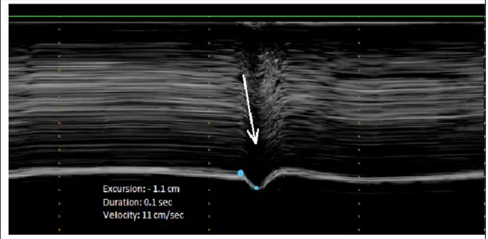
During quiet breathing, in most of cases, no detectable motion was observed on the paralyzed hemidiaphragm (Figure 1a). In some patients, a slight paradoxical displacement (less than 0.5cm) was observed (Figure 1b). During voluntary sniffing, a paradoxical motion
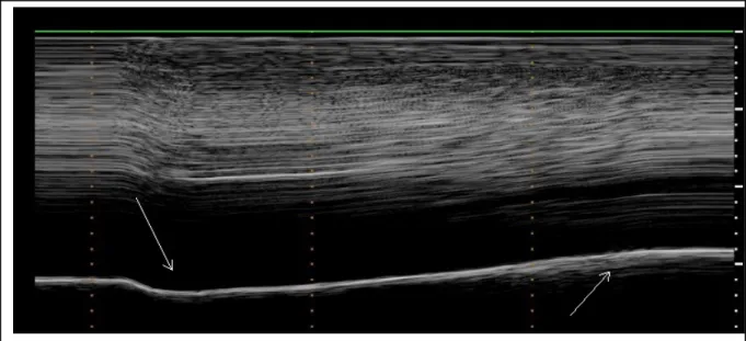
was recorded in all patients (Figure 2). The motion recorded appeared as a quick negative excursion measured at around 1cm of magnitude. During deep breathing, the motion recorded showed a paradoxical motion at the beginning of the inspiration followed by a reestablishment of the motion in the cranio-caudal direction (Figure 3).
Motion of the hemidiaphragm on the healthy side (Table 2)
Diaphragmatic motions during quiet breathing and VS were successfully recorded in all patients. During deep breathing, diaphragmatic motion could be measured in 12 patients out of 13 on the right side and in 9 patients out of 12 on the left side.
These measurements were compared with the control group of 170 healthy volunteers. Patients and controls were well matched for gender (69 vs 71% of men), age (58±16 vs 55±12 years), height (168±8 vs 167±9cm) and weight (72±14 vs 72±13kg).
When compared with the measurements performed in the control group, excursions recorded on the healthy side, in patients suffering from hemidiaphragm paralysis, were significantly increased during quiet breathing in both, right side (3±1.1 vs 1.8±0.3cm - p<0.001) and left side (2.8±0.7 vs 1.8±0.4cm - p<0.001), respectively. The velocity of contraction was also increased in patients on right side (3±1.1 vs 1.7±1cm/sec - p<0.001) and left side (3.8±1.6 vs 1.8±0.5cm/sec - p<0.001), respectively. In contrast, the duration of contraction was shortened in patients when compared with controls on right (0.3±0.1 vs 1.2±0.3sec - p<0.001) and left side (0.25±0.1 vs 1±0.3sec - p<0.001).
These significant differences were also observed during voluntary sniffing on the right hemidiaphragm (4±0.8 in patients vs 3±0.6cm in controls - p<0.001) and on the left hemidiaphragm (4.3±1.4 in patients vs 3.1±0.6cm in controls - p<0.001).
During deep breathing, the excursions recorded were not significantly different in patients and controls on the right side 6.2±1.7 vs 6.6±1.2cm and on the left side 6.4±1.2 vs 6.8±1.3cm, respectively.
On the healthy side, 18 patients out of 25 had a diaphragmatic motion during quiet breathing larger than upper normal values. The percentage of subjects reporting diaphragmatic
excursion upper than normal values was significantly increased in patients (72%) when compared with controls (7%) – p<0.001.
Discussion
In the studied population, the pulmonary function tests were compatible with a restrictive pattern which may result from cumulating factors such as the diaphragmatic dysfunction itself, the thoracic and pulmonary alterations induced by the recent thoracic surgery and in some subjects the presence of pleural fluid accumulation (Van Belle et al., 1992; Ng et al., 2002; Westerdahl et al., 2003; Roncada et al., 2015).
The current study offers new data on ultrasonic imaging of the paralyzed
a slight paradoxical displacement of the paralyzed diaphragm were recorded. During voluntary sniffing, a cranial displacement was observed in all patients. This paradoxical movement was a quick displacement (mean velocity: 8.4cm/sec) reaching around -1cm (from -0.5 to -2.3cm). These findings are in agreement with the results of Lloyd et al. (2006)
performed in a population of 6 patients with neurologic disorders and suffering from
diaphragmatic paralysis. It has been reported, using fluoroscopy, that some healthy subjects had a paradoxical movement of the diaphragm during sniffing (Alexander, 1966).
Consequently, the third maneuver used in our study, i.e. a deep slow inspiration, might be useful. In our population, a biphasic movement, with an initial paradoxical cranial movement and a terminal caudal displacement, was recorded on the side of the paralyzed
hemidiaphragm. Excursions of the cranial and caudal displacements were both measured at about 1cm, but in some patients the caudal displacement could either be lacking or reach more than 2cm. It has been suggested that the bell wall relaxation might contribute to the caudal displacement of the paralyzed diaphragm during inspiration(McCool & Mead, 1989).
Consequently, the time course of the displacement during deep breathing should be carefully examined and the presence of a terminal caudal displacement should not lead the investigator to conclude there is normal function of the hemidiaphragm.
In patients suffering from hemidiaphragm paralysis, the measurement of the excursions recorded on the healthy side can be also interesting. Indeed, the excursions recorded during quiet breathing and voluntary sniffing, were larger than the excursions of a group of 170 healthy volunteers. In the case of suspected unilateral diaphragmatic
dysfunction, an excursion larger than normal values can support the diagnosis. In our population, 18 patients out of 25 had hemidiaphragmatic motion larger than upper normal values. These findings were in agreement with previous experimental studies that reported a
compensatory increase in neural drive to the functioning hemidiaphragm after unilateral diaphragm paralysis(Katagiri et al., 1994). Furthermore, in patients suffering from unilateral diaphragmatic dysfunction, Houston et al. (1995) have previously reported large excursions of the normal side in relation to a compensatory mechanism. Nevertheless, after thoracic
surgery, diaphragmatic dysfunction is frequent and can be bilateral (Wheeler et al., 1985; Aguirre et al., 2013). In our population, one patient suffered from right hemidiaphragm paralysis and left hemidiaphragm paresis. In these circumstances, the magnitude of the excursions of the non-paralyzed hemidiaphragm is not informative.
One could object that the measurement of the diaphragmatic thickness in the zone of apposition of the diaphragm to the rib cage, using B-mode ultrasonography, has previously demonstrated its interest in the diagnosis of diaphragmatic dysfunction. A paralyzed
diaphragm does not thicken during inspiration(Gottesman & McCool, 1997). Consequently, the benefit of the assessment of diaphragmatic motion by M-mode US can be questioned. In fact, both methods have some limitations and it would be interesting to combine it.
Firstly, the measurement of diaphragmatic thickness can be difficult in some patients; a poor acoustic window has been reported in 2 to 10% of subjects (Kim et al., 2011; Vivier et al., 2012). In the intensive care unit, the failure of measurement of the diaphragmatic
thickness at end expiration was around 5% on the right side and 15% on the left side (Goligher et al., 2015). In contrast, the study of the diaphragmatic motion during quiet breathing and voluntary sniffing is easy to perform and the percentage of failure is very low. In a previous study performed in 210 healthy volunteers(Boussuges et al., 2009), right and left diaphragmatic motions were successfully accomplished during quiet breathing in the whole population. During voluntary sniffing, measurement was always possible on the right side and, in 208 out of 210 volunteers, on the left side. During deep breathing, in some
subjects the measurement of diaphragmatic motion was difficult to perform, the diaphragm being obscured by the descending lung, particularly on the left side. In patients suffering from diaphragmatic paralysis or paresis, the caudal and cranial excursions are weak and should be easier to record.
Secondly, the failure of the paralyzed diaphragm to thicken led to a decrease in the thickening ratio i.e. the ratio between the diaphragmatic thickness measured at the end of maximal inspiration and the diaphragmatic thickness at the end of expiration. A change in diaphragm thickness of 28-96% has been reported in healthy volunteers, with a change of -35 % to 5% in those with a paralyzed diaphragm. Achange in diaphragmatic thickness during inspiration of less than 20% has been suggested to predict diaphragm paralysis (Gottesman & McCool, 1997). Nevertheless, some healthy subjects could have a thickness ratio close to this threshold (since the lower normal range is 28%) and in such patients, the diagnosis of paralysis
remained uncertain. Furthermore, the criteria for distinguishing between diaphragmatic paralysis and diaphragmatic paresis have not yet been established. In these circumstances, the complementary study of diaphragmatic motion by M-mode ultrasonography can be useful; a decrease in diaphragmatic excursions, without paradoxical movement, suggests
hemidiaphragm paresis rather than paralysis (Kim et al., 2011). Lastly, when a
hemidiaphragm paralysis is recognized by M-mode ultrasonography it would be interesting to assess the thickening ratio to support this diagnosis. Moreover, the measurement of the diaphragmatic thickness can be useful in assessing the date of the injury. Indeed, it has been demonstrated that a chronically paralyzed diaphragm is atrophic (thickness lower than 0.2cm) in contrast to an acutely paralyzed diaphragm that may present normal thickness.
The present study was not designed to detect diaphragm paralysis but to extensively describe the ultrasonographic characteristics in patients suffering from diaphragm paralysis. Only the results for patients with a final diagnosis of hemidiaphragm paralysis were reported. Diaphragmatic paralysis is frequently not diagnosed because of its non-specific presentation. During the same 5-year period, other patients admitted to the two rehabilitation centers might have been underdiagnosed. Consequently, the frequency of this disease could not be assessed by our study. It would be interesting to perform a prospective study including the systematic use of reference method such as diaphragm electromyography with the measurement of phrenic nerve conduction time to determine the value of ultrasonography in the detection of diaphragm paralysis. If this study demonstrates a good diagnostic value, the systematic use of ultrasonography in high risk patients should improve the early diagnosis of hemidiaphragm paralysis.
In the present work, diaphragmatic dysfunction was frequently due to a phrenic nerve injury related to the cardiac interventions and only 26 patients were investigated. It would be interesting to investigate a larger population of patients suffering from diaphragmatic
dysfunction due to other underlying conditions.
Conclusion
The present study, using M-mode ultrasonography, provides the results of the analysis of the diaphragmatic kinetics in patients suffering from hemidiaphragm paralysis. No
movement during quiet breathing and a paradoxical movement during voluntary sniffing are commonly recorded. During deep breathing an initial paradoxical (cranial) displacement followed by a caudal displacement of the hemidiaphragm are observed. The study of the
healthy side can be also informative. Indeed, a compensatory phenomenon frequently leads to an increase in the excursions of the leaves during quiet breathing, when compared to normal values. The combination of these maneuvers associated with the measurement of the
diaphragmatic thickness should be an interesting tool to detect hemidiaphragm paralysis in subjects at high risk, such as thoracic surgery patients.
Acknowledgments: we thank Ibtissem Smaali of the Ylang-Ylang Center (Le Port, Reunion Island).
This work was supported by the French Ministry of Defense (PPRC n°11G-A01299) The authors have no conflicts of interest.
References
1. Aguirre VJ, Sinha P, Zimmet A, Lee GA, Kwa L, Rosenfeldt F. Phrenic nerve injury during cardiac surgery: mechanisms, management and prevention. Heart Lung Circ. (2013); 22: 895-902.
2. Alexander C. Diaphragm movements and the diagnosis of diaphragmatic paralysis. Clin Radiol. (1966); 17: 79-83.
3. Boussuges A, Gole Y, Blanc P. Diaphragmatic motion studied by M-mode
ultrasonography: methods, reproducibility, and normal values. Chest. (2009); 135: 391-400.
4. Boussuges A, Chaumet G, Poirette L. Interest of ultrasonographic assessment of diaphragmatic function in cardiac rehabilitation center: a case report. Medicine (Baltimore). (2015); 94: e801.
5. Epelman M, Navarro OM, Daneman A, Miller SF. M-mode sonography of diaphragmatic motion: description of technique and experience in 278 pediatric patients. Pediatr Radiol. (2005); 35: 661–667.
6. Goligher EC, Laghi F, Detsky ME, Farias P, Murray A, Brace D, Brochard LJ, Bolz SS, Rubenfeld GD, Kavanagh BP, Ferguson ND. Measuring diaphragm thickness with ultrasound in mechanically ventilated patients: feasibility, reproducibility and validity. Intensive Care Med. (2015); 41: 642-649.
7. Gottesman E, McCool FD. Ultrasound evaluation of the paralyzed diaphragm. Am J Respir Crit Care Med. (1997); 155: 1570-1574.
8. Houston JG, Fleet M, Cowan MD, McMillan NC. Comparison of ultrasound with fluoroscopy in the assessment of suspected hemidiaphragmatic movement
abnormality. Clin Radiol. (1995); 50: 95-98.
9. Jammes Y, Budin-Poirier C, Brégeon F. Electromyographic tools to assess hemidiaphragm paralysis. Clin Physiol Funct Imaging. (2010); 30:107-115.
10. Kantarci F, Mihmanli I, Demirel MK,Harmanci K, Akman C, Aydogan F, Mihmanli A, Uysal O. Normal diaphragmatic motion and the effects of body composition: determination with M-mode sonography. J Ultrasound Med. (2004); 23: 255–260. 11. Katagiri M, Young RN, Platt RS, Kieser TM, Easton PA. Respiratory muscle
compensation for unilateral or bilateral hemidiaphragm paralysis in awake canines. J Appl Physiol. (1994); 77: 1972-1982.
12. Kim WY, Suh HJ, Hong SB, Koh Y, Lim CM. Diaphragm dysfunction assessed by ultrasonography: influence on weaning from mechanical ventilation. Crit Care Med. (2011); 39: 2627–2630.
13. Lloyd T, Tang YM, Benson MD, King S. Diaphragmatic paralysis: the use of M mode ultrasound for diagnosis in adults. Spinal Cord. (2006); 44: 505-508.
14. McCool FD, Mead J. Dyspnea on immersion: mechanisms in patients with bilateral diaphragm paralysis. Am Rev Respir Dis. (1989); 139: 275–276.
15. Miller MR, Hankinson J, Brusasco V, Burgos F, Casaburi R, Coates A, Crapo R, Enright P, van der Grinten CP, Gustafsson P, Jensen R, Johnson DC, MacIntyre N, McKay R, Navajas D, Pedersen OF, Pellegrino R, Viegi G, Wanger J. Standardisation of spirometry. Eur Respir J. (2005); 26: 319-338.
16. Nason LK, Walker CM, McNeeley MF, Burivong W, Fligner CL, Godwin JD.
17. Ng CS, Wan S, Yim AP, Arifi AA. Pulmonary dysfunction after cardiac surgery. Chest. (2002); 121: 1269-1277.
18. Patel AS, O'Donnell C, Parker MJ, Roberts DH. Diaphragm paralysis definitively diagnosed by ultrasonography and postural dependence of dynamic lung volumes after seven decades of dysfunction. Lung. (2007); 185: 15-20.
19. Quanjer PH, Tammeling GJ, Cotes JE, Pedersen OF, Peslin R, Yernault JC. Lung volumes and forced ventilatory flows. Report working party: Standardization of lung function testing. Eur Respir J. (1993); 6: 5–40.
20. Roncada G, Dendale P, Linsen L, Hendrikx M, Hansen D. Reduction in pulmonary function after CABG surgery is related to postoperative inflammation and
hypercortisolemia. Int J Clin Exp Med. (2015); 8: 38-46.
21. Scillia P, Cappello M, De Troyer A. Determinants of diaphragm motion in unilateral diaphragmatic paralysis. J Appl Physiol. (2004); 96: 96-100.
22. Targhetta R, Chavagneux R, Ayoub J, Lemerre C, Préfaut C, Bourgeois JM, Balmes P. Right diaphragmatic kinetics measured by M-mode ultrasonography with concomitant spirometry in normal subjects and asthmatic patients. Preliminary results. Rev Med Interne. (1995); 16: 819–826.
23. Van Belle AF, Wesseling GJ, Penn OC, Wouters EF. Postoperative pulmonary function abnormalities after coronary artery bypass surgery. Respir Med. (1992); 86: 195-199.
24. Vivier E, Mekontso-Dessap A, Dimassi S, Vargas F, Lyazidi A, Thille AW, Brochard L. Diaphragm ultrasonography to estimate the work of breathing during non-invasive ventilation. Intensive Care Med. (2012); 38: 796-803.
25. Westerdahl E, Lindmark B, Bryngelsson I, Tenling A. Pulmonary function 4 months after coronary artery bypass graft surgery. Respir Med. (2003); 97: 317-322.
26. Wheeler WE, Rubis LJ, Jones CW, Harrah JD. Etiology and prevention of topical hypothermia-induced phrenic nerve injury and left lower lobe atelectasis during cardiac surgery. Chest. (1985); 88: 680–683.
Figure captions : Hemidiaphragmatic paralysis studied by M-mode ultrasonography
Figure 1: M-mode ultrasonography of a paralyzed hemidiaphragm during quiet breathing. No motion (figure 1a) or a paradoxical motion (arrows – figure 1b) was observed.
Figure 1a
Figure 2: M-mode ultrasonography of a paralyzed hemidiaphragm during voluntary sniffing showing a paradoxical motion (arrow).
Figure 3: During deep breathing, a biphasic movement was observed on M-mode ultrasonography with an initial paradoxical movement (left arrow) and a terminal caudal displacement (right arrow).
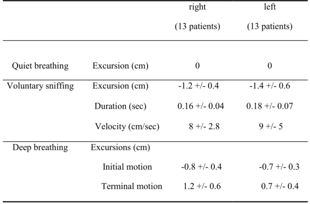
Table 1: Hemidiaphragm motion measurements on paralyzed side (mean +/- SD)
right (13 patients)
left (13 patients)
Quiet breathing Excursion (cm) 0 0
Voluntary sniffing Excursion (cm) -1.2 +/- 0.4 -1.4 +/- 0.6 Duration (sec) 0.16 +/- 0.04 0.18 +/- 0.07 Velocity (cm/sec) 8 +/- 2.8 9 +/- 5 Deep breathing Excursions (cm)
Initial motion -0.8 +/- 0.4
-0.7 +/- 0.3 Terminal motion 1.2 +/- 0.6 0.7 +/- 0.4
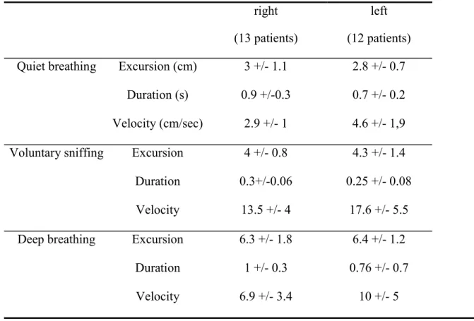
Table 2: Hemidiaphragm motion measurements on healthy side (mean +/- SD)
right (13 patients)
left (12 patients) Quiet breathing Excursion (cm) 3 +/- 1.1 2.8 +/- 0.7
Duration (s) 0.9 +/-0.3 0.7 +/- 0.2 Velocity (cm/sec) 2.9 +/- 1 4.6 +/- 1,9 Voluntary sniffing Excursion 4 +/- 0.8 4.3 +/- 1.4
Duration 0.3+/-0.06 0.25 +/- 0.08 Velocity 13.5 +/- 4 17.6 +/- 5.5 Deep breathing Excursion 6.3 +/- 1.8 6.4 +/- 1.2 Duration 1 +/- 0.3 0.76 +/- 0.7 Velocity 6.9 +/- 3.4 10 +/- 5