HAL Id: hal-03009179
https://hal.sorbonne-universite.fr/hal-03009179
Submitted on 17 Nov 2020
HAL is a multi-disciplinary open access
archive for the deposit and dissemination of
sci-entific research documents, whether they are
pub-lished or not. The documents may come from
teaching and research institutions in France or
abroad, or from public or private research centers.
L’archive ouverte pluridisciplinaire HAL, est
destinée au dépôt et à la diffusion de documents
scientifiques de niveau recherche, publiés ou non,
émanant des établissements d’enseignement et de
recherche français ou étrangers, des laboratoires
publics ou privés.
Testing for ROS1, ALK, MET, and HER2
rearrangements and amplifications in a large series of
biliary tract adenocarcinomas
Jeremy Augustin, Caroline Gabignon, Aurélie Scriva, Laëtitia Menu, Claire
Calmel, Olivier Scatton, François Paye, Jean-François Fléjou, Françoise Praz,
Pascale Cervera, et al.
To cite this version:
Jeremy Augustin, Caroline Gabignon, Aurélie Scriva, Laëtitia Menu, Claire Calmel, et al.. Testing
for ROS1, ALK, MET, and HER2 rearrangements and amplifications in a large series of biliary tract
adenocarcinomas. Virchows Archiv, Springer Verlag, 2020, 477 (1), pp.33-45.
�10.1007/s00428-020-02822-8�. �hal-03009179�
1
Testing for ROS1, ALK, MET and HER2 rearrangements and amplifications in a large series of biliary tract adenocarcinomas.
Jeremy Augustin 1,2, Caroline Gabignon 1, Aurélie Scriva 1, Laëtitia Menu 2, Claire Calmel 2, Olivier Scatton 3,2, François Paye 4,2, Jean-François Fléjou 1,2, Françoise Praz 2,5, Pascale Cervera 1,2,* and Dominique Wendum 1,2,*.
1 AP-HP, Hôpital Saint Antoine, Service d’Anatomie et Cytologie Pathologiques, 75012 Paris, France.
2 Faculté de Médecine Sorbonne Université ; INSERM UMR_S 938
,
Centre de Recherche Saint-Antoine (CRSA), 75012 Paris, France.3 AP-HP, Hôpital Saint Antoine, Service de Chirurgie Hépato-Biliaire et Transplantation Hépatique, 75012 Paris, France.
4 AP-HP, Hôpital Saint Antoine, Service de Chirurgie Digestive, 75012 Paris, France.
5 Centre National de la Recherche Scientifique (CNRS), Paris, France.
*Pascale Cervera and Dominique Wendum contribute equally to the work mentorship.
Corresponding author: Pascale Cervera pascale.cervera@aphp.fr Phone: 00 33 149282172 Fax: 00 33 1 49282878 https://orcid.org/0000-0001-6048-2214 Acknowledgements
This work was financially supported by AFEF (Association Française pour l’Etude du Foie).
We thank Sylvie Dumont and Fatiha Merabtene (“Plateforme d’Histomorphologie St-Antoine", Sorbonne Université, UMS30 LUMIC) for technical assistance.
2
Abstract
Biliary tract carcinomas are divided into intrahepatic, perihilar, distal extrahepatic cholangiocarcinomas, and gallbladder adenocarcinomas. Therapies targeting ROS1, ALK, MET and HER2 alterations are currently evaluated in clinical trials. We assessed ROS1, ALK translocations/amplifications as well as MET, and HER2 amplifications for each tumor subtype by fluorescent in situ hybridization (FISH) and immunohistochemistry (IHC) in 73 intrahepatic, 40 perihilar bile duct , 36 distal extrahepatic cholangiocarcinomas, and 45 gallbladder adenocarcinomas (n=194). By FISH, we detected targetable alterations in 5.2% of cases (n=10): HER2 and MET amplifications were found in 4.1% (n=8) and 1.0% (n=2), respectively. The HER2 amplified cases were mostly gallbladder adenocarcinomas (n = 5,). The MET and HER2 amplified cases were all positive by IHC. Fourteen cases without MET amplification were positive by IHC, whereas HER2 over-expression was detected by IHC only in HER2 amplified cases. We detected no ALK or ROS1 translocation or amplification. Several alterations were consistent with aneuploidy: 24 cases showed only one copy of ROS1 gene, 4 cases displayed a profile of chromosomal instability, and an over-representation of centromeric alpha-satellite sequences was found in five cases.
We confirm a relatively high rate of HER2 amplifications in gallbladder adenocarcinomas and the efficacy of IHC to screen these cases. Our results also suggest the value of IHC to screen MET amplification. Contrary to initial publications, ROS1 rearrangements seem to be very rare in biliary tract adenocarcinomas. We confirm a relatively high frequency of aneuploidy and chromosomal instability, and reveal the over-representation of centromeric alpha-satellite sequences in intrahepatic cholangiocarcinomas. .
Keywords:
3
Introduction
Although biliary tract adenocarcinoma is a rare cancer (five cases per 100,000 inhabitants in industrialized countries) [1], its incidence has been increasing during the last thirty years particularly for intrahepatic cholangiocarcinomas [2]. Biliary tract adenocarcinomas can rarely be surgically resected and more than 50% of the patients will relapse [3]. The use of adjuvant treatments is still controversial with a median survival of only 9 to 15 months given the chemoresistance of this cancer [4]. In this context, other therapies are needed and characterization of molecular alterations that may be potential targets for therapy is crucial.
Several theranostic alterations have been targeted in clinical trials like ACSé (NCT02034981) evaluating crizotinib safety and efficacy in patients with locally advanced rearranged cancers with ALK, MET, and ROS1 rearrangements, or NCT02836847 and NCT03093870 trials that investigated trastuzumab and varlitinib efficacy in HER2 amplified biliary tract adenocarcinomas [5].
However, there are few data regarding these alterations in biliary tract adenocarcinomas and previous studies reported contradictory results (Table 1 of the supplementary data) [6-19]. FIG-ROS1 fusions were detected by PCR in 8.7% cholangiocarcinomas [6] and 9.2%, biliary tract carcinomas (exclusively in extrahepatic biliary cholangiocarcinomas and gallbladder carcinomas)[ 7]. The frequency of ROS1 rearrangements was lower with 3 cases of ROS1 rearrangements out of 261 cholangiocarcinomas (1.1%) using fluorescent in situ hybridization (FISH), and exclusively in intrahepatic tumors [8]. A recent report mentioned the absence of ROS1 rearrangement out of 110 biliary tract adenocarcinomas assessed by FISH [9]. In this study, one case with ALK rearrangement was detected using FISH and MET expression was explored using only immunohistochemistry (IHC). HER2 amplification has been described in biliary tract adenocarcinomas with a frequency around 3% in distal extrahepatic cholangiocarcinomas and 10% in gallbladder carcinomas [10, 12, 13, 16, 18-21].
Biliary tract adenocarcinomas are gathered together in studies although it is well known that they do not represent a single tumor type, as they arise from different biliary cells and display different mutational profiles [5, 22-24]. They are classified according to their anatomical location into intrahepatic, extrahepatic cholangiocarcinomas (perihilar or distal) and gallbladder carcinomas. Morphomolecular subtypes (small and large duct type) are now integrated for intrahepatic cholangiocarcinomas in the fifth edition of the WHO classification of digestive system tumors, [25, 26]. Therefore, in order to have a precise view of some molecular targets in biliary tract adenocarcinomas, we assessed ROS1 and ALK rearrangements and amplifications, as well as MET and HER2 amplifications using FISH as gold standard in a large French series of biliary tract carcinomas that are
well-4
characterized in terms of location, pathology and staging. We also performed immunohistochemical stainings in order to determine which method, IHC or FISH, was more effective for screening these tumors.
Patients and methods
Case selection
One hundred and ninety-four (194) cases of histologically-confirmed biliary tract adenocarcinomas diagnosed on surgically-resected specimens between 2005 and 2019 were retrieved from the Saint-Antoine Hospital database. Only cases with invasive carcinomas were included. Informed consent was obtained for the study.
Age, gender, tumor location, associated risk factors of liver or biliary tract disease were recorded. Tumor stage, nodal status, vascular invasion, perineural invasion and status of the resection margins were retrieved from pathologic reports. Tumor stage was defined for all cases according the 7th edition of the TNM classification.
Biliary tract adenocarcinoma specimens and tissue microarray construction
All specimens were formalin-fixed and paraffin-embedded. Based on radiologic and pathologic findings, biliary tract adenocarcinomas were subdivided into four subtypes according to their location: intrahepatic, perihilar bile duct, distal extrahepatic bile duct cholangiocarcinomas, and gallbladder adenocarcinomas. For each case, one representative tumor block was selected for tissue microarray construction. Two to five 0.6-mm tissue cores were used and hematoxylin and eosin (HE) staining was performed on each tissue microarray slide to confirm the presence of tumor areas. Whole tissue sections were used for the 36 distal extrahepatic bile duct cholangiocarcinomas.
Morphological analysis of the tumors
HE-stained sections were reviewed to specify histological type and grade according to the WHO classification [27]. We also subclassified intrahepatic cholangiocarcinomas into cholangiolar and bile duct type using morphological criteria described in Liau et al. [25], which are similar to the WHO criteria’s defining the small and large duct types [26].
Fluorescent in situ hybridization for ROS1, ALK, MET and HER2,
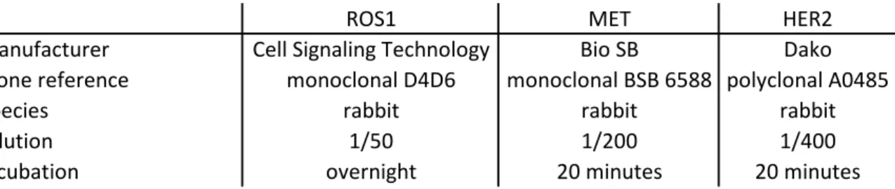
Probes and technical details for each probe are listed in Table 2 of the supplementary data. FISH was performed on tissue microarrays using six probes to detect ROS1 rearrangement or amplification, cMET amplification, HER2
5
amplification, ALK rearrangement or amplification, according to the manufacturers' instructions. Briefly, whatever the probe used, 3-µm slides from tissue microarray blocks were deparaffinized using xylene washes. After rehydration, slides were microwaved preprocessed and after pepsine digestion, dried before co-denaturation and overnight hybridization using a hybridizer system (Dako, Agilent, CA, United States). Post-hybridization washes and counterstain were performed according to each of the probes that were used.
For ROS1 break-apart probes detection, FISH slides were scanned using PathScan® FISH (Excilone, EXCS-PS-F, Elancourt, France) at x60 objective, after delimitating regions of interest on HE slides. Scanned FISH slides were visualized on PathScan® Viewer (Excilone, Elancourt, France), selecting appropriate channels. FISH signal abnormalities were confirmed using a fluorescence microscope with appropriate channels. For all other probes, FISH slides were directly visualized on fluorescence microscope. Forty non-overlapping nuclei from three tumor areas were evaluated for each probe at x100 oil immersion objective. ROS1 translocation was defined as the presence of green and red signals separated by at least two signal diameters in the cell nucleus, with still a fused red and green signal. ROS1 monosomy was defined as a unique fused red and green signal with two HER2, MET and ALK signals, in most nuclei. HER2 amplification was defined by HER2/CEP17 ratio higher than 2, with no
HER2 signals less than 6 [28]. As proposed by some authors for lung adenocarcinoma, MET amplification was
defined by the existence of 6 copies or more of MET per cell [29], and because the probe used did not target centromere 7, MET/CEP7 ratio was not evaluated. Because there are no official guidelines to evaluate ROS1 amplification, we defined ROS1 amplification as for MET. ALK amplification was defined by an ALK/CEP2 ratio higher than 2 [30].
Aneuploidy was defined by an unbalanced number of chromosomes or loss of large portions of chromosomes; polysomy as a type of aneuploidy, with three or more copies of one or more chromosomes. Because we used dual probes, one locus-specific and one centromeric, one fused signal loss might suggest whole or segmental chromosomal aneuploidy or monosomy [31].
Unbalanced ratios of specific gene loci and centromeric alpha-satellite sequences were analyzed, by enumeration of the fluorescent signals in the tumor cells, considering cell-to-cell variability, a feature of dynamic chromosomal instability [32].
Centromeric alpha-satellite sequences over-representation was defined as a number of alpha-satellites signals greater than 2 with a number of specific genes loci signals equal to 2.
6
IHC detection of ROS1, MET, and HER2 proteinsAntibodies used for IHC procedures are summarized in Table 3 supplementary data. IHC procedures were performed on 3-µm deparaffinized tissue microarray sections. For antigen retrieval, we used a pH 8.0 EDTA solution (15 minutes of boiling water). IHC against ROS1 protein was performed manually whereas IHC against MET and HER2 was performed on Bond autostainer (Leica Biosystems, Wetzlar, Germany). For ROS1 IHC, HCC78 lung adenocarcinoma and U-138-MG glioblastoma cell lines were used as positive controls. These two cell lines express an SLC34A2-ROS1 fusion and a FIG-ROS1 fusion, respectively. Mz-ChA1, a gallbladder adenocarcinoma cell line, which is not ROS1 rearranged and does not express ROS1 protein was used as a negative control. RT-PCR evaluating levels of ROS1 5’ and 3’ gene regions and anti-ROS1 immunoblotting have been conducted in the laboratory to confirm these data (data not shown). A diffuse cytoplasmic staining was considered as positive. For HER2 IHC, a breast HER2 positive (3+) adenocarcinoma was used as a positive control. A strong complete, basolateral or lateral membranous HER2 staining in more than 10% of tumor cells was considered as strongly positive (equivalent to 3+ score). A weak to moderate complete, basolateral or lateral membranous staining in more than 10% of tumor cells was considered weakly positive (equivalent to 2+ score). Faint or barely perceptible membranous staining in more than 10% (equivalent to 1+ score) and no or staining in less than 10% of tumor cells were both considered negative [33]. For MET IHC, a MET strongly positive lung adenocarcinoma was used as control. Intensity of membranous and cytoplasmic immunostaining was scored according to a four-tier system: no staining (0); weak (1+); moderate (2+); and strong (3+) [34]. Cases with no or weak staining (0 and 1+) were considered negative, and cases with moderate and strong staining (2+ and 3+) were considered positive.
Results
Population
Main clinical characteristics of our series are summarized in Table 1.
One hundred and ninety-four (194) cases of surgically resected biliary tract adenocarcinomas were included in this study. Sex ratio (men to women) was 1.3. Mean age was 65.5 years (SD= 10.9 years) and median age was 66 years (range 29 to 88 years).
Tumor characteristics
Characteristics of tumors according to their location and type are summarized in Table 1.
7
The chromosomal alterations detected by FISH and the clinical and pathological features associated with those cases are specified in Table 2.
ROS1 chromosomal alterations
In the whole series of 194 biliary tract adenocarcinomas, we did not observe any case of ROS1 rearrangement or amplification. However, we detected 24 (12.4%) cases with chromosome 6 monosomy or large deletion. For each of these cases, most of tumor cells had only one fused signal of ROS1 with the ROS1 Dual Color Break Apart Probe and obvious single ROS1 and CEP6 signals with the ROS1/CEN 6 Dual Color Probe (Table 2), whereas they contained two HER2, MET and ALK signals. One example of ROS1 monosomy is shown in Figure 1.
MET amplification
Two of the 194 tumors harbored a MET amplification. These tumors were respectively a poorly differentiated intrahepatic cholangiocarcinoma and a well /moderately differentiated perihilar cholangiocarcinoma. Figures 2a, 2b and 2c illustrate the intrahepatic cholangiocarcinoma with MET amplification.
ALK alterations
Using the ALK corresponding probes, we did not detect any amplification or rearrangement in the whole series.
HER2 amplification
HER2 amplified tumors were more often located in gallbladder adenocarcinoma. Out of the 73 intrahepatic
cholangiocarcinomas, using FISH with the HER2 IQFISH pharmDx™, we detected one case (1.4%) with HER2 amplification (Table 2). This case developed within a mucinous cystic neoplasm and was classified as bile duct type. Out of the 36 distal extrahepatic bile duct cholangiocarcinomas, we detected 2 cases (5.5%) with HER2 amplification, one developed in an intraductal papillary neoplasm. Out of the 45 gallbladder adenocarcinomas, we detected 5 cases (11.1%) with HER2 amplification (Table 2), of which 3 were poorly differentiated. None of the 40 perihilar bile duct cholangiocarcinomas harbored HER2 amplification. An example of a HER2 amplified tumor is shown in Figures 2d, 2e and 2f.
Unbalanced HER2/CEP17 and ROS1/CEP6 signal ratios and polysomy
Using dual probes with specific probe spanning HER2 and ROS1 and their corresponding centromeric probes, we observed three cases (Table 2), with unbalanced ratio of HER2/CEP17 and ROS1/CEP6, with cell-to-cell variations within tumors. In addition, one of these three cases was also polysomic showing multiple copies of chromosome
8
6. At last, one case was polysomic with multiple copies of chromosome 17, with a cell-to-cell variation. The percentage of tumor cells with different ratios are summarized in Table 2.
Centromeric alpha-satellite sequences over-representation
We detected five cases with centromeric alpha-satellite sequences over-representation (Table 2). For each of these cases, we observed two specific signals corresponding to HER2 or ROS1, whereas more than 2 centromeric signals were observed in tumor cells. All these cases were intrahepatic cholangiocarcinomas (cholangiolar type n=3, bile duct type n=2).
Correlation between fluorescent in situ hybridization and immunohistochemistry
All cases that carried amplification of MET (n=2) or HER2 (n=8) were positive by IHC. Examples of MET and
HER2 positive immunostainings are respectively shown in Figures 2c and 2f.
All amplified HER2 cases were negative by IHC. On the contrary, IHC was positive in 14 (7, 4%) non-amplified MET cases. IHC against ROS1 protein did not show any positivity in the whole series (n=194). Cases with unbalanced ratios of HER2/CEP17 and ROS1/CEP6 chromosomal and case with polysomy showed a negative immunostaining using anti-ROS1, anti-HER2 and anti-MET antibodies.
DISCUSSION
In this series of 194 biliary tract adenocarcinomas, using FISH assays with probes against ROS1, MET, ALK and
HER2 loci, we detected targetable alterations in 5.1% of cases (n=10). From these, 8 cases were HER2 amplified,
while the remaining 2 cases displayed MET amplification. The HER2 amplified cases were mostly gallbladder adenocarcinomas (11.1%). The three other cases were distal extrahepatic and intrahepatic (bile duct type) cholangiocarcinomas, in accordance with other studies [12, 13, 35, 36].
The two cases displaying MET amplification were an intrahepatic (bile duct type) and an extrahepatic perihilar cholangiocarcinoma, in accordance with the recent literature suggesting that MET amplification is a rare event in biliary tract adenocarcinomas. Indeed, using hybrid capture sequencing, in a large series, Javle et al. detected MET amplifications in 2% of intrahepatic cholangiocarcinomas and in 1% of gallbladder adenocarcinomas, whereas none of the 57 extrahepatic bile duct cholangiocarcinomas displayed MET amplification [14].
9
We found no ROS1 rearrangement in our series of 194 biliary tract adenocarcinomas. This result is in contradiction with the initial studies using a PCR method that reported 8.7 and 9.2% of ROS1 rearrangements in 23 cholangiocarcinomas and 65 biliary adenocarcinomas respectively [6, 7]. In large series (1.4% of 208 intahepatic cholangiocarcinomas, 1% of 100 cholangiocarcinomas without anatomical or subtype specification), using FISH, with or without an IHC pre-screening, no or very few ROS1 rearrangement was evidenced, which is in accordance with our findings [
8-
10]. Therefore, ROS1 rearrangement is probably a very rare targetable alteration in biliary tract adenocarcinomas.We did not detect any case of ALK rearrangement in our series, which is in accordance with previous studies [9]. We detected ROS1 monosomy in 13.7% of intrahepatic cholangiocarcinomas, 10% of perihilar cholangiocarcinomas, 16.7% of distal cholangiocarcinomas and 8.9% of gallbladder carcinomas in our series. As we detected one copy of ROS1 gene and one copy of chromosome 6 centromere, complete loss or extensive deletion of chromosome 6, likely occurred. Partial losses of chromosome 6 have already been described in the literature, but we did not find reports of complete loss [37, 38].
Using FISH and focusing on a subset of target genes, we evidenced four cases with chromosomal alterations with a cell-to-cell variability suggestive of dynamic chromosomal instability. Among these cases, which were mostly gallbladder carcinomas (3/4) two showed unbalanced ratios of ROS1/CEP6 and HER2/CEP17; one showed both unbalanced ROS1/CEP6 and HER2/CEP17 ratios and polysomy with multiple copies of chromosome 6 centromere; one showed polysomy with multiple copies of chromosome 17 centromere, without HER2 over-representation: this last occurrence may also be a pattern of numerical chromosomal instability [41].
Only intrahepatic cholangiocarcinomas (n=5, three cholangiolar and two bile duct type), showed centromeric alpha-satellite sequences over-representation. These sequences are essential elements that stabilize interactions with DNA binding proteins, maintain heterochromatin architecture [42] and over-representation of these sequences seems to be characteristic of some tumors [43]. Such repeats appear to be involved in the development of breast adenocarcinoma [44] and associated with aneuploidy [45], but they have not been reported in biliary tract adenocarcinoma.
Our observations confirm the complexity of genetic abnormalities involved in the process of biliary tract neoplasia, which may underlie the mechanisms of resistance to EGFR inhibitors in cholangiocarcinoma [46, 47]. The balanced rearrangements are probably not the primary or major event, initiating tumor emergence, in cholangiocarcinoma.
10
The design of the study allowed us to conclude that screening of HER2 and MET amplifications may reliably be performed by immunohistochemistry. IHC seems to be a sensitive and specific method to detect HER2 amplifications in biliary tract adenocarcinomas. Conversely, MET IHC is a sensitive though not specific method to detect MET amplification because 7.3% of the non-amplified MET cases were positive by IHC. Therefore, additional FISH technique is mandatory in MET immunostaining positive cases. The value of IHC to detect ROS1 or ALK rearrangement or amplification, could not be assessed in this study.
The main strength of this study is its large number of cases and the precise anatomical distribution according to WHO and TNM classifications in addition to the main characteristics of the tumors, including their morphological pattern [25, 26, 39]. Indeed, the four types of biliary tract adenocarcinomas have different biological behavior with specific molecular alterations [22, 24]. Therefore, we separated these types in order to obtain accurate data for each type of carcinoma. When analyzing our results by tumor type, we confirm the predominance of HER2 amplification in gallbladder carcinomas and distal extrahepatic cholangiocarciomas. Centromeric alpha-satellite sequences over-representation seems to concern only intrahepatic cholangiocarcinomas and the frequency of ROS1 monosomy seems to be quite comparable between the different types of tumor types (8.9% to 16,7%).
Moreover, the design of the study was uniform and systematic: we performed FISH for all targeted chromosomal alterations (HER2 and MET amplifications, ROS1 and ALK rearrangements and amplifications), in our whole series with IHC in parallel. Assessment of IHC was performed blinded to FISH results at the time of evaluation. Because there are currently no official guidelines defining how to assess HER2, MET, ROS1 and ALK status using FISH and IHC in biliary tract adenocarcinomas, positivity was defined based on well-established criteria, well-known by pathologists, and applied for other organs. Several studies looked for targetable chromosomal alterations in biliary tract adenocarcinomas, but authors often used IHC, RT-PCR, or NGS based methods rather than FISH which is the standard for detecting chromosome rearrangements in pathology laboratories [9, 14, 22, 40]. In some studies, FISH was used, but not systematically which hinders the comparison between the diagnostic tools [9, 10, 18]. In Chiang et al. study, ROS1 and ALK rearrangements were examined using FISH only for tumors over-expressing ROS1 or ALK whereas these authors used IHC only to detect MET amplifications, with possible non-amplified IHC positive cases [9].
11
However, our study has some limitations. First, the small number of tumors with MET alterations found (n=2) does not allow a comparison between the tumor types. Second, there is also a low number (n=9) of bile duct type intrahepatic cholangiocarcinomas in our study which leads to the same limitations.
Moreover, our study is monocentric. Therefore, pre-analytical phases, known to influence IHC and FISH assays were comparable for all the samples. On the one hand, this allowed us to investigate these alterations in well-defined experimental conditions. On the other hand, it did not allow us to evaluate the robustness of these techniques between laboratories. The value of IHC for MET and HER2 amplification screening should be confirmed by other studies. Finally, rearrangements have only been studied by FISH which is less informative than RNA sequencing.
To conclude, by studying a subset of target genes in a large series of well-defined biliary tract adenocarcinomas, we confirm the existence of a spectrum of molecular and chromosomal alterations varying with the tumor location, the relatively high rate of HER2 amplification in gallbladder adenocarcinomas and the efficacy of IHC to screen these cases as to screen MET amplification, although very rare in biliary tract adenocarcinomas. ROS1 rearrangements cannot be regarded as a molecular hallmark of biliary tract adenocarcinomas and, contrary to the initial published data, it seems to be a very rare event. Our data confirm the frequent existence of aneuploidy and chromosomal instability in biliary tract adenocarcinomas and reveal the existence of centromeric alpha-satellite sequences over-representation in intrahepatic cholangiocarcinomas.
Declarations
Funding: This study was funded by the academic grant from AFEF (AAP 2014- R14192DD).
Conflict of Interest: The authors declare that they have no conflict of interest.
Availability of data and material (data transparency): on supplementary data. Material is available in AP-HP, Hôpital Saint Antoine, Service d’Anatomie et Cytologie Pathologiques, 75012 Paris, France.
12
Contributions: Dominique Wendum and Pascale Cervera conceived and designed the study. Jeremy Augustin, Pascale Cervera and Dominique Wendum wrote the manuscript. Pascale Cervera and Jeremy Augustin analysed in situ hybridization data. Jeremy Augustin, Pascale Cervera and Dominique Wendum analysed immunohistochemical data. Françoise Praz supervised RT-PCR and Western blotting experiments. Jean-François Fléjou, Olivier Scatton, François Paye and Françoise Praz edited and reviewed the manuscript. Caroline Gabignon, Aurélie Scriva, Claire Calmel and Laetitia Menu performed and interpreted experiments, and reviewed the manuscript. All authors gave final approval for publication.
Jeremy Augustin, Pascale Cervera and Dominique Wendum take full responsibility for the work as a whole, including the study design, access to data and the decision to submit and publish the manuscript.
13
REFERENCES
1. Groot Koerkamp B, Fong Y (2014) Outcomes in biliary malignancy. J Surg Oncol 110 (5):585-591. https://doi: 10.1002/jso.23762
2. Shaib Y, El-Serag HB (2004) The epidemiology of cholangiocarcinoma. Semin Liver Dis 24 (2):115-125. https://doi: 10.1055/s-2004-828889
3. DeOliveira ML, Cunningham SC, Cameron JL, Kamangar F, Winter JM, Lillemoe KD, Choti MA, Yeo CJ, Schulick RD (2007) Cholangiocarcinoma: thirty-one-year experience with 564 patients at a single institution. Ann Surg 245 (5):755-762. https://doi: 10.1097/01.sla.0000251366.62632.d3
4. Abdel-Rahman O, Elsayed Z, Elhalawani H (2018) Gemcitabine-based chemotherapy for advanced biliary tract carcinomas. Cochrane Database Syst Rev 4:Cd011746. https://doi:
10.1002/14651858.CD011746.pub2
5. Rizvi S, Gores GJ (2017) Emerging molecular therapeutic targets for cholangiocarcinoma. J Hepatol 67 (3):632-644. https://doi: 10.1016/j.jhep.2017.03.026
6. Gu TL, Deng X, Huang F, Tucker M, Crosby K, Rimkunas V, Wang Y, Deng G, Zhu L, Tan Z, Hu Y, Wu C, Nardone J, MacNeill J, Ren J, Reeves C, Innocenti G, Norris B, Yuan J, Yu J, Haack H, Shen B, Peng C, Li H, Zhou X, Liu X, Rush J, Comb MJ (2011) Survey of tyrosine kinase signaling reveals ROS kinase fusions in human cholangiocarcinoma. PLoS One 6 (1):e15640. https://doi:
10.1371/journal.pone.0015640
7. Peraldo Neia C, Cavalloni G, Balsamo A, Venesio T, Napoli F, Sassi F, Martin V, Frattini M, Aglietta M, Leone F (2014) Screening for the FIG-ROS1 fusion in biliary tract carcinomas by nested PCR. Genes Chromosomes Cancer 53 (12):1033-1040. https://doi: 10.1002/gcc.22212
8. Lim SM, Yoo JE, Lim KH, Meng Tai DW, Cho BC, Park YN (2017) Rare Incidence of ROS1 Rearrangement in Cholangiocarcinoma. Cancer research and treatment : official journal of Korean Cancer Association 49 (1):185-192. https://doi: 10.4143/crt.2015.497
9. Chiang NJ, Hsu C, Chen JS, Tsou HH, Shen YY, Chao Y, Chen MH, Yeh TS, Shan YS, Huang SF, Chen LT (2016) Expression levels of ROS1/ALK/c-MET and therapeutic efficacy of cetuximab plus
chemotherapy in advanced biliary tract cancer. Sci Rep 6:25369. https://doi: 10.1038/srep25369 10. Graham RP, Barr Fritcher EG, Pestova E, Schulz J, Sitailo LA, Vasmatzis G, Murphy SJ, McWilliams
RR, Hart SN, Halling KC, Roberts LR, Gores GJ, Couch FJ, Zhang L, Borad MJ, Kipp BR (2014) Fibroblast growth factor receptor 2 translocations in intrahepatic cholangiocarcinoma. Human pathology 45 (8):1630-1638. https://doi: 10.1016/j.humpath.2014.03.014
11. Voss JS, Holtegaard LM, Kerr SE, Fritcher EG, Roberts LR, Gores GJ, Zhang J, Highsmith WE, Halling KC, Kipp BR (2013) Molecular profiling of cholangiocarcinoma shows potential for targeted therapy treatment decisions. Human pathology 44 (7):1216-1222. https://doi:
10.1016/j.humpath.2012.11.006
12. Albrecht T, Rausch M, Roessler S, Geissler V, Albrecht M, Halske C, Seifert C, Renner M, Singer S, Mehrabi A, Vogel MN, Pathil-Warth A, Busch E, Kohler B, Rupp C, Weiss KH, Springfeld C, Rocken C, Schirmacher P, Goeppert B (2019) HER2 gene (ERBB2) amplification is a low-frequency driver with potential predictive value in gallbladder carcinoma. Virchows Archiv : an international journal of pathology. https://doi: 10.1007/s00428-019-02706-6
13. Albrecht T, Rausch M, Rossler S, Albrecht M, Braun JD, Geissler V, Mehrabi A, Vogel MN, Pathil-Warth A, Mechtersheimer G, Renner M, Rupp C, Weiss KH, Busch E, Kohler B, Springfeld C,
Schirmacher P, Goeppert B (2019) HER2 gene (ERBB2) amplification is a rare event in non-liver-fluke associated cholangiocarcinogenesis. BMC cancer 19 (1):1191. https://doi: 10.1186/s12885-019-6320-y
14
14. Javle M, Bekaii-Saab T, Jain A, Wang Y, Kelley RK, Wang K, Kang HC, Catenacci D, Ali S, Krishnan S, Ahn D, Bocobo AG, Zuo M, Kaseb A, Miller V, Stephens PJ, Meric-Bernstam F, Shroff R, Ross J (2016) Biliary cancer: Utility of next-generation sequencing for clinical management. Cancer 122 (24):3838-3847. https://doi: 10.1002/cncr.30254
15. Kim Y, Bang SS, Jee S, Park S, Shin SJ, Jang K (2019) Prevalence and Clinicopathological Significance of MET Overexpression and Gene Amplification in Patients with Gallbladder Carcinoma. Cancer research and treatment : official journal of Korean Cancer Association. https://doi: 10.4143/crt.2019.370 16. Nakazawa K, Dobashi Y, Suzuki S, Fujii H, Takeda Y, Ooi A (2005) Amplification and overexpression
of c-erbB-2, epidermal growth factor receptor, and c-met in biliary tract cancers. The Journal of pathology 206 (3):356-365. https://doi: 10.1002/path.1779
17. Shafizadeh N, Grenert JP, Sahai V, Kakar S (2010) Epidermal growth factor receptor and HER-2/neu status by immunohistochemistry and fluorescence in situ hybridization in adenocarcinomas of the biliary tree and gallbladder. Human pathology 41 (4):485-492. https://doi:
10.1016/j.humpath.2009.10.002
18. Yoshida H, Shimada K, Kosuge T, Hiraoka N (2016) A significant subgroup of resectable gallbladder cancer patients has an HER2 positive status. Virchows Archiv : an international journal of pathology 468 (4):431-439. https://doi: 10.1007/s00428-015-1898-1
19. Yoshikawa D, Ojima H, Iwasaki M, Hiraoka N, Kosuge T, Kasai S, Hirohashi S, Shibata T (2008) Clinicopathological and prognostic significance of EGFR, VEGF, and HER2 expression in cholangiocarcinoma. Br J Cancer 98 (2):418-425. https://doi: 10.1038/sj.bjc.6604129
20. Pellino A, Loupakis F, Cadamuro M, Dadduzio V, Fassan M, Guido M, Cillo U, Indraccolo S, Fabris L (2018) Precision medicine in cholangiocarcinoma. Transl Gastroenterol Hepatol 3:40. https://doi: 10.21037/tgh.2018.07.02
21. Wiggers JK, Ruys AT, Groot Koerkamp B, Beuers U, ten Kate FJ, van Gulik TM (2014) Differences in immunohistochemical biomarkers between intra- and extrahepatic cholangiocarcinoma: a systematic review and meta-analysis. J Gastroenterol Hepatol 29 (8):1582-1594. https://doi: 10.1111/jgh.12620 22. Nakamura H, Arai Y, Totoki Y, Shirota T, Elzawahry A, Kato M, Hama N, Hosoda F, Urushidate T,
Ohashi S, Hiraoka N, Ojima H, Shimada K, Okusaka T, Kosuge T, Miyagawa S, Shibata T (2015) Genomic spectra of biliary tract cancer. Nat Genet 47 (9):1003-1010. https://doi: 10.1038/ng.3375 23. Banales JM, Cardinale V, Carpino G, Marzioni M, Andersen JB, Invernizzi P, Lind GE, Folseraas T,
Forbes SJ, Fouassier L, Geier A, Calvisi DF, Mertens JC, Trauner M, Benedetti A, Maroni L, Vaquero J, Macias RI, Raggi C, Perugorria MJ, Gaudio E, Boberg KM, Marin JJ, Alvaro D (2016) Expert
consensus document: Cholangiocarcinoma: current knowledge and future perspectives consensus statement from the European Network for the Study of Cholangiocarcinoma (ENS-CCA). Nat Rev Gastroenterol Hepatol 13 (5):261-280. https://doi: 10.1038/nrgastro.2016.51
24. Kendall T, Verheij J, Gaudio E, Evert M, Guido M, Goeppert B, Carpino G (2019) Anatomical, histomorphological and molecular classification of cholangiocarcinoma. 39 Suppl 1:7-18. https://doi: 10.1111/liv.14093
25. Liau JY, Tsai JH, Yuan RH, Chang CN, Lee HJ, Jeng YM (2014) Morphological subclassification of intrahepatic cholangiocarcinoma: etiological, clinicopathological, and molecular features. Mod Pathol 27 (8):1163-1173. https://doi: 10.1038/modpathol.2013.241
26. Paradis V, Fukayama M, Park Y, Schirmacher P (2019) Tumours of the liver and intrahepatic bile ductsWHO classification of tumours : Digestive system tumours., 5th.edn., pp. 216-264
27. WHO Classification of Tumours Edittorial Board (2019) Digestive system tumours, 5th edn, IARC,
Lyon, France.
28. Ruschoff J, Dietel M, Baretton G, Arbogast S, Walch A, Monges G, Chenard MP, Penault-Llorca F, Nagelmeier I, Schlake W, Hofler H, Kreipe HH (2010) HER2 diagnostics in gastric cancer-guideline
15
validation and development of standardized immunohistochemical testing. Virchows Archiv : an international journal of pathology 457 (3):299-307. https://doi: 10.1007/s00428-010-0952-2
29. Koeppen H, Yu W, Zha J, Pandita A, Penuel E, Rangell L, Raja R, Mohan S, Patel R, Desai R, Fu L, Do A, Parab V, Xia X, Januario T, Louie SG, Filvaroff E, Shames DS, Wistuba I, Lipkind M, Huang J, Lazarov M, Ramakrishnan V, Amler L, Phan SC, Patel P, Peterson A, Yauch RL (2014) Biomarker analyses from a placebo-controlled phase II study evaluating erlotinib+/-onartuzumab in advanced non-small cell lung cancer: MET expression levels are predictive of patient benefit. Clin Cancer Res 20 (17):4488-4498. https://doi: 10.1158/1078-0432.ccr-13-1836
30. Peretti U, Ferrara R, Pilotto S, Kinspergher S, Caccese M, Santo A, Brunelli M, Calio A, Carbognin L, Sperduti I, Garassino M, Chilosi M, Scarpa A, Tortora G, Bria E (2016) ALK gene copy number gains in non-small-cell lung cancer: prognostic impact and clinico-pathological correlations. Respir Res 17 (1):105. https://doi: 10.1186/s12931-016-0422-8
31. Zasadil LM, Britigan EM, Weaver BA (2013) 2n or not 2n: Aneuploidy, polyploidy and chromosomal instability in primary and tumor cells. Semin Cell Dev Biol 24 (4):370-379. https://doi:
10.1016/j.semcdb.2013.02.001
32. Geigl JB, Obenauf AC, Schwarzbraun T, Speicher MR (2008) Defining 'chromosomal instability'. Trends Genet 24 (2):64-69. https://doi: 10.1016/j.tig.2007.11.006
33. Bartley AN, Washington MK, Colasacco C, Ventura CB, Ismaila N, Benson AB, 3rd, Carrato A, Gulley ML, Jain D, Kakar S, Mackay HJ, Streutker C, Tang L, Troxell M, Ajani JA (2017) HER2 Testing and Clinical Decision Making in Gastroesophageal Adenocarcinoma: Guideline From the College of American Pathologists, American Society for Clinical Pathology, and the American Society of Clinical Oncology. J Clin Oncol 35 (4):446-464. https://doi: 10.1200/jco.2016.69.4836
34. Xu Y, Peng Z, Li Z, Lu M, Gao J, Li Y, Li Y, Shen L (2015) Expression and clinical significance of c-Met in advanced esophageal squamous cell carcinoma. BMC cancer 15:6. https://doi: 10.1186/s12885-014-1001-3
35. Roa I, de Toro G, Schalper K, de Aretxabala X, Churi C, Javle M (2014) Overexpression of the HER2/neu Gene: A New Therapeutic Possibility for Patients With Advanced Gallbladder Cancer. Gastrointest Cancer Res 7 (2):42-48
36. Yan M, Schwaederle M, Arguello D, Millis SZ, Gatalica Z, Kurzrock R (2015) HER2 expression status in diverse cancers: review of results from 37,992 patients. Cancer Metastasis Rev 34 (1):157-164. https://doi: 10.1007/s10555-015-9552-6
37. Sia D, Hoshida Y, Villanueva A, Roayaie S, Ferrer J, Tabak B, Peix J, Sole M, Tovar V, Alsinet C, Cornella H, Klotzle B, Fan JB, Cotsoglou C, Thung SN, Fuster J, Waxman S, Garcia-Valdecasas JC, Bruix J, Schwartz ME, Beroukhim R, Mazzaferro V, Llovet JM (2013) Integrative molecular analysis of intrahepatic cholangiocarcinoma reveals 2 classes that have different outcomes. Gastroenterology 144 (4):829-840. https://doi: 10.1053/j.gastro.2013.01.001
38. Rijken AM, Hu J, Perlman EJ, Morsberger LA, Long P, Kern SE, Hruban RH, Yeo CJ, Griffin CA (1999) Genomic alterations in distal bile duct carcinoma by comparative genomic hybridization and karyotype analysis. Genes Chromosomes Cancer 26 (3):185-191
39. Klimstra D, Lam A, Paradis V, Schirmacher P (2019) Tumours of the gallbladder and extrahepatic bile ductsWHO classification of tumours : Digestive system tumours. pp. 266-294
40. Churi CR, Shroff R, Wang Y, Rashid A, Kang HC, Weatherly J, Zuo M, Zinner R, Hong D, Meric-Bernstam F, Janku F, Crane CH, Mishra L, Vauthey JN, Wolff RA, Mills G, Javle M (2014) Mutation profiling in cholangiocarcinoma: prognostic and therapeutic implications. PLoS One 9 (12):e115383. https://doi: 10.1371/journal.pone.0115383
16
41. Potapova TA, Zhu J, Li R (2013) Aneuploidy and chromosomal instability: a vicious cycle driving cellular evolution and cancer genome chaos. Cancer Metastasis Rev 32 (3-4):377-389. https://doi: 10.1007/s10555-013-9436-6
42. Garrido-Ramos MA (2017) Satellite DNA: An Evolving Topic. Genes (Basel) 8 (9). https://doi: 10.3390/genes8090230
43. Eymery A, Horard B, El Atifi-Borel M, Fourel G, Berger F, Vitte AL, Van den Broeck A, Brambilla E, Fournier A, Callanan M, Gazzeri S, Khochbin S, Rousseaux S, Gilson E, Vourc'h C (2009) A
transcriptomic analysis of human centromeric and pericentric sequences in normal and tumor cells. Nucleic Acids Res 37 (19):6340-6354. https://doi: 10.1093/nar/gkp639
44. Ichida K, Suzuki K, Fukui T, Takayama Y, Kakizawa N, Watanabe F, Ishikawa H, Muto Y, Kato T, Saito M, Futsuhara K, Miyakura Y, Noda H, Ohmori T, Konishi F, Rikiyama T (2018) Overexpression of satellite alpha transcripts leads to chromosomal instability via segregation errors at specific chromosomes. Int J Oncol. https://doi: 10.3892/ijo.2018.4321
45. McNulty SM, Sullivan BA (2018) Alpha satellite DNA biology: finding function in the recesses of the genome. 26 (3):115-138. https://doi: 10.1007/s10577-018-9582-3
46. Vaquero J, Lobe C, Fouassier L (2018) Unveiling resistance mechanisms to EGFR inhibitors in cholangiocarcinoma. Oncotarget 9 (99):37274-37275. https://doi: 10.18632/oncotarget.26403
47. Fouassier L, Marzioni M, Afonso MB, Dooley S, Gaston K, Giannelli G (2019) Signalling networks in cholangiocarcinoma: Molecular pathogenesis, targeted therapies and drug resistance. 39 Suppl 1:43-62. https://doi: 10.1111/liv.14102
17
Captions
Table S1: overview of studies on ROS1, ALK, MET and HER2 alterations in biliary tract adenocarcinomas. Table S2: probes and protocols used for fluorescent in situ hybridization analysis.
Table S3: characteristics of antibodies for the immunohistochemistry analyses. Table 1: clinical and pathological characteristics of study groups.
Table 2: clinicopathologic details of cases with chromosomal abnormalities and corresponding gene copies number, highlighted by fluorescent in situ hybridization.
Fig. 1. Example of ROS1 monosomy. Well to moderately differentiated intrahepatic cholangiocarcinoma (1a) with only one red green fused signal per cell (1b).
Fig. 2 a-c. Example of MET amplification. Poorly differentiated intrahepatic cholangiocarcinoma (2a); MET amplification with more than 6 red signals per cell (2b); MET immunohistochemistry showing a positive cell membrane and cytoplasmic staining (2c). d-e Example of HER2 amplification. Well to moderately differentiated gallbladder adenocarcinoma (2d); HER2 amplification with more than 15 red HER2 signals for 2 green centromere signals per cell (2e); HER2 immunohistochemistry showing a positive cell membrane staining (2f).
cholangiolar (n=64)
N (%)
Age
Median
64
Mean (sd)
64.7 (10.6)
Sex
Male
38 (59.4)
Female
26 (40.6)
Associated risk factors
None
33 (51.6)
PSC
0 (0)
Mucinous cystic neoplasm
0 (0)
Hemochromatosis
2 (3.1)
Lithiasis
2 (3.1)
MDR3 deficiency
1 (1.6)
Fatty liver disease
9 (12.5)
Alcoholic liver disease
5 (7.8)
HBV
5 (7.8)
HBV+Alcohol
1 (1.6)
HCV
6 (9.4)
HCV+Alcohol
0 (0)
Intraductal papillary neoplasm
0 (0)
Liver fibrosis
F0
22 (34.3)
F1
9 (14.1)
F2
10 (15.6)
F3
5 (7.8)
F4
NA
13 (20.3)
NA
5 (7.8)
Tumor size (mm)
Mean (sd)
70.5 (40.8)
NA
Multiple tumors
No
36 (56.2)
Yes
28 (43.8)
T stage *
T1
21 (32.8)
T2
34 (53.1)
T3
7 (10.9)
Intrahepa
Table 1: clinica
T4
1 (1.6)
NA
1 (1.6)
Histologic type
Cholangiocarcinoma / biliary type adenocarcinoma
(NOS)
54 ** (84.4)
Mixed neuroendocrine non-neuroendocrine neoplasm
(adenocarcinoma)
0 (0)
Adenosquamous carcinoma
0 (0)
Combined hepatocellular cholangiocarcinoma
9 (14.1)
Lymphoepithelioma-like cholangiocarcinoma
1 (1.6)
Poorly cohesive (with or without signet ring cells)
0
Intestinal type adenocarcinoma
0
Tumor differentiation
Well or moderate
34 (53.1)
Poor
30 (46.9)
NA
0 (0)
Microvascular invasion
No
20 (31.2)
Yes
44 (68.8)
Perineural invasion
No
49 (76.6)
Yes
15 (23.4)
Lymph Node infiltration
N0
47 (73.4)
N1
14 (21.9)
NA
3 (4.7)
Resection margins
R0
42 (65.6)
R1
22 (34.4)
NA
0 (0)
Chromosomal abnormalities
None found
50 (78.1)
HER2 amplification
0
ROS1 translocation
0
ROS1 amplification
0
ALK translocation
0
ALK amplification
0
MET amplification
1 (1.6)
CASOR
3 (4.7)
Chromosomic instability
10 (15.6)
ROS1 monosomy
9 (14)
aneuploidy
1 (1.6)
sd, standard deviation; PSC, primary sclerosing cholangitis; HBV, hepatitis B virus; HCV
* T stage of the 7th TNM UICC classification
bile duct type (n=9)
perihilar (n=40)
distal extra hepatic (n=36)
N (%)
N (%)
N (%)
65
68
67
61.8 (10.4)
67.3 (11.4)
65.1 (12.0)
6 (66.7)
19 (47.5)
29 (80.6)
3 (33.3)
21 (52.5)
7 (19.4)
5 (55.6)
39 (97.5)
32 (88.9)
1 (11.1)
0
1 (2.8)
1 (11.1)
0
0
0 (0)
0
0
0 (0)
0
1 (2.8)
1 (11.1)
0
0
0 (0)
1 (2.5)
0
0 (0)
0
1 (2.8)
0 (0)
0
0
0 (0)
0
0
0 (0)
0
0
1 (11.1)
0
0
0 (0)
0
1 (2.8)
5 (55.6)
9 (22.5)
NA
1 (11.1)
9 (22.5)
NA
0
3 (7.5)
NA
1 (11.1)
9 (22.5)
NA
1 (11.1)
4 (10)
NA
1 (11.1)
6 (15)
NA
63.3 (17.8)
30.9 (13.5)
23.8 (11.9)
0
6
8
3 (33.3)
40 (100)
36 (100)
6 (66.7)
0
0
1 (11.1)
2 (5)
3 (8.3)
6 (66.7)
33 (82.5)
8 (22.2)
0
5 (12.5)
25 (69.4)
atic (n=73)
Extrahepatic (n=76)
2 (22.2)
0
0
0
0
0
8 (88.9)
39 (97.5)
36 (100)
0
0
0
0
0
0
1 (11.1)
0
0
0
0
0
0
1 (2.5)
0
0
0
0
6 (66.7)
36 (90)
17 (47.2)
3 (33.3)
4 (10)
19 (52.8)
0 (0)
0 (0)
0 (0)
5 (55.6)
18 (45)
9 (25)
4 (44.4)
22 (55)
27 (75)
6 (66.7)
4 (10)
7 (19.4)
3 (33.3)
36 (90)
29 (80.6)
6 (66.7)
22 (55)
14 (38.9)
3 (33.3)
16 (40)
22 (61.1)
0 (0)
2 (5)
0 (0)
4 (44.4)
25 (62.5)
24 (66.7)
5 (55.6)
15 (37.5)
11 (30.6)
0 (0)
0 (0)
1 (2.8)
5 (55.6)
35 (87.5)
28 (77.8)
1 (11.1)
0
2 (5.6)
0
0
0
0
0
0
0
0
0
0
0
0
0
1 (2.5)
0 (0)
2 (22.2)
0 (0)
0
1 (11.1)
4 (10)
6 (16.7)
1 (11.1)
4 (10)
6 (16.7)
0
0 (0)
0 (0)
V, hepatitis C virus; CASOR, centromeric alpha-satellite sequences over-representation; NA, data no
Gallbladder (n=45)
N (%)
67
66.1 (11.1)
18 (40)
27 (60)
28 (62.2)
4 (8.9)
0
0
13 (28.9)
0
0
0
0
0
0
0
0
NA
NA
NA
NA
NA
NA
35.3 (19)
15
45 (100)
0
5 (11.1)
14 (31.1)
20 (44.4)
5 (11.1)
1 (2.2)
41 (91.1)
1 (2.2)
1 (2.2)
0
0
1 (2.2)
1 (2.2)
26 (57.8)
18 (40)
1 (2.2)
19 (42.2)
26 (57.8)
24 (53.3)
21 (46.7)
11 (24.4)
18 (40)
16 (35.6)
24 (53.3)
18 (40)
3 (6.6)
33 (73.3)
5 (11.1)
0
0
0
0
0 (0)
0
7 (15.6)
4 (8.9)
3 (6.7)
ot
available
ROS1 translocation
ROS1 amplification
Manufacturer
Zytovision
Zytovision
ZytoLight ® SPEC
ROS1 Dual Color Break Apart Probe
ROS1/CEN 6 Dual Color Probe
ZytoLight ® SPEC
Reference
Z-2144-200
Z-2162-200
Digestion
7 minutes
7 minutes
Denaturation
10 minutes; 75°C
10 minutes; 75°C
Hybridization
16 hours; 37°C
16 hours; 37°C
Green spot
6q22.1 proximal
6q22.1
Red spot
6q22.1 distal
alpha-satellites seq on Chr6
Table S2: probes and protocols used for fulorescent in situ hybridization analysis
cMET amplification
HER2 amplification
Vysis
DAKO
Vysis MET Spectrum
Red FISH Probe
HER2 IQFISH pharmDx™
06N05-020
K5731
7 minutes
7 minutes
5 minutes; 73°C
5 minutes; 85°C
16 hours; 37°C
16 hours; 45°C
-
Centromeric region of chromosome 17 (accorgind to manufacturer)
spectrum red 7q31.2
218kb region encompassing HER2 gene (according to manufacturer)
ALK translocation
ALK amplification
Zytovision
Zytovision
ZytoLight ® SPEC
ALK Dual Color Break Apart Probe
ALK/2q11 Dual Color Probe
ZytoLight ® SPEC
Z-2124-200
Z-2161-200
7 minutes
7 minutes
10 minutes; 75°C
10 minutes; 75°C
16 hours; 37°C
16 hours; 37°C
2p23.1 et p23.2
2p23.2
2p23.2 distale
2q11.2
Molecular alteration Meta Analysis in biliary tract adenocarcinomas: PCR, immunohistochemistry, In situ hybridization
Cancer type and localisation HER2 MET ALK ROS Case number Authors journal
intrahepatic cholangiocarcinoma NR 58% (15) NR NR 26 Terada T, Nakanuma Y, Sirica AE (1994) Hum Pathol;29:175–180.
intrahepatic cholangiocarcinoma 0- NR 21,4%- 0% NR NR 28 Nakazawa K, Dobashi Y, Suzuki S, Fujii H, Takeda Y, Ooi A (2005) J Pathol. ;206:356–365
extrahepatic cholangiocarcinoma 11,5-8,5% 0- NR NR NR 78
Gallbladder adenocarcinoma 15,7-21,6% 0- NR NR NR 89
intrahepatic cholangiocarcinoma 0,009 NR NR 106 Yoshikawa D, Ojima H, Iwasaki M, Hiraoka N, Kosuge T, Kasai S, Hirohashi S, Shibata T (2008) Br J Cancer;98:418–425
extrahepatic cholangiocarcinoma 8.5% NR NR NR 130
adenocarcinomas of the biliary tree (45) and gallbladder (6) 2 (4%)- polysomy 16; Amplification 1/51 (2%) NR NR NR 51 Shafizadeh N, Grenert JP, Sahai V, Kakar S. (2010) Hum Pathol.; 41(4):485-92.
adenocarcinomas of the extrahepatic biliary tree 0-0 NR NR NR 10 Pignochino Y, Sarotto I, Peraldo-Neia C, Penachioni JY, Cavalloni G, et al. (2010) BMC Cancer 18;10:631
adenocarcinomas of the intrahepatic biliary tree 5 (26,3%)-1 with amplification (5%) and 4 with polysomy (21%) 19
gallbladder 1(10%)-1 (10%) NR NR NR 10
NR NR NR 2 FIG-ROS1 fusion 23 Gu T-L, Deng X, Huang F, et al (2011) PLoS ONE 6:e15640
Gallbladder adenocarcinoma 12,80% NR NR NR 187 Roa I, de Toro G, Schalper K, et al (2014) Advanced Gallbladder Cancer. Gastrointest Cancer Res
biliary tract carcinomas NR NR NR FIG-ROS1 fusion : 6 (9%) 65 Peraldo Neia C, Cavalloni G, Balsamo A, et al (2014) Genes Chromosomes Cancer
intrahepatic cholangiocarcinoma NR 80/110 Chiang N-J, Hsu C, Chen J-S, et al (2016) Sci Rep. 3;6:25369.
Gallbladder adenocarcinoma 16,6-11,8% NR NR NR 211 Yoshida H, Shimada K, Kosuge T, Hiraoka N.(2016) Virchows Arch;468(4):431-9
Cholangiocarcinoma NR NR NR 38 (19,1%)-3 (1,1%) intrahépatic 261 Lim SM, Yoo JE, Lim KH, Meng Tai DW, Cho BC, Park YN (2017) Cancer Res Treat.;49(1):185-192
intrahepatic cholangiocarcinoma 4.8 %-17,9 % NR NR NR 27 with elegible criteria/440 Galdy S, Lamarca A, McNamara MG, et al (2017) Review Cancer and Metastasis Reviews 36:141–157
extrahepatic cholangiocarcinoma 19.9 %-57.6 % NR NR NR
Pellino A, Loupakis F, Cadamuro M, et al (2018) review Translational Gastroenterology and Hepatology
gallbladder adenocarcinoma NR NR NR 2 14
extrahepatic cholangiocarcinoma NR NR NR 4 25
intrahepatic cholangiocarcinoma NR NR NR 0 26
Gallbladder adenocarcinoma NR 37 (39,8%)-17 (18,3%) NR NR 113 Y. Kim, S. Bang, S. Jee, S. Park, S. Shin, S. S. Paik (2019) Cancer Res Treat.2019.370
Gallbladder adenocarcinoma 10-10 (5,4%) NR NR NR 186 Albrecht T, Rausch M, Roessler S, et al. (2019) Virchows Arch. 14 decembre
intrahepatic cholangiocarcinoma 1-1 (micopapillary morphology) NR NR NR 155 Albrecht T, Rausch M, Roessler S, et al. (2019) BMC Cancer 19:1191
proximal extrahepatic cholangiocarcinoma 2-2 NR NR NR 155
distal extrahepatic cholangiocarcinoma 3-3 (1,4%) NR NR NR 126
Location ROS1 ALK MET
(methods / results) (methods / results) (methods / results)
Gu et al (2011) [6] PLoS ONE 6 (1):e15640
Not specified (Cholangiocarcinomas) (n=23) PCR (after immunoaffinity phosphoproteomic screening) FIG-ROS1 fusion 8,7% Peraldo Neia et al (2014) [7]
Genes Chromosomes Cancer 53 (12):1033-1040
gallbladder carcinoma (n=14) PCR FIG-ROS1 fusion 14,3% extrahepatic cholangiocarcinoma (n=25) PCR FIG-ROS fusion 16% intrahepatic cholangiocarcinoma (n=26) PCR FIG-ROS fusion 0 Lim et al (2017) [8]
Cancer research and treatment 49 (1):185-192
extrahepatic cholangiocarcinoma (n=53)
FISH (break apart) 0 (n=53)
intrahepatic cholangiocarcinoma (n=208)
FISH (break apart) 1,4% Chiang et al (2016) [9] Scientific reports 6:25369. gallbladder carcinoma + extrahepatic cholangiocarcinoma (n=30)
FISH (pre-screening by IHC) 0
FISH (pre-screening by IHC) 0
IHC 0
intrahepatic cholangiocarcinoma (n=80)
FISH (pre-screening by IHC) 0
FISH (pre-screening by IHC) 1,2% IHC 7,5% Graham et al (2014) [10] Human Patholology 45 (8):1630-1638 Cholangiocarcinoma Anatomical location not systematically specified (n=100)
FISH (break apart) 1% Voss et al (2013) Human Pathology 44: 1216-1222 intrahepatic cholangiocarcinoma (n=67) MassARRAY (mutation) 4,5% extrahepatic cholangiocarcinoma (n=27) 3,70% Nakazawa et al (2005) J Pathol. ;206:356–365 gallbladder carcinoma (n=89) FISH (pre-screening by IHC) 0
extrahepatic cholangiocarcinoma
(n=78) 0
intrahepatic cholangiocarcinoma
(n=28) 0
Yoshikawa et al (2008)
British Journal of cancer 98; 418-425
extrahepatic cholangiocarcinoma (n=130) intrahepatic cholangiocarcinoma (n=106) Shafizadeh et al ((2010) Human Pathology 41: 485-492 gallbladder carcinoma (n=6) extrahepatic cholangiocarcinoma (n=19) intrahepatic cholangiocarcinoma (n=26) Yoshida et al (2016) Virchows Archivs;468(4):431-9 gallbladder carcinoma (n=211) Albrecht et al (2019) BMC Cancer 19:1191 distal extrahepatic cholangiocarcinoma (n=126) perihilar extrahepatic cholangiocarcinoma (n=155) intrahepatic cholangiocarcinoma (n=155) Albrecht et al (2019) Virchows archiv doi: 10.1007/s00428-019-02706-6. gallbladder carcinoma (n=186)
Kim et al. (2019) Cancer Res Treatment
doi: https://doi.org/10.4143/crt.2019.370 gallbladder carcinoma (n= 93) IHC (overexpression) 39,8% FISH (amplification) 9.7 to 18;3% according to the interpretation rules Javle et al (2016) [32] Cancer 122 (24) : 3838-47 gallbladder carcinoma (n=85) 1% extrahepatic cholangiocarcinoma (n=57) 0 intrahepatic cholangiocarcinoma (n=412) 2%
HER2
(methods / results)
FISH (pre-screening by IHC) 3%
(2 extrahepatic, 1 intrahepatic)
FISH (pre-screening by IHC) 10%
4% 0 IHC 8.5% IHC 0.9% IHC 0 10,5% 0
FISH (pre-screening by IHC) 16,6%
FISH (pre-screening by IHC) 2,4%
1,30%
0,60%
FISH (pre-screening by IHC) 5,4%
Next‐generation sequencing 16%
11% 3%