HAL Id: hal-02380728
https://hal.archives-ouvertes.fr/hal-02380728
Submitted on 26 Nov 2019
HAL is a multi-disciplinary open access
archive for the deposit and dissemination of
sci-entific research documents, whether they are
pub-lished or not. The documents may come from
teaching and research institutions in France or
abroad, or from public or private research centers.
L’archive ouverte pluridisciplinaire HAL, est
destinée au dépôt et à la diffusion de documents
scientifiques de niveau recherche, publiés ou non,
émanant des établissements d’enseignement et de
recherche français ou étrangers, des laboratoires
publics ou privés.
AuroraB prevents chromosome arm separation defects
by promoting telomere dispersion and disjunction.
Céline Reyes, Céline Serrurier, Tiphaine Gauthier, Yannick Gachet, Sylvie
Tournier
To cite this version:
Céline Reyes, Céline Serrurier, Tiphaine Gauthier, Yannick Gachet, Sylvie Tournier. AuroraB prevents
chromosome arm separation defects by promoting telomere dispersion and disjunction.. Journal of
Cell Biology, Rockefeller University Press, 2015, 208 (6), pp.713-27. �10.1083/jcb.201407016�.
�hal-02380728�
JCB:
Article
The Rockefeller University Press $30.00
*C. Reyes and C. Serrurier contributed equally to this paper.
Correspondence to Sylvie Tournier: sylvie.tournier-gachet@univ-tlse3.fr; or Yannick Gachet: yannick.gachet@univ-tlse3.fr
Abbreviations used in this paper: CPC, chromosomal passenger complex; HP1, heterochromatin protein 1; rDNA, ribosomal DNA; SMC, structural mainte-nance of chromosome; SPB, spindle pole body.
Introduction
The spatiotemporal control of mitotic events is essential to
pre-vent the formation of aneuploid cells. During spindle formation,
the kinetochores, protein structures that assemble at the
centro-meres of each pair of sister chromatids, attach to microtubules
from opposite spindle poles. Subsequently, chromosomes align
at the metaphase plate and once correctly bioriented, sister
chromatids separate and move toward the poles because of the
proteolytic cleavage of cohesin by the cysteine protease
sepa-rase. Cohesin is a conserved and essential protein complex
re-quired for sister chromatid cohesion, which is composed of the
two structural maintenance of chromosome (SMC) family
pro-teins SMC1 and SMC3 and the two non-SMC subunits Rad21
(Uhlmann et al., 1999). Sister chromatid cohesion defines the
correct back-to-back arrangement of sister kinetochores and
prevents chromosome attachment defects such as merotelic
at-tachment in which one kinetochore is attached to both poles
(Gregan et al., 2007, 2011; Courtheoux et al., 2009; Sakuno
et al., 2009). In fission yeast, the recruitment of cohesin at the
mating type locus, at outer repeat regions of centromeres, and
at telomeric sites is dependent on the fission yeast
heterochro-matin protein 1 (HP1) homologue Swi6 through an interaction
with Psc3 (Bernard et al., 2001; Nonaka et al., 2002) and the
loading factor Mis4 (Fischer et al., 2009). HP1 proteins were
originally identified as critical components in
heterochromatin-mediated gene silencing (James et al., 1989; Clark and Elgin,
1992). These proteins possess an N-terminal chromodomain
that specifically reads methylation of histone H3 at lysine 9
(H3K9me) and a chromoshadow domain that interacts with
var-ious effectors such as cohesin.
The Aurora B kinase is required for the fidelity and the
coordination of mitotic processes. Aurora B is one of the
chro-mosomal passenger complex (CPC) proteins, first identified in
vertebrates as proteins that move from centromeres to the
spin-dle midzone at the metaphase-to-anaphase transition (Carmena
et al., 2012). The CPC regulates key mitotic events such as
chromosome compaction, correction of chromosome–microtubule
attachment errors, spindle assembly checkpoint control, central
spindle formation, and the regulation of furrow ingression and
T
he segregation of centromeres and telomeres at
mitosis is coordinated at multiple levels to prevent
the formation of aneuploid cells, a phenotype
fre-quently observed in cancer. Mitotic instability arises from
chromosome segregation defects, giving rise to
chroma-tin bridges at anaphase. Most of these defects are
cor-rected before anaphase onset by a mechanism involving
Aurora B kinase, a key regulator of mitosis in a wide range
of organisms. Here, we describe a new role for Aurora B
in telomere dispersion and disjunction during fission
yeast mitosis. Telomere dispersion initiates in metaphase,
whereas disjunction takes place in anaphase. Dispersion
is promoted by the dissociation of Swi6/HP1 and
cohe-sin Rad21 from telomeres, whereas disjunction occurs at
anaphase after the phosphorylation of condensin subunit
Cnd2. Strikingly, we demonstrate that deletion of Ccq1,
a telomeric shelterin component, rescued cell death after
Aurora inhibition by promoting the loading of condensin
on chromosome arms. Our findings reveal an essential
role for telomeres in chromosome arm segregation.
Aurora B prevents chromosome arm separation defects
by promoting telomere dispersion and disjunction
Céline Reyes,
1,2* Céline Serrurier,
1,2* Tiphaine Gauthier,
1,2Yannick Gachet,
1,2and Sylvie Tournier
1,21Laboratoire de biologie cellulaire et moléculaire du contrôle de la prolifération, Université de Toulouse, F-31062 Toulouse, France 2Centre National de la Recherche Scientifique, LBCMCP-UMR5088, F-31062 Toulouse, France
© 2015 Reyes et al. This article is distributed under the terms of an Attribution–Noncommercial– Share Alike–No Mirror Sites license for the first six months after the publication date (see http://www.rupress.org/terms). After six months it is available under a Creative Commons License (Attribution–Noncommercial–Share Alike 3.0 Unported license, as described at http://creativecommons.org/licenses/by-nc-sa/3.0/).
THE
JOURNAL
OF
CELL
BIOLOGY
Results
Telomere clusters dissociate in two steps during mitosis
To investigate whether telomere disjunction is subject to cell
cycle regulation, we performed live imaging of cells
endoge-nously expressing a nuclear envelope marker, Amo1-rfp (Pardo
and Nurse, 2005), a spindle pole body (SPB) marker, Cdc11-cfp
(Tournier et al., 2004), and a telomere marker protein,
Taz1-gfp—a homologue of mammalian TRF1/TRF2 that binds
exclu-sively to telomere repeats (Cooper et al., 1997; Vassetzky et al.,
1999; Chikashige and Hiraoka, 2001; Fig. 1 A). Fission yeast
has three chromosomes. During interphase, telomeres
orga-nized in one to three clusters in 96% of cells (n = 456; Fig. 1 A),
but as cells progressed through mitosis, the number of Taz1 spots
gradually increased to a maximum of 12 as a function of spindle
length (Fig. 1 B). In early mitosis, the telomere clusters were still
intact (Fig. 1 B, red diamonds), suggesting that cluster dispersion
may occur at the metaphase-to-anaphase transition. To address
this point, we followed individual taz1-gfp cdc11-cfp–expressing
cells through mitosis. In early mitosis, also called phase 1 in
fission yeast, a short spindle (up to 2.0 µm) is formed, as judged
by SPB separation. In phase 2, this spindle length is maintained
until sister chromatid separation (anaphase A). Phase 3 consists
entirely of anaphase B, during which the spindle elongates along
the long axis of the cell. We find that telomere cluster
disper-sion is initiated (from one to two foci) in phases 1 and 2, before
anaphase A, and clusters fully dissociate during anaphase B
(phase 3) when the chromosome arms are separated (telomere
disjunction, from 6 to 12 foci; Fig. 1, C and D). An example of
mitotic telomere dispersion and disjunction is shown in a single
cell analysis (Fig. 1 C) together with a quantification of Taz1
foci during mitotic progression (Fig. 1 D).
During telophase, the spindle disassembles, the two SPBs
move back to the center of the cell, and the telomeres form new
clusters (
Fig. S1 A
). Quantification of Taz1 foci confirmed that
the reestablishment of Taz1 clusters occurs after spindle
disas-sembly and the onset of cytokinesis, as judged by the movement
of the SPBs toward the cell center (Fig. S1, B and C). Together,
these experiments suggest that telomere clustering is regulated
through the cell cycle and that cluster separation occurs in two
steps, namely, telomere dispersion before anaphase onset
fol-lowed by telomere disjunction during anaphase B.
Aurora B kinase controls sister chromatid telomere dispersion and disjunction
We performed live imaging of cells simultaneously expressing
markers for the SPBs (Cdc11-cfp), telomeres (Taz1-rfp), and
Aurora B (Ark1-gfp). In addition to its well-known localization
at the kinetochore and the spindle midzone (Adams et al., 2001;
Giet and Glover, 2001), Aurora B also colocalized, transiently,
to the telomeres in metaphase (Fig. 2 A, 0–3 min, arrows).
Local-ization to the telomeres was not observed after anaphase onset
(Fig. 2 A, 4 and 5 min).
To test the function of Aurora B in telomere disjunction,
we used ATP analogue sensitive alleles of Aurora B (Shokat
mutant ark1-as3; Hauf et al., 2007). Cdc25-22 ark1-as3 cells
abscission (Sampath et al., 2004; Kotwaliwale et al., 2007;
Mora-Bermúdez et al., 2007; Steigemann et al., 2009; Tada
et al., 2011). The substrates of Aurora B in these different steps
of mitosis are beginning to be elucidated (Koch et al., 2011;
Carmena et al., 2012). Notably, the CPC regulates the binding of
condensin, a multimeric protein complex essential for the
main-tenance of mitotic chromosome architecture (Giet and Glover,
2001; Morishita et al., 2001; Ono et al., 2004). In fission yeast,
Aurora B–dependent phosphorylation of the kleisin Cnd2
pro-motes condensin recruitment to chromosomes (Nakazawa et al.,
2011; Tada et al., 2011). Similarly, in human cells,
phosphory-lation of kleisin by Aurora B promotes efficient association of
condensin I to mitotic chromosomes (Ono et al., 2004; Lipp
et al., 2007; Tada et al., 2011).
In Schizosaccharomyces pombe, homologues of the CPC,
Bir1/Survivin, Pic1/INCENP, and Ark1/Aurora B are major
reg-ulators of mitosis (Morishita et al., 2001; Petersen et al., 2001;
Leverson et al., 2002). Inhibition of Aurora B using a Shokat
mutant leads to chromosome segregation defects (Koch et al.,
2011), such as merotelic or syntelic attachment (Gay et al.,
2012), and condensin is thought to be the main target of Aurora B
at the kinetochore (Nakazawa et al., 2011; Tada et al., 2011).
Aurora B colocalizes with kinetochores during metaphase and
redistributes to the spindle midzone at anaphase onset in several
organisms including fission yeast (Petersen et al., 2001). In
ad-dition, it has been shown to colocalize with telomeres in
mito-sis using either microscopy or chromatin immunoprecipitation
approaches (Vanoosthuyse et al., 2007; Yamagishi et al., 2010).
Although the kinetochore function of Aurora B in chromosome
attachment and segregation has been extensively studied in
several organisms (Tanaka et al., 2002; Ditchfield et al., 2003;
Pinsky et al., 2006; Hauf et al., 2007), the role of Aurora B at
telomeres is presently unknown.
The organization of telomeres into foci within the nuclear
space is essential for the control of gene activation, gene
silenc-ing, and DNA repair (Palladino et al., 1993; Gotta and Gasser,
1996; Maillet et al., 1996; Hediger et al., 2002; Sexton et al.,
2007; Misteli and Soutoglou, 2009). In fission yeast, the
telo-meres are located at the nuclear periphery in two to four
clus-ters (Funabiki et al., 1993). This focal organization depends
on heterochromatin (Ekwall et al., 1996; Alfredsson-Timmins
et al., 2007) and is affected by mutations in the key RNAi factors
Ago1, Dcr1, and Rdp1 (Hall et al., 2003). How the disjunction
of telomere foci is controlled during chromosome segregation
remains unknown.
In this study, we demonstrate that the fission yeast
Au-rora kinase Ark1 promotes telomere dispersion and disjunction
in mitosis. Aurora controls telomere dispersion in metaphase
by promoting the dissociation of Swi6/HP1 and cohesin Rad21
from telomeres. Subsequently, Aurora promotes telomere
dis-junction and chromosome arm separation by
phosphorylat-ing the condensin subunit Cnd2. Strikphosphorylat-ingly, deletion of the
telomere-specific shelterin complex component Ccq1 is
suffi-cient to bypass Aurora B–dependent defects in telomere
disper-sion and disjunction. Hence, this telomere-specific mechanism
can prevent the formation of anaphase chromatin bridges in
dividing cells.
kinase activity controls telomere dispersion, sister chromatid
telomere disjunction, and nucleolar segregation in anaphase.
Telomere nondisjunction mechanically disturbs spindle elongation and leads to the formation of anaphase chromatin bridges
Aurora B inhibition causes several types of chromosome
at-tachment defects in mitosis, e.g., merotelic atat-tachments (Tanaka
et al., 2002; Cimini et al., 2006; Knowlton et al., 2006), which
antagonize the spindle elongation rate (Courtheoux et al., 2009).
Because merotelic attachments are partly corrected during
ana-phase B by spindle elongation forces (Courtheoux et al., 2009),
it seems unlikely that the lethality of Aurora-inhibited cells
arises from this type of defect. We thus analyzed the impact of
telomere nondisjunction on spindle elongation. We followed the
changes in spindle elongation in single cells (cdc25-22 ark1-as3
synchronised in G2) expressing markers for telomeres (Fig. 3 A,
taz1-gfp
, green), kinetochores (ndc80-gfp, green), centromeres
(mis6-rfp, red), and SPBs (cdc11-cfp, blue). In control cells
(Fig. 3, A and B, gray), the spindle elongation rate was 0.87 ±
0.09 µm/min. As expected, inhibition of Aurora B in metaphase
led to the appearance of merotelic attachments in anaphase as
judged by the appearance of stretched kinetochores (Fig. 3,
A and B, red). Concomitantly, the spindle elongation rate was
halved (0.39 ± 0.12 µm/min). In cells lacking merotelic
attach-ments it was possible to observe the impact of the nondisjunction
of chromosome arms on mitotic progression (Fig. 3, A and B,
green). In such cells, DAPI staining revealed the presence
of fine chromatin bridges with fully separated kinetochores
expressing telomere (taz1-gfp, green; pot1-rfp, red),
kineto-chore (ndc80-gfp, green), centromere (mis6-rfp, red), and SPB
markers (cdc11-cfp, blue) were synchronized in G2 (36°C) and
released into mitosis (25°C) for 10 min (>80% of cells were in
phase 1; Fig. 2 B). Aurora B activity was specifically inhibited
in early mitosis by adding the ATP analogue Napp1 (10 µM;
Fig. 2 B). Single cell analysis of telomere disjunction with
re-spect to spindle size shows that Aurora inhibition significantly
reduced the number of Taz1 foci, even after full spindle
elon-gation (Fig. 2 C). In cells with fully elongated spindles,
ki-netochores were correctly segregated in 48 ± 0.7% of the cells
(n = 179; as judged by the Mis6/Ndc80 kinetochore signals) but
telomeres remained as clusters at the spindle midzone (Fig. 2 D).
Interestingly, although Pot1 was lost from telomeres in control
cells during mitosis, it remained strongly associated with
telo-meres when Aurora was inhibited.
In fission yeast, both arms of chromosome III interact
directly with the nucleolus, the main bulk of the ribosomal
DNA (rDNA) being located close to the telomeres (Uzawa
and Yanagida, 1992). To investigate the position of telomere
clusters relative to the nucleolus, we performed live imaging
of cells simultaneously expressing the fission yeast fibrillarin
(
Fig. S2 A
, Fib1-rfp, red; Beauregard et al., 2009), an SPB
marker (Cdc11-cfp, blue), and a telomere protein marker
(Taz1-gfp, green). In wild-type cells, telomeres localized in close
proximity to the nucleolus and full telomere disjunction was
always coordinated with nucleolar segregation (Fig. S2 A, left).
Interestingly, Aurora B inhibition blocked both telomere
dis-junction and nucleolar segregation in 86 ± 2% (n = 293) of cells
(Fig. S2 A, right). These experiments demonstrate that Aurora B
Figure 1. Telomere cluster dispersion and disjunction occurs gradually in mitosis from metaphase up to anaphase B. (A) Image of a wild-type cell expressing Taz1-gfp (telomeres, green), Cdc11-cfp (SPB, blue), and Amo1-rfp (nuclear envelope, red) during interphase. (B) Analysis of the number of Taz1 foci ac-cording to spindle length before telomere re-clustering. The data shown are from a single representative experiment out of three repeats. For the experiment shown, n = 119. The red dots represent clusters of telomeres (defined as a maximum of one to three foci), whereas blue dots represent the initiation of telomere disper-sion and disjunction (more than three foci). (C) Movie of a cell expressing Taz1-gfp (green) and Cdc11-cfp (blue) through mitotic progres-sion. (D) Analysis of the cell shown in C illustrat-ing the precise timillustrat-ing of telomere dispersion and disjunction according to spindle length. The number of Taz1 foci is shown with a color code for each nucleus. Multiple cells were ana-lyzed and behave in a similar manner. Note that during phase 1 (prophase) and phase 2 (metaphase) a single nucleus is observed, whereas in phase 3 (anaphase B) the number of Taz1 foci for each nucleus is shown.
gray or white bars) but, unlike Aurora inhibition, no defects
in telomere disjunction (Fig. 3 D, light gray bars). Together,
these experiments reveal that telomere nondisjunction and/or
chromosome arm separation defects mechanically block
chro-mosome segregation and lead to the formation of anaphase
chromatin bridges independently of merotelic attachment.
The role of Aurora B in telomere dispersion is Rap1 independent but Swi6/HP1 dependent
In fission yeast, Taz1 binds to double-stranded telomere DNA
(Cooper et al., 1997) and recruits Rap1. Rap1 is part of the
shelterin complex that protects chromosome ends and
telo-mere length in a Taz1-dependent manner (Cooper et al., 1998;
Chikashige and Hiraoka, 2001; Ferreira and Cooper, 2001;
Kanoh and Ishikawa, 2001; Kanoh et al., 2005; Miller et al.,
2005). Recent investigation suggests that Rap1
phosphoryla-tion by the Cdk1 kinase (Cdc2) induces telomere dissociaphosphoryla-tion
(Fig. 3 C, note the presence of telomere signals at the cell
midzone). The spindle elongation rate was reduced (0.4 ±
0.06 µm/min; Fig. 3, A and B, green) and spindles often collapsed
(
Fig. S3 A
). Eventually, cell death occurred after cell abscission
(Fig. S3 B), similarly to the so-called cut phenotype (the
forma-tion of the septum between daughter cells in the absence of normal
nuclear division; Hirano et al., 1986). Thus, telomere
nondisjunc-tion and/or defects in chromosome arm separanondisjunc-tion can impact
mitotic progression independently of merotelic attachment.
Monopolin is a kinetochore protein that promotes
ki-netochore microtubule orientation in mitosis (and in meiosis I)
and maintains rDNA integrity (Tóth et al., 2000; Poon and
Mekhail, 2011). Deletion of the monopolin complex subunit
Pcs1 in S. pombe results in merotelic attachments (Corbett
et al., 2010; Rumpf et al., 2010; Gregan et al., 2011). We also
observed a high frequency of chromosome attachment defects
(including merotelic attachment) in pcs1 cells (Fig. 3 D, dark
Figure 2. Aurora B kinase colocalizes with telomeres in metaphase and controls sister chromatid telomere dispersion and disjunction. (A) Movie of a cell expressing Taz1-rfp (telo-meres, red), Ark1-gfp (Aurora, green), and Cdc11-cfp (SPB, blue) during mitotic progres-sion. In metaphase (first four panels), Aurora transiently colocalizes to telomeres (arrows), whereas in anaphase (last two panels), it is absent from telomeres and present at the spin-dle midzone. (B) Schematic description of the procedure used to specifically inhibit Aurora kinase in early mitosis. (C) cdc25-22 ark1-as3 (red, n = 98, and black, n = 62) or cdc25-22 (gray, n = 60) cells were synchronized in G2 (36°C) and released in metaphase (25°C) be-fore adding 10 µM Napp1 (Aurora inhibition). Synchronized mitotic cells were filmed through mitosis in the presence (Aurora inhibition, red) or absence (No inhibition or control, gray and black) of 10 µM Napp1 and the number of Taz1 dots was counted according to spindle length. The data shown are from a single rep-resentative experiment out of three repeats. (D) Example of the phenotypes seen in cdc25-22
ark1-as3 cells treated or not with 10 µM
Napp1. Note that after Aurora inhibition, in a proportion of cells, centromeres are separated (Mis6 signal, left) but telomeres (Taz1 signal, left, or Pot1 signal, right) remain as clusters.
The heterochromatin protein Swi6/HP1 localizes to
telo-meres, centrotelo-meres, and the mating type locus (Ekwall et al.,
1995; Nakayama et al., 2001). To address the role of Swi6 in
sister chromatid telomere disjunction, we performed single
cell analysis in ark1-as3 swi6 cells (Fig. 4 C). The
num-ber of Taz1 foci were scored with respect to spindle length in
swi6
cells with (Napp1; Fig. 4, C and D, red dots) or without
Aurora inhibition (Fig. 4, C and D, control, black dots). Unlike
rap1
or wild-type cells, telomere dispersion occurred more
or less normally in swi6 cells after Aurora inhibition
(Fig. 4, C and D, red dots; and Fig. S4 D), although these
cells showed high levels of merotelic attachment (Fig. 4 E,
dark gray bars). Further, telomere dispersion was not
suffi-cient to promote telomere disjunction and nucleolar
segrega-tion after Aurora inhibisegrega-tion in swi6. Rather, cells revealed
decondensed chromosome arms, separated telomeres, and
nonsegregated nucleoli, leading to anaphase chromatin bridges
and cell death (Fig. 4 E, light gray bars; and Fig. S2 B).
Finally, Swi6/HP1 deletion was sufficient to rescue the telomere
dispersion defects of rap1 cells as shown by the increased
number of foci per nuclei in the double mutant rap1 swi6
(Fig. S4 E). These experiments suggest that Aurora B controls
telomere dispersion and disjunction through separate
mecha-nisms and that telomere dispersion is Swi6/HP1 dependent.
Aurora B controls the dissociation of the cohesin Rad21 and Swi6/HP1 from telomeres at anaphase
Previous work has established that cohesin participates in
subtelo-meric heterochromatin maintenance in fission yeast, probably
from the nuclear envelope (Fujita et al., 2012). We tested the
role of Rap1 in telomere disjunction by single cell analysis
before or after Aurora B inhibition using ark1-as3 rap1 cells
(Fig. 4, A and B). The number of Taz1 foci were scored with
respect to spindle length in rap1 cells with (Napp1; Fig. 4,
A and B, red dots) or without Aurora inhibition (Fig. 4,
A and B, control, black dots). Aurora inhibition blocked both
telomere dispersion and disjunction (only up to four Taz1
dots) and nucleolar segregation (Fig. S2 B), suggesting that
Aurora B controls telomere dispersion and disjunction in a
Rap1-independent manner.
Telomere disjunction and nucleolar segregation were
largely unaffected in the absence of Rap1. Indeed, we observed
the presence of stretched telomeres in only 5% (n = 500) of
mitotic cells during anaphase, whereas the nucleolus and the
kinetochores segregated normally (
Fig. S4 A
). In those cells
showing stretched telomeres, spindle elongation was reduced
to 0.48 ± 0.04 µm/min, demonstrating that sister telomere
nondisjunction alone can affect spindle elongation (Fig. S4 B).
Stretched telomeres eventually relaxed to single dots that
moved to the spindle poles (Fig. S4 A). In addition to these rare
telomere nondisjunction defects, we noticed a severe defect in
telomere dispersion in rap1 cells. A maximum of 9 Taz1 dots
were observed after full spindle elongation instead of the 12 dots
in the wild type (Fig. 4 A, black dots). Statistical analysis of a
population of rap1 cells in early anaphase (before telomere
reclustering) confirmed that telomeres failed to fully separate
(Fig. S4 C). Thus, telomere disjunction can be uncoupled from
telomere dispersion, suggesting that independent mechanisms
control these events.
Figure 3. Telomere nondisjunction mechani-cally disturbs spindle elongation and leads to the formation of anaphase chromatin bridges. (A) Single cell analysis of spindle elongation during mitotic progression in the absence (gray lines, n = 22) or in the presence of 10 µm Napp1 (red, n = 13, and green,
n = 4 lines). In the presence of 10 µm Napp1,
cells displaying telomere segregation defects and merotelic attachments are shown in red, whereas cells with telomere nondisjunction defects are shown in green. (B) Mean rate of spindle elongation for the different phe-notypes shown in A. (C) Example of a fine chromatin bridge seen after Aurora inhibi-tion with correctly segregated kinetochores. (D) Percentage of anaphase chromatin bridges, merotely, and aneuploidy phenotypes in con-trol (n = 206), after Aurora inhibition (n = 293), or in cells deleted for the monopolin subunit Pcs1 (n = 221). Light gray, anaphase bridges; dark gray, merotely; white, aneu-ploidy. Error bars indicate SD obtained from three independent experiments.
(Figs. 5 C and S5 B, black line). This suggests that Swi6
re-moval occurs at the metaphase-to-anaphase transition (Fig. 5 C).
Subsequently, Swi6 progressively reappears at the telomeres
during anaphase B (Fig. 5, B–D; and Fig. S5, A and B) and
re-mains so in G2 (Fig. 5 C, blue line) or when Aurora is inhibited
in mitotic cells, even after full spindle elongation (Fig. 5, B–D).
This transient dissociation of Swi6/HP1 from the telomeres in
mitosis was confirmed by analysis of multiple cells (Fig. 5 D).
Finally, we found that the cohesin Rad21 was not with
telo-meres in swi6 cells even after Aurora inhibition (Fig. S5 C).
Together, these experiments suggest that Aurora-dependent
re-moval of Swi6/HP1 and consequently cohesin Rad21 from
telo-meres in early mitosis contributes to telomere dispersion.
Aurora B–dependent phosphorylation of condensin bypasses telomere nondisjunction in mitosis
To clarify the mechanisms leading to Aurora B–dependent
telo-mere disjunction, we tested several key players in chromosome
segregation. Recent studies in fission yeast have established that
acting locally on Swi6/HP1 binding in the subtelomeric region
(Nonaka et al., 2002; Dheur et al., 2011). To better understand
the intricate relationship between Aurora B and its telomeric
target, we analyzed the impact of Aurora inhibition on both
cohesin Rad21 and Swi6/HP1 localization. We performed live
imaging of ark1-as3 cells simultaneously expressing
Rad21-gfp (cohesin subunit) or Swi6-Rad21-gfp (heterochromatin HP1)
to-gether with markers for the SPBs (Cdc11-cfp) and kinetochores
(Mis6-rfp). In control cells, we observed that cohesin Rad21
concentrates into discrete foci in early mitosis, which
disap-pear at anaphase onset (Fig. 5 A). These foci colocalize with
telomere and centromere markers (unpublished data). When
Aurora was inhibited in mitotic cells, Rad21 remained
specifi-cally associated with telomere foci, but not centromeres, after
full spindle elongation (Fig. 5 A). Like Rad21, Swi6 localizes
to telomeres and centromeres in interphase cells (Ekwall et al.,
1995; Nakayama et al., 2001; Fig. 5 B and
Fig. S5 A
). In control
cells, Swi6/HP1 transiently dissociates from telomeres during
mitosis (Fig. 5 C, green line; and Fig. S5 B, red line),
con-comitantly with the increase in the rate of spindle elongation
Figure 4. The role of Aurora B in telomere disjunction is Rap1 independent but Swi6/HP1 dependent. (A) Rap1-deleted cells were filmed through mitosis in the presence (Aurora inhibi-tion, red, n = 139) or absence (control, black,
n = 111) of 10 µM Napp1 and the number of
Taz1 dots was counted according to spindle length. Note that clusters do not fully dissociate in Rap1-deleted cells as compared with wild type (light gray in the graph). The data shown are from a single representative experiment out of three repeats. (B) Example of pheno-types seen in ark1-as3 rap1 cells treated or not with 10 µM Napp1. (C) Swi6-deleted cells were filmed through mitosis in the presence (Aurora inhibition, red, n = 127) or absence (control, black, n = 70) of 10 µM Napp1 and the number of Taz1 dots was counted accord-ing to spindle length. The data shown are from a single representative experiment out of three repeats. (D) Example of phenotypes seen in ark1-as3 swi6 cells treated or not with 10 µM Napp1. (E) Percentage of anaphase chromatin bridges (light gray) and merotely phenotype (dark gray) before or after Aurora inhibition in rap1 or swi6 cells. Light gray, anaphase bridges; dark gray, merotely. Error bars indicate SD.
These observations suggest that Aurora B controls
telo-mere disjunction through condensin phosphorylation but that
telomere dispersion is independent of condensin.
The role of Aurora B in telomere dispersion and disjunction requires the shelterin
component Ccq1
Our results suggest that Aurora B may play a specific role at
telomeres to participate in chromosome arm separation.
Chro-mosome ends are capped by a protein complex known as
shel-terin (de Lange, 2009; Nandakumar and Cech, 2013). In fission
yeast, one of the proteins of this complex, Ccq1, is required for
telomerase recruitment. Because telomere length is reduced in
ccq1
cells, chromosomes undergo circularization after several
rounds of division and display DNA damage phenotypes
(elon-gated cells and G2 delays; Tomita and Cooper, 2008). To select
a population of ccq1 cells with noncircular chromosomes and
no chromatin bridges, experiments were performed rapidly after
strain selection (see Material and methods). At this early stage,
ccq1
cells still exhibit telomeric Taz1 and normal cell size.
We addressed the role of Ccq1 in sister chromatid telomere
dis-junction by performing single cell analysis in a population of
ark1-as3 ccq1
cells (Fig. 7, A and B). Taz1 foci were scored
with respect to spindle length with (Napp1; Fig. 7, A and B, red
dots) or without Aurora inhibition (control, black dots).
Telo-mere dispersion occurred in a majority of cells deleted for Ccq1
and the Aurora-dependent nondisjunction defects were partially
rescued (Fig. 7, A–C, red dots). Statistical analysis revealed that
a condensin subunit (Cnd2) is an Aurora B substrate (Nakazawa
et al., 2011; Tada et al., 2011). A condensin phosphomimetic
mutant (cnd2-3E) alleviates the chromosome segregation
de-fects in cells where Aurora B has been inactivated (Nakazawa
et al., 2011; Tada et al., 2011). Hence, condensin may be the
essential target of Aurora B at the fission yeast kinetochore. We
speculated that phosphorylation of Cnd2 by Aurora B may also
be sufficient to promote telomere dispersion or disjunction. To
address this point, we used cnd2-3E ark1-as3 mutant cells that
also expressed SPB (Cdc11-cfp), kinetochore (Ndc80-gfp and
Mis6-rfp), and telomere (Taz1-gfp) markers. We scored the
number of Taz1 foci relative to spindle length in wild-type or
cnd2-3E
cells after Aurora inhibition (Fig. 6 A, red dots). We
found that the cnd2-3E mutant partially bypassed the telomere
nondisjunction effects imposed by Aurora inhibition (Fig. 6,
A and B; and Fig. S5 D) and was also able to rescue nucleolar
segregation in 75.6 ± 3.2% (n = 97) of cells observed (Fig. S2 B).
However, although in wild-type cells telomere
disper-sion was initiated in phases 1 and 2, before telomere disjunction
(Fig. 1, C and D; and Fig. 6, C and D), cnd2-3E mutant undergoes
telomere disjunction in the absence of full dispersion after Aurora
inhibition. An example of this is shown by single cell analysis in
Fig. 6 (C and D), together with a quantification of Taz1 foci
dur-ing spindle elongation. Conversely, telomere disjunction defects
were observed in a phospho-null mutant of Cnd2 (cnd2-3A) even
after full spindle elongation (Fig. 6 B, right). Dispersion seems to
occur normally in cnd2-3A, which remains viable, as opposed to
Aurora inhibition (Fig. 6 B, right).
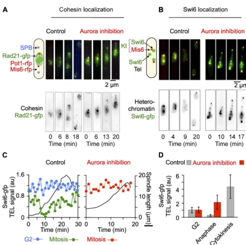
Figure 5. Aurora B controls the dissociation of cohesin Rad21 and Swi6/HP1 from telomeres at anaphase. (A) Rad21-gfp mis6-rfp pot1-rfp
ark1-as3 cells were filmed through mitosis in
the presence (Aurora inhibition, red) or ab-sence (control, black) of 10 µM Napp1. Rad21 remains associated to telomeres as judged by its colocalization with the telomeric Pot1 signal after Aurora inhibition. (B) swi6-gfp mis6-rfp
ark1-as3 cells were filmed through mitosis in the
presence (Aurora inhibition, red) or absence (control, black) of 10 µM Napp1. (C) Quan-tification of the normalized mean fluorescence intensity of Swi6-gfp at telomeres during mitotic progression as a function of spindle length (in black) in the cells shown in A. In control mitotic cells (green), the decrease in Swi6-gfp is con-comitant to the increase in spindle length during anaphase onset (black). During anaphase B, Swi6-gfp progressively reaccumulates at telo-meres. In the presence of Napp1 (Aurora inhibition, red), Swi6-gfp remains constant at telomeres during spindle elongation (in black). The intensity of Swi6-gfp at telomeres in G2 cells simultaneously filmed within the same field of cells than in A is shown as a control (blue). Multiple cells were analyzed and behave in a similar manner. (D) Statistical analysis show-ing the mean intensity of Swi6-gfp signal at telomeres as a function of the cell cycle stage (G2, anaphase, or cytokinesis) in the presence (red) or absence (gray) of Napp1. Error bars indicate SD.
(Nakazawa et al., 2011; Tada et al., 2011). To clarify how
Ccq1 deletion rescued Aurora inhibition, we performed single
cell analysis of condensin (cnd1-gfp) recruitment on
chro-mosomes during mitosis in control cells, ccq1 cells, and
phosphomimetic cnd2-3E mutant. As previously described,
condensin colocalizes to several nuclear compartments
dur-ing mitosis (Nakazawa et al., 2008), includdur-ing the nucleolus
(rDNA) and kinetochores. At anaphase onset, condensin
lo-calization switches to chromosome arms (Fig. 8 D, bottom,
Hoechst) and the spindle midzone (Fig. 8, B and D). Transient
localization of condensin with telomere clusters was also
oc-casionally observed during metaphase (Fig. 8 D). In ccq1
cells and cnd2-3E mutant, this mitotic pattern of condensin
lo-calization was unchanged (Fig. 8, C and E, top). However, we
noted that although Pot1 decreased in mitosis in control and
ccq1
cells, it remained strongly associated with telomeres in
the cnd2-3E mutant.
The effect of Aurora inhibition on condensin dynamics
was investigated (Fig. 8, B, C, and E, Aurora inhibition). In
wild-type cells, condensin recruitment to chromosome arms in
both ccq1 deletion and the condensin cnd2-3E phosphomimetic
mutant rescued to some extent the formation of anaphase
chro-matin bridges (Fig. 7 C, light gray bars). In the condensin
mu-tant, the rescue was not specific to chromosome arm segregation
defects as the appearance of merotelic attachment after Aurora
inhibition was also bypassed. Instead, deletion of Ccq1
specifi-cally reduced the appearance of anaphase chromatin bridges but
not kinetochore attachment defects (Fig. 7 C, dark gray bars).
Finally, the cnd2-3A strain showed a high percentage
of anaphase chromatin bridges but few merotelic attachments
(Fig. 7 C). In agreement with these findings, both condensin
phosphomimetic mutant and ccq1 deletion rescued cell viability
after Aurora inhibition (Fig. 7 D) and also bypassed the defect
in nucleolar segregation (not depicted).
Deletion of Ccq1 rescues cell death after Aurora inhibition by promoting the loading of condensin onto chromosome arms
In fission yeast, Aurora B–dependent phosphorylation of the
kleisin Cnd2 promotes condensin recruitment to chromosomes
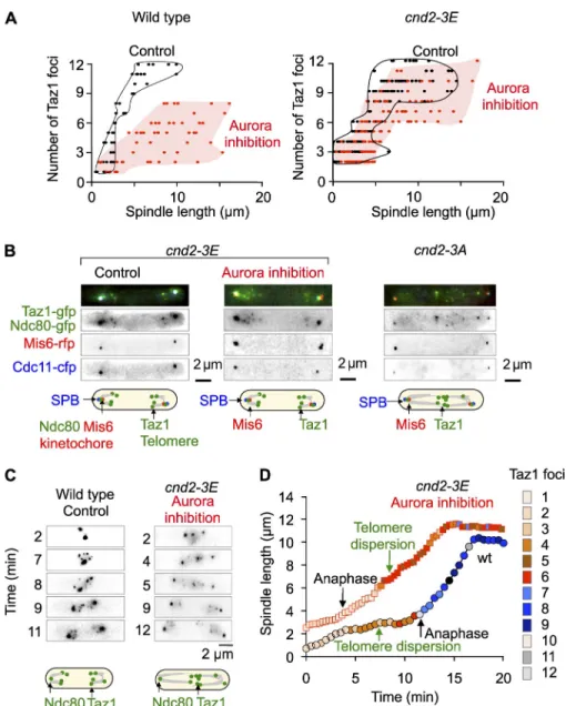
Figure 6. Aurora B–dependent phosphoryla-tion of condensin bypasses telomere nondis-junction but not dispersion. (A) Wild-type or the phosphomimetic mutant cnd2-3E cells were filmed through mitosis in the presence (Aurora inhibition, red, n = 48 control; n = 110
cnd2-3E) or absence (control, black, n = 42 control; n = 92 cnd2-3E) of 10 µM Napp1 and the
number of Taz1 dots was counted according to spindle length. The data shown are from a single representative experiment out of three repeats. (B) Example of phenotypes seen in
cnd2-3E ark1-as3 or cnd2-3A ark1-as3 cells
treated or not with 10 µM Napp1. (C) Exam-ple of a wild-type cell control and a cnd2-3E
ark1-as3 with Napp1 during mitotic
progres-sion. (D) The number of Taz1 foci (color code shown on the right) according to spindle length is analyzed from the movies shown in C. Note that in control cells (circles), telomere disper-sion occurs before anaphase as opposed to
cnd2-3E cells (squares) in the presence of
Au-rora inhibitor. Multiple cells were analyzed and display defects in telomere dispersion.
dissociation from the nuclear envelope (unpublished data). Our
findings are thus not related to previous studies describing an
important role for telomere dissociation from the nuclear
enve-lope in the control of chromosome arm separation (Fujita et al.,
2012; Titos et al., 2014).
We demonstrate that Aurora B targets spatially distinct
heterochromatin domains, centromeres, and telomeres to
con-trol chromosome segregation. Interestingly, Aurora localization
to telomeres is both Bub1 and Swi6 independent (unpublished
data). These findings are in agreement with previous work
performed by chromatin immunoprecipitation on the CPC
component Bir1 (Yamagishi et al., 2010). Other studies have
reported that the Shugoshin protein Sgo2 is required for the
proper recruitment of Aurora at centromeres (Hauf et al., 2007;
Vanoosthuyse et al., 2007). We found that the CPC was still able
to localize to both centromeres and telomeres in the absence of
Sgo2 although the signal was somewhat reduced. As expected,
sgo2
cells are not defective in telomere disjunction, show
very few merotelic attachments, and are viable, phenotypes not
observed in Aurora-depleted cells (unpublished data).
anaphase was clearly reduced after Aurora inhibition (Fig. 8 B,
bottom, arrows; Nakazawa et al., 2011; Tada et al., 2011).
Con-versely, condensin remained correctly loaded on chromosome
arms in the absence of Ccq1 or in the phosphomimetic mutant
cnd2-3E
after Aurora inhibition (Fig. 8, C and E, bottom,
ar-rows). Together these experiments suggest that Aurora plays a
specific role at telomeres to promote telomere dispersion and
disjunction and that this mechanism contributes to correct
con-densin loading and chromosome arm separation.
Discussion
Our study demonstrates that the physical association of
meres is tightly cell cycle regulated. During mitosis,
telo-mere foci first dissociate from the nuclear envelope (Fujita
et al., 2012) and then undergo separation in two discrete steps.
Telomere dispersion (up to six dots) occurs during metaphase,
before chromosome segregation, whereas sister chromatid
telomere disjunction (up to 12 dots) is achieved during mid-
anaphase. Aurora B is required for both steps but not for telomere
Figure 7. The role of Aurora B in telomere dispersion and disjunction requires the shel-terin component Ccq1. (A) Ccq1-deleted cells were filmed through mitosis in the presence (Aurora inhibition, red, n = 92) or absence (control, black, n = 79) of 10 µM Napp1 and the number of Taz1 dots was counted accord-ing to spindle length. The data shown are from a single representative experiment out of three repeats. (B) Example of phenotypes seen in
ark1-as3 ccq1 cells treated or not with 10 µM
Napp1. (C) Percentage of anaphase chromatin bridges and merotely phenotype before or after Aurora inhibition in ccq1 (n = 210), cnd2-3E (n = 250), or cnd2-3A (n = 46) cells. Light gray, anaphase bridges; dark gray, merotely. Error bars indicate SD. (D) Sensitivity of wild-type,
ccq1, rap1, and cnd2-3E mutant cells to low
doses of Napp1 (0.8 µM or 1 µM) was evalu-ated after serial dilutions of cells on plates. Note that under these conditions ccq1 or cnd2-3E cells survive as opposed to wild type.
(Moser et al., 2011; Yamazaki et al., 2012). Ccq1 also
partici-pates in the telomeric recruitment of a multi-enzyme complex
(termed SHREC) that mediates heterochromatic transcriptional
gene silencing (Sugiyama et al., 2007). The dissociation of
shelterin components from telomeres during mitosis was
pre-viously demonstrated using chromatin immunoprecipitation
(Chang et al., 2013). Our study provides the timing for this
dissociation with respect to chromosome segregation and
telomere dispersion. It also suggests that Aurora controls the
dissociation of Pot1 or Ccq1 at the metaphase-to-anaphase
tran-sition (Fig. 9).
In parallel to its role in dissociating Swi6/HP1, Pot1,
or Ccq1 from telomeres and promoting telomere dispersion,
Aurora B promotes chromosome arm separation by
phosphory-lating the condensin subunit Cnd2 (Nakazawa et al., 2011; Tada
et al., 2011). Condensins are universal organizers of chromosomes
that participate in several processes, such as condensation and
segregation of rDNA, clustering of tRNA genes, axial
com-paction of pericentric chromatin, and recoiling of stretched
In addition to its role in kinetochore–microtubule
attach-ment, Aurora B promotes the transient dissociation of Swi6/
HP1 from the telomeres at the metaphase-to-anaphase
transi-tion. This event likely contributes to telomere dispersion, as the
cohesin subunit Rad21 also remains strongly associated with
telomere clusters in a Swi6-dependent manner. In contrast,
Rad21 is not retained on chromosome arms and this observation
is also confirmed by chromatin immunoprecipitation data
indi-cating that Rad21 is specifically lost from heterochromatic sites
but not other sites on chromosome arms in swi6 cells (Bernard
et al., 2001; Nonaka et al., 2002). Hence, the absence of
telo-meric heterochromatin (Swi6/HP1 deletion) is sufficient to
by-pass the telomere dispersion defects seen after Aurora inhibition
or after deletion of Rap1.
Chromosome end protection in S. pombe is similar to that
seen in mammals and a recent study has identified several
pro-teins including Pot1 or Ccq1 that form the shelterin complex
(Miyoshi et al., 2008). Ccq1 is the S. pombe–specific
compo-nent of this complex and is required for telomerase recruitment
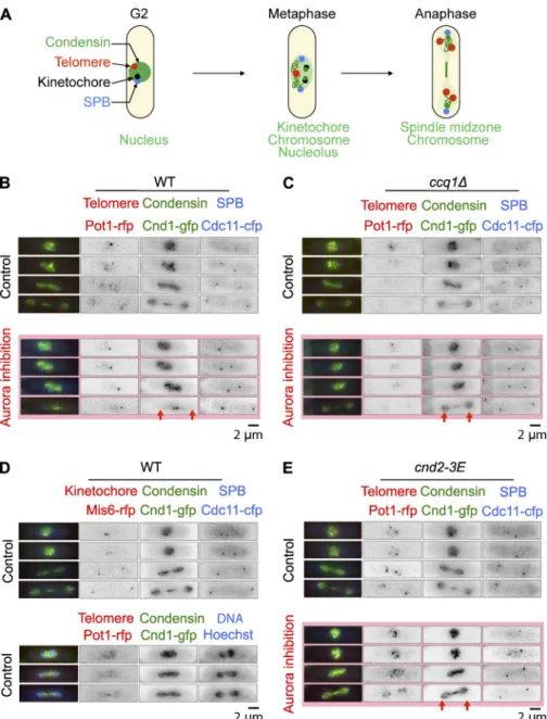
Figure 8. Deletion of Ccq1 rescues cell death after Aurora inhibition by promoting the loading of condensin on chromosome arms. (A) Schematic representation illustrating the differ-ent localization of condensin throughout mitosis. (B) Single cell analysis of ark1-as pot1-rfp
cnd1-gfp cdc11-cfp cells imaged in the presence or
absence of 10 µM Napp1 (Aurora B inhibition). (C) Single cell analysis of ccq1 ark1-as pot1-rfp
cnd1-gfp cdc11-cfp cells imaged in the presence
or absence of 10 µM Napp1 (Aurora B inhibi-tion). (D) Single cell analysis of ark1-as mis6-rfp
gfp cdc11-cfp or ark1-as pot1-rfp cnd1-gfp cdc11-cfp cells imaged in the presence of
Hoechst. (E) Single cell analysis of cnd2-3E
ark1-as pot1-rfp cnd1-gfp cdc11-cfp cells imaged in
the presence or absence of 10 µM Napp1. In B, C, and E, the red arrows highlight the presence or absence of condensin localization to chromo-some arms.
clustering in interphase fission yeast cells (unpublished data)
and Rad21 is maintained at telomeres when Aurora is inhibited.
If this is the case, the initial step in cluster dissociation in
meta-phase cells may be reminiscent of the prometa-phase dissociation
pathway that is seen in higher eukaryotes (Waizenegger et al.,
2000). Intriguingly, it has been shown that two pathways are
re-quired to dissociate cohesin from chromosomes in such cells.
The first, in prophase, requires the activity of Aurora B and
polo-like kinase (Losada et al., 2002; Sumara et al., 2002;
Giménez-Abián et al., 2004), whereas the second requires the
proteins Wapl and Pds5 (Gandhi et al., 2006; Kueng et al.,
2006; Shintomi and Hirano, 2009). Further experiments will be
required to establish whether telomere dispersion in mitosis in
fission yeast is homologous to the prophase dissolution
path-way seen elsewhere.
Our work raises the following important question: what
are the respective roles for telomere dispersion and disjunction
in chromosome segregation? When Aurora B is inactivated,
sister chromatids are entangled along the chromosome arms,
including the telomere-adjacent regions. Thus, telomere
non-disjunction might be an indirect effect of arm nonnon-disjunction.
However, because the telomespecific component Ccq1 is
re-quired for telomere nondisjunction and for cell viability after
Aurora inhibition, we propose that the dissociation of the
shel-terin complex in early mitosis initiates telomere dispersion,
pro-motes condensin loading, and participates to chromosome arm
separation. How Ccq1 deletion triggers condensin loading and
chromosome arm separation after Aurora inhibition remains to
be determined but a more thorough examination of CPC
func-tion in Ccq1-deleted cells should shed light on such quesfunc-tions.
One source of mitotic instability is the presence of
chro-mosomes with dysfunctional telomeres. This defect could give
rise to chromatin bridges at anaphase, leading to chromatin
fragmentation or chromosome loss (Gisselsson and Höglund,
2005). Our study reveals a novel mechanism that controls
chromosome arms in anaphase (Carmena et al., 2012). Our study
shows that a phosphomimetic mutant of the condensin subunit
Cnd2 (cnd2-3E) is unable to rescue the telomere dispersion
de-fects seen after Aurora inhibition. Our study therefore suggests
that telomere dispersion is promoted by a separate mechanism
involving the removal of Swi6/HP1. Rather, condensin controls
telomere disjunction and nucleolar segregation by mechanically
pulling on chromosome arms. In agreement with this model,
Swi6/HP1 and Pot1 remain associated with telomeres when
Aurora is inhibited in a cnd2-3E mutant even after telomere
disjunction. In addition, the condensin mutant cut3 arrests with
dispersed telomeres (up to six dots) but nonseparated
chromo-some arms (unpublished data).
The removal of Swi6/HP1 from heterochromatin in
mito-sis has been observed in several organisms. Whereas in higher
eukaryotes the biological significance of this event remains to
be determined, in budding yeast Aurora B–dependent
phos-phorylation of H3S10 is thought to contribute to chromosome
compaction during anaphase (Neurohr et al., 2011). This
modi-fication causes the removal of HP1/Swi6 from H3K9me3,
which facilitates the dissociation of the CPC from chromosome
arms and its enrichment at centromeres (Nozawa et al., 2010).
In fission yeast, Aurora B also controls the phosphorylation of
histone H3 on Serine 10 (Petersen et al., 2001) and the removal
of Swi6/HP1 from telomeres (Chen et al., 2008; this study).
Whether or not this pathway is responsible for telomere
separa-tion in fission yeast is currently unknown.
It is tempting to speculate that the different states of
telo-mere clustering observed during the fission yeast cell cycle
re-flects the dynamics of cohesion at telomeres. Indeed, a recent
study established that cohesin participates in subtelomeric
het-erochromatin maintenance in fission yeast, probably by acting
locally on Swi6/HP1 binding in the subtelomeric region (Dheur
et al., 2011). In agreement with this hypothesis, neither rad21-K1
cohesin mutant nor the swi6 mutant show correct telomere
Figure 9. Model summarizing the different state in telomere clustering throughout mito-sis. Schematic representation illustrating the regulation of telomere dissociation throughout mitosis. (left) Telomere foci undergo separation in two discrete steps. Telomere cluster disper-sion (up to 6 dots) occurs during metaphase, before chromosome segregation, whereas sister chromatid telomere disjunction (up to 12 dots) is achieved during mid-anaphase. Finally, telomere reclustering occurs during cytokinesis (subsequent G1/S phase). Au-rora B is required for telomere dispersion and disjunction. (right) At the level of telomeres, Aurora promotes in metaphase the delocaliza-tion of several telomere/subtelomere compo-nents, such as shelterin components (brown), Swi6/HP1 (blue), or cohesin Rad21 (green). At anaphase, Aurora favors the loading of condensin (pink). Our model suggests that the delocalization of telomere components such as the shelterin protein Ccq1 promotes telomere dispersion and Aurora-dependent loading of condensin to chromosome arms.
Cnd2-deleted cells (Tada et al., 2011). The integration at cnd2::ura4 locus was selected by 5-fluoroorotic acid resistance and confirmed by PCR. A similar technique was used to construct the cnd2-3E strain (Tada et al., 2011).
The ark1-as3 strain was created from ark1-as2 by additionally mu-tating Ser229 to Ala (gift from S. Hauf, Virginia Polytechnic Institute and State University, Blacksburg, VA). To generate the ark1-as2 allele (Ark1-Leu166Ala), the ark1 gene was PCR mutagenized from a strain into which a hygromycin-resistance cassette (hygR) had been integrated 400 bp 5 of the ark1+ open reading frame (Hauf et al., 2007). The hygR-ark1-as2 con-struct was integrated in a wild-type strain at the endogenous locus. Both the
ark1-as2 and the ark1-as3 strains contain the additional amino acid
muta-tions Gln28Arg and Gln176Arg, which were unintentionally inserted dur-ing the first PCR mutagenesis (Hauf et al., 2007). Both alleles displayed sensitivity to the ATP analogue Napp1.
Aurora inhibition
Aurora inhibition was performed using analogue-sensitive alleles of Ark1 (ark1-as3), which allows rapid and specific inactivation of Aurora kinase using the ATP analogue Napp1 (Hauf et al., 2007). As specified in the fig-ure legends, asynchronous or synchronous populations of cells were filmed in the presence of 10 µM Napp1 and followed through mitosis. To quantify the mitotic defects seen after Aurora inhibition, the temperature-sensitive double mutant cdc25-22 ark1-as3 was synchronized by incubation at 36°C to accumulate cells in G2 phase, and then released into early mitosis by incubation at the permissive temperature of 25°C (>80% of cells in phase 1) and adding 10 µM Napp1. Progress of the cells through mitosis was followed and attachment defects in anaphase were quantified by live cell imaging.
Live cell imaging
Live cell analysis was performed in an imaging chamber (CoverWell PCI-2.5; Grace Bio-Labs) filled with 1 ml of 1% agarose in minimal medium and sealed with a 22 × 22-mm glass coverslip. Time-lapse images of Z stacks (maximum five stacks of 0.3–0.4-µm steps, to avoid photobleaching) were taken at 15-s intervals or as indicated in the relevant figure legend. Exposure times were 300–500 ms with a HIGHlite light source (Roper Sci-entific) reduced to 30% to avoid photo toxicity and photobleaching. Either the image with the best focal plane or projected images were prepared for each time point. Images were visualized with a charge coupled device CoolSNAP HQ camera (Roper Scientific) fitted to a DM6000 upright mi-croscope (Leica) with a 100× (1.4 NA) objective and SEMROCK filters and were recorded using the MetaMorph software package (Molecular Devices). Intensity adjustments were made using the MetaMorph, ImageJ, and Adobe Photoshop packages (Adobe Systems).
Analysis of spindle elongation and kinetochore misattachments
The position of the SPBs, kinetochores, and telomeres was determined by visualization of the Cdc11–cfp, Ndc80–gfp/Mis6-rfp, and Taz1/Pot1 sig-nals and captured using MetaMorph. Maximum intensity projections were prepared for each time point, with the images from each channel being combined into a single RGB image. These images were cropped around the cell of interest, and optional contrast enhancement was performed in MetaMorph where necessary. The cropped images were exported to IGOR (version Pro6; WaveMetrics) as 8-bit RGB-stacked TIFF files with each frame corresponding to one image of the time-lapse series. For both chan-nels, custom peak detection was performed. The successive positions of the SPBs and kinetochores were determined. The data generated were used to calculate the rate of spindle elongation and the number of lagging chores and telomeres. A merotelic kinetochore is defined as a single kineto-chore (Ndc80-gfp) within an anaphase spindle that undergoes splitting while a corresponding single centromeric signal (Mis6-rfp) remains barely unstretched (Courtheoux et al., 2009; Gay et al., 2012). Thus, imaging these two kinetochore and centromere markers simultaneously allows us to discriminate between merotelic attachment or lagging chromosomes at-tached to a single pole.
Cell fixation
To determine the percentage of aneuploidy and cut phenotype, cells were fixed in 3.7% formaldehyde for 7 min at room temperature, washed once in PBS, and observed in the presence of DAPI/calcofluor.
Quantification of Swi6 fluorescence intensity at telomeres
To analyze the fluorescence intensity of Swi6 at telomeres in movies, we designed several regions of interests around Swi6 telomere foci and mea-sured the total light content of these spots. An equivalent number of regions
chromosome segregation, which requires telomere integrity
in-dependently of the segregation of other heterochromatin loci
such as centromeres.
Materials and methods
Cell culture
Media, growth, maintenance of strains, and genetic methods were as de-scribed previously (Moreno et al., 1991). Cells were grown at 25°C in yeast extract and centrifuged 30 s at 3,000 g before mounting onto an im-aging chamber. All strains used in the study were isogenic to wild-type 972 and are listed in Table S1. Strains from genetic crosses were selected by random spore or tetrad dissection and selected in the presence of appropriate supplements or drugs, or screened visually for the presence of the appropriate fluorescent markers. Visualization of the nucleolus was achieved by the expression of the plasmid pREP41x/fib1-mRFP (Beauregard et al., 2009). Yeast transformations were performed by electroporation in the presence of thiamine (repressive condition). Fib1-mRFP protein was induced by growing the cells in the absence of thiamine for 24 h. For Aurora inhibition rescue, ccq1 colonies were taken at an early stage of selection before chromosome circularization (as judged by the lack of sen-sitivity to methyl methanesulfonate). At this early stage (between 2 to 7 d of growth), Taz1 signal was still observed and cell size was comparable to wild type. Ccq1 colonies with circular chromosomes (as judged by sensi-tivity to methyl methanesulfonate) did not rescue Aurora inhibition. The presence of different types of colonies in ccq1 or tpz1 mutants has been previously characterized (Harland et al., 2014).
Mutant and tagged strains
Standard genetic and PCR-based gene targeting methods were used to construct S. pombe strains (Table S1). All fluorescent tagged (gfp, rfp, and cfp) and truncated genes (full-length truncations) are under the con-trol of their endogenous promoters and integrated at their native chromo-somal loci. Most of the deleted or epitope-tagged strains were thus produced by homologous recombination using modified pFA6a series of plasmids carrying hygromycin-resistance gene (hyg in strain list; Hentges et al., 2005) or previously described pFA6a series of plasmids carrying kanamycin-resistance gene (kanR in strain list; Bähler et al., 1998). This includes taz1-gfp (gift from J. Cooper, Center for Cancer Research, Na-tional Institutes of Health, Bethesda, MD), amo1-rfp (gift from P. Nurse, Cancer Research UK, London, Enlgand, UK), mis6-2rfp (gift from T. Toda, Cancer Research UK, London, England, UK), ndc80-gfp cdc11-cfp (Tournier et al., 2004), taz1-mrfp and ccq1 (gift from M. Flory, Wesleyan Uni-versity, Middletown, CT; Motwani et al., 2010), rad21-gfp (gift from J.P. Javerzat, Institut de Biochimie et Genetique Cellulaires, Bordeaux, France, and P. Bernard, Université Claude Bernard Lyon 1, Lyon, France), pot1-rfp (gift from V. Geli, Centre de Recherche en Cancérologie de Marseille, Marseille, France), and Cnd1-gfp (gift from M. Yanagida, Osaka University, Osaka, Japan).
The cnd1-gfp strain is a full-length fusion created by homologous re-combination and integrated at its native chromosomal loci (Sutani et al., 1999). The following deletions are full-length deletions created by homolo-gous recombination: rap1::ura4 (gift from J. Kanoh, Institute for protein re-search, Osaka University, Osaka, Japan) and swi6::his1 and pcs1::ura4 (gift from J.P. Javerzat).
The temperature-sensitive cdc25-22 allele (Thuriaux et al., 1980) was used for fission yeast synchronization in G2 (http://www.pombase .org/spombe/result/SPAC24H6.05).
Fibrillarin fused with the mRFP (R.Y. Tsien, University of California, San Diego, La Jolla, CA) was constructed in two steps (Beauregard et al., 2009). First, mRFP was amplified using a pair of oligonucleotides: mRFP1-forw2 con-taining a XhoI restriction site and a PmeI restriction site (bold; 5-CCGCTC-GAGGTTTAAACGCCTCCTCCGA-3), and mRFP1-reverse, containing a BamHI site (5-CGGGATCCTTAGGCGCCGGTGGAGTG-3). Subsequently, the PCR product was digested with XhoI and BamHI and cloned in pREP41X. Fibrillarin was amplified from genomic DNA with Fib1-XhoI forward (XhoI site; 5-AGACTCGAGATGGCATATACACCAGGTTCA-3) and Fib1-PmeI-no-stop reverse (PmeI site in bold characters; 5-AGAGTTTAAACCCTGATGTCTCAAG-TATTTTCCTAC-3). After which, the PCR product was digested with XhoI and PmeI and inserted into pREP41X-mRFP, previously digested with the same en-zymes (gift from L.A. Rokeach, Montreal University, Montreal, Canada).
The cnd2-3A strain (gift from Y. Watanabe, University of Tokyo, Tokyo, Japan) was created by changing S5, S41, and S52 to alanines and the genomic fragments carrying the mutations were transformed into