Affects Conjugation in Bacillus subtilis
The MIT Faculty has made this article openly available. Please share how this access benefits you. Your story matters.Citation Johnson, Christopher M., and Alan D. Grossman. “The Composition of the Cell Envelope Affects Conjugation in Bacillus Subtilis.” Ed. P. J. Christie. Journal of Bacteriology 198.8 (2016): 1241–1249.
As Published http://dx.doi.org/10.1128/jb.01044-15 Publisher American Society for Microbiology Version Author's final manuscript
Citable link http://hdl.handle.net/1721.1/107191
Terms of Use Creative Commons Attribution-Noncommercial-Share Alike Detailed Terms http://creativecommons.org/licenses/by-nc-sa/4.0/
1
The composition of the cell envelope affects conjugation in Bacillus subtilis
2 3
Christopher M. Johnson and Alan D. Grossman* 4
Department of Biology, Massachusetts Institute of Technology, Cambridge, 5
Massachusetts, USA 6
7
running head: Effects of membrane phospholipids on conjugation 8 9 10 11 *Corresponding author: 12 Department of Biology 13 Building 68-530 14
Massachusetts Institute of Technology 15 Cambridge, MA 02139 16 phone: 617-253-1515 17 E-mail: [email protected] 18 19 20
Abstract 21
Conjugation in bacteria is the contact-dependent transfer of DNA from one cell to 22
another via donor-encoded conjugation machinery. It is a major type of horizontal gene 23
transfer between bacteria. Conjugation of the integrative and conjugative element 24
ICEBs1 into Bacillus subtilis is affected by the composition of phospholipids in the cell 25
membrane of the donor and recipient. We found that reduction (or elimination) of lysyl-26
phosphatidylglycerol caused by loss of mprF caused a decrease in conjugation 27
efficiency. Conversely, alterations that caused an increase in lysyl-28
phosphatidylglycerol, including loss of ugtP or overproduction of mprF, caused an 29
increase in conjugation efficiency. In addition, we found that mutations that alter 30
production of other phospholipids, e.g., loss of clsA and yfnI, also affected conjugation, 31
apparently without substantively altering levels of lysyl-phosphatidylglycerol, 32
indicating that there are multiple pathways by which changes to the cell envelope affect 33
conjugation. We found that the contribution of mprF to conjugation was affected by the 34
chemical environment. The presence of certain salts altered conjugation, and wild type 35
cells were generally more responsive to conditions that enhanced conjugation whereas 36
mprF mutant cells were more sensitive to some conditions that inhibited conjugation. 37
Our results indicate that mprF and lysyl-phosphatidylglycerol allow cells to maintain 38
relatively consistent conjugation efficiencies in a variety of ionic conditions. 39
40 41
Importance 42
Horizontal gene transfer is a driving force in microbial evolution, enabling cells that 43
receive DNA to acquire new genes and phenotypes. Conjugation, the contact-44
dependent transfer of DNA from a donor to a recipient by a donor-encoded secretion 45
machine, is a prevalent type of horizontal gene transfer. Although critically important, 46
it is not well understood how the recipient influences the success of conjugation. We 47
found that the composition of phospholipids in the membrane of donors and recipients 48
influences the success of transfer of the integrative and conjugative element ICEBs1 in 49
Bacillus subtilis. Specifically, the presence of lysyl-phosphatidylglycerol enables 50
relatively constant conjugation efficiencies in a range of diverse chemical environments. 51
Introduction 53
Conjugation is one of several processes bacteria use to acquire new genes. During 54
conjugation a donor bacterium transfers DNA directly to a recipient bacterium in a 55
contact dependent manner. The conjugation machinery is typically encoded by a mobile 56
genetic element, which itself is frequently transferred during conjugation. Conjugation 57
can also deliver genes that are not directly involved in the conjugation process, but that 58
are located on the mobile genetic element or on other DNA elements that are 59
transferred. These genes are known to confer a wide variety of phenotypes to cells and 60
their transfer can allow recipients to rapidly acquire new characteristics. For example, 61
conjugative elements are widely involved in the spread of antibiotic resistances 62
(reviewed in 1, 2, 3). 63
ICEBs1 is an integrative and conjugative element (ICE) found in Bacillus subtilis (4, 64
5). ICEs are widespread and found in many bacterial species (6). Unlike conjugative 65
plasmids, ICEs integrate into the host chromosome where they are maintained during 66
chromosomal replication, segregation, and cell division, much like a transposon or 67
phage lysogen (reviewed in 3, 7). Under certain circumstances, ICEs can excise from the 68
chromosome forming a plasmid intermediate that can then be transferred to recipient 69
cells by the element-encoded conjugation machinery. 70
ICEBs1 is found integrated in the trn-leu2 gene in the B. subtilis chromosome and 71
becomes activated in response to extracellular signaling, starvation, or DNA damage 72
(4). The regulatory genes of ICEBs1 involved in cell-cell signaling (rapI, phrI) have been 73
defined (4, 8). Overexpression of RapI leads to excision of ICEBs1 in >90% of cells in a 74
growing population, allowing a high frequency of experimentally-induced conjugation 75
(4, 8, 9). ICEBs1 encodes a type IV secretion system that transfers DNA from the donor 76
to a recipient. Type IV secretion systems are found in other ICEs and conjugative 77
plasmids, both in Gram positive and Gram negative bacteria (10). 78
During conjugation, DNA is transferred from the cytoplasm of the donor to that of 79
the recipient, crossing the envelope of each to generate a transconjugant. The 80
composition of the cell envelope of both the donor and recipient influences the success 81
conjugation. For example, in Gram negative bacteria, the outer membrane protein 82
OmpR and the lipopolysaccharide are important for formation of mating pairs (11-16). 83
In Enterococcus faecalis, lipoteichoic acids may be important for mating pair formation 84
(17-19). Recently, we found that in B. subtilis, the phospholipid head groups of the 85
membrane bilayer make important contributions to conjugation (20). 86
The cell envelope of B. subtilis and other Gram positive bacteria contains a single 87
lipid bilayer. The lipids of this membrane vary in the composition of their fatty acid 88
tails and their head groups (reviewed in 21). The most abundant phospholipids in the 89
membrane of B. subtilis are the negatively-charged phosphatidylglycerol, zwitterionic 90
phosphatidylethanolamine and neutral glycolipids, negatively charged cardiolipin, and 91
positively charged lysyl-phosphatidylglycerol (reviewed in 22). The membrane of B. 92
subtilis carries a net negative charge. 93
Although the conjugation machinery is encoded by the conjugative element, host 94
genes, in both the donor and recipient, are also important for successful transfer of 95
conjugative DNA. Previously, we used Tn-seq to identify mutations that increased or 96
decreased the ability of cells to act as recipients in conjugation (20). We found that 97
deletion of genes involved in the synthesis of various phospholipids have distinct 98
effects on the ability of B. subtilis to act as a recipient in conjugation. Several of the 99
mutations (ugtP, yfnI, mprF) that affect conjugation affect consumption of the 100
phospholipid phosphatidylglycerol (Fig. 1). 101
Here, we analyze these mutants to evaluate the effects of phospholipids on 102
conjugation. We used double mutant analysis to determine epistasis between several of 103
the phospholipid mutations. Our results indicate that lysyl-phosphatidylglycerol 104
stimulates conjugation and that other phospholipids are also important for conjugation, 105
independently of lysyl-phosphatidylglycerol. We also found that the phenotype caused 106
by loss of mprF (needed for production of lysyl-phosphatidylglycerol) was enhanced by 107
some environmental conditions and suppressed by others. Our results indicate the 108
ability of cells to function in conjugation is buffered against some chemical variations in 109
the environment by lysyl-phosphatidylglycerol. 110
111 112
Materials and Methods 113
Media and growth conditions 114
Escherichia coli cells were grown at 37°C in LB medium. B. subtilis cells were grown at 115
37°C in LB medium or S750 defined minimal medium with 0.1% glutamate and 40
116
µg/ml required amino acids (23). Arabinose (1% w/v) was used as a carbon source. 117
Isopropyl-ß-D-thiogalactopyranoside (IPTG, 1 µg/ml) was used to induce expression of 118
Pspank(hy)-mprF. Xylose (1% w/v) was used to induce expression of Pxyl-rapI. 119
Ampicillin was used at 100 µg/ml for E. coli. Antibiotics were used at the following 120
concentrations for B. subtilis: spectinomycin (100 µg/ml) kanamycin (5 µg/ml), 121
chloramphenicol (5 µg/ml) and a combination of erythromycin (0.5 µg/ml) and 122
lincomycin (12.5 µg/ml) to select for macrolide-lincosamide-streptogramin (MLS) 123
resistance. 124
Strains and alleles 125
B. subtilis strains are listed in Table 1. Strains with trp phe alleles are derived from 126
JH642 (trpC2, pheA1). rapI was overexpressed from the xylose-inducible fusion Pxyl-rapI 127
(integrated in the chromosome at amyE) to activate ICEBs1 in donor cells,. The ∆(rapI-128
phrI)160::cat allele was constructed with the same genomic boundaries as the ∆(rapI-129
phrI)342::kan allele (4). Upstream and downstream genomic DNA fragments and the 130
chloramphenicol resistance gene cat were amplified by PCR and joined together by 131
isothermal (Gibson) assembly (24). This product was used to transform naturally 132
competent B. subtilis cells, a chloramphenicol resistant isolate was selected and the 133
allelic exchange verified by PCR. 134
The unmarked ∆mprF459 allele was constructed by replacing mprF with cat flanked 135
by lox sites to generate strain CMJ459, then recombining out the lox-cat allele using Cre 136
recombinase expressed from pDR244, as previously described (20, 25). The genomic 137
boundaries of this allele are the same as for the ∆mprF125::mls and ∆mprF162::spc alleles. 138
For MprF overexpression studies, mprF was cloned into a plasmid that carried 139
Pspank(hy), lacI and mls situated between genomic sequence from lacA. mprF was 140
placed under control of the promoter Pspank(hy). This plasmid was transformed into 141
naturally competent B. subtilis cells and Pspank(hy)-mprF, lacI and mls introduced by 142
double cross over at lacA. Expression of mprF was induced by the addition of 1 mM 143
IPTG. 144
Thin layer chromatography 145
Lipids were extracted from cells using a modified Bligh-Dyer method (26). We grew 146
cells in minimal medium to an OD600 of ~1, sampled 1 ml of culture and pelleted the 147
cells, removed the supernatant, resuspended in 1 ml water, pelleted the cells and 148
resuspended in 100 µl 1 M perchloric acid and incubated for 30 minutes on ice. Lipids 149
were extracted by adding 1 ml 2:6:2 methanol:chloroform:water and 0.625 µg of a 150
phosphatidylserine standard (Sigma Aldrich) to each sample and incubating at 4°C 151
overnight on a rocking platform. Lipids were recovered by adding 300 µl H2O and 300
152
µl chloroform and incubating the samples for 30 min at -20°C, then centrifuging for 5 153
min at 720x g. The organic (bottom) phase was recovered, dried under nitrogen and the 154
extracted lipids resuspended in 12 µl 2:1 chloroform methanol. 155
The total volume of each sample was spotted on silica 60 plates (Angela) along with 156
lysyl-phosphatidylglycerol (0.63 µg - 2.5 µg) (Avanti Polar Lipids) and 157
phosphatidylserine (0.25 µg - 1 µg) standards and developed in a thin layer 158
chromatography chamber with 60:35:5 chloroform:methanol:water. The plates were 159
dried, stained with ninhydrin (1.5 mg/ml ninhydrin in water-saturated butanol with 160
3% v/v acetic acid) and charred. The plates were scanned on a flat bed scanner and 161
analyzed with ImageJ (27). Standard curves were generated for lysyl-162
phosphatidylglycerol (0.16 µg - 5 µg) and phosphatidylserine (0.25 µg - 1 µg) to ensure 163
that the amount of each phospholipid in the samples was within the linear range of the 164
assay. 165
Mating assays 166
Mating assays were performed on filters as previously described (9, 20). Briefly, 167
donor and recipient cells were grown separately in minimal medium with 1% arabinose 168
as a carbon source. Donors were induced with 1% (w/v) xylose for 2 hours to induce 169
expression of Pxyl-rapI, thereby activating ICEBs1 gene expression. An equal number of 170
donors and recipients was mixed, collected on a mating filter and placed on a mating 171
support consisting of 1.5% agar with a buffered salt solution (see below) for 90 minutes. 172
Mating filters were typically placed on SMS agar (28) unless otherwise specified. TSS 173
agar (28) was used as an alternate buffer in the mating support in some experiments. 174
TSS was further amended in some experiments, as noted in the text. Cells were then 175
rinsed off the filter, diluted and spread on LB plates with selective antibiotics to 176
determine the numbers of transconjugants, donors and-or recipients. 177
178 179
Results 180
Effects of genes involved in phospholipid biosynthesis on conjugation 181
Previously, we found that mprF and several other genes involved in the synthesis of 182
phospholipids affect the efficiency of conjugation (20). MprF catalyzes addition of a 183
lysyl group from lys-tRNALys to phosphatidylglycerol to form
phosphatidylglycerol (Fig. 1) (26, 29, 30). Loss of mprF in donors and-or recipients 185
causes a decrease in conjugation of ICEBs1 (Fig. 2) (20), indicating that elimination of 186
lysyl-phosphatidylglycerol is detrimental for conjugation. 187
In contrast to the loss of mprF, we found that overexpression of mprF in recipients 188
caused an increase in the acquisition of ICEBs1 via conjugation. We fused mprF to the 189
LacI-repressible, IPTG-inducible promoter Pspank(hy) at an ectopic location (amyE) on 190
the chromosome in a mutant missing the normal copy of mprF. We found that 191
expression of Pspank(hy)-mprF in recipients caused an increase in mating efficiency 192
(Fig. 2A). When mprF was similarly overexpressed in the donor (strain CMJ248) and 193
mated to a wild type recipient (CAL89), the mating efficiency was 7-8-fold greater than 194
that of the wild type donor (KM250) mated to the same recipient Together with 195
previous findings on the effects of loss of mprF on conjugation (20), our results indicate 196
that both loss and overproduction of mprF affect conjugation efficiencies. Since the only 197
known role of mprF in B. subtilis is in the production of lysyl-phosphatidylglycerol from 198
phosphatidylglycerol and charged lysyl-tRNA, our results indicate that the amount of 199
lysyl-phosphatidylglycerol, or other compounds derived from phosphatidylglycerol, 200
affect conjugation. If these effects are due to lysyl-phosphatidylglycerol, then this 201
phospholipid appears to stimulate conjugation. 202
Other genes affecting phospholipid biosynthesis that were previously identified as 203
having an effect on conjugation include lysA, ugtP, and yfnI (Fig. 1) (20). Similar to loss 204
of mprF, loss of lysA in either the donor or the recipient inhibits conjugation (Fig. 2E and 205
20). lysA encodes diaminopimelate decarboxylase, which catalyzes synthesis of L-lysine 206
from meso-diaminopimelate (31). lysA is essential for synthesis of lysine, used in the 207
production of lysyl-phosphatidylglycerol, so lysA mutations might affect conjugation by 208
altering lysyl-phosphatidylglycerol production. In contrast, loss of ugtP or yfnI enhances 209
the ability of cells to act as recipients in conjugation (Fig. 2 and reference 20). ugtP is 210
involved in synthesis of glycolipid, a component of the membrane that also acts as a 211
precursor in the synthesis of lipoteichoic acids (Fig. 1) (32). yfnI is one of four genes with 212
overlapping roles in lipoteichoic acid synthesis in B. subtilis (33). Like MprF, The 213
products of ugtP and yfnI consume phosphatidylglycerol. 214
Based on the functions of the genes described above, and their consumption of 215
phosphatidylglycerol, we decided to test the effects of clsA on conjugation. The clsA 216
gene product, cardiolipin synthetase, consumes phosphatidylglycerol during the 217
synthesis of cardiolipin, another phospholipid of the membrane bilayer. clsA was not 218
identified previously in our mutant hunt because the apparent effect on conjugation 219
was below the cutoff used to identify candidate genes (20). 220
We found that loss of clsA in recipients caused an increase in the acquisition of 221
ICEBs1 via conjugation (Fig. 2C). This increase was similar to that caused by a ugtP null 222
mutation. Together, these results indicate that phosphatidylglycerol or derivatives of 223
phosphatidylglycerol can stimulate and-or inhibit the efficiency of conjugation. 224
Double mutant analysis of phospholipid biosynthesis mutants 225
Deletion of individual genes encoding phospholipid synthetases that consume 226
phosphatidylglycerol (Fig. 1) resulted in opposite affects on conjugation efficiency, 227
depending on which gene was deleted. For example, deletion of mprF caused a 228
decrease and deletion of ugtP, yfnI, or clsA caused an increase in conjugation. There are 229
two simple models to explain these effects. 1) Lysyl-phosphatidylglycerol might 230
enhance conjugation. In this model, loss of mprF (needed to make lysyl-231
phosphatidylglycerol) causes a decrease in conjugation because of loss of lysyl-232
phosphatidylglycerol. In addition, loss of ugtP, yfnI, and clsA might cause an increase 233
in phosphatidylglycerol (substrate for MprF) and a subsequent increase in lysyl-234
phosphatidylglycerol, thereby causing an increase in conjugation. 2) Alternatively (or 235
in addition), cardiolipin, glycolipids and lipoteichoic acids might act individually or 236
together to inhibit conjugation. For example, phospholipids and teichoic acids can 237
interfere with hydrolase activity (34-39) and might inhibit the cell wall hydrolase CwlT 238
that is encoded by and needed for transfer of ICEBs1. Loss of clsA (cardiolipin) and yfnI 239
(lipoteichoic acids), and perhaps ugtP (glycolipids), relieves this inhibition causing an 240
increase in conjugation. In this model, loss of mprF leads to an increase in 241
phosphatidylglycerol and a possible increase in the inhibitory molecule(s) and thus a 242
decrease in conjugation. To test these models we generated strains in which multiple 243
phospholipid synthetases were inactivated and tested them as recipients in conjugation 244
experiments (Fig. 2). Results described below indicate that lysyl-phosphatidylglycerol 245
enhances conjugation. 246
We found that the decrease in conjugation frequency caused by loss of mprF was 247
epistatic to the increase in conjugation frequency due to loss of ugtP (Fig. 2B). We 248
measured the conjugation efficiencies using standard mating assays between a wild 249
type donor (KM250) and recipients carrying the mutation(s) of interest. An mprF ugtP 250
double mutant recipient had essentially the same phenotype as the mprF single mutant 251
recipient (Fig. 2B). This result indicates that mprF is needed for the increase in 252
conjugation caused by loss of ugtP, and that the ugtP phenotype is likely due to an 253
increase in the level of lysyl-phosphatidylglycerol. 254
We also made double mutants between mprF and clsA (CMJ332), yfnI (CMJ132), and 255
lysA (CMJ336). We used the double mutants as recipients in conjugation experiments 256
and directly compared the results to that of the single mutants. The conjugation 257
efficiency of ICEBs1 into the mprF clsA double mutant was about half (0.45) of that into 258
wild type recipients. This appeared to be partly (mostly) additive between the 259
conjugation efficiencies of the single mutants: an approximately 6-fold reduction (0.17) 260
and an approximately 4-fold increase (4.4) for mprF and clsA, respectively (expect: 0.17 x 261
4.4 = 0.73) (Fig. 2C). The conjugation efficiency of the mprF yfnI double mutant was also 262
about half (0.45) that of wild type recipients, indicative of additive effects of the 6-fold 263
decrease (0.17) and 2-fold increase (2.1) in the mprF and yfnI single mutants (expect: 0.17 264
x 2.1 = 0.36) (Fig. 2D). The conjugation efficiency of the mprF lysA double mutant was 265
decreased 70-fold (0.014) and appeared to be fully additive between the effects of each 266
of the single mutants 6-fold (0.17) and ~11 fold (0.087) decrease of the mprF and lysA 267
mutants (expect: 0.17 x 0.087 = 0.014) (Fig. 2E). Although it is difficult to determine if 268
the phenotypes of the double mutants are precisely additive, the data clearly indicate 269
that mprF is epistatic to ugtP and not to clsA, yfnI, and lysA. 270
Together, the results of the double mutant analyses indicate that: 1) loss of ugtP and 271
mprF likely affect conjugation by affecting levels of lysyl-phosphatidylglycerol; 2) loss 272
of clsA, yfnI, and lysA probably do not affect levels of lysyl-phosphatidylglycerol and 273
their effects on conjugation are likely by altering other components of the cell 274
membrane. 275
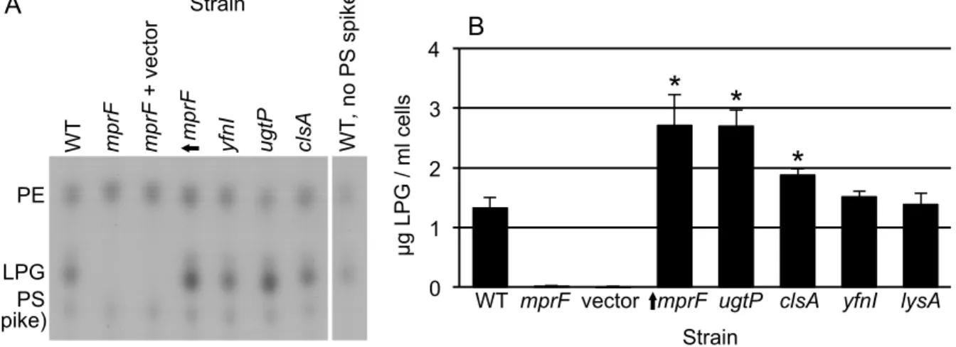
Analysis of lysyl-phosphatidylglycerol levels in mutant cells 276
To test the inferences from the genetic analyses describe above, we measured the 277
amount of lysyl-phosphatidylglycerol in each of the different phospholipid synthesis 278
mutants (Fig. 3). We grew cells in defined minimal medium, extracted phospholipids 279
and used thin-layer chromatography to measure lysyl-phosphatidylglycerol (Fig. 3, 280
LPG). As expected (26), there was no detectable lysyl-phosphatidylglycerol in the mprF 281
mutant (Fig. 3). In contrast, overproduction of MprF caused an increase in the amount 282
of lysyl-phosphatidylglycerol above that found in otherwise wild type cells (Fig. 3). We 283
found that the ugtP null mutation, and to a lesser extent the clsA null mutation, also 284
caused an increase in the amount of lysyl-phosphatidylglycerol (Fig. 3). The simplest 285
interpretation of these results is that the increase in lysyl-phosphatidylglycerol in the 286
ugtP mutant, and perhaps the clsA mutant, is likely causing the increase in conjugation 287
efficiency. However, the double mutant analysis described above demonstrated that 288
mprF was epistatic to ugtP and apparently additive with clsA. The smaller effect of clsA 289
compared to ugtP on the level of lysyl-phosphatidylglycerol and the double mutant 290
phenotypes indicate that the conjugation phenotype of ugtP, but not that of clsA, was 291
due to an increase in lysyl-phosphatidylglycerol. 292
In contrast to the mutations that affected levels of lysyl-phosphatidylglycerol, yfnI or 293
lysA null mutations caused no detectable change in levels of lysyl-phosphatidylglycerol 294
(Fig. 3). The results of the conjugation and thin-layer chromatography experiments are 295
summarized in Table 2. Together with the analysis of double mutants (Fig. 2, Table 2), 296
these results indicate that the conjugation phenotypes caused by mutations in mprF and 297
ugtP are likely due to changes in levels of lysyl-phosphatidylglycerol and that the 298
conjugation phenotypes caused by mutations in clsA, yfnI, and lysA are most likely not 299
due to changes in levels of lysyl-phosphatidylglycerol. 300
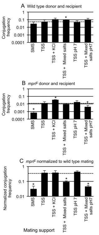
The mating defect of mprF mutants is affected by the chemical environment 301
During the course of our investigations, we noticed that the composition of the agar 302
surface on which the filter paper for mating was placed (the mating support) influenced 303
the magnitude of the conjugation phenotype caused by loss of mprF. Specifically, loss of 304
mprF from both donors (CMJ476) and recipients (CMJ162) caused a pronounced 305
conjugation defect (0.031, ~30 fold) compared to a cross between wild type donors 306
(CMJ348) and recipients (CMJ161), similar to previously reported results (20). This 307
drop in conjugation was observed when matings were performed on Spizizen’s 308
minimal salts (SMS) agar. SMS agar contains 15 mM ammonium sulfate, 80 mM dibasic 309
potassium phosphate, 44 mM monobasic potassium phosphate, 3.4 mM trisodium 310
citrate, 0.8 mM magnesium sulfate, and 1.5% agar at pH 7.0 (28). 311
In contrast to the ~30-fold decrease in conjugation between mprF mutants on SMS 312
agar, there was a much smaller effect when matings were done on agar containing 313
Spizizen’s salts and Tris (TSS) (Fig. 4). TSS agar is buffered with Tris instead of 314
potassium phosphate and contains 37 mM ammonium chloride, 2 mM dibasic 315
potassium phosphate, 50 mM Tris base, 1 mM magnesium sulfate, 0.004% iron(III) 316
chloride, 0.004% trisodium citrate, 1.5% agar, pH 7.5 (28)). Under these conditions, the 317
conjugation frequency of mprF mutant cells was reduced by approximately 3-fold (0.37) 318
compared to that of wild type donors and recipients. We ruled out the possibility that 319
production of lysyl-phosphatidylglycerol was restored in the mprF mutant on the TSS 320
agar support; there was no detectable lysyl-phosphatidylglycerol under these 321
conditions in the mutant. These findings indicate that there is something about TSS that 322
suppresses, or something about SMS that exacerbates the conjugation defect of the mprF 323
mutant. 324
We investigated what aspect of the different mating supports accounted for the 325
magnitude of the mprF mutant phenotype. Since mating in the mprF mutants was much 326
lower in SMS than TSS, we postulated that the lower pH and-or some of the additional 327
ions in SMS were inhibiting conjugation of mprF mutants. There are several differences 328
between TSS and SMS. Notably, SMS contains a higher total concentration of different 329
salts compared to TSS and a lower pH (7 versus 7.5). SMS has higher concentrations of 330
potassium (204 mM vs 4 mM), phosphate (124 mM vs 2 mM), sulfate (16 mM vs 1 mM) 331
and citrate (3 mM vs 0.1 mM). 332
We measured mating efficiencies on TSS agar as the base support with additions to 333
make it more closely resemble SMS. Addition of potassium chloride (125 mM) or mixed 334
salts (106 mM sodium phosphate, 14 mM sodium sulfate, and 3 mM trisodium citrate) 335
increased the conjugation frequency in matings between wild type cells (Fig. 4A). 336
Adjustment of the pH to 7.0 (without other changes) had little or no detectable effect 337
and had no additional effect in the presence of mixed salts (Fig. 4A). 338
As with wild type cells, the addition of potassium chloride also increased the 339
conjugation frequency in matings between mprF mutant cells (Fig. 4B) and adjustment 340
of the pH to 7.0 had little or no effect (Fig. 4B). However, unlike the effect on wild type 341
cells, addition of mixed salts did not cause an increase in the conjugation efficiency in 342
matings between mprF mutants at either pH (Fig. 4B). 343
Direct comparison of the conjugation frequencies for wild type cells (Fig. 4A) and 344
mprF cells (Fig. 4B) showed that mprF caused a more severe phenotype when matings 345
were performed on TSS with mixed salts (at pH 7.5 and pH 7) compared to TSS (Fig. 346
4C). Based on these results, we conclude that the salts found in SMS contributed to the 347
defect in mating caused by loss of mprF, particularly at pH 7. 348
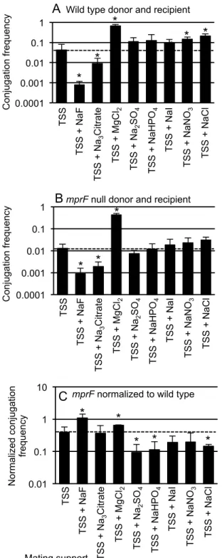
Ion-specific effects on conjugation and the effects of mprF 349
Based on the above results, we wondered if other salts might affect wild type and 350
mprF mutants differently. To test this, we used TSS agar as our base medium and 351
supplemented it with 125 mM of different salts, including: sodium fluoride, trisodium 352
citrate, magnesium chloride, sodium sulfate, dibasic sodium phosphate titrated with 353
monobasic sodium phosphate to give a pH of 7.5 and a phosphate concentration of 125 354
mM, sodium iodide, sodium nitrate or sodium chloride. We measured mating 355
efficiencies of wild type cells (Fig. 5A), mprF mutants (Fig. 5B) and then directly 356
compared mprF to wild type (Fig. 5C). 357
We found that addition of sodium fluoride or sodium citrate to TSS caused a 358
decrease in mating efficiency of wild type cells (Fig. 5A). There was also a decrease in 359
the mating efficiency of mprF mutants (Fig. 5B). With sodium fluoride, this decrease 360
was somewhat less for the mprF mutants than for wild type cells (Fig. 5C). With sodium 361
citrate, the decrease was about the same for mprF and wild type (Fig. 5C). 362
In contrast, we found that addition of magnesium chloride to TSS caused an increase 363
of 16-fold in mating efficiency of wild type cells (Fig. 5A). Likewise, there was a similar 364
or somewhat greater increase in the mating efficiency of mprF mutants (33-fold, Fig. 5B, 365
C). These results indicate that the use of TSS supplemented with magnesium chloride 366
as a solid support for filter matings allows for highly efficient conjugation. 367
Addition of several other salts, including sodium sulfate, sodium phosphate, 368
sodium iodide, sodium nitrate, and sodium chloride, to TSS either stimulated or had 369
relatively little effect on the mating efficiency of wild type cells (Fig. 5A). The 370
stimulatory effects were less than that of magnesium chloride. The same salts had little 371
or no effect or caused a small increase in the mating efficiency of mprF mutants (Fig. 372
5B). The stimulatory effects on wild type cells were larger than the effects on mprF 373
mutants and this is most easily seen in the ratio of mating efficiencies of mprF to wild 374
type (Fig. 5C). These ratios are <0.37, the ratio of efficiencies when mating is done on 375
TSS without any modifications. 376
Together, our results (Figs. 4, 5) indicate that mating efficiencies are affected by the 377
external ionic environment and that several salts that enhance conjugation of wild type 378
cells do not have the same stimulatory effect on mprF mutants. Since mprF mutants do 379
not produce lysyl-phosphatidylglycerol, we infer that the different effects of salts is due 380
to the presence or absence of this phospholipid. The presence of mprF and hence lysyl-381
phosphatidylglycerol enables cells to have efficient conjugation in a variety of different 382 ionic conditions. 383 384 385 Discussion 386
Our findings indicate that lysyl-phosphatidylglycerol plays a role in stimulating 387
conjugation. Preventing or reducing lysyl-phosphatidylglycerol synthesis in either the 388
donor or the recipient reduces conjugation. Overproduction of lysyl-389
phosphatidylglycerol in either partner enhances conjugation. Accumulation of lysyl-390
phosphatidylglycerol was eliminated in mprF null mutants and increased in ugtP 391
mutants or upon overexpression of mprF. Our results also indicate that alterations in 392
phospholipid content that do not detectably affect lysyl-phosphatidylglycerol also alter 393
conjugation efficiencies. 394
mprF and ugtP
395
We found that mprF is epistatic to ugtP for the conjugation phenotype. That is, the 396
mprF ugtP double mutant had the same phenotype as the mprF single mutant. This is 397
consistent with the interpretation that the conjugation phenotypes of ugtP and mprF 398
mutants are due to alterations in lysyl-phosphatidylglycerol and that loss of ugtP causes 399
an increase in phosphatidylglycerol, which then leads to an increase in lysyl-400
phosphatidylglycerol (Fig. 1). mprF is epistatic because it is needed to make lysyl-401
phosphatidylglycerol. 402
Loss of ugtP caused an increase in the amount of lysyl-phosphatidylglycerol, 403
indicating that UgtP normally plays a role limitingthe amount of lysyl-404
phosphotidylglycerol in the cell. ugtP is also known to affect cell division (40), 405
primarily by directly interacting with and inhibiting the cell division protein FtsZ (40, 406
41). The effects of ugtP on cell division and conjugation are most likely not related. We 407
infer this mainly because the effects on cell division appear to be direct and the effects 408
on conjugation are likely through mprF. 409
ugtP mutants also appear to have many alterations in gene expression in rich 410
medium (26). The effects of mprF mutations on gene expression are not known, but 411
based on analyses of an mprF pssA ywnE (clsA) triple mutant, there are fewer effects 412
than in a ugtP single mutant (26). It is possible that the effects of mprF and ugtP on 413
conjugation are due to alterations in gene expression. However, the simplest model is 414
that these genes affect conjugation due to alterations in lysyl-phosphotidylglycerol and 415
that the composition of the cell envelope directly affects activity of the conjugation 416
machinery (see below). 417
mprF and lysyl-phosphatidylglycerol enable efficient conjugation in various ionic
418
conditions 419
Our results demonstrate that the effects of lysyl-phosphatidylglycerol on 420
conjugation are dependent on the environmental conditions. That is, the ratio of mating 421
efficiencies comparing mprF mutants and wild type cells was affected by the ionic 422
conditions used for mating. For example, the mprF mutants had a much more severe 423
mating defect (~30-fold) on SMS agar compared to the modest defect (~3-fold) on TSS 424
agar. Together with analysis of the differences between SMS and TSS, our results 425
indicate that mprF and lysyl-phosphotidylglycerol normally facilitate efficient mating 426
under a variety of external ionic conditions. We suggest that the presence of lysyl-427
phosphatidylglycerol buffers conjugation against the some of the otherwise inhibitory 428
effects of different salts and enhances conjugation in the presence of others, allowing 429
the conjugation machinery to function reasonably well in a range of different ionic 430
conditions. 431
mprF homologs, and by extension lysyl-phosphatidylglycerol, affect cell surface 432
properties of other organisms. For example, mprF in Staphylococcus aureus acts as a 433
virulence factor and potentiates resistance to several cationic antimicrobials, including 434
those produced by potential human hosts (reviewed in 22). mprF homologs impact the 435
ability of Enterococcus faecium and Listeria monocytogenes to adapt to different 436
environmental conditions (42, 43). We suggest that in Gram positive bacteria, mprF and 437
lysyl-phosphatidylglycerol ensures that the cell envelope is buffered from some of the 438
variations in the chemistry of the environment, and enables the cell to perform 439
physiological functions in a regular manner under different environmental conditions. 440
A model for how membrane phospholipids affect conjugation efficiencies 441
We suspect that alterations in the phospholipid content of the recipient (and donor) 442
might affect the function of the conjugation machinery. This could be through changes 443
to the physical properties of the membrane (e.g., fluidity) that might affect assembly of 444
the machinery. This could also be through inhibition of a component of the machinery. 445
Transfer of DNA through the ICEBs1-encoded conjugation machinery depends on CwlT 446
(44), a secreted cell wall hydrolase encoded by ICEBs1 (44, 45). Components of the cell 447
envelope, including lipoteichoic acids (34, 35), wall teichoic acids (36, 37) and the 448
phospholipids cardiolipin (35), phosphatidylglycerol (38) and lysyl-449
phosphatidylglycerol (35, 39), can modulate the function of at least some cell wall 450
hydrolases. Cell wall teichoic acids inhibit hydrolase activity, at least in part, by 451
preventing hydrolase binding to the peptidoglycan of the cell wall (36, 37). 452
Phospholipids can stimulate or inhibit the function of particular hydrolases; for 453
example, phosphatidylglycerol can either enhance or inhibit the N-acetylmuramoyl-L-454
alanine amidase of E. coli, depending on concentration, but has no effect on the major 455
autolysin of Clostridium acetobutylicum under the conditions tested (38, 39). Altering the 456
phospholipid content of the donor and-or recipient may affect a postulated interaction 457
between the cell wall hydrolase CwlT and the cell envelope, either enhancing or 458
inhibiting the ability of conjugation machinery to deliver DNA. This interaction could 459
be binding of the conjugation machinery to the recipient cell envelope and-or digestion 460
of the donor and recipient cell wall. If this model is correct, it strongly predicts that the 461
cell wall hydrolase acts on both donor and recipient cells. 462
Cell wall hydrolases are encoded by many conjugative elements (10, 46-49). Where 463
tested, they are critical for efficient conjugation. Based on this conservation, it seems 464
likely that the composition of the cell wall affects the efficiencies of many different 465
conjugative elements. Perhaps the cell wall hydrolases have evolved in ways that help 466
determine the host range of the cognate element. 467
Acknowledgments 469
We thank Tony DeBono (Anthony Sinsky lab) for help with thin layer 470
chromatography, John Helmann for strains, Suzanne Walker, Bernhardt Trout, Barbara 471
Imperialli, and Thomas Bernhardt for helpful conversations and Laurel Wright and 472
Monika Avello for comments on the manuscript. Research reported here is based upon 473
work supported, in part, by the National Institute of General Medical Sciences of the 474
National Institutes of Health under award number R01GM050895 to ADG. Any 475
opinions, findings, and conclusions or recommendations expressed in this report are 476
those of the authors and do not necessarily reflect the views of the National Institutes of 477 Health. 478 479 References 480
1. Garriss Gv, Waldor MK, Burrus V. 2009. Mobile antibiotic resistance encoding 481
elements promote their own diversity. PLoS Genetics 5:e1000775. 482
2. Toleman MA, Walsh TR. 2011. Combinatorial events of insertion sequences and 483
ICE in Gram-negative bacteria. FEMS Microbiology Reviews 35:912-935. 484
3. Johnson CM, Grossman AD. 2015. Integrative and Conjugative Elements (ICEs): 485
What They Do and How They Work. Annu Rev Genet 49:577-601. 486
4. Auchtung JM, Lee CA, Monson RE, Lehman AP, Grossman AD. 2005. Regulation 487
of a Bacillus subtilis mobile genetic element by intercellular signaling and the global 488
DNA damage response. Proc Natl Acad Sci U S A 102:12554-12559. 489
5. Burrus V, Pavlovic G, Decaris B, Guedon G. 2002. The ICESt1 element of 490
Streptococcus thermophilus belongs to a large family of integrative and conjugative 491
elements that exchange modules and change their specificity of integration. 492
Plasmid 48:77-97. 493
6. Guglielmini J, Quintais L, Garcillan-Barcia MP, de la Cruz F, Rocha EP. 2011. The 494
repertoire of ICE in prokaryotes underscores the unity, diversity, and ubiquity of 495
conjugation. PLoS Genet 7:e1002222. 496
7. Wozniak RA, Waldor MK. 2010. Integrative and conjugative elements: mosaic 497
mobile genetic elements enabling dynamic lateral gene flow. Nat Rev Microbiol 498
8:552-563. 499
8. Auchtung JM, Lee CA, Garrison KL, Grossman AD. 2007. Identification and 500
characterization of the immunity repressor (ImmR) that controls the mobile genetic 501
element ICEBs1 of Bacillus subtilis. Mol Microbiol 64:1515-1528. 502
9. Lee CA, Auchtung JM, Monson RE, Grossman AD. 2007. Identification and 503
characterization of int (integrase), xis (excisionase) and chromosomal attachment 504
sites of the integrative and conjugative element ICEBs1 of Bacillus subtilis. Mol 505
Microbiol 66:1356-1369. 506
10. Alvarez-Martinez CE, Christie PJ. 2009. Biological diversity of prokaryotic type IV 507
secretion systems. Microbiol Mol Biol Rev 73:775-808. 508
11. Perez-Mendoza D, de la Cruz F. 2009. Escherichia coli genes affecting recipient 509
ability in plasmid conjugation: are there any? BMC Genomics 10:71. 510
12. Watanabe T, Arai T, Hattori T. 1970. Effects of cell wall polysaccharide on the 511
mating ability of Salmonella typhimurium. Nature 225:70-71. 512
13. Skurray RA, Hancock RE, Reeves P. 1974. Con- mutants: class of mutants in 513
Escherichia coli K-12 lacking a major cell wall protein and defective in conjugation 514
and adsorption of a bacteriophage. J Bacteriol 119:726-735. 515
14. Havekes L, Tommassen J, Hoekstra W, Lugtenberg B. 1977. Isolation and 516
characterization of Escherichia coli K-12 F- mutants defective in conjugation with an 517
I-type donor. J Bacteriol 129:1-8. 518
15. Sanderson KE, Janzer J, Head J. 1981. Influence of lipopolysaccharide and protein 519
in the cell envelope on recipient capacity in conjugation of Salmonella typhimurium. J 520
Bacteriol 148:283-293. 521
16. Ishiwa A, Komano T. 2004. PilV adhesins of plasmid R64 thin pili specifically bind 522
to the lipopolysaccharides of recipient cells. J Mol Biol 343:615-625. 523
17. Trotter KM, Dunny GM. 1990. Mutants of Enterococcus faecalis deficient as 524
recipients in mating with donors carrying pheromone-inducible plasmids. Plasmid 525
24:57-67. 526
18. Bensing BA, Dunny GM. 1993. Cloning and molecular analysis of genes affecting 527
expression of binding substance, the recipient-encoded receptor(s) mediating 528
mating aggregate formation in Enterococcus faecalis. J Bacteriol 175:7421-7429. 529
19. Ehrenfeld EE, Kessler RE, Clewell DB. 1986. Identification of pheromone-induced 530
surface proteins in Streptococcus faecalis and evidence of a role for lipoteichoic acid 531
in formation of mating aggregates. J Bacteriol 168:6-12. 532
20. Johnson CM, Grossman AD. 2014. Identification of host genes that affect 533
acquisition of an integrative and conjugative element in Bacillus subtilis. Mol 534
Microbiol 93:1284-1301. 535
21. Parsons JB, Rock CO. 2013. Bacterial lipids: metabolism and membrane 536
homeostasis. Prog Lipid Res 52:249-276. 537
22. Ernst CM, Peschel A. 2011. Broad-spectrum antimicrobial peptide resistance by 538
MprF-mediated aminoacylation and flipping of phospholipids. Mol Microbiol 539
80:290-299. 540
23. Jaacks KJ, Healy J, Losick R, Grossman AD. 1989. Identification and 541
characterization of genes controlled by the sporulation-regulatory gene spo0H in 542
Bacillus subtilis. J Bacteriol 171:4121-4129. 543
24. Gibson DG, Young L, Chuang RY, Venter JC, Hutchison CA, 3rd, Smith HO. 544
2009. Enzymatic assembly of DNA molecules up to several hundred kilobases. Nat 545
Methods 6:343-345. 546
25. Meisner J, Montero Llopis P, Sham LT, Garner E, Bernhardt TG, Rudner DZ. 547
2013. FtsEX is required for CwlO peptidoglycan hydrolase activity during cell wall 548
elongation in Bacillus subtilis. Mol Microbiol 89:1069-1083. 549
26. Salzberg LI, Helmann JD. 2008. Phenotypic and transcriptomic characterization of 550
Bacillus subtilis mutants with grossly altered membrane composition. J Bacteriol 551
190:7797-7807. 552
27. Schneider CA, Rasband WS, Eliceiri KW. 2012. NIH Image to ImageJ: 25 years of 553
image analysis. Nat Methods 9:671-675. 554
28. Harwood CR, Cutting SM. 1990. Molecular Biological Methods for Bacillus. John 555
Wiley & Sons, Chichester. 556
29. Peschel A, Jack RW, Otto M, Collins LV, Staubitz P, Nicholson G, Kalbacher H, 557
Nieuwenhuizen WF, Jung G, Tarkowski A, van Kessel KP, van Strijp JA. 2001. 558
Staphylococcus aureus resistance to human defensins and evasion of neutrophil 559
killing via the novel virulence factor MprF is based on modification of membrane 560
lipids with l-lysine. J Exp Med 193:1067-1076. 561
30. Oku Y, Kurokawa K, Ichihashi N, Sekimizu K. 2004. Characterization of the 562
Staphylococcus aureus mprF gene, involved in lysinylation of phosphatidylglycerol. 563
Microbiology 150:45-51. 564
31. Yamamoto J, Shimizu M, Yamane K. 1991. Molecular cloning and analysis of 565
nucleotide sequence of the Bacillus subtilis lysA gene region using B. subtilis phage 566
vectors and a multi-copy plasmid, pUB110. Agric Biol Chem 55:1615-1626. 567
32. Jorasch P, Wolter FP, Zahringer U, Heinz E. 1998. A UDP glucosyltransferase from 568
Bacillus subtilis successively transfers up to four glucose residues to 1,2-569
diacylglycerol: expression of ypfP in Escherichia coli and structural analysis of its 570
reaction products. Mol Microbiol 29:419-430. 571
33. Wormann ME, Corrigan RM, Simpson PJ, Matthews SJ, Grundling A. 2011. 572
Enzymatic activities and functional interdependencies of Bacillus subtilis 573
lipoteichoic acid synthesis enzymes. Mol Microbiol 79:566-583. 574
34. Holtje JV, Tomasz A. 1975. Lipoteichoic acid: a specific inhibitor of autolysin 575
activity in Pneumococcus. Proc Natl Acad Sci U S A 72:1690-1694. 576
35. Cleveland RF, Wicken AJ, Daneo-Moore L, Shockman GD. 1976. Inhibition of 577
wall autolysis in Streptococcus faecalis by lipoteichoic acid and lipids. J Bacteriol 578
126:192-197. 579
36. Yamamoto H, Miyake Y, Hisaoka M, Kurosawa S, Sekiguchi J. 2008. The major 580
and minor wall teichoic acids prevent the sidewall localization of vegetative DL-581
endopeptidase LytF in Bacillus subtilis. Mol Microbiol 70:297-310. 582
37. Schlag M, Biswas R, Krismer B, Kohler T, Zoll S, Yu W, Schwarz H, Peschel A, 583
Gotz F. 2010. Role of staphylococcal wall teichoic acid in targeting the major 584
autolysin Atl. Mol Microbiol 75:864-873. 585
38. Vanderwinkel E, De Vlieghere M. 1985. Modulation of Escherichia coli N-586
acetylmuramoyl-L-alanine amidase activity by phosphatidylglycerol. Biochim 587
Biophys Acta 838:54-59. 588
39. Croux C, Canard B, Goma G, Soucaille P. 1992. Autolysis of Clostridium 589
acetobutylicum ATCC 824. J Gen Microbiol 138:861-869. 590
40. Weart RB, Lee AH, Chien A-C, Haeusser DP, Hill NS, Levin PA. 2007. A 591
metabolic sensor governing cell size in bacteria. Cell 130:335-347. 592
41. Chien AC, Zareh SK, Wang YM, Levin PA. 2012. Changes in the oligomerization 593
potential of the division inhibitor UgtP co-ordinate Bacillus subtilis cell size with 594
nutrient availability. Mol Microbiol 86:594-610. 595
42. Smith AM, Harrison JS, Sprague KM, Roy H. 2013. A conserved hydrolase 596
responsible for the cleavage of aminoacylphosphatidylglycerol in the membrane of 597
Enterococcus faecium. J Biol Chem 288:22768-22776. 598
43. Dare K, Shepherd J, Roy H, Seveau S, Ibba M. 2014. LysPGS formation in Listeria 599
monocytogenes has broad roles in maintaining membrane integrity beyond 600
antimicrobial peptide resistance. Virulence 5:534-546. 601
44. DeWitt T, Grossman AD. 2014. The bifunctional cell wall hydrolase CwlT is 602
needed for conjugation of the integrative and conjugative element ICEBs1 in 603
Bacillus subtilis and B. anthracis. J Bacteriol 196:1588-1596. 604
45. Fukushima T, Kitajima T, Yamaguchi H, Ouyang Q, Furuhata K, Yamamoto H, 605
Shida T, Sekiguchi J. 2008. Identification and characterization of novel cell wall 606
hydrolase CwlT: a two-domain autolysin exhibiting n-acetylmuramidase and DL-607
endopeptidase activities. J Biol Chem 283:11117-11125. 608
46. Koraimann G. 2003. Lytic transglycosylases in macromolecular transport systems 609
of Gram-negative bacteria. Cell Mol Life Sci 60:2371-2388. 610
47. Scheurwater EM, Burrows LL. 2011. Maintaining network security: how 611
macromolecular structures cross the peptidoglycan layer. FEMS Microbiol Lett 612
318:1-9. 613
48. Zahrl D, Wagner M, Bischof K, Bayer M, Zavecz B, Beranek A, Ruckenstuhl C, 614
Zarfel GE, Koraimann G. 2005. Peptidoglycan degradation by specialized lytic 615
transglycosylases associated with type III and type IV secretion systems. 616
Microbiology 151:3455-3467. 617
49. Abajy MY, Kopec J, Schiwon K, Burzynski M, Doring M, Bohn C, Grohmann E. 618
2007. A type IV-secretion-like system is required for conjugative DNA transport of 619
broad-host-range plasmid pIP501 in gram-positive bacteria. J Bacteriol 189:2487-620
2496. 621
50. Menard KL, Grossman AD. 2013. Selective pressures to maintain attachment site 622
specificity of integrative and conjugative elements. PLoS Genet 9:e1003623. 623
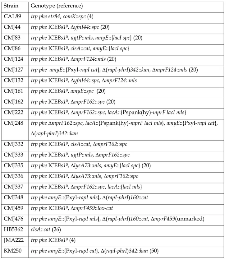
Table 1. B. subtilis strains used. 625
626
Strain Genotype (reference) CAL89 trp phe str84, comK::spc (4)
CMJ44 trp phe ICEBs10, ∆yfnI44::spc (20)
CMJ83 trp phe ICEBs10, ugtP::mls, amyE::{lacI spc} (20)
CMJ86 trp phe ICEBs10, clsA::cat, amyE::{lacI spc}
CMJ124 trp phe ICEBs10, ∆mprF124::mls (20)
CMJ127 trp phe amyE::{Pxyl-rapI cat}, ∆(rapI-phrI)342::kan, ∆mprF124::mls (20) CMJ132 trp phe ICEBs10, ∆yfnI44::spc, ∆mprF124::mls
CMJ161 trp phe ICEBs10, amyE::spc (20)
CMJ162 trp phe ICEBs10, ∆mprF162::spc (20)
CMJ222 trp phe ICEBs10, ∆mprF162::spc, lacA::{Pspank(hy)-mprF lacI mls}
CMJ248 trp phe ∆mprF162::spc, lacA::{Pspank(hy)-mprF lacI mls}, amyE::{Pxyl-rapI cat}, ∆(rapI-phrI)342::kan
CMJ332 trp phe ICEBs10, clsA::cat, ∆mprF162::spc
CMJ333 trp phe ICEBs10, ugtP::mls, ∆mprF162::spc
CMJ335 trp phe ICEBs10, ∆lysA73::mls, amyE::{lacI spc} (20)
CMJ336 trp phe ICEBs10, ∆lysA73::mls, ∆mprF162::spc
CMJ337 trp phe ICEBs10, ∆mprF162::spc, lacA::{lacI mls}
CMJ348 trp phe amyE::{Pxyl-rapI mls}, ∆(rapI-phrI)160::cat CMJ459 trp phe ICEBs10, ∆mprF459::lox-cat
CMJ476 trp phe amyE::{Pxyl-rapI mls}, ∆(rapI-phrI)160::cat, ∆mprF459(unmarked) HB5362 clsA::cat (26)
JMA222 trp phe ICEBs10 (4)
KM250 trp phe amyE::{Pxyl-rapI cat}, ∆(rapI-phrI)342::kan (50) 627
628 629
Table 2. Summary of mutations affecting conjugation and phospholipid synthesis. 630
631
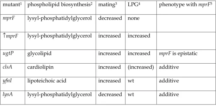
mutant1 phospholipid biosynthesis2 mating3 LPG4 phenotype with mprF5
mprF lysyl-phosphatidylglycerol decreased none ↑mprF lysyl-phosphatidylglycerol increased increased
ugtP glycolipid increased increased mprF is epistatic
clsA cardiolipin increased (increased) additive
yfnI lipoteichoic acid increased wt additive
lysA lysyl-phosphatidylglycerol decreased wt additive 632
1All are null mutations except ↑mprF, which indicates overexpression of mprF.
633
2The phospholipid whose synthesis depends on the indicated gene (Fig. 1) is indicated.
634
3The effect of the mutation on conjugation is indicated.
635
4The relative amount of lysyl-phosphatidylglycerol (LPG) produced in the indicated
636
mutant compared to wild type (wt) cells. "None" indicates that there was no 637
detectable LPG. Parentheses around increased (increased) indicates a possible effect, 638
but on the edge of statistical significance (Fig. 3). ND = not determined 639
5The phenotype of the double mutant (with mprF) with respect to conjugation. mprF is
640
epistatic indicates that the phenotype of the double mutant is the same as the mprF 641
single mutant (Fig. 2) 642
Figure Legends 644
645
Figure 1. Pathways of phospholipid biosynthesis that affect conjugation of 646
ICEBs1. Some of the pathways involved in phospholipid biosynthesis are shown. 647
Genes relevant to this work are indicated above the arrows. 648
649
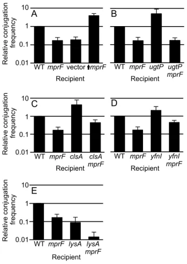
Figure 2. Effects of mutations in recipients on acquisition of ICEBs1. The relative 650
conjugation frequency (y-axis) is shown for each of the indicated recipients (x-axis). The 651
same donor strain (KM250) was used for all experiments and ICEBs1 was induced in the 652
donor by overproduction of the activator RapI (Materials and Methods). The relative 653
conjugation frequency (y-axis) is the number of transconjugants per donor crossed to 654
the indicated recipient strain, normalized to that of the wild type recipient (CMJ161) in 655
each experiment. The wild type conjugation efficiency was approximately 4% 656
transconjugants per donor in these experiments. Conjugation frequencies measured 657
with recipients that are null for mprF, ugtP, yfnI and lysA are similar to those previously 658
reported (20) and were included in these experiments to allow direct comparison with 659
the appropriate double mutants. The graph shows means and standard deviation from 660
≥3 experiments. The conjugation efficiency for each single mutant is statistically 661
different from that of wild type (p <0.05). Data for wild type (CMJ161) and an mprF null 662
mutant recipient (CMJ162) are included in all panels for comparison. 663
A. vector (CMJ337) contains Pspank(hy) with no insert; ↑mprF (CMJ222) is an mprF
664
null mutant with Pspank(hy) driving expression of mprF. 665
B. ugtP (CMJ83); ugtP mprF double mutant (CMJ333 p<0.05 vs. ugtP). 666
C. clsA (CMJ86); clsA mprF double mutant (CMJ332, p<0.05 vs. clsA and mprF). 667
D. yfnI (CMJ44); yfnI mprF double mutant (CMJ132, p<0.05 vs. yfnI and mprF). 668
E. lysA (CMJ335); lysA mprF double mutant (CMJ336, p<0.05 vs. lysA and mprF). 669
These strains were grown with 40 µg/ml lysine. 670
Figure 3. Effects of mutations on the level of lysyl-phosphatidylglycerol. The 672
amount of lysyl-phosphatidylglycerol (LPG) recovered from a 1 ml culture of cells at an 673
OD600 of 1 was determined from the indicated strains: WT, wild type (CMJ161); mprF 674
(CMJ162); mprF + vector (CMJ337); ↑mprF (CMJ222) is mprF null with Pspank(hy) 675
driving expression of mprF; yfnI (CMJ44); ugtP (CMJ83); clsA (CMJ86); lysA (CMJ335) 676
grown with 40 µg/ml lysine (in panel B only). 677
A. LPG was extracted from cell membranes and examined using thin-layer 678
chromatography (Materials and Methods). LPG and phosphatidylethanolamine (PE) 679
standards were used to identify the LPG and PE bands. Phosphatidylserine (PS) was 680
added to samples as an internal standard. The locations of the LPG, PE and PS bands 681
are indicated. The last part of the panel shows the wild type sample with no added PS. 682
B. The LPG content of each strain was quantified from ≥3 experiments. Asterisks 683
indicate a significant difference in the amount of LPG recovered compared to that from 684
the wild type strain (p <0.05, t-test). 685
686 687
Figure 4. The chemical composition of the mating support affects conjugation. 688
Standard filter matings were performed on supports with different chemical 689
compositions. Donor and recipient cells were mixed in equal numbers, then collected on 690
a filter that was placed on a mating support with the indicated composition. KCl was 691
added to 125 mM. Mixed salts contained 106 mM sodium phosphate, 14 mM sodium 692
sulfate, and 3 mM trisodium citrate. The dashed horizontal line in each panel marks the 693
value for mating on TSS. The mean and standard deviation from ≥3 experiments for 694
each condition are shown. Asterisks indicate that the difference in conjugation 695
frequency on the given support compared to conjugation frequency on TSS is 696
statistically significant (p < 0.05, t-test). 697
A, B. The conjugation frequency is shown as transconjugants per donor for A) a wild 698
type donor (CMJ348) and recipient (CMJ161); B) an mprF null mutant donor (CMJ476) 699
and recipient (CMJ162). 700
C. Conjugation frequencies obtained in A and B are directly compared. The ratio of 701
the conjugation frequencies of the mprF mutant (B) to that of the wild type strain (A) 702
under each of the indicated conditions is shown. 703
Figure 5. Some salts enhance conjugation of wild type, but not mprF cells. Filter 704
matings were performed as described in Materials and Methods. Equal numbers of 705
donor and recipient cells were mixed, collected on a filter and placed on a mating 706
support with the indicated composition. Chemicals supplements were added at 125 707
mM. The samples tested with TSS + NaHPO4 also contains dibasic sodium phosphate
708
titrated with monobasic sodium phosphate to give a pH of 7.5. The dashed horizontal 709
line in each panel indicates the conjugation frequency on TSS. The mean and standard 710
deviation from ≥3 experiments is shown for each condition. Asterisks indicate that the 711
difference in conjugation frequency on the given support compared to conjugation 712
frequency on TSS is statistically significant (p < 0.05, t-test). 713
A, B. The conjugation frequency (transconjugants per donor) is shown for A) wild 714
type donor (CMJ348) and wild type recipient (CMJ161); B) an mprF null mutant donor 715
(CMJ476) and an mprF null mutant recipient (CMJ162). 716
C. The conjugation frequencies obtained in A and B are directly compared and 717
plotted as the ratio of the mprF mutants (B) to that of wild type strains (A) under each of 718
the indicated conditions. 719
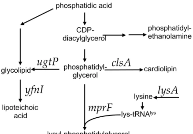
Figure 1. Pathways of phospholipid biosynthesis that affect conjugation of ICEBs1. Some of the pathways involved in phospholipid biosynthesis are shown. Genes relevant to this work are indicated above the arrows.
diacylglycerol glycolipid ethanolamine phosphatidyl- glycerol lipoteichoic acid cardiolipin lysyl-phosphatidylglycerol ugtP clsA mprF yfnI lys-tRNAlys lysA lysine
R el at ive co nj fre qu en 0.1 0.01 WT R el at ive co nj ug at io n fre qu en cy 10 1 0.1 0.01 R el at ive co nj ug at io n fre qu en cy 10 1 0.1 0.01 Recipient Recipient Recipient Recipient Recipient C D E
mprF vector mprF WT mprF ugtP ugtP mprF WT mprF clsA clsA mprF WT mprF yfnI yfnI mprF WT mprF lysA lysA mprF
Figure 2. Effects of mutations in recipients on acquisition of ICEBs1. The relative conjugation frequency (y-axis) is shown for each of the indicated recipients (x-axis). The same donor strain (KM250) was used for all experiments and ICEBs1 was induced in the donor by overproduction of the activator RapI (Materials and Methods). The relative conjugation frequency (y-axis) is the number of transconjugants per donor crossed to the indicated recipient strain, normalized to that of the wild type recipient (CMJ161) in each experiment. The wild type conjugation efficiency was approximately 4% transconjugants per donor in these experiments. Conjugation frequencies measured with recipients that are null for mprF, ugtP, yfnI and lysA are similar to those previously reported [Johnson, 2014, tn-seq] and were included in these experiments to allow direct comparison with the appropriate double mutants. The graph shows means and standard deviation from ≥3 experiments. The conjugation efficiency for each single mutant is statistically different from that of wild type (p <0.05). Data for wild type (CMJ161) and an mprF null mutant recipient (CMJ162) are included in all panels for comparison.
A. vector (CMJ337) contains Pspank(hy) with no insert; ↑mprF (CMJ222) is an mprF null mutant with Pspank(hy) driving expression of mprF.
B. ugtP (CMJ83); ugtP mprF double mutant (CMJ333 p<0.05 vs. ugtP).
C. clsA (CMJ86); clsA mprF double mutant (CMJ332, p<0.05 vs. clsA and mprF). D. yfnI (CMJ44); yfnI mprF double mutant (CMJ132, p<0.05 vs. yfnI and mprF).
E. lysA (CMJ335); lysA mprF double mutant (CMJ336, p<0.05 vs. lysA and mprF). These strains were grown with 40 µg/ml lysine.
0 1 2 3 4 µ g LPG / ml ce lls
*
*
*
WT mprF vector mprF ugtP clsA yfnI lysA ykuC Strain WT mp rF mp rF + ve ct mp rF yf nI ugtP clsA PE LPG PS (spike) WT , n o PS
Figure 3. Effects of mutations on the level of phosphatidylglycerol. The amount of lysyl-phosphatidylglycerol (LPG) recovered from a 1 ml culture of cells at an OD600 of 1 was determined from the indicated strains: WT, wild type (CMJ161); mprF (CMJ162); mprF + vector (CMJ337); ↑mprF (CMJ222) is mprF null with Pspank(hy) driving expression of mprF; yfnI (CMJ44); ugtP (CMJ83); clsA (CMJ86); lysA (CMJ335) grown with 40 µg/ml lysine (in panel B only).
A. LPG was extracted from cell membranes and examined using thin-layer chromatography
(Materials and Methods). LPG and phosphatidylethanolamine (PE) standards were used to identify the LPG and PE bands. Phosphatidylserine (PS) was added to samples as an internal standard. The locations of the LPG, PE and PS bands are indicated. The last part of the panel shows the wild type sample with no added PS.
B. The LPG content of each strain was quantified from ≥3 experiments. Asterisks indicate a
significant difference in the amount of LPG recovered compared to that from the wild type strain (p <0.05, t-test).
Figure 4. The chemical composition of the mating support affects conjugation. Standard filter matings were performed on supports with different chemical compositions. Donor and recipient cells were mixed in equal numbers, then collected on a filter that was placed on a mating support with the indicated
composition. KCl was added to 125 mM. Mixed salts contained 106 mM sodium phosphate, 14 mM sodium sulfate, and 3 mM trisodium citrate. The dashed horizontal line in each panel marks the value for mating on TSS. The mean and standard deviation from ≥3 experiments for each condition are shown. Asterisks indicate that the difference in conjugation frequency on the given support compared to conjugation frequency on TSS is statistically significant (p < 0.05, t-test).
A, B. The conjugation frequency is shown as transconjugants per donor for A) a wild type donor (CMJ348) and recipient (CMJ161); B) an mprF null mutant donor (CMJ476) and recipient (CMJ162).
C. Conjugation frequencies obtained in A and B are directly compared. The ratio of the conjugation
frequencies of the mprF mutant (B) to that of the wild type strain (A) under each of the indicated conditions is shown. 0.0001 0.001 0.01 0.1 Conjugation fre qu en cy SMS TSS T SS + KC l T SS + Mi xe d sa lts T SS pH 7 T SS + Mi xe d sa lts pH 7 0.0001 0.001 0.01 0.1 1
B mprF donor and recipient
Conjugation fre qu en cy SMS TSS T SS + KC l T SS + Mi xe d sa lts T SS pH 7 T SS + Mi xe d sa lts pH 7 0.01 0.1 1
C mprF normalized to wild type mating
N orma lize d co nj ug at io n fre qu en cy SMS TSS T SS + KC l T SS + Mi xe d sa lts T SS pH 7 T SS + Mi xe d sa lts pH 7 Mating support * * * * * *
0.01 0.1 1 10 0.0001 0.001 0.01 0.1 1
Figure 5. Some salts enhance conjugation of wild type, but not mprF cells. Filter matings were performed as described in Materials and Methods. Equal numbers of donor and recipient cells were mixed, collected on a filter and placed on a mating support with the indicated composition. Chemicals supplements were added at 125 mM. The samples tested with TSS + NaHPO4 also contains dibasic sodium phosphate titrated with monobasic sodium phosphate to give a pH of 7.5. The dashed horizontal line in each panel indicates the conjugation frequency on TSS. The mean and standard deviation from ≥3 experiments is shown for each condition. Asterisks indicate that the difference in conjugation frequency on the given support compared to conjugation frequency on TSS is statistically significant (p < 0.05, t-test).
A, B. The conjugation frequency (transconjugants per donor) is shown for A) wild type donor (CMJ348) and wild type recipient (CMJ161); B) an mprF null mutant donor (CMJ476) and an mprF null mutant recipient (CMJ162).
C. The conjugation frequencies obtained in A and B are directly compared and plotted as the ratio of the
mprF mutants (B) to that of wild type strains (A) under each of the indicated conditions.
Mating support * * * * T SS T SS + NaF T SS + N a3 C itra te T SS + Mg C l2 T SS + N a2 SO 4 T SS + N aH PO 4 T SS + NaI T SS + N aN O3 T SS + NaCl C N orma lize d co nj ug at io n fre qu en cy
mprF normalized to wild type B mprF null donor and recipient
C on ju ga tio n fre qu en cy * * * T SS T SS + NaF T SS + N a3 C itra te T SS + Mg C l2 T SS + N a2 SO 4 T SS + N aH PO 4 T SS + NaI T SS + N aN O3 T SS + NaCl 0.0001 0.001 0.01 0.1 C on ju ga tio n fre qu * * * * T SS T SS + NaF T SS + N a3 C itra te T SS + Mg C l2 T SS + N a2 SO 4 T SS + N aH PO 4 T SS + NaI T SS + N aN O3 T SS + NaCl * * *