HAL Id: hal-02406408
https://hal.archives-ouvertes.fr/hal-02406408
Submitted on 17 Nov 2020
HAL is a multi-disciplinary open access
archive for the deposit and dissemination of sci-entific research documents, whether they are pub-lished or not. The documents may come from teaching and research institutions in France or
L’archive ouverte pluridisciplinaire HAL, est destinée au dépôt et à la diffusion de documents scientifiques de niveau recherche, publiés ou non, émanant des établissements d’enseignement et de recherche français ou étrangers, des laboratoires
Role of the Metal Ion in Bio-Inspired Hydrogenase
Models: Investigation of a Homodinuclear FeFe Complex
vs Its Heterodinuclear NiFe Analogue
Lianke Wang, Marcello Gennari, Alexandre Barrozo, Jennifer Fize, Christian
Philouze, Serhiy Demeshko, Franc Meyer, Maylis Orio, Vincent Artero, Carole
Duboc
To cite this version:
Lianke Wang, Marcello Gennari, Alexandre Barrozo, Jennifer Fize, Christian Philouze, et al.. Role of the Metal Ion in Bio-Inspired Hydrogenase Models: Investigation of a Homodinuclear FeFe Complex vs Its Heterodinuclear NiFe Analogue. ACS Catalysis, American Chemical Society, 2020, 10 (1), pp.177-186. �10.1021/acscatal.9b03212�. �hal-02406408�
Role of the metal ion in bio-inspired hydrogenase models: investigation of a homodinuclear FeFe complex vs its heterodinuclear NiFe analogue
Lianke Wang,a Marcello Gennari,a,* Alexandre Barrozo,b Jennifer Fize,c Christian Philouze,a Serhiy Demeshko,d Franc Meyer,d Maylis Orio,b Vincent Artero,bCarole Duboca,*
a
Univ. Grenoble Alpes, UMR CNRS 5250, Département de Chimie Moléculaire, 38000 Grenoble, France.
b
Aix Marseille Univ, CNRS, Centrale Marseille, iSm2, Marseille (France)
c
Univ. Grenoble Alpes, CNRS, CEA, IRIG, Laboratoire de Chimie et Biologie des Métaux, 38000 Grenoble, France
d
Universität Göttingen, Institut für Anorganische Chemie, Tammannstrasse 4, D-37077 Göttingen, Germany
Abstract
In Nature, dihydrogen is catalytically produced by the [FeFe] and [NiFe] hydrogenases. Despite common structural features in their dinuclear active site, i.e. a thiolate-rich coordination sphere and CO/CN- ligation, the synergetic way, in which the two metal
sites act during catalysis, is specific for each enzyme. With the aim of understanding the role of the nature of the metal (Fe vs Ni), we report on a homodinuclear FeFe complex, a parent of a previously reported NiFe complex, in order to compare their electrocatalytic activity for H2 production. The diiron complex [(CO)LN2S2FeIIFeII(CO)Cp]+ (with LN2S2 =
2,2′-(2,2′-bipyridine-6,6′-diyl)bis(1,1-diphenylethanethiolate and Cp =
cyclopentadienyl), has been synthesized and fully characterized. In the solid state, it contains two CO ligands, one bound to the {FeCp} moiety in a semi-bridging manner and one terminally bound to the {FeLN2S2} moiety. This dinuclear iron complex is thus not
isostructural to [LN2S2NiIIFeII(CO)Cp]+, which contains a single CO ligand terminally
bound to the Fe site. However, in low-concentrated MeCN solutions, the CO ligand
coordinated to the {FeLN2S2} moiety is removed, and the CO ligand bound to the {FeCp}
moiety becomes fully bridging between the two Fe sites. Under such conditions, the diiron complex displays similar catalytic performances to the parent NiFe complex (a
comparable overpotential, = 730 mV and 690 mV and TON = 15 and 16, respectively).
Cyclic voltammetry data give direct experimental evidence for an E[ECEC] mechanism, which was also previously proposed for the NiFe complex. However, the structure of the one-electron reducedspecies the entry point of the catalytic cycle, slightly differs for the two systems: in [LN2S2NiI(CO)FeIICp], this is valence localized species on the site Ni and
the CO ligand bridges the two metallic sites, while in [(CO)LN2S2FeFeCp], this is a type II
Introduction
Nature designed two distinct dinuclear catalysts to reversibly reduce protons into dihydrogen. Two metal ions act in synergy in such {MFeS2} cores (M = Ni or Fe, Scheme 1), present in the active site of the [NiFe] and [FeFe] hydrogenases (H2ases),
respectively.1-6 Although both enzymes involve a similar ECEC mechanism7 to produce
H2 (E = electron transfer, C = chemical reaction, here corresponding to a proton transfer),
different types of intermediates and strategies are involved.1, 8 For the [NiFe] H2ase, only
the Ni ion participates in the redox chemistry by cycling between oxidation states ranging from +I to +III. The Fe site remains in the +II oxidation states but contributes in the stabilization of hydride ligands, bridging both metal centers in two key intermediates in the catalytic cycle.9-12 Regarding [FeFe] H2ase, the redox chemistry also
occurs at a single metal site (the distal iron center, Fed). It only cycles between two
oxidation states (+I and +II), and is involved in the stabilization of a terminal hydride ligand. The second iron center (proximal Fe site, Fep) acts as a scaffold through the
positioning of the doubly-bridging azadithiolate ligand that plays a key role as proton relay. Besides it also connects Fed with a [4Fe4S] cluster that acts as an electron
reservoir and cycles between +1 and +2 charge states during the catalytic cycle.13-15
In this context, chemists applied two main approaches to mimic the active sites of these two enzymes.2, 16-17 Bio-inspired NiFe catalysts for H2 production have been mainly
designed with a {NiFeS2} core consisting of a Ni site in a thiolate-rich environment and a Fe site with CO, CN-, Cp- or carbene ligands. The main objective was to localize the redox
chemistry at the Ni site.18 In the case of the FeFe mimics, even if the majority also
displays a {Fe2S2} core, a special interest has been focused on the bridging azadithiolate ligand that bears an amine proton relay. When possible, the incorporation of an electron transfer mediator in the second coordination sphere, such as a [4Fe4S] cluster19 or a
ferrocenyl group,20 has also been considered.21 Due to these specific requirements, a
direct comparison between the hydrogen evolution reaction (HER) activity of NiFe and
FeFe H2ase mimics with the same coordination environment has not been reported so
far. Two series of NiFe and FeFe complexes with similar environments have been
described but the complexes are not isostructural: [(dppe)Ni(
-pdt)Fe(CO)3]/[(dppe)(CO)Fe(-pdt)Fe(CO)3]17, 22-23 and [(L’N2S2)NiFe(CO)Cp]+/
[(L’N2S2)(Fe-NO)Fe(CO)Cp]+(L’N2S2 = bismercaptoethyl diazacycloheptane)24 (Scheme 1).
Regarding the first series, even if both complexes can generate similar hydride intermediates, only the NiFe derivate catalyses H2 production. As for the second series, a
{Fe(NO)}7 is required to stabilize the FeFe complex in such an environment. Because of
the peculiar redox character of the {Fe(NO)}n unit, it is difficult to conclude on the effect
of the nature of the metal, Ni vs Fe, on the catalytic process in this specific case.
We have recently investigated the propensity of a NiFe complex, [LN2S2NiIIFeIICp(CO)]+
( , with LN2S2 = 2,2’-(2,2’-bipyridine-6,6’-diyl)bis(1,1’-diphenylethanethiolate),
chemistry mainly centered on the Ni-N2S2 site, and two catalytic intermediates, including a hydride species, have been identified in homogeneous conditions.25-28 In this
work, we report the structure, properties and HER reactivity in homogeneous conditions of the parent FeFe complex, (Scheme 1), with the aim of comparing it with its NiFe counterpart and of understanding the role of the metal in the N2S2 site, Ni vs Fe.
Scheme 1. Active sites of the [FeFe] and [NiFe] H2ases (resting state), selected examples
of mimics discussed in the text.
Results and Discussion
Synthesis and solid-state characterization of .The tetrafluoroborate salt of
the diiron complex [(CO)LN2S2FeIIFeII(CO)Cp]+ (with LN2S2 =
2,2′-(2,2′-bipyridine-6,6′-diyl)bis(1,1-diphenylethanethiolate and Cp = cyclopentadienyl), , was
previously reported dinuclear FeII complex [FeII2(LN2S2)(HLN2S2)]BF4 ([Fe2SH]BF4)29 in
MeCN at room temperature (Scheme 2). The formation of is the result of the
cleavage of the [Fe2SH]+ complex making two thiolate-donor ligands available for binding
the [CpFe(CO)]+ moiety. Two iron-bound CO molecules are present in the structure of
(see below), so that the use of an excess of [CpFe(CO)]+ (4 equiv. vs [Fe2SH]+)
as CO source is required for its synthesis. The diffusion of Et2O into an MeCN solution of
in the presence of tetrabutylammonium perchlorate provides black needle crystals suitable for single-crystal X-ray crystallographic analysis.
Scheme 2. Synthesis of the diiron complexes discussed in this work. The notations Fe
and Fe' refer to the {FeLN2S2} and {FeCp} moieties, respectively.
The X-ray structure of the perchlorate salt of (Figure 1, left, and Tables
S1-S2) reveals a {Fe2S2} core in which two inequivalent iron(II) centers are thiolate-bridged. The Fe1 ion (corresponding to the Fe unit in ) is coordinated by one terminally bound CO molecule, two sulfur and two nitrogen atoms of LN2S2, resulting in a
distorted square pyramidal geometry with a τ5 value of 0.12. The Fe2 ion
(corresponding to the Fe’ unit in ) is surrounded by a CO molecule, two bridging sulfur atoms of LN2S2 and a 5-coordinated Cp ligand. No Fe…Fe interaction is
present, in agreement with the observed Fe…Fe distance of 2.5611(12) Å. The latter is
similar to the Fe…Fe distances found in [FeFe] hydrogenases in the resting Hox state
(2.55-2.62 Å).5, 15 The Fe1S1S2 plane is almost perpendicular to the Fe2S1S2 plane with
a dihedral angle of 87.93(6) °. The CO ligand coordinated to Fe2 is semi-bridging between the two iron centers (Fe1···C44 and Fe2-C44 distances of 2.345(6) Å and 1.770(6) Å, respectively), like in the enzyme. Indeed, the crystallographic structure of the [FeFe] hydrogenase from D. desulfuricans (presence of two non-equivalent molecules) displays Fe-CO distances of 2.40 Å (2.59 Å) and 1.69 Å (1.69 Å) in the Hred
state.30 N N FeII S FeII S S S Ph Ph Ph Ph Ph Ph Ph Ph N N H + 2 [CpFe(CO)(MeCN)2]+ FeII S FeII S OC C O Ph Ph N N Ph Ph + [Fe2SH]+ FeII S FeII S C O Ph Ph N N Ph Ph + (MeCN) CoCp2 MeCN, RT MeCN, RT Fe1.5+ S Fe1.5+ S OC Ph Ph N N Ph Ph {FeFe'}+ {FeFe'}+ 2CO CO {FeFe'}CO - CO + CO present in equilibrium only form CH2Cl2 in MeCN 1/2
Figure 1. (left) X-ray crystal structure of ClO4·MeCN with partial thermal
ellipsoids drawn at 30% probability. For clarity, the co-crystallized solvent and the perchlorate counterion have been omitted and the aromatic groups are drawn as lines. (right) Zero-field 57Fe Mössbauer spectrum (experimental + simulation) of solid
recorded at 80 K.
Figure 2. (left) IR spectra of in the solid state (black), in CH2Cl2 (0.4 mM, red),
and in MeCN (1.0 mM, blue; 0.2 mM, pink) solutions; (right) UV-vis spectra of in the solid state (black), in CH2Cl2 (0.4 mM, 2 mm path length, red), and in MeCN (1.0
mM, 2 mm path length, blue; 0.2 mM, 10 mm path length, pink) solutions.
The powder IR spectrum of (Figure 2, left) displays two CO stretching
vibration bands at 1959 and 1918 cm-1, assigned to the terminal and semi-bridging CO
ligands, respectively (Table 1). The powder zero-field 57Fe Mössbauer spectrum of
recorded at 80 K (Figure 1, right) exhibits two quadrupole doublets with δ = 0.23 mm.s-1; ∆EQ = 0.69 mm.s-1 for FeA and δ = 0.35 mm.s-1; ∆EQ = 2.32 mm.s-1 for FeB,
consistent with two distinct low spin iron(II) centers (Table 1). Based on the Mossbauer parameters of the FeII center from the previously reported parent
complex (δ
= 0.39 mm.s-1 and ∆EQ = 1.82 mm.s-1),25 FeB can be assigned to the FeII ion located in the
Fe’ subunit. Consequently, FeA corresponds to the FeII ion in the Fe subunit. The solid
381 nm and 483 nm with a broad shoulder around 650 nm. The EPR spectrum of is silent, in agreement with the low spin character of both FeII ions.
Table 1. Experimental and (TD)-DFT predicted spectroscopic parameters of the
different characterized complexes and DFT-optimized structures (*).
Experimental values Calculated values
IR (CO in cm-1) (*) 1959, 1918 (solid) 1960, 1900 (*) 1822 (MeCN) 1801 (*) 1822 (MeCN) 1838 (*) 1896 (solid)a 1904 NIR (in nm) (*) 1015 1017b Mossbauer (δc (∆EQ) in mm.s-1) (*) Fe 0.23(0.69) Fe' 0.35(2.32) Fe 0.26 (0.82) Fe' 0.39 (2.37) (*) Fe 0.33(0.44) Fe' 0.51(2.19) Fe 0.27 (0.64) Fe' 0.58 (1.91) EPR (g) (*) @ 100 K, giso =2.064 @ 22 K, gǁ = 2.262 g⊥= 1.947 2.088 (1.991, 2.124, 2.149)
a the corresponding spectrum is displayed in the SI. b the NIR vibration corresponds to an
intervalence charge transfer transition (see Figure 7).
Spectroscopic characterization of in solution. The IR spectrum of
dissolved in CH2Cl2 displays CO stretching vibration bands at 1971 cm-1 and
1918 cm-1, similar to those in powder spectrum (Figure 2, left). The solid-state structure
is thus retained in CH2Cl2 solution. On the other hand, the IR spectrum of
dissolved in MeCN is more complex to interpret. In a concentrated MeCN solution (1.0 mM), the two νCO bands are still present, but with an additional feature at 1822 cm-1,
corresponding to a bridging CO. Conversely, in a diluted MeCN solution (0.2 mM), the νCO
band at 1822 cm-1 is almost exclusively observed. The disappearance of the bands
associated with the terminal and semi-bridging CO ligands of together with the concomitant appearance of a bridging CO ligand vibration can be attributed to the formation of a new species, named , in which one of the CO has been released, and the other has been shifted towards a bridging position. Based on the fact that this reactivity is observed in MeCN but not in CH2Cl2, we believe that MeCN is involved in the
decoordination of the CO ligand, either indirectly (polarity effect) or by direct coordination through exchange with one CO. Scheme 2 displays this proposed equilibrium reaction.
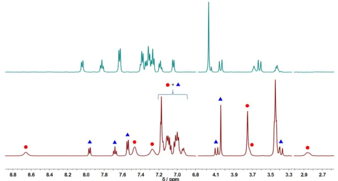
Figure 3. 1H NMR (500 MHz) spectra of
dissolved in CD2Cl2 (0.4 mM, above,
all the shown peaks are assigned to ) or CD3CN (1.0 mM, below, peak
attribution: blue triangles = ; red circles = ).
Accordingly, in the 1H NMR spectrum of
in CD2Cl2 only one set of signals is
observed, in accordance with the molecular structure observed in solid state, while two set of signals are present in CD3CN (1.0 mM solution), corresponding to and
(Figure 3).
The UV-vis signature of in CH2Cl2 and MeCN solutions (Figure 2, right)
confirms the speciation determined by IR and 1H NMR. The spectrum in CH2Cl2 (main
bands: 382 nm, 4110 M-1·cm-1 and 475 nm, 1800 M-1·cm-1) is similar to the solid-state
spectrum, confirming that the structure of is retained in CH2Cl2 solution. In
MeCN, at high concentration (1.0 mM), the three absorption bands at 381 nm, 429 nm
and 536 nm correspond to a mixture of both and , whereas at low
concentration (0.2 mM), the spectrum shows only the absorption features of ,
i.e. two bands at 434 nm (2989 M-1·cm-1) and 536 nm (2024 M-1·cm-1).
The equilibrium shown in Scheme 2 can be directed towards the formation of either or by tuning the experimental conditions. When CO is bubbled into the mixture of both species in MeCN (0.5 mM ), the band at 381 nm increases, while those at 429 and 536 nm disappear with the concomitant appearance of an additional absorption band at 472 nm (Figure 4, left). The resulting UV-vis spectrum is similar to that of in CH2Cl2, indicating that CO bubbling leads to the
quantitative formation of the complex also in MeCN. When CO is removed by
argon bubbling from the CO-saturated MeCN solution of (Figure 4, right), the
two bands at 381 and 472 nm disappear and the two bands characteristic of
newly appear. In both forward and backward processes, an isosbestic point at 415 nm is observed, demonstrating that only the two and species are involved in the equilibrium shown in Scheme 2.
Figure 4. (left) UV-vis spectra of dissolved in MeCN (0.5 mM, 5 mm path length) before (blue line), during (grey lines) and after (red line) bubbling CO. The
CO-saturated solution of is successively bubbled with argon and the
corresponding UV-vis monitoring is shown on the right (before Ar-bubbling: red line, during: grey lines, after: magenta line).
To further investigate the structural and spectroscopic properties of , DFT calculations have been performed. We initially validated our approach through the study of . Its optimized structure, *, displays structural parameters (Table 1) and predicted CO vibrations that are fully consistent with the experimental
data (less than 8 pm of difference between predicted and experimental metal–ligand bond lengths; COcalc at 1900 and 1960 cm-1 vs COexp at 1918 and 1959 cm-1). The
Mössbauer parameters have been also predicted by DFT and the calculated parameters are well consistent the experimental data confirming our previous attribution: for the Fe unit, δcalc = 0.26 mm.s-1; ∆EQcalc = 0.82 mm.s-1 and for the Fe' unit, δcalc = 0.39 mm.s-1; ∆EQcalc
= 2.37 mm.s-1 (Table 1).
For , several structures containing only one metal-bound CO have been optimized with or without an iron-bound MeCN molecule. Two calculated structures are consistent with the experimental CO vibration at 1822 cm-1: (i) one with a bridging CO,
COcalc = 1801 cm-1 and no coordinated MeCN ( *) and (ii) one with a bridging
CO and a MeCN molecule coordinated to the Fe center, COcalc = 1838 cm-1
( (*)). Considering the acetonitrile binding energy, the calculated free
energy difference (1.9 kcal.mol-1)between these two species is consistent with the
presence of both complexes in equilibrium (for the sake of simplicity it will be referred to in the rest of the manuscript).
Electrochemical properties of . The electrochemical properties of the
shown by IR. The CV (Figure 5) exhibits two diffusion-controlled reversible one-electron reduction waves at E1/2 = –1.21 V (∆E = 91 mV) and E1/2 = –1.65 V (∆E = 90 mV) versus
Fc+/0. The first cathodic process is assigned to the reduction of
into the
mixed-valence complex (Scheme 3), which has been isolated and fully characterized
spectroscopically (see below). A similar cathodic process was previously observed for at a close potential (–1.29 V), but with the reduction occurring on the Ni site (NiII to NiI). This indicates that the nature of the metal does not notably affect the redox
properties of this class of dinuclear complexes.
The second one-electron reduction process is proposed to mainly occur at the bipyridine moiety of the LN2S2 ligand, as shown in the case of
(for which E1/2 = –1.90 V).25
The bipyridine is reduced at a noticeably less negative potential in the case of the FeFe compound, presumably because of structural and electronic differences in the one-electron reduced species (see below).
Even if the CV displays a reversible redox system for the second reduction process, the proposed radical-based intermediate is too unstable to be spectroscopically
detected, as for the NiFe complex.ref nat chem
The CV of also presents an irreversible anodic signal at 0.10 V (SI), assigned to the FeII to FeIII oxidation in the Fe unit. The corresponding cathodic peak at -0.62 V is
located at the same reduction potential of the previously reported mononuclear [LN2S2FeIIIMeCN]+ complex.31 This implies that the electrochemically oxidized form of
is not stable on the CV timescale and undergoes a fast dissociation into [LN2S2FeIIIMeCN]+ and [CpFe(CO)(MeCN)2]+. Such oxidation process was not observed in
the case of .
Figure 5. CVs of (0.2 mM) in the absence (black) and presence of various amounts of Et3NHBF4 (red: 1 equiv.; blue: 2 equiv.; green: 5 equiv.; magenta: 10 equiv.;
orange: 15 equiv.; cyan: 20 equiv.; dark yellow: 30 equiv.) in MeCN. Glassy carbon working electrode, 100 mV·s-1, 0.1 M n-Bu4NClO4. Inset: CV of Et3NHBF4 (0.2 mM) in
MeCN in the absence (black) and presence (red) of one equivalent of .
Spectroscopic characterization of the one-electron reduced species . On
the basis of the electrochemical properties of , cobaltocene (CoCp2, E1/2 = -1.33
V) was used to chemically reduce it to generate the mixed-valence .
This compound could be isolated as a powder and characterized (Table 1). The νCO band
at 1896 cm-1 in the IR spectrum suggests the presence of one CO ligand terminally bound
to one iron center. We ruled out the coordination of an MeCN ligand because of the absence of a CN vibration in the infrared spectrum (see SI). The zero-field Mössbauer spectrum (powder, 80 K, Figure 6, left) of exhibits two main quadrupole doublets, with FeA: δ = 0.51 mm.s-1, ∆EQ =2.19 mm.s-1 (35 %) and FeB: δ = 0.33 mm.s-1, ∆EQ =0.44 mm.s-1 (41 %), which can be assigned to the dinuclear mixed valence complex.
The attribution of these parameters to a specific Fe site is arduous and has been made with a combined DFT analysis (see below). The sample also contains two additional
minor Fe-based species probably arising from the decomposition of ,
characterized by two doublets δ3(∆EQ3) =0.10 (2.16) mm.s-1 (17%) and δ4(∆EQ4) = 1.17
(3.29) mm.s-1 (7%). The former displays parameters close to those of the
[CpFe(CO)2CH2SiMe3] complex (δ(∆EQ) =0.099 (1.75) mm.s-1),3 and could then be
attributed to a mononuclear [FeCOCp(solvent)2]+ complex. The latter parameters are
typical of a high spin FeII species and comparable to those found for
[FeII2(LN2S2)(HLN2S2)]BF4 (δ(∆EQ) 0.87 (3.76) mm.s-1 indicating that this species could
correspond to a mononuclear Fe complex containing the LN2S2 ligand.
Figure 6. Zero-field 57Fe Mössbauer spectrum (left) at 80 K of
in solid state.
X-band EPR spectra (right) of (black solid, in solid state at 22 K; black dash, in frozen MeCN solution at 7 K; red dash, in frozen THF solution at 100 K; red solid, in solid at 100 K. 2000 3000 4000 5000 -400000 -300000 -200000 -100000 0 100000 Field / G -4 -2 0 2 4 0.990 0.992 0.994 0.996 0.998 1.000 Relative tr an smission Velocity / mm-1
11 The powder EPR spectrum of recorded at 22 K exhibits an axial signal with gǁ
= 2.262 and g⏊= 1.947, corresponding to a localized S = 1/2 spin system (Figure 6, right).
In contrast, the EPR spectrum recorded on the powder at 100 K features a broader and more isotropic signal (gi = 2.064), consistent with a delocalized mixed-valence species.
Based on this temperature behavior, the electronic structure of can be described as a class II delocalized mixed-valence FeIFeII species.
The identity and valence delocalization of has been confirmed also in solution both by EPR and Near Infrared (NIR) spectroscopy. The EPR spectra of in THF/MeCN recorded at 7 K and 100 K are similar to the corresponding powder spectra (Figure 6). The NIR spectrum displays a band centered at 1015 nm (9852 cm-1), 1/2 =
2280 cm-1 in agreement with a mixed-valence compound (see below).
To gain more insights on the structural and electronic properties of , different possible structures have been optimized by DFT. Only one computed structure, *, which displays a terminally bound CO to Fe, is consistent with the
experimental IR data (COcalc = 1904 cm-1, νCOexp = 1896 cm-1). All spectroscopic
parameters have been then predicted theoretically for * (Table 1). TD-DFT
calculations predict a NIR transition at 1017 nm (f = 0.025) that perfectly matches the experimental band. This transition is characterized by a strong metal-based character consistent with an intervalence charge transfer (IVCT) transition (Figure 7). The
Mössbauer parameters predicted for * match well with the experimental data,
and can thus be assigned to each iron center: for the Fe unit, δcalc(exp) = 0.27(0.33) mm.s-1; ∆EQcalc(exp) = 0.64(0.44) mm.s-1 andfor the Fe' unit, δcalc(exp) = 0.58(0.51) mm.s-1; ∆EQcalc(exp)
= 1.91(2.19) mm.s-1. The Mülliken spin population is consistent with a valence
delocalized species with 0.40 and 0.40 for Fe and Fe', respectively (0.03, each N and 0.14, remaining). Finally the g isotropic value obtained from DFT is 2.088, with a rather small g anisotropy (g1 = 1.991; g2 = 2.124; g3 = 2.149) compared to the experimental axial EPR
signal recorded at low temperature (g = 0.31). The combination of all these
experimental data and theoretical information are consistent with a class II delocalized mixed-valence species with a structure in which the terminal CO is bound the Fe site and not bridging the two metal centers as in the case of the parent complex.
EPR parameters of [COax-Fe1.5-Fe1.5]
g tensor
Calc. Exp
1.991; 2.124; 2.149 (2.088) (2.064)
UV-vis features of [COax-Fe 1.5
-Fe1.5]
Wavelength (nm)
Calc. Exp
1017 (f = 0.025) 1015
Transition density for 1017 nm band : IVCT character as metal centers exclusively involved
color code is yellow for donor state and red for acceptor state
M etrical parameters
Fe1-Fe2 Fe1-N1 Fe1-N2 Fe1-S1 Fe1-S2 Fe1-C1 Fe2-S1 Fe2-S2 2.647 1.961 1.961 2.233 2.229 1.750 2.221 2.208 I R features µCO (cm-1) Calc. Exp 1904 1896 M ossbauer parameters DEQ(mm.s-1) Calc. Exp
0.64 (FeN2S2) 1.91 (FeCp) 2.19 & 0.44
d(mm.s-1)
Calc. Exp
0.27 (FeN2S2) 0.58 (FeCp) 0.51 & 0.33
Electronics of [COax-Fe1.5-Fe1.5] – delocalized M V species
Figure 7. (left) Difference electron densities sketch of the NIR TDDFT-predicted
transition for * at 1017 nm (yellow = negative, red = positive), (right) localized
SOMO of *.
The terminal coordination of CO to the [FeLN2S2] moiety in
(*) is consistent with
the redox properties of the / system: although this reduction is
proposed to mainly occur at the bipyridine site of LN2S2 as in
, its potential is
significantly higher (E1/2 = -1.65 V for vs E1/2 = -1.90 V for ). The fact
that is easier to reduce than (i.e. the one-electron reduced form of ) may be due to the stronger back-bonding of the terminal CO ligand in vs the bridging CO in .
Electrocatalytic activity for H2 production of . The electrocatalytic behavior
of for H2 production has been evaluated using Et3NHBF4 as a mild proton
source in MeCN (pKa = 18.6),32 conditions that are identical to those used during the
investigation of .(Natchem) In the presence of increasing amounts of Et3NHBF4
(0.2-6 mM), a catalytic process (Ecat/2 in the range −1.89 to −1.94 V for a concentration in
Et3NHBF4 in the range 1 to 6 mM Et3NHBF4, vs Eno catalyst = -2.16 V vs Fc+/0, potentials
being measured at the half-wave) develops at potential ~250 mV more cathodic than the second process generating (Figure 5). The lower value of E
cat/2 compared to E1/2 / (see the inset of Figure 5) indicates that the two-electron reduced species reacts with a proton to generate an intermediate that needs to be further reduced at a lower potential before to react with a second proton to generate H2
(see below). The plots of catalytic current intensity (icat) versus the concentration of
added Et3NHBF4 provide a linear correlation indicating a diffusion controlled regime for
catalysis under these conditions (SI).5 Bulk electrolysis experiments performed at ~ –1.8
V vs Fc+/0 on a Hg-pool cathode (SI) confirmed the electrocatalytic production of H2
(detected by GC) in the presence of (~15 turnovers achieved with ~60%
Faradaic yield within ~20 min, ~60% conversion from 10 mM Et3NHBF4).
Mechanistic considerations. The parent electrocatalyst was shown to follow
an E(ECEC) mechanism for H2 production.25, 27 The complex undergoes a
one-electron reduction at the Ni center to afford [LN2S2NiIFeIICp(CO)] (
, which is
further reduced to produce the radical based [LN2S2°NiIFeIICp(CO)]- complex that is then
protonated on Fe affording a semi-bridging hydride [LN2S2NiII(H)FeIICp(CO)]-.
[LN2S2NiII(H)FeIICp(CO)]-. The complex then needs to be further reduced before to react
with a second proton to produce H2.
Based on our present experimental results and on the mechanism of the parent catalyst,25-27 a similar E(ECEC) mechanism is proposed for electrocatalytic H2
Scheme 3. Proposed catalytic pathways for H2 evolution with catalyst
(potentials are referred to Fc+/0).
The first reduction generates the spectroscopically characterized mixed-valence
complex (see above). This species undergoes a second one-electron reduction
to form . The fact that the catalytic wave occurs at a reduction potential lower
than E1/2 = -1.65 V versus Fc+/0 (corresponding to the / redox
system) evidences that: (i) is protonated to generate either a Fe-hydride or
thiol-containing intermediate, and (ii) this protonated species needs to be reduced before reacting with a second proton to produce H2. is thus
regenerated for another electrocatalytic cycle.
Interestingly, for the FeFe electrocatalyst the E(ECEC) cycle can be experimentally validated, while for the parent NiFe complex it was mainly predicted by DFT calculations. In the case of the predicted potential of the third reduction process (that occurs after the first protonation step) is higher than the second one (E0calc = -1.42 V and
E0calc= -1.77 V vs Fc+/0, respectively)27 and thus was not detectable.25 In contrast, for
the third reduction process can be directly observed since it occurs at a lower reduction potential (Ecat/2 = −1.94 V vs E1/2 = -1.65 V).
Discussion
In the present work, we investigated a FeIIFeII complex as the homodinuclear
counterpart of the efficient heterodinuclear NiFe HER electrocatalyst
[LN2S2NiIIFeIICp(CO)]+, in which the NiII ion has now been replaced by FeII. The objective
of this study is to evaluate the influence of the nature of the metal ion located in the N2S2 site on the HER reactivity.
Structural differences between the two MIIFeII complexes, both in the solid state and in
solution, have been observed. In the solid state, the FeFe complex could not be isolated in an isostructural form of since it contains two CO molecules: one bound to
Fe’ ({FeCp}) in a semi-bridging manner and one terminally bound to the Fe center
({FeLN2S2}). In low-concentrated MeCN solutions, one CO ligand is removed and the
remaining CO ligand is bridging, whereas in it is terminally-bound to the Fe’ moiety. Stabilizing a four-coordinate FeII ion in the Fe site is difficult. In the specific case
{FeFe'} -+ FeII N N S C O FeII Cp S N Fe1.5+ N S CO Fe1.5+ Cp S e- e -e -H+ H+ H2 E1/2 = -1.21 V E1/2 = -1.65 V Ecat/2 = -1.94 V {FeFe'}CO+ {FeFe'}CO CO {FeFe'}
of the LN2S2 ligand, even if mononuclear tetracoordinated [MLN2S2] complexes have been
previously isolated with M = CoII,33 CuII,34 ZnII 35 and NiII,36 we did not succeed in
isolating the corresponding [FeLN2S2] complex. Instead, the mononuclear [FeLN2S2(Cl)]
-and the dinuclear [Fe2(LN2S2)(LN2S2H)]+ complexes were obtained, in which the FeII ions
are five-coordinated.29, 31 Likewise, in [(N2S2)(Fe-NO)Fe(CO)Cp]+ (Scheme 1)
Darensbourg et al. needed a NO co-ligand to complete the N2S2 coordination of one Fe center.24 For [(dppe)Ni(-pdt)Fe(CO)3], an additional CO was required to isolate the
homobimetallic congener [(dppe)(CO)Fe(-pdt)Fe(CO)3], as in the present case.17
Since our aim is to evaluate the role of the metal in the catalytic process, the most relevant species to be compared are the one-electron reduced complexes, , which represent the entrance in the proposed catalytic cycles. Even if only one CO is present in both structures, it is terminally bound to the {FeLN2S2} moiety in
,
while it bridges the Ni and Fe ions in . The electronic structure is also notably different since displays a mixed-valence character of type II, whereas in the spin-density is almost exclusively localized on the Ni site.
Concerning the electrocatalytic HER process, it occurs in both cases through an E(ECEC)
mechanism: for , cyclic voltammetry experiments carried out in the presence
of Et3NH+ (see Figure 5) demonstrate experimentally this pathway, while for it
was mainly supported by DFT. The overpotential requirement of the
electrocatalyst ( = 730 mV) is slightly higher than that of ( = 690 mV). Indeed, even if the second E-step occurs at a less negative potential for , the catalytic wave develops at a lower potential for the FeFe compound (see above). Finally, the FeFe and NiFe systems display comparable catalytic stabilities in MeCN solution (TON = 15, TON = 16).
All together, these data suggest that even if the electronic and structural parameters of the key intermediates, i.e. the one-electron reduced species slightly differs, the
nature of the metal ion located in the N2S2 ligand does not significantly impact the catalytic HER efficiency and an E(ECEC) pathway is proposed in both cases. The reactivity is most likely dominated by the coordinating and electronic properties of the LN2S2 ligand itself, especially owing to its non-innocent redox character. In fact, for both
NiFe and FeFe electrocatalysts, the second reduction process is proposed to occur on the bipyridine unit of LN2S2. In addition, DFT calculations carried out for the NiFe system
predict that the third reduction process also occurs on the bipyridine unit. Since, a similar mechanism is suggested for the FeFe system, it clearly appears that the redox activity of the LN2S2 ligand, especially its bipyridine unit, partly drives the entire catalytic
process.
Interestingly, the present study describes a rare example of an [FeFe] H2ase model that
mimics the iron oxidation states relevant to enzyme reactivity. The mimics the Hox state (scheme 1) and an ECEC pathway generates H2 from both and
the Hox state. By contrast, most of the reported [FeFe] H2ase models involve more
proposed to be involved during the catalytic cycle of [FeFe] hydrogenase.40 In this case, the bipyridine moiety acts as an electron reservoir for the FeFe catalyst, like the proximal [4Fe4S] cluster in [FeFe] H2ase.41, 42
Regarding the protonation steps, especially the one that is proposed to occur after the generation of the two electron reduced species, ,it can lead to a Fe-hydride
species or to a thiol-containing intermediate, . Based on several DFT investigations, it has been proposed that such type of (hetero)dinuclear complexes with bridging thiolates display the propensity to protonate a thiolate during the catalytic process followed by the decoordination of the corresponding thiol, because of the hemilability properties of these metallodithiolate ligands.(darensbourg et hall, acs catal)
24, 26-27, 37. In contrast, in the present case of
, we propose the generation of a
terminal hydride on the Fe' site based on the fact that (i) a metal-hydride is generated during the first protonation of the parent electrocatalyst, and (ii) the Fe' site is proposed to have a vacant site based on the DFT-predicted structure of .
Conclusion
In summary, we succeed to compare the activity between a [FeFe] H2ase mimic and its
previously reported [NiFe] analogue, two dinuclear HER catalysts in which only the nature of the metal (Ni vs Fe) in the {MLN2S2} metallodithiolate ligand is modified.
Although the structure of the catalytic intermediates, especially the one electron reduced species, notably differs, the activity of the two electrocatalysts is similar. This might be explained by the non-innocent redox activity of the LN2S2 ligand that is
proposed to dominate the catalytic process. Further studies are needed at probing the full catalytic cycle, deciphering between terminal or bridging hydride ligands as catalytic intermediates and investigating if hemilability of the metallodithiolate ligand is at play in the H2 evolution process mediated by .
Acknowledgements. The authors gratefully acknowledge research support of this work
by the China Scholarship Council (LW), the French National Agency for Research in the framework of the "Investissements d’avenir” program (ANR-15-IDEX-02), the Labex ARCANE, the CBH-EUR-GS (ANR-17-EURE-0003) and the ANR-DFG (ANR-16-CE92_0012_01), and the Deutsche Forschungsgemeinschaft (DFG Me1313/14-1, NiFeMim).
References
1. Lubitz, W.; Ogata, H.; Rüdiger, O.; Reijerse, E., Hydrogenases. Chem. Rev. 2014,
2. Schilter, D.; Camara, J. M.; Huynh, M. T.; Hammes-Schiffer, S.; Rauchfuss, T. B., Hydrogenase Enzymes and Their Synthetic Models: The Role of Metal Hydrides. Chem.
Rev. 2016, 116, 8693-8749.
3. Nicolet, Y.; Piras, C.; Legrand, P.; Hatchikian, C. E.; Fontecilla-Camps, J. C., Desulfovibrio desulfuricans iron hydrogenase: the structure shows unusual coordination to an active site Fe binuclear center. Structure 1999, 7, 13-23.
4. Chen, Z.; Lemon, B. J.; Huang, S.; Swartz, D. J.; Peters, J. W.; Bagley, K. A., Infrared Studies of the CO-Inhibited Form of the Fe-Only Hydrogenase from Clostridium pasteurianum I: Examination of Its Light Sensitivity at Cryogenic Temperatures.
Biochemistry 2002, 41, 2036-2043.
5. Peters, J. W.; Lanzilotta, W. N.; Lemon, B. J.; Seefeldt, L. C., X-ray Crystal Structure of the Fe-Only Hydrogenase (CpI) from Clostridium pasteurianum to 1.8 Angstrom Resolution. Science 1998, 282, 1853.
6. Volbeda, A.; Charon, M.-H.; Piras, C.; Hatchikian, E. C.; Frey, M.; Fontecilla-Camps, J. C., Crystal structure of the nickel-iron hydrogenase from Desulfovibrio gigas. Nature
1995, 373, 580-587.
7. Coutard, N.; Kaeffer, N.; Artero, V., Molecular engineered nanomaterials for catalytic hydrogen evolution and oxidation. Chem. Commun. 2016, 52, 13728-13748.
8. Can, M.; Armstrong, F. A.; Ragsdale, S. W., Structure, Function, and Mechanism of
the Nickel Metalloenzymes, CO Dehydrogenase, and Acetyl-CoA Synthase. Chem. Rev.
2014, 114, 4149-4174.
9. Ash, P. A.; Hidalgo, R.; Vincent, K. A., Proton Transfer in the Catalytic Cycle of [NiFe] Hydrogenases: Insight from Vibrational Spectroscopy. ACS Catal. 2017, 7, 2471-2485.
10. Ogata, H.; Lubitz, W.; Higuchi, Y., Structure and function of [NiFe] hydrogenases. J.
Biochem. 2016, 160, 251-258.
11. Hidalgo, R.; Ash, P. A.; Healy, A. J.; Vincent, K. A., Infrared Spectroscopy During Electrocatalytic Turnover Reveals the Ni-L Active Site State During H2 Oxidation by a NiFe Hydrogenase. Angew. Chem. Int. Ed. 2015, 54, 7110-7113.
12. Ogata, H.; Nishikawa, K.; Lubitz, W., Hydrogens detected by subatomic resolution
protein crystallography in a [lsqb]NiFe[rsqb] hydrogenase. Nature 2015, 520, 571-574. 13. Tard, C.; Pickett, C. J., Structural and Functional Analogues of the Active Sites of the [Fe]-, [NiFe]-, and [FeFe]-Hydrogenases. Chem. Rev. 2009, 109, 2245-2274.
14. Adamska, A.; Silakov, A.; Lambertz, C.; Ruediger, O.; Happe, T.; Reijerse, E.; Lubitz, W., Identification and Characterization of the "Super-Reduced" State of the H-Cluster in FeFe Hydrogenase: A New Building Block for the Catalytic Cycle? Angew. Chem. Int. Ed.
2012, 51, 11458-11462.
15. Mulder, D. W.; Ratzloff, M. W.; Shepard, E. M.; Byer, A. S.; Noone, S. M.; Peters, J. W.; Broderick, J. B.; King, P. W., EPR and FTIR Analysis of the Mechanism of H2 Activation by [FeFe]-Hydrogenase HydA1 from Chlamydomonas reinhardtii. J. Am. Chem.
Soc. 2013, 135, 6921-6929.
16. Simmons, T. R.; Berggren, G.; Bacchi, M.; Fontecave, M.; Artero, V., Mimicking hydrogenases: From biomimetics to artificial enzymes. Coord. Chem. Rev. 2014, 270–271, 127-150.
17. Gloaguen, F.; Rauchfuss, T. B., Small molecule mimics of hydrogenases: hydrides
and redox. Chem. Soc. Rev. 2009, 38, 100-108.
18. Kaur-Ghumaan, S.; Stein, M., [NiFe] hydrogenases: how close do structural and functional mimics approach the active site? Dalton Trans. 2014, 43, 9392-9405.
19. Tard, C.; Liu, X.; Ibrahim, S. K.; Bruschi, M.; Gioia, L. D.; Davies, S. C.; Yang, X.; Wang, L.-S.; Sawers, G.; Pickett, C. J., Synthesis of the H-cluster framework of iron-only hydrogenase. Nature 2005, 433, 610-613.
20. Camara, J. M.; Rauchfuss, T. B., Combining acid-base, redox and substrate binding
functionalities to give a complete model for the FeFe -hydrogenase. Nature Chem. 2012,
4, 26-30.
21. Rauchfuss, T. B., Diiron Azadithiolates as Models for the [FeFe]-Hydrogenase Active Site and Paradigm for the Role of the Second Coordination Sphere. Acc. Chem. Res.
2015, 48, 2107-2116.
22. Barton, B. E.; Whaley, C. M.; Rauchfuss, T. B.; Gray, D. L., Nickel−Iron Dithiolato Hydrides Relevant to the [NiFe]-Hydrogenase Active Site. J. Am. Chem. Soc. 2009, 131, 6942-6943.
23. Zhu, W.; Marr, A. C.; Wang, Q.; Neese, F.; Spencer, D. J. E.; Blake, A. J.; Cooke, P. A.; Wilson, C.; Schröder, M., Modulation of the electronic structure and the Ni–Fe distance in heterobimetallic models for the active site in [NiFe]hydrogenase. Proc. Natl. Acad. Sci. U.
S. A. 2005, 102, 18280-18285.
24. Ding, S.; Ghosh, P.; Lunsford, A. M.; Wang, N.; Bhuvanesh, N.; Hall, M. B.; Darensbourg, M. Y., Hemilabile Bridging Thiolates as Proton Shuttles in Bioinspired H2 Production Electrocatalysts. J. Am. Chem. Soc. 2016, 138, 12920-12927.
25. Brazzolotto, D.; Gennari, M.; Queyriaux, N.; Simmons, T. R.; Pécaut, J.; Demeshko,
S.; Meyer, F.; Orio, M.; Artero, V.; Duboc, C., Nickel-centred proton reduction catalysis in a model of [NiFe] hydrogenase. Nature Chem. 2016, 8, 1054–1060.
26. Brazzolotto, D.; Wang, L. K.; Tang, H.; Gennari, M.; Queyriaux, N.; Philouze, C.; Demeshko, S.; Meyer, F.; Orio, M.; Artero, V.; Hall, M. B.; Duboc, C., Tuning Reactivity of Bioinspired NiFe -Hydrogenase Models by Ligand Design and Modeling the CO Inhibition Process. ACS Catal. 2018, 8, 10658-10667.
27. Tang, H.; Hall, M. B., Biomimetics of [NiFe]-Hydrogenase: Nickel- or Iron-Centered
Proton Reduction Catalysis? J. Am. Chem. Soc. 2017, 139, 18065-18070.
28. Ahmed, M. E.; Chattopadhyay, S.; Wang, L. K.; Brazzolotto, D.; Pramanik, D.; Aldakov, D.; Fize, J.; Morozan, A.; Gennari, M.; Duboc, C.; Dey, A.; Artero, V., Hydrogen Evolution from Aqueous Solutions Mediated by a Heterogenized NiFe -Hydrogenase Model: Low pH Enables Catalysis through an Enzyme-Relevant Mechanism. Angew.
Chem. Int. Ed. 2018, 57, 16001-16004.
29. Wang, L.; Gennari, M.; Cantú Reinhard, F. G.; Gutiérrez, J.; Morozan, A.; Philouze, C.; Demeshko, S.; Artero, V.; Meyer, F.; de Visser, S. P.; Duboc, C., A Non-Heme Diiron Complex for (Electro)catalytic Reduction of Dioxygen: Tuning the Selectivity through Electron Delivery. J. Am. Chem. Soc. 2019, 141, 8244-8253.
30. Nicolet, Y.; de Lacey, A. L.; Vernède, X.; Fernandez, V. M.; Hatchikian, E. C.; Fontecilla-Camps, J. C., Crystallographic and FTIR Spectroscopic Evidence of Changes in Fe Coordination Upon Reduction of the Active Site of the Fe-Only Hydrogenase from Desulfovibrio desulfuricans. J. Am. Chem. Soc. 2001, 123, 1596-1601.
31. Wang, L. K.; Reinhard, F. G. C.; Philouze, C.; Demeshko, S.; de Visser, S. P.; Meyer, F.; Gennari, M.; Duboc, C., Solvent- and Halide-Induced (Inter)conversion between Iron(II)-Disulfide and Iron(III)-Thiolate Complexes. Chem. Eur. J. 2018, 24, 11973-11982. 32. Izutsu, K., Acid-Base Dissociation Constants in Dipolar Aprotic Solvents. Blackwell Scientific: Oxford, U.K. , 1990.
33. Gennari, M.; Gerey, B.; Hall, N.; Pécaut, J.; Vezin, H.; Collomb, M. N.; Orio, M.; Duboc, C., Structural, spectroscopic and redox properties of a mononuclear Co-II thiolate
complex - the reactivity toward S-alkylation: an experimental and theoretical study.
Dalton Trans. 2012, 41, 12586-12594.
34. Gennari, M.; Pécaut, J.; Collomb, M. N.; Duboc, C., A Copper Thiolate Centre for Electron Transfer: Mononuclear versus Dinuclear Complexes. Dalton Trans. 2012, 41, 3130-3133
35. Gennari, M.; Retegan, M.; DeBeer, S.; Pecaut, J.; Neese, F.; Collomb, M. N.; Duboc, C., Experimental and Computational Investigation of Thiolate Alkylation in Ni-II and Zn-II Complexes: Role of the Metal on the Sulfur Nucleophilicity. Inorg. Chem. 2011, 50, 10047-10055.
36. Gennari, M.; Orio, M.; Pécaut, J.; Neese, F.; Collomb, M.-N.; Duboc, C., Reversible Apical Coordination of Imidazole between the Ni(III) and Ni(II) Oxidation States of a Dithiolate Complex: A Process Related to the Ni Superoxide Dismutase. Inorg. Chem.
2010, 49, 6399-6401.
37. Ding, S.; Ghosh, P.; Darensbourg, M. Y.; Hall, M. B., Interplay of hemilability and redox activity in models of hydrogenase active sites. Proc. Natl. Acad. Sci. USA 2017, 114, E9775-E9782.