HAL Id: inserm-02190367
https://www.hal.inserm.fr/inserm-02190367
Submitted on 22 Jul 2019HAL is a multi-disciplinary open access
archive for the deposit and dissemination of sci-entific research documents, whether they are pub-lished or not. The documents may come from teaching and research institutions in France or abroad, or from public or private research centers.
L’archive ouverte pluridisciplinaire HAL, est destinée au dépôt et à la diffusion de documents scientifiques de niveau recherche, publiés ou non, émanant des établissements d’enseignement et de recherche français ou étrangers, des laboratoires publics ou privés.
Plasmodium ovale curtisi and Plasmodium ovale
wallikeri
Naowarat Saralamba, François Nosten, Colin Sutherland, Ana Paula Arez,
Georges Snounou, Nicholas White, Nicholas Day, Arjen Dondorp, Mallika
Imwong
To cite this version:
Naowarat Saralamba, François Nosten, Colin Sutherland, Ana Paula Arez, Georges Snounou, et al.. Genetic dissociation of three antigenic genes in Plasmodium ovale curtisi and Plasmodium ovale wallik-eri. PLoS ONE, Public Library of Science, 2019, 14 (6), pp.e0217795. �10.1371/journal.pone.0217795�. �inserm-02190367�
Genetic dissociation of three antigenic genes
in Plasmodium ovale curtisi and Plasmodium
ovale wallikeri
Naowarat SaralambaID1,2*, Francois NostenID3, Colin J. SutherlandID4, Ana Paula ArezID5, Georges Snounou6, Nicholas J. WhiteID2,7, Nicholas P. J. Day2,7, Arjen M. Dondorp2,7, Mallika Imwong1,2
1 Department of Molecular Tropical Medicine and Genetics, Faculty of Tropical Medicine, Mahidol University, Bangkok, Thailand, 2 Mahidol-Oxford Tropical Medicine Research Unit, Faculty of Tropical Medicine, Mahidol University, Bangkok, Thailand, 3 Shoklo Malaria Research Unit, Mahidol-Oxford Tropical Medicine Research Unit, Bangkok, Thailand, 4 Immunology Unit, Department of Infectious and Tropical Diseases, London School of Hygiene and Tropical Medicine, London, United Kingdom, 5 Global Health and Tropical Medicine, GHTM, Instituto de Higiene e Medicina Tropical, IHMT, Universidade Nova de Lisboa, UNL, Lisbon, Portugal, 6 CEA-Universite´ Paris Sud 11-INSERM U1184, Immunology of Viral Infections and Autoimmune Diseases (IMVA), IDMIT Department, IBFJ, DRF, Fontenay-aux-Roses, Paris, France, 7 Centre for Tropical Medicine and Global Health, Nuffield Department of Medicine, University of Oxford, Oxford, United Kingdom
*naowarat.sar@mahidol.ac.th
Abstract
Plasmodium ovale curtisi and Plasmodium ovale wallikeri are two sympatric human malaria
species prevalent in Africa, Asia and Oceania. The reported prevalence of both P. ovale spp. was relatively low compared to other malaria species, but more sensitive molecular detection techniques have shown that asymptomatic low-density infections are more com-mon than previously thought. Whole genome sequencing of both P. ovale spp. revealed genetic dissociation between P. ovale curtisi and P. ovale wallikeri suggesting a species bar-rier. In this study we further evaluate such a barrier by assessing polymorphisms in the genes of three vaccine candidate surface protein: circumsporozoite protein/
thrombospon-din-related anonymous-related protein (ctrp), circumsporozoite surface protein (csp) and merozoite surface protein 1 (msp1). The complete coding sequence of ctrp and csp, and a
partial fragment of msp1 were isolated from 25 P. ovale isolates and compared to previously reported reference sequences. A low level of nucleotide diversity (Pi = 0.02–0.10) was observed in all three genes. Various sizes of tandem repeats were observed in all ctrp, csp and msp1 genes. Both tandem repeat unit and nucleotide polymorphism in all three genes exhibited clear dimorphism between P. ovale curtisi and P. ovale wallikeri, supporting evi-dence of non-recombination between these two species.
Introduction
Plasmodium ovale curtisi and Plasmodium ovale walllikeri are two sympatric species of malaria
parasites found across many malaria endemic countries in Africa, Asia and Oceania [1–3]. a1111111111 a1111111111 a1111111111 a1111111111 a1111111111 OPEN ACCESS
Citation: Saralamba N, Nosten F, Sutherland CJ,
Arez AP, Snounou G, White NJ, et al. (2019) Genetic dissociation of three antigenic genes in
Plasmodium ovale curtisi and Plasmodium ovale wallikeri. PLoS ONE 14(6): e0217795.https://doi. org/10.1371/journal.pone.0217795
Editor: E´rika Martins Braga, Universidade Federal
de Minas Gerais, BRAZIL
Received: January 29, 2019 Accepted: May 17, 2019 Published: June 6, 2019
Copyright:© 2019 Saralamba et al. This is an open access article distributed under the terms of the Creative Commons Attribution License, which permits unrestricted use, distribution, and reproduction in any medium, provided the original author and source are credited.
Data Availability Statement: All relevant data are
within the manuscript and its Supporting Information files. All DNA sequences are available from the NCBI database (accession number MK403987- MK404049).
Funding: This research project is supported by
Mahidol University, and was part of the Wellcome Trust Mahidol University-Oxford Tropical Medicine Research Programme supported by the Wellcome Trust of Great Britain. The funders had no role in
Although morphological features ofP. ovale curtisi and P. ovale wallkeri are indistinguishable,
these twoP. ovale species are genetically distinct, and there is evidence of differences in latency
and clinical presentation [4–6]. Nuclear genome sequences ofP. ovale curtisi and P. ovale wall-ikeri were recently reported and revealed different expansion in some gene families [7]. Cur-rently the target genes used for discriminating betweenP. ovale curtisi and P. ovale wallikeri
are theSSU rRNA gene [8],tryptophan rich antigen (potra) [9],reticulocyte-binding protein 2
(porbp2) [9], and some sexual stage proteins [9]. Sequence polymorphisms in the cell-surface associated proteins that are candidate targets for vaccine development have only been studied rarely. The current study assessed genetic diversity in a highly polymorphic region of the blood stage merozoite surface protein genemsp1, and in two genes encoding sexual stage and
sporozoite proteins,ctrp and csp respectively, in P. ovale curtisi and P. ovale wallikeri.
CTRP is a member of the micronemal and cell-surface associated proteins. InP. falciparum
disruption of thectrp gene prevents oocyst development in the anopheline mosquito [10], indicating that CTRP is important for mosquito midgut development. For this reason CTRP has been proposed as a transmissions-blocking vaccine candidate. CSP is the major surface protein on thePlasmodium sporozoite. It is a candidate target for pre-erythrocytic stage
vac-cine development. Genetic polymorphism within thecsp gene have been investigated in most
human malaria species includingP. falciparum [11,12],P. vivax [13,14],P. malariae [15,16], andP. knowlesi [17], but not inP. ovale. MSP1 is one of the predominant antigen expressed in
the erythrocytic stage ofPlasmodium spp. The msp1 gene is highly polymorphic and has been
well characterized inP. falciparum [18,19] andP. vivax [13,14]. A study ofP. ovale isolates
from Thailand revealed low diversity in themsp1 gene [20].
The current study evaluates sequence diversity ofctrp, csp and msp1, in a wider collection
ofP. ovale isolates collected from Thailand and African countries. Assessing diversity in these
surface proteins is important for defining vaccine candidates, and to further assess the species barrier betweenP. ovale curtisi and P. ovale wallikeri. In the current era of malaria elimination,
the better understanding ofP. ovale curtisi and P. ovale wallikeri is essential to ensure success
against all human malaria species.
Materials and methods
Samples
Twenty-five samples ofP. ovale (14 P. ovale wallikeri and 11 P. ovale curtisi) were collected
from Thailand and African countries during 1995–2010 (S1 Table). All samples were obtained from patients enrolled in previous studies who gave written informed consent to blood sam-pling. Parasitaemia of these samples varied from 1 per 500 WBC to 198 per 500 WBC. The pro-tocol for this study was reviewed and approved (reference number MUTM2001-049-04) by the ethics committee of the Faculty of Tropical Medicine, Mahidol University, Thailand. Genomic DNA of all samples was confirmed for the present ofP. ovale. Nested PCR of the SSU rRNA gene was performed with primer rPLU1/rPLU5 in the primary reaction and with
primer rOVA1/rPLU2 in the secondary reaction [21]. A nested PCR protocol based on the linker region ofdhfr-ts gene was applied with primer Pla-DHFR-F/Pla-TS-R in the primary
reaction and with primer PO-Lin-F/PO-Lin-R in the secondary reaction [22]. In addition, a semi-nested PCR ofpotra gene was performed with a primer specific to both P. ovale spp.
(PoTRA-F/PoTRA rev3) in the primary reaction and with specificP. ovale curtisi (PoTRA-F/
PocTRA-R) andP. ovale wallikeri (PoTRA-F/PowTRA-R) primers in the secondary reaction
[23].
study design, data collection and analysis, decision to publish, or preparation of the manuscript.
Competing interests: The authors have declared
Isolation of
poctrp and pocsp gene
Specific primers targetingpoctrp and pocsp genes were designed to obtain the full length of
those two gene sequences (Table 1). A semi-nested PCR approach was used for amplification of each fragment with PCR conditions as presented inTable 1. All PCR reactions were per-formed with 10 mM Tris-HCl, pH 8.3, 50 mM KCl, 2 mM MgCl2, 125μM dNTPs, 250 nM of
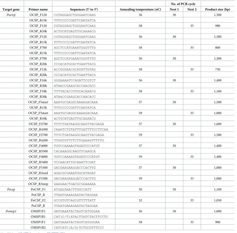
Table 1. Primer sequences and PCR conditions for isolation ofpoctrp, pocsp and pomsp1 genes.
No. of PCR cycle
Target gene Primer name Sequences (5’ to 3’) Annealing temperature (oC) Nest 1 Nest 2 Product size (bp)
Poctrp OCSP_F120 CGTAGGAGCTGGGAATCAAG 56 30 1,500 OCSP_R15k TTTCCCCCGATTCAATATCA OCSP_F120 CGTAGGAGCTGGGAATCAAG 58 35 980 OCSP_R10k ACTGCATGAGTTGCAAAACG OCSP_F120 CGTAGGAGCTGGGAATCAAG 56 30 1,500 OCSP_R15k TTTCCCCCGATTCAATATCA OCSP_F700 AGCTCCATGAAATGGGTTTG 58 35 800 OCSP_R15k TTTCCCCCGATTCAATATCA OCSP_F700 AGCTCCATGAAATGGGTTTG 56 30 1,300 OCSP_R20k CCCACATGCACTGAATTACG OCSP_F12k ACCGGGAACGCATATTGTAG 58 35 750 OCSP_R20k CCCACATGCACTGAATTACG OCSP_F16k GGGAAAATCCAGATTCGTCT 56 30 1,400 OCSP_R30k ATAACCGAAGCACCAACACC OCSP_F18k TTTTACACCGTGCACAAACG 58 35 1,160 OCSP_R30k ATAACCGAAGCACCAACACC OCSP_F5start AAATGCGAGGCAAAAGACAAA 57 30 1,500 OCSP_R15k TTTCCCCCGATTCAATATCA OCSP_F5start AAATGCGAGGCAAAAGACAAA 59 35 1,000 OCSP_R10k ACTGCATGAGTTGCAAAACG OCSP_F2700 TTTCTGATAAGGCAAGTTACGAGA 57 30 1,600 OCSP_R4300 CAAATCTGTATTTGATTTTCCTTCAA OCSP_F2700 TTTCTGATAAGGCAAGTTACGAGA 59 35 1,500 OCSP_R4200 TTGGTGTTTTCTTGAAAGTTTTTG OCSP_F4000 TGTCCAAAAGTAGATCCCATGT 57 30 1,400 OCSP_R5500 CACAAAGGCAAGTTCAAGCA OCSP_F4000 TGTCCAAAAGTAGATCCCATGT 59 35 1,400 OCSP_R5400 TCCAACATTGCAAATTCGAT OCSP_F5300 GACGAAGAAGGACCCACTTG 57 30 1,000 OCSP_R3end AGACGCGAAATGGCATAGAT OCSP_F5300 GACGAAGAAGGACCCACTTG 59 35 1,000 OCSP_R3stop GAAGAACTGACGCGGAAAAA
Pocsp PoCSP_F1 ATGAGGAACTTGGCCATT 50 30 1,100 PoCSP_R TTAATGAAAGAATACTAGGAA
PoCSP_F2 GCCGTGTCAGCGTTTTTATT 52 35 1,050
PoCSP_R TTAATGAAAGAATACTAGGAA
Pomsp1 OMSP1F1 GATGAAATACTAGTCATGGGAA 56 30 1,000 OMSP1R1 CAT(C/T)ATACTTATCTACTTCCTC
OMSP1F1 GATGAAATACTAGTCATGGGAA 58 35 900
OMSP1R2 CATCATC(A/G)TCTGCGTTTCCC https://doi.org/10.1371/journal.pone.0217795.t001
each primer and 4 unit of Taq Polymerase (Kapa biosystems, USA). PCR products were then purified by Gel/PCR purification kit (Favogen, Taiwan), before being submitted for DNA sequencing.
Analysis of variable region in
pomsp1 gene
Twelve availablepomsp1 sequences from both P. ovale curtisi and P. ovale wallikeri were
retrieved from the NCBI database (accession number FJ824670, FJ824671, KC137340— KC137349) and multiple sequence alignments were performed. A highly polymorphic region withinpomsp1 was observed between amino acid residues 700 to 1,000. The primers OMSP1.
F1, OMSP1.R1, and OMSP1.R2, were designed for a semi-nested PCR approach to analyses this polymorphic domain in 25P. ovale samples (Table 1). Positive PCR products were then purified by Gel/PCR purification kit (Favogen, Taiwan), before being submitted for DNA sequencing. Allpomsp1 sequences obtained in this study were analyzed together with the
pre-vious reports.
Sequence analysis and phylogenetic tree reconstruction
Nucleotide polymorphisms ofpoctrp, pocsp and pomsp1 from P. ovale curtisi and P. ovale wallikeri were analyzed with ClustalW multiple alignment using BioEdit version 7.2.6.1 [24]. Nucleotide sequences ofpoctrp, pocsp and pomsp1 were translated to deduced amino acid
sequences using BioEdit version 7.2.6.1 [24]. The sequences obtained from 25 samples ofP. ovale spp. were analyzed in comparison with the previously reported sequences from the
NCBI database (poctrp: accession number LT594512, LT594589, pocsp: accession number
SBT72933, SBT84923,pomsp1: accession number LT594511, LT594588, KX672044,
KX672045, FJ824670, FJ824671, KC137340—KC137349). Genetic variability including average pairwise nucleotide diversity (Pi), haplotype diversity, and sliding plot nucleotide diversity with a window length of 100 bp and 25 bp step size withinpoctrp, pocsp and pomsp1 from P. ovale curtisi and P. ovale wallikeri was obtained from DnaSP 6.10.4 [25]. The ratio of non-syn-onymous to synnon-syn-onymous (dN/dS) within eachP. ovale spp. was measured by DnaSP 6.10.4
[25]. Tests for neutral evolution were assessed with Tajima’s D, Fu and Li’s D, and Fu and Li’s F tests using DnaSP 6.10.4 [25].
A neighbor-joining (NJ) phylogenetic tree was constructed from concatenated CTRP, CSP and MSP1 protein sequences to assess relationships betweenP. ovale curtisi and P. ovale walli-keri. A bootstrap test (1,000 replicates) was applied under the Jones-Taylor-Thornton (JTT)
model of evolution using MEGA7 [26].
Results
Isolation and analysis of
poctrp
The complete coding sequence ofpoctrp gene was obtained from 11 P. ovale curtisi and 14 P. ovale wallikeri isolates (accession number MK403987-MK404009). It revealed that the poctrp
genes for bothspecies has only one exon encoding for 2,007 to 2,047 amino acids. Sequence
alignment of these 25poctrp sequences, together with another two poctrp sequences (accession
number LT594512 and LT594589) available in the NCBI database, and otherctrp sequences
from the otherPlasmodium spp. that infect humans, showed that poctrp is composed of a
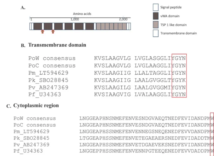
sig-nal peptide, six vWA domains, seven TSP1 domains, transmembrane domain, and a cyto-plasmic region (Fig 1). Alignment of the CTRP of all humanPlasmodium spp. revealed highly
conserved transmembrane (TM) and cytoplasmic regions (Fig 1). A conserved amino acid sequence YGYN/K for the tyrosine-based TM motif involved in cellular trafficking, and the
cytoplasmic domain tryptophan residue (Fig 1) which is the key interaction to drive parasite motility were conserved between all studied humanPlasmodium spp. Multiple alignment of
the full-length CTRP among all human-infectingPlasmodium spp. also identified a highly
con-served region close to the C-terminus.
Allpoctrp sequences including the two reference sequences were translated to deduce their
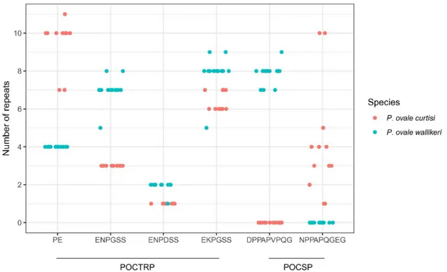
corresponding amino acids and analyzed for intra- and inter-specific sequence diversity at this locus. The deduced amino acid alignment of PoCTRP showed two prominent regions. The first region is located around 300–320 amino acids between the vWA1 and vWA2 domains.P. ovale curtisi isolates carries two amino acids repeats “PE” with 7–11 copies, while all P. ovale wallikeri isolates had 4 “PE” repeat units (Fig 2). The second region is located between codons 570 and 600, where a tandem repeat of six amino acids was identified. Three patterns of six amino acids repeats were observed: ENPDSS, EKPGSS, and ENPGSS. Different numbers of repeat units were presented in theP. ovale isolates (Fig 2). The repeat EKPGSS is the most fre-quent in bothP. ovale curtisi and P. ovale wallikeri. This region showed a marked difference in
Fig 1. ThePlasmodium ctrp gene. (A) Schematic representation of the domain structure of Plasmodium ctrp gene. The Plasmodium CTRP composes
of N-terminal signal peptide, six vWA domain, seven tandemly arrayed TSP 1- like domains, and the C-terminal transmembrane domain. Based onP. falciparum CTRP domain structure analysis, the PoCTRP domains could be drawn from multiple sequence alignment. Amino acid sequence signature
for eachPlasmodium species were observed between vWA 1–2 domains and between vWA 2–3 domains as indicated by red arrows. (B) Amino acid
alignment of the transmembrane (TM) domain with the box representing the conserved tyrosine-based motif involved in cellular trafficking. (C) Amino acid alignment of the cytoplasmic region. The conserved tryptophan residue that interacts with motility actomyosin machinery was marked with the box.
length, providing a potential additional genotypic marker to differentiateP. ovale curtisi
fromP. ovale wallikeri. Multiple sequence alignment of CTRP of all human Plasmodium spp.
revealed species-specific regions forP. ovale spp. at codons 512–538 and codons 573–599.
PCR amplification ofP. ovale spp. with primers targeting those two regions are useful to
distin-guishP. ovale curtisi from P. ovale wallikeri.
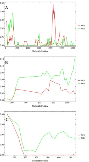
The availablepoctrp genes were analysed in a sliding plot for nucleotide diversity between P. ovale wallikeri and P. ovale curtisi (Fig 3).P. ovale curtisi showed higher diversity around
the first 1 kb whereP. ovale wallikeri showed higher diversity at 4 kb—5 kb of poctrp (Fig 3). For this gene, the average nucleotide diversity ofP. ovale curtisi is slightly lower than that of P. ovale wallikeri, and combined analysis of all 27 P. ovale sequences showed a higher diversity
value than that calculated from each species alone (Table 2), which indicates distinct distribu-tions of diversity across thepoctrp locus in the two species.
Isolation and analysis of
pocsp
Thepocsp gene was successfully amplified from 14 P. ovale wallikeri and 11 P. ovale curtisi
iso-lates (accession number MK404010-MK404031). The completepocsp gene varied in size from
1,020 to 1,185 bp, and the size variation resulted from variable tandem repeats in the central repeat region. Thepocsp sequences were analyzed together with two other sequences available
in NCBI databases and those of thecsp of the other human Plasmodium spp. The protein
domain architecture ofpocsp was determined based on homologous CSP proteins alignment
with other humanPlasmodium spp. The pocsp structure domain was similar to that of the csp
from the otherPlasmodium spp. Four domains in the conserved N-terminus domain
(con-served region I) and in the con(con-served C-terminus domain (Th2R, con(con-served region II, and Th3R) were of particular interest. A summary of the amino acid patterns in each of these
Fig 2. Distribution of the major tandem repeat units in PoCTRP and PoCSP.
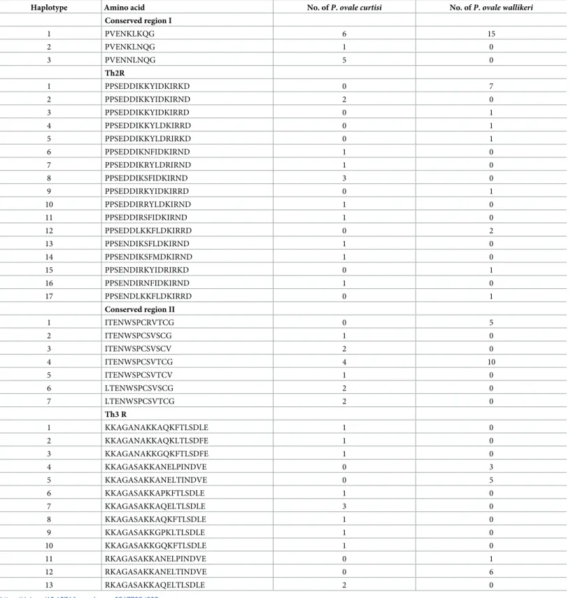
domains is presented inTable 3. Overall, a higher number of haplotypes was observed inP. ovale curtisi as compared to P. ovale wallikeri. Whereas P. ovale wallikeri showed only one
hap-lotype in conserved region I and two in conserved region II, three and six, respectively, were observed forP. ovale curtisi. A high number of haplotypes was observed in the Th2R and Th3R
domains for bothP. ovale curtisi and P. ovale wallikeri, but it is interesting to note that none
were shared by both species. The central repeat region ofpocsp was also analyzed. Several
pat-terns of nine amino acid repeats were observed. A specific repeat unit was observed forP. ovale wallikeri (DPPAPVPQG), and for P. ovale curtisi NPPAPQGEG, with the latter showing
a higher diversity in repeat unit numbers (Fig 2).
Fig 3. Sliding window plot of nucleotide diversity. Sliding plot with a window length of 100 bp and 25 bp step size
using DnaSP v5 revealed nucleotide diversity betweenP. ovale wallikeri (Pi 1) and and P. ovale cursiti (Pi 2). The
nucleotide diversity is calculated fromctrp (A), csp (B), and partial msp1 gene (C).
Thepocsp gene was evaluated for nucleotide diversity between P. ovale curtisi and P. ovale wallikeri. Sliding plots of nucleotide diversity revealed overall higher nucleotide diversity in P. ovale curtisi than in P. ovale wallikeri (Fig 3). The estimated synonymous (dS) and nonsynon-ymous (dN) substitution was also found at higher value inP. ovale curtisi than that of P. ovale wallikeri (Table 2). Combined analysis of bothP. ovale spp. showed significantly positive
val-ues (p<0.05) for Fu and Li’s D and Fu and Li’s F tests, suggesting population bottlenecks or balancing selections in these two species (Table 2).
Genetic analysis of
pomsp1
The sequences for the variable regions within thepomsp1 gene covering amino acids 710 to
1,020 were obtained from 14P. ovale wallikeri and 11 P. ovale curtisi isolates (accession
num-ber MK404032-MK404049). Apart from this, sixteen sequences ofpomsp1 gene (accession
number LT594511, LT594588, KX672044, KX672045, FJ824670, FJ824671, KC137340— KC137349) were available in the NCBI database. Taken together, 41 PoMSP1 sequences were used in the alignment. A clear dimorphic pattern was observed betweenP. ovale curtisi and P. ovale wallikeri. Amino acid tandem repeat patterns were found in P. ovale spp. The tandem
repeats are characteristic for the two differentP. ovale spp. There were three arrangement
pat-terns of three 5-amino acid repeat units (PGAGG, PGAAG, and PGVPG) found exclusively inP. ovale wallikeri isolates Whereas, nine arrangement patterns of six 4-amino acids repeat
units (QAAT, QTAT, HAST, QATT, QVTT, QSAT) were observed specifically in theP. ovale curtisi isolates (S2 Table).
Analysis of gene diversity and haplotype diversity at thepomsp1 locus showed that P. ovale curtisi has higher diversity than that of P. ovale wallikeri (Table 2). Sliding window plots showed higher overall nucleotide diversity inP. ovale curtisi than in P. ovale wallikeri (Fig 3). The ratio of synonymous (dS) and nonsynonymous (dN) substitutions was higher inP. ovale wallikeri than P. ovale curtisi (Table 2).
Comparative analysis of
P. ovale curtisi and P. ovale wallikeri
Genetic analysis ofP. ovale curtisi and P. ovale wallikeri based on three surface protein genes
revealed clear dissociation between these two species. Analysis within each species was per-formed though sequence diversity and amino acid patterns. The sequence polymorphism in
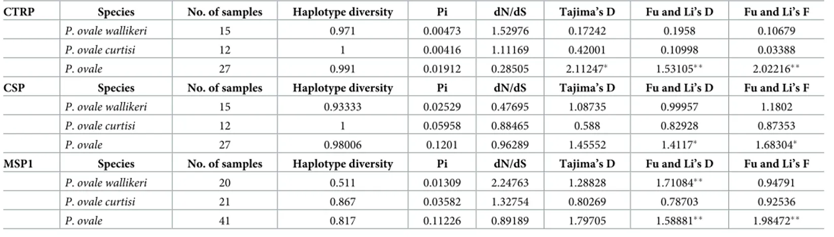
Table 2. Nucleotide diversity and natural selection inP. ovale spp.
CTRP Species No. of samples Haplotype diversity Pi dN/dS Tajima’s D Fu and Li’s D Fu and Li’s F
P. ovale wallikeri 15 0.971 0.00473 1.52976 0.17242 0.1958 0.10679
P. ovale curtisi 12 1 0.00416 1.11169 0.42001 0.10998 0.03388
P. ovale 27 0.991 0.01912 0.28505 2.11247� 1.53105�� 2.02216��
CSP Species No. of samples Haplotype diversity Pi dN/dS Tajima’s D Fu and Li’s D Fu and Li’s F
P. ovale wallikeri 15 0.93333 0.02529 0.47695 1.08735 0.99957 1.1802
P. ovale curtisi 12 1 0.05958 0.88465 0.588 0.82928 0.87353
P. ovale 27 0.98006 0.1201 0.96289 1.45552 1.4117� 1.68304�
MSP1 Species No. of samples Haplotype diversity Pi dN/dS Tajima’s D Fu and Li’s D Fu and Li’s F
P. ovale wallikeri 20 0.511 0.01309 2.24763 1.28828 1.71084�� 0.94791 P. ovale curtisi 21 0.867 0.03582 1.32754 0.80269 0.78703 0.92536 P. ovale 41 0.817 0.11226 0.89189 1.79705 1.58881�� 1.98472�� �P<0.05, ��P<0.02 https://doi.org/10.1371/journal.pone.0217795.t002
Table 3. Sequence polymorphism in the conserved regions ofP. ovale CSP.
Haplotype Amino acid No. ofP. ovale curtisi No. ofP. ovale wallikeri Conserved region I 1 PVENKLKQG 6 15 2 PVENKLNQG 1 0 3 PVENNLNQG 5 0 Th2R 1 PPSEDDIKKYIDKIRKD 0 7 2 PPSEDDIKKYIDKIRND 2 0 3 PPSEDDIKKYIDKIRRD 0 1 4 PPSEDDIKKYLDKIRRD 0 1 5 PPSEDDIKKYLDRIRKD 0 1 6 PPSEDDIKNFIDKIRND 1 0 7 PPSEDDIKRYLDRIRND 1 0 8 PPSEDDIKSFIDKIRND 3 0 9 PPSEDDIRKYIDKIRRD 0 1 10 PPSEDDIRRYLDKIRND 1 0 11 PPSEDDIRSFIDKIRND 1 0 12 PPSEDDLKKFLDKIRRD 0 2 13 PPSENDIKSFLDKIRND 1 0 14 PPSENDIKSFMDKIRND 1 0 15 PPSENDIRKYIDRIRKD 0 1 16 PPSENDIRNFIDKIRND 1 0 17 PPSENDLKKFLDKIRRD 0 1 Conserved region II 1 ITENWSPCRVTCG 0 5 2 ITENWSPCSVSCG 1 0 3 ITENWSPCSVSCV 2 0 4 ITENWSPCSVTCG 4 10 5 ITENWSPCSVTCV 1 0 6 LTENWSPCSVSCG 2 0 7 LTENWSPCSVTCG 2 0 Th3 R 1 KKAGANAKKAQKFTLSDLE 1 0 2 KKAGANAKKAQKLTLSDFE 1 0 3 KKAGANAKKGQKFTLSDFE 1 0 4 KKAGASAKKANELPINDVE 0 3 5 KKAGASAKKANELTINDVE 0 5 6 KKAGASAKKAPKFTLSDLE 1 0 7 KKAGASAKKAQELTLSDLE 3 0 8 KKAGASAKKAQKFTLSDLE 1 0 9 KKAGASAKKGPKLTLSDLE 1 0 10 KKAGASAKKGQKFTLSDLE 1 0 11 RKAGASAKKANELPINDVE 0 1 12 RKAGASAKKANELTINDVE 0 6 13 RKAGASAKKAQELTLSDLE 2 0 https://doi.org/10.1371/journal.pone.0217795.t003
poctrp, pocsp and pomsp1 showed more divergence in P. ovale curtisi than in P. ovale wallikeri
(Fig 3,Table 2). The test for neutrality (Tajima’s D, Fu and Li’s D, and Fu and Li’s F tests) was applied topoctrp, pocsp and pomsp1 to compare observed polymorphism frequencies with
expected frequencies. Significantly positive values were obtained from Fu and Li’s D and Fu and Li’s F whenP. ovale spp. were analyzed as one group (Table 2). These statistics reflect higher than expected frequencies of alleles, which might have resulted from population bottle-necks or balancing selections.P. ovale curtisi had a higher number of different haplotypes in
all conserved domains of the CSP (Table 3). This suggests thatP. ovale curtisi is intrinsically
more genetically diverse thanP. ovale wallikeri, but may also represent limitations of our
sample.
Some of the studiedP. ovale curtisi and P. ovale wallikeri infections were mixed with other
human malaria spp., which might have impacted the characteristics of CTRP, CSP and MSP1. Therefore the genetic analysis of CTRP, CSP and MSP1 was compared between single and mixed infections. There were four mixed infection found inP. ovale wallikeri, in which all four
samples were collected from Thailand. ForP. ovale curtisi, most samples were collected from
Africa with four single infections and five mixed infections. There was no significant difference in average nucleotide diversity (Pi), haplotype diversity, and dN/dS between single and mixed infections. Statistical testing for neutrality (Tajima’s D, Fu and Li’s D, and Fu and Li’s F tests) was also not significantly different (S3 Table). In addition, the pattern of tandem repeats in CTRP, CSP and MSP1 inP. ovale spp. showed no difference either between single and mixed
infections or between Asia and Africa isolates.
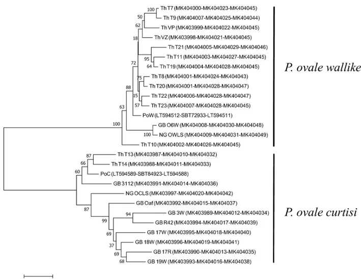
To infer genetic relationships ofP. ovale curtisi and P. ovale wallikeri, a phylogenetic tree
was reconstructed based on the three cell-surface associated proteins, CTRP, CSP and MSP1. The Neighbour-Joining method [27] was used to infer the evolutionary history of each species. Based on CTRP, CSP and MSP1 (Fig 4),P. ovale curtisi and P. ovale wallikeri clustered
accord-ing to species, and the tree topologies inferred from each gene showed similar features of grouping intoP. ovale curtisi and P. ovale wallikeri. This suggested that the genes of P. ovale curtisi and P. ovale wallikeri do not recombine and show distinct characteristics (Fig 4).
Discussion
In addition to the earlier described polymorphisms inpomsp1, the current study provides the
genetic characterization for two more cell-surface associated proteins:ctrp and csp. Analysis of
the completectrp gene from all human Plasmodium spp., including the P. ovale species
pre-sented here, revealed a strongly conserved region in the CTRP protein, likely related to its importance for parasite survival. The highly conserved transmembrane and cytoplasmic regions are likely associated with cellular trafficking and parasite development. The C-terminal part containing residues and domains crucial for CTRP function for all humanPlasmodium
spp. were also conserved. This protein could therefore be a candidate target for vaccine devel-opment. Analysis of thecsp gene in P. ovale curtisi and P. ovale wallikeri revealed a similar
gene structure compared to that of the other human malaria species. The amino acid haplo-types observed in the conserved region of thecsp gene were nearly all specific to either one or
other species, with only 1/3 and 1/7 shared for conserved region I and conserved region II, respectively. Interestingly, no overlap was observed for the 17 Th2R and 13 Th3R haplotypes detected. This could imply a species-specific immune interactions with these T helper epitopes, or indicate a distinct biologically functional constraint. As the two species harbor distinct
pocsp repeat regions, NPPAPQGEG and DPPAPVPQG, respectively, these peptides may
pro-vide useful species-specific targets for the development of antibody reagents for serological dis-tinction of sporozoites from the two ovale species.
The study also provided addition information on the cell-surface associated protein, MSP1. Analysis of the variable region withinpomsp1 of 25 P. ovale samples in this study
supple-mented with 16pomsp1 from previous reports showed a clear distinction between P. ovale curtisi and P. ovale wallikeri. Alignment of the MSP1 from all human Plasmodium spp.
showed the interspecies conserved blocks corresponding to previous characterizations [28]. Sequence polymorphisms of CTRP, CSP and MSP1 from eachP. ovale spp. can be used for
determination of parasite evolutionary relationships. Phylogenetic tree reconstruction based on concatenated CTRP, CSP and MSP1 clearly showed thatP. ovale curtisi and P. ovale walli-keri are cluster separately, consistent with previous reports [29,30].
Analogies in the reported surface proteins inP. ovale with other human Plasmodium
spe-cies could help selecting potential vaccine candidates. For instance, CTRP affects oocyst devel-opment ofP. falciparum in Anopheles mosquitoes [10], and conserved regions within CTRP across humanPlasmodium spp. could provide candidate targets for transmission-blocking
vaccine. In MSP1, domain architectures are similar between all humanPlasmodium spp.,
and our study of PoMSP1 revealed an interspecies conserved domains 6 (residues 812–911)
Fig 4. Phylogenetic analysis ofP. ovale spp. Phylogenetic tree inferred using the Neighbor-Joining method based on concatenated CTRP, CSP, and
MSP1 proteins. The percentage of replicate trees in which the associated taxa clustered together in the bootstrap test (1,000 replicates) are shown next to the branches. The evolutionary distances were computed using the JTT matrix-based method and are in the units of the number of amino acid substitutions per site. Accession numbers of CTRP-CSP-MSP1 of each sample is shown in the bracket.
between thePlasmodium spp., which could be candidates for a trans-species malaria vaccine.
Our data could also provide the basis for development of new serological reagents for distin-guishing the two species, and for identifying individuals with a history of exposure toP. ovale
spp. carrying species-specific serum antibodies. In addition to the genes evaluated in this study, other important polymorphic genes have been used for discrimination between the two
P. ovale spp., including the surfin variant gene family and the Plasmodium interspersed repeat
(pir) superfamily, which showed expansion in both P. ovale spp.[7]. Additional genes encoding potential targets for vaccine development warrant further study, including genes encoding reticulocyte binding proteins and tryptophan-rich domains [31].
In summary, this study showed conserved domains in thepoctrp and pocsp genes which
code for potential targets for future vaccines. Quantifying polymorphism in nucleotide sequences and the tandem repeat diversity betweenP. ovale curtisi and P. ovale wallikeri
showed absence of recombination, supporting their designation as distinct species. Within the three analysed genes, diversity was higher inP. ovale curtisi than in P. ovale wallikeri. However,
this will need to be confirmed in a larger sample size with better comparison between the geo-graphical areas where the strains were collected. In the current sample mostP. ovale curtisi
was collected from highly endemic African countries whereas mostP. ovale wallikeri were
col-lected in Thailand which has low endemicity.
Supporting information
S1 Table. List of samples used in the study.
(XLSX)
S2 Table. Amino acid pattern of partial MSP1 inP. ovale spp.
(XLSX)
S3 Table. Comparative analysis of single and mixed infectionsP. ovale spp.
(XLSX)
Acknowledgments
We would like to thank all the patients and the other support staff for the samples from Shoklo Malaria Research Unit, Tak, Thailand. This research project is supported by Mahidol Univer-sity, and was part of the Wellcome Trust Mahidol University-Oxford Tropical Medicine Research Programme supported by the Wellcome Trust of Great Britain.
Author Contributions
Conceptualization: Naowarat Saralamba, Francois Nosten, Colin J. Sutherland, Georges
Snounou, Arjen M. Dondorp.
Investigation: Naowarat Saralamba.
Resources: Francois Nosten, Colin J. Sutherland, Ana Paula Arez, Georges Snounou. Supervision: Nicholas J. White, Nicholas P. J. Day, Arjen M. Dondorp, Mallika Imwong. Writing – original draft: Naowarat Saralamba.
Writing – review & editing: Francois Nosten, Colin J. Sutherland, Ana Paula Arez, Georges
References
1. Oguike MC, Betson M, Burke M, Nolder D, Stothard JR, Kleinschmidt I, et al. Plasmodium ovale curtisi and Plasmodium ovale wallikeri circulate simultaneously in African communities. International Journal for Parasitology. 2011; 41(6):677–83.https://doi.org/10.1016/j.ijpara.2011.01.004PMID:21315074
2. Roucher C, Rogier C, Sokhna C, Tall A, Trape JF. A 20-Year Longitudinal Study of Plasmodium ovale and Plasmodium malariae Prevalence and Morbidity in a West African Population. Plos One. 2014; 9 (2).https://doi.org/10.1371/journal.pone.0087169PMID:24520325
3. Sutherland CJ, Tanomsing N, Nolder D, Oguike M, Jennison C, Pukrittayakamee S, et al. Two Nonre-combining Sympatric Forms of the Human Malaria Parasite Plasmodium ovale Occur Globally. Journal of Infectious Diseases. 2010; 201(10):1544–50.https://doi.org/10.1086/652240PMID:20380562
4. Nabarro LEB, Nolder D, Broderick C, Nadjm B, Smith V, Blaze M, et al. Geographical and temporal trends and seasonal relapse in Plasmodium ovale spp. and Plasmodium malariae infections imported to the UK between 1987 and 2015. Bmc Medicine. 2018; 16. https://doi.org/10.1186/s12916-018-1204-6PMID:30477484
5. Nolder D, Oguike MC, Maxwell-Scott H, Niyazi HA, Smith V, Chiodini PL, et al. An observational study of malaria in British travellers: Plasmodium ovale wallikeri and Plasmodium ovale curtisi differ signifi-cantly in the duration of latency. Bmj Open. 2013; 3(5).https://doi.org/10.1136/bmjopen-2013-002711
PMID:23793668
6. Rojo-Marcos G, Rubio-Munoz JM, Angheben A, Jaureguiberry S, Garcia-Bujalance S, Tomasoni LR, et al. Prospective comparative multi-centre study on imported Plasmodium ovale wallikeri and
Plasmo-dium ovale curtisi infections. Malaria Journal. 2018; 17.https://doi.org/10.1186/s12936-018-2544-6
PMID:30376868
7. Ansari HR, Templeton TJ, Subudhi AK, Ramaprasad A, Tang JX, Lu F, et al. Genome-scale compari-son of expanded gene families in Plasmodium ovale wallikeri and Plasmodium ovale curtisi with
Plas-modium malariae and with other PlasPlas-modium species. International Journal for Parasitology. 2016; 46
(11):685–96.https://doi.org/10.1016/j.ijpara.2016.05.009PMID:27392654
8. Calderaro A, Piccolo G, Perandin F, Gorrini C, Peruzzi S, Zuelli C, et al. Genetic polymorphisms influ-ence Plasmodium ovale PCR detection accuracy. Journal of clinical microbiology. 2007; 45(5):1624–7.
https://doi.org/10.1128/JCM.02316-06PMID:17360843.
9. Oguike MC, Sutherland CJ. Dimorphism in genes encoding sexual-stage proteins of Plasmodium ovale
curtisi and Plasmodium ovale wallikeri. International Journal for Parasitology. 2015; 45(7):449–54. https://doi.org/10.1016/j.ijpara.2015.02.004PMID:25817462
10. Templeton TJ, Kaslow DC, Fidock DA. Developmental arrest of the human malaria parasite
Plasmo-dium falciparum within the mosquito midgut via CTRP gene disruption. Mol Microbiol. 2000; 36(1):1–9.
PMID:10760158.
11. Le HG, Kang JM, Moe M, Jun H, Thai TL, Lee J, et al. Genetic polymorphism and natural selection of circumsporozoite surface protein in Plasmodium falciparum field isolates from Myanmar. Malaria Jour-nal. 2018; 17. ARTN 361https://doi.org/10.1186/s12936-018-2513-0PMID:30314440
12. Zeeshan M, Alam MT, Vinayak S, Bora H, Tyagi RK, Alam MS, et al. Genetic Variation in the
Plasmo-dium falciparum Circumsporozoite Protein in India and Its Relevance to RTS,S Malaria Vaccine. Plos
One. 2012; 7(8).https://doi.org/10.1371/journal.pone.0043430PMID:22912873
13. Bonilla JA, Validum L, Cummings R, Palmer CJ. Genetic diversity of Plasmodium vivax PVCSP and PVMSP1 in Guyana, south America. American Journal of Tropical Medicine and Hygiene. 2006; 75 (5):830–5.https://doi.org/10.4269/ajtmh.2006.75.830PMID:17123973
14. Imwong M, Pukrittayakamee S, Gruner AC, Renia L, Letourneur F, Looareesuwan S, et al. Practical PCR genotyping protocols for Plasmodium vivax using Pvcs and Pvmsp1. Malaria Journal. 2005; 4.
https://doi.org/10.1186/1475-2875-4-20PMID:15854233
15. Lo E, Nguyen K, Nguyen J, Hemming-Schroeder E, Xu JB, Etemesi H, et al. Plasmodium malariae Prevalence and csp Gene Diversity, Kenya, 2014 and 2015. Emerging Infectious Diseases. 2017; 23 (4):601–10.https://doi.org/10.3201/eid2304.161245PMID:28322694
16. Saralamba N, Mayxay M, Newton PN, Smithuis F, Nosten F, Archasuksan L, et al. Genetic polymor-phisms in the circumsporozoite protein of Plasmodium malariae show a geographical bias. Malaria Journal. 2018; 17.https://doi.org/10.1186/s12936-018-2413-3PMID:30012172
17. Fong MY, Ahmed MA, Wong SS, Lau YL, Sitam F. Genetic Diversity and Natural Selection of the
Plas-modium knowlesi Circumsporozoite Protein Nonrepeat Regions. Plos One. 2015; 10(9).https://doi.org/ 10.1371/journal.pone.0137734PMID:26379157
18. Chen JT, Li J, Zha GC, Huang G, Huang ZX, Xie DD, et al. Genetic diversity and allele frequencies of
Plasmodium falciparum msp1 and msp2 in parasite isolates from Bioko Island, Equatorial Guinea.
19. Mwingira F, Nkwengulila G, Schoepflin S, Sumari D, Beck HP, Snounou G, et al. Plasmodium
falcipa-rum msp1, msp2 and glurp allele frequency and diversity in sub-Saharan Africa. Malaria Journal. 2011;
10.https://doi.org/10.1186/1475-2875-10-79PMID:21470428
20. Putaporntip C, Hughes AL, Jongwutiwes S. Low level of sequence diversity at merozoite surface pro-tein-1 locus of Plasmodium ovale curtisi and P. ovale wallikeri from Thai isolates. PloS one. 2013; 8(3): e58962.https://doi.org/10.1371/journal.pone.0058962PMID:23536840.
21. Snounou G, Singh B. Nested PCR analysis of Plasmodium parasites. Methods in molecular medicine. 2002; 72:189–203.https://doi.org/10.1385/1-59259-271-6:189PMID:12125116.
22. Tanomsing N, Imwong M, Theppabutr S, Pukrittayakamee S, Day NP, White NJ, et al. Accurate and sensitive detection of Plasmodium species in humans by use of the dihydrofolate reductase-thymidylate synthase linker region. Journal of clinical microbiology. 2010; 48(10):3735–7.https://doi.org/10.1128/ JCM.00898-10PMID:20702666.
23. Tanomsing N, Imwong M, Sutherland CJ, Dolecek C, Hien TT, Nosten F, et al. Genetic marker suitable for identification and genotyping of Plasmodium ovale curtisi and Plasmodium ovale wallikeri. Journal of clinical microbiology. 2013; 51(12):4213–6.https://doi.org/10.1128/JCM.01527-13PMID:24068009. 24. Hall TA. BioEdit: a user-friendly biological sequence alignment editor and analysis program for Window
95/98/NT. NuclAcidsSympSer. 1999; 41:95–8.
25. Rozas J, Ferrer-Mata A, Sanchez-DelBarrio JC, Guirao-Rico S, Librado P, Ramos-Onsins SE, et al. DnaSP 6: DNA Sequence Polymorphism Analysis of Large Data Sets. Molecular Biology and Evolution. 2017; 34(12):3299–302.https://doi.org/10.1093/molbev/msx248PMID:29029172
26. Kumar S, Stecher G, Tamura K. MEGA7: Molecular Evolutionary Genetics Analysis Version 7.0 for Big-ger Datasets. Molecular Biology and Evolution. 2016; 33(7):1870–4.https://doi.org/10.1093/molbev/ msw054PMID:27004904
27. Saitou N, Nei M. The neighbor-joining method: a new method for reconstructing phylogenetic trees. Molecular biology and evolution. 1987; 4(4):406–25.https://doi.org/10.1093/oxfordjournals.molbev. a040454PMID:3447015.
28. del Portillo HA, Longacre S, Khouri E, David PH. Primary structure of the merozoite surface antigen 1 of
Plasmodium vivax reveals sequences conserved between different Plasmodium species. Proceedings
of the National Academy of Sciences of the United States of America. 1991; 88(9):4030–4.https://doi. org/10.1073/pnas.88.9.4030PMID:2023952.
29. Escalante AA, Ayala FJ. Phylogeny of the malarial genus Plasmodium, derived from rRNA gene sequences. Proceedings of the National Academy of Sciences of the United States of America. 1994; 91(24):11373–7.https://doi.org/10.1073/pnas.91.24.11373PMID:7972067.
30. Escalante AA, Freeland DE, Collins WE, Lal AA. The evolution of primate malaria parasites based on the gene encoding cytochrome b from the linear mitochondrial genome. Proceedings of the National Academy of Sciences of the United States of America. 1998; 95(14):8124–9.https://doi.org/10.1073/ pnas.95.14.8124PMID:9653151.
31. Garrido-Cardenas JA G-C L, Manzano-Agugliaro F, Mesa-Valle C. Plasmodium genomics: an approach for learning about and ending human malaria. Parasitol Res. 2019; 118(1):1–27.https://doi. org/10.1007/s00436-018-6127-9PMID:30402656