HAL Id: inserm-02440804
https://www.hal.inserm.fr/inserm-02440804
Submitted on 15 Jan 2020
HAL is a multi-disciplinary open access
archive for the deposit and dissemination of
sci-entific research documents, whether they are
pub-lished or not. The documents may come from
teaching and research institutions in France or
abroad, or from public or private research centers.
L’archive ouverte pluridisciplinaire HAL, est
destinée au dépôt et à la diffusion de documents
scientifiques de niveau recherche, publiés ou non,
émanant des établissements d’enseignement et de
recherche français ou étrangers, des laboratoires
publics ou privés.
Peptidyl arginine deiminase immunization induces
anticitrullinated protein antibodies in mice with
particular MHC types
Fanny Arnoux, Charlotte Mariot, Elisa Peen, Nathalie Lambert, Nathalie
Balandraud, Jean Roudier, Isabelle Auger
To cite this version:
Fanny Arnoux, Charlotte Mariot, Elisa Peen, Nathalie Lambert, Nathalie Balandraud, et al.. Peptidyl
arginine deiminase immunization induces anticitrullinated protein antibodies in mice with particular
MHC types. Proceedings of the National Academy of Sciences of the United States of America ,
Na-tional Academy of Sciences, 2017, 114 (47), pp.E10169-E10177. �10.1073/pnas.1713112114�.
�inserm-02440804�
Peptidyl arginine deiminase immunization induces
anticitrullinated protein antibodies in mice with
particular MHC types
Fanny Arnouxa, Charlotte Mariota,1, Elisa Peena,1, Nathalie C. Lamberta, Nathalie Balandrauda,b, Jean Roudiera,b,2,
and Isabelle Augera
aINSERM UMR 1097, Aix Marseille University, 13009 Marseille, France; andbService de Rhumatologie, Hôpital Sainte Marguerite, Assistance Publique
Hôpitaux de Marseille, 13009 Marseille, France
Edited by Dennis A. Carson, University of California, San Diego, La Jolla, CA, and approved October 18, 2017 (received for review July 26, 2017)
Autoantibodies to citrullinated proteins (ACPAs) are present in two-thirds of patients with rheumatoid arthritis (RA). ACPAs are produced in the absence of identified T cell responses for each citrullinated protein. Peptidyl arginine deiminase 4 (PAD4), which binds proteins and citrullinates them, is the target of autoanti-bodies in early RA. This suggests a model for the emergence of ACPAs in the absence of detectable T cells specific for citrullinated antigens: ACPAs could arise because PADs are recognized by T cells, which help the production of autoantibodies to proteins bound by PADs, according to a“hapten/carrier” model. Here, we tested this model in normal mice. C3H are healthy mice whose IEβk chain is highly homologous to theβ1 chain HLA-DRB1*04:01, the allele most strongly associated with RA in humans. C3H mice im-munized with PADs developed antibodies and T cells to PAD and IgG antibodies to citrullinated fibrinogen peptides, in the absence of a T cell response to fibrinogen. To analyze the MHC background effect on hapten/carrier immunization, we immunized DBA/2 mice (whose IEβd chain is similar to that of DRB1*04:02, an HLA-DR4 subtype not associated with RA). DBA/2 mice failed to de-velop antibodies to citrullinated fibrinogen peptides. Thus, T cell immunization to PAD proteins may trigger ACPAs through a hap-ten/carrier mechanism. This may constitute the basis for a new mouse model of ACPA-positive RA.
rheumatoid arthritis
|
PAD protein|
anticitrullinated protein antibody|
mouse model|
MHCR
heumatoid arthritis (RA), a chronic inflammatory joint dis-ease, is associated with HLA-DRB1 alleles expressing the “shared epitope”-like HLA-DRB1*04:01 and HLA-DRB1*01:01 (1–4). RA is usually preceded by the development of anticitrulli-nated protein autoantibodies (ACPAs) (5, 6). ACPAs recognize citrullins [citrullin is an amino acid obtained by posttranslational modification of arginin by enzymes called Peptidyl Arginyl Dei-minases (PADs)] on many different proteins like fibrin, vimentin, enolase, collagen, and so forth (7–10).The function of HLA-DR molecules is to present peptides to helper T cells to help the production of IgG antibodies by B cells. Therefore, an obvious explanation for the association of RA with HLA-DR and ACPAs could be that HLA-DR–restricted T cells might help antibody responses to the many different citrullinated proteins known to be recognized by ACPAs. However, T cells specific for citrullinated proteins have not been identified so far. Furthermore, this explanation implies that RA-associated HLA-DR alleles bind citrullinated peptides to allow their presentation to T cells. This “citrullinated peptide binding” hypothesis is controversial, supported by very limited binding data (11) and contradicted by extensive binding data on fibrinogen, vimentin, collagen, and Epstein–Barr virus peptides under native and cit-rullinated forms (12–14).
While trying to identify new autoantibodies in patients with early or late RA by using human protein chips, we found that IgG antibodies to PAD4, one of the five isotypes of PAD, are
present in 20% of patients with early and 40% of patients with late RA (15). This finding led us to propose the hypothesis that PAD4 is the T cell target whose recognition provides help for the production of autoantibodies to citrullinated proteins (ACPAs), according to a classical hapten/carrier model. Here, we demon-strate this model in normal mice.
Results
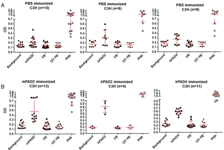
Anti-PAD Antibodies in C3H Mice Immunized with PADs. C3H mice were immunized with murine or human PAD2, or murine or human PAD4, or PBS in Freund’s complete adjuvant (CFA). Three booster injections with PAD or PBS in Freund’s incomplete adjuvant (IFA) were given s.c. 15, 35, and 55 d later (Fig. 1). IgG responses to PADs were analyzed by ELISA. Sera were diluted at 1/40. Positive sera were defined by an OD value higher than twice the background OD (obtained by adding each serum to a well without PAD).
Only murine PAD2, human PAD2, and PAD4 were immunogenic. C3H mice immunized with PBS did not develop anti-PAD an-tibodies (Fig. 2A). The average background OD for wells without PAD was 0.06, and the average OD for wells with PADs was 0.07. In C3H mice immunized with murine PAD2 or human PAD2 or human PAD4, anti-PAD antibodies were detected after first immunization and persisted over time in 28/28 mice immunized with either murine PAD2 or human PAD2 or human PAD4 versus 0/20 mice immunized with PBS (Fisher’s test, P = 6× 10⁻¹⁴) (Fig. 2).
Significance
The presence and development of autoantibodies to citrulli-nated proteins (ACPAs) are highly associated with rheumatoid arthritis (RA). The mechanisms leading to the production of ACPAs are unknown. Here, we propose a model to explain the emergence of anticitrullinated protein autoantibodies in RA. Indeed, we could trigger the development of anticitrullinated fibrinogen autoantibodies in normal mice by immunization with peptidyl arginine deiminase (PAD), most likely through a hapten/carrier mechanism in which the carrier is the PAD en-zyme that performs citrullination and the hapten is any protein being citrullinated (hence bound) by PAD. Our results allow us to understand the birth of anticitrullin autoimmunity.
Author contributions: J.R. and I.A. designed research; F.A., C.M., E.P., and I.A. performed research; N.C.L., N.B., J.R., and I.A. analyzed data; and J.R. and I.A. wrote the paper. The authors declare no conflict of interest.
This article is a PNAS Direct Submission.
This open access article is distributed underCreative Commons
Attribution-NonCommercial-NoDerivatives License 4.0 (CC BY-NC-ND).
1C.M. and E.P. contributed equally to this work.
2To whom correspondence should be addressed. Email: jean.roudier@inserm.fr.
This article contains supporting information online atwww.pnas.org/lookup/suppl/doi:10.
1073/pnas.1713112114/-/DCSupplemental.
MEDICAL
SCIENCES
PNAS
In C3H mice immunized with murine PAD2, the average back-ground OD was 0.06, and the average test OD was 1 after the first immunization, 1.4 after the second immunization, 1.8 after the third immunization, and 1.7 after the fourth immunization (Fig. 2B).
In C3H mice immunized with human PAD2, the average back-ground OD was 0.06, and the average test OD was 0.8 after the first immunization, 0.9 after the second immunization, 1.5 after the third immunization, and 1.6 after the fourth immunization (Fig. 2B).
Fig. 2. IgG responses to PADs in C3H mice. IgG responses to murine PAD2 or human PAD2 or human PAD4 were analyzed by ELISAs. Plates were coated with PADs and blocked with BSA. Sera from primed mice were obtained at 15, 35, 55, and 65 d postimmunization and were diluted at 1/40. (A) For PBS-immunized mice, 15 sera were tested for murine PAD2, 8 sera were tested for human PAD2, and 9 sera were tested for human PAD4. (B) For PAD-immunized mice, each serum was tested against the same PAD used for each immunization. After washing, peroxidase-conjugated antimurine IgG was added. The OD was read at 405 nm. The background OD was obtained by adding each serum to a well without PAD (negative). Positive sera were defined as an OD value higher than twice the background OD.
In C3H mice immunized with human PAD4, the average background OD was 0.07, and the average test OD was 1.1 after the first immunization, 1.8 after the second immunization, 1.6 af-ter the third immunization, and 1.3 afaf-ter the fourth immunization (Fig. 2B).
The presence of anti-PAD antibodies was confirmed by titra-tion assays for 12 C3H mice immunized with PADs (Fig. S1).
T Cell Responses to PADs in C3H Mice Immunized with PADs.Spleen and lymph nodes were obtained 65 d after immunizations with murine PAD2 or human PAD2 or human PAD4 or PBS. T cell proliferation to PADs, native or citrullinated fibrinogen, or phytohemagglutinin (PHA) was evaluated by bromodeoxyrur-idine incorporation. Positive responses were defined by OD values higher than twice the background OD obtained for cells cultured without antigen.
T cell responses to PADs were detected in 24/28 C3H mice immunized with PADs versus 4/20 C3H mice immunized with PBS (Fisher’s test, P = 6 × 10⁻⁶) (Fig. 3).
Indeed, 11/13 C3H mice immunized with murine PAD2, 4/4 C3H mice immunized with human PAD2, and 9/11 C3H mice immunized with human PAD4 developed immunizing PAD-specific T cell responses (Fig. 3B). No T cell response was de-tected for native or citrullinated fibrinogen in mice immunized with PADs or PBS. Among the four mice immunized with PBS who responded to PADs, two recognized murine PAD2, one recognized human PAD2, and one recognized murine PAD2, human PAD2, and human PAD4 (Fig. 3A).
In C3H mice immunized with PBS, the average background OD was 0.17 for cells cultured without antigen, and the average test OD was 0.26 for cells cultured with murine PAD2 or human PAD2 or human PAD4, 0.16 for cells cultured with native or cit-rullinated fibrinogen, and 1.1 for cells cultured with PHA (Fig. 3A). In C3H mice immunized with murine PAD2, the average background OD was 0.18 for cells cultured without antigen, and the average test OD was 0.48 for cells cultured with murine PAD2, 0.17 for cells cultured with native or citrullinated fibrinogen, and 1.1 for cells cultured with PHA (Fig. 3B).
In C3H mice immunized with human PAD2, the average background OD was 0.19 for cells cultured without antigen, and the average test OD was 0.82 for cells cultured with human PAD2, 0.19 for cells cultured with native or citrullinated fibrinogen, and 1.8 for cells cultured with PHA (Fig. 3B).
In C3H mice immunized with human PAD4, the average background OD was 0.25 for cells cultured without antigen, and the average test OD was 0.6 for cells cultured with human PAD4, 0.22 for cells cultured with native or citrullinated fibrinogen, and 1.3 for cells cultured with PHA (Fig. 3B).
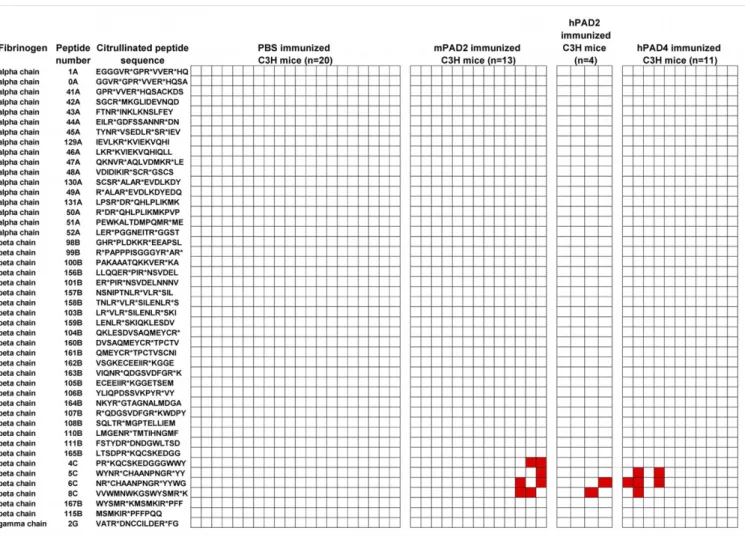
Antibodies to Citrullinated Peptides from Human Fibrinogen in C3H Mice Immunized with PADs. To test whether anticitrullinated peptide antibodies were produced in C3H mice after PAD im-munization, we screened sera from 28 mice immunized with murine PAD2 or human PAD2 or human PAD4 and sera from 20 mice immunized with PBS with 46 citrullinated peptides from the alpha, beta, and gamma chain of human fibrinogen. These
Fig. 3. T cell responses to PADs in C3H mice. Spleen and lymph nodes were obtained at 65 d postimmunization for PBS-immunized mice (A) or PAD-immunized mice (B). Cells were extracted and cultured at 5× 10⁶cells per well with 2μg of proteins or PHA. T cell responses were evaluated by
bromo-deoxyruridine incorporation. Positive responses were defined as an OD value higher than twice the OD for cells cultured without protein.
MEDICAL
SCIENCES
PNAS
brinogen (locus NP_000499) with 50–100% identity with their murine counterparts, 28 15-mers from the beta chain of human fibrinogen (locus NP_005132) with 70–100% identity with their murine counterparts, and one 15-mer from the gamma chain of human fibrinogen (locus NP_068656.2) with 100% identity with its murine counterpart.
We identified four major citrullinated epitopes: peptides 4C, 5C, 6C, and 8C (Fig. 4). These peptides encompassing residues 420–479 of the beta chain of human fibrinogen were recognized by 8/28 sera from mice immunized with PADs versus 0/20 sera from mice immunized by PBS (Fisher’s test, P = 0.014).
The presence of anticitrullinated peptide antibodies was con-firmed by titration assays for six C3H mice immunized with PADs (Figs. S2–S4).
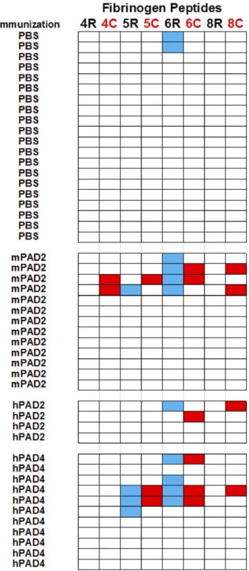
To check for the presence of citrullin residue-specific anti-bodies, we then screened the same sera from 28 mice immunized with murine PAD2 or human PAD2 or human PAD4 and from 20 mice immunized with PBS, with peptides 4, 5, 6, and 8 under their native and citrullinated form (Fig. 5).
IgG responses to native peptides were detected in 10/28 mice immunized with PADs versus 2/20 mice immunized with PBS (Fisher’s test, P = 0.05) (Fig. 5).
6/28 mice immunized with PADs versus 0/20 mice immunized with PBS (Fisher’s test, P = 0.03) (Fig. 5).
Peptide 8C was the most interesting because it was only rec-ognized under its citrullinated form by the sera from 4/28 mice immunized with murine PAD2 or human PAD2 or human PAD4 (Fig. 5).
Influence of MHC Background on Anticitrullinated Peptide Immunization.
To test whether polymorphism of the IEβ chain influences the development of T cell responses to PAD and antibody responses to citrullinated peptides from fibrinogen, we immunized mice express-ing an IEβ allele other than IEβk with PAD.
We chose DBA2 mice whose IEβd chain is similar to that of non-RA–associated HLA-DRB1*0402 (16) (Fig. S5). IgG re-sponses to PADs were analyzed by ELISA. Sera were diluted at 1/40. DBA/2 mice were immunized with murine PAD2 or human PAD4 proteins, which had given the highest anti-PAD antibody titers in C3H mice after PAD immunization.
We detected IgG responses to PADs in 10/10 DBA/2 mice immunized with murine PAD2 and human PAD4 and 0/9 DBA/2 mice immunized with PBS (Fisher’s test, P = 1 × 10⁻⁵) (Fig. 6).
In DBA/2 mice immunized with PBS, the average background OD was 0.06, and the average test OD was 0.06 when adding each serum to a well with murine PAD2 or human PAD4 (Fig. 6A).
Fig. 4. Citrullinated peptides recognized by the sera of C3H mice immunized with PADs. ELISA plates were coated with 46 citrullinated peptides and blocked with BSA. Sera from primed mice obtained at 55 and 65 d postimmunization were diluted at 1/80. After washing, peroxidase-conjugated antimurine IgG was added. The OD was read at 405 nm. The background OD was obtained by adding each serum to a well without peptide. Positive sera were defined as an OD value higher than twice the background OD. A column corresponded to one mouse. IgG to citrullinated peptide is indicated in red.
In DBA/2 mice immunized with murine PAD2, the average background OD was 0.05, and the average test OD was 0.27 after
the first immunization, 0.77 after the second immunization, 0.7 after the third immunization, and 0.8 after the fourth im-munization (Fig. 6B).
In DBA/2 mice immunized with human PAD4, the back-ground OD was 0.06, and the average test OD was 0.44 after the first immunization, 0.47 after the second immunization, 0.88 af-ter the third immunization, and 0.93 afaf-ter the fourth immuni-zation (Fig. 6B).
Anti-PAD antibody levels (as indicated by test ODs) were lower in DBA/2 than in C3H mice after immunization with PADs.
The presence of anti-PAD antibodies was confirmed by titra-tion assays for 10 DBA/2 mice immunized with PADs (Fig. S6). T cell responses to PADs were detected in 8/10 DBA/2 mice immunized with murine PAD2 and human PAD4 and 3/9 DBA/2 mice immunized with PBS (Fisher’s test, P = 0.07) (Fig. 7).
Indeed, in DBA/2 mice immunized with PBS, the average background OD was 0.15 in cells cultured without antigen, and the average test ODs were 0.2 in cells cultured with murine PAD2 or human PAD4, 0.14 in cells cultured with native or citrullinated fibrinogen, and 0.35 in cells cultured with PHA (Fig. 7).
In DBA/2 mice immunized with murine PAD2, the average background OD was 0.18 in cells cultured without antigen, and the average test OD was 0.37 in cells cultured with murine PAD2, 0.17 in cells cultured with native or citrullinated fibrin-ogen, and 0.6 in cells cultured with PHA (Fig. 7).
In DBA/2 mice immunized with human PAD4, the average background OD was 0.18 in cells cultured without antigen, and the test ODs were 0.53 obtained in cells cultured with human PAD4, 0.18 in cells cultured with native or citrullinated fibrin-ogen, and 0.61 in cells cultured with PHA (Fig. 7).
T cell responses (as indicated by test ODs) to murine PAD2 were lower in DBA/2 mice immunized with murine PAD2 than in C3H mice immunized with murine PAD2. T cell responses (as indicated by test ODs) to human PAD4 were similar in DBA/2 and C3H mice immunized with human PAD4. No T cell re-sponses were detected in native or citrullinated fibrinogen in DBA/2 mice immunized with PADs or PBS. T cell responses to PHA (as indicated by test ODs) were lower in DBA/2 than in C3H mice. DBA/2 mice are known to have the lowest PHA responsiveness of all classical mouse strains, whereas C3H are among the low responders (17).
Finally, we screened sera from mice immunized with PBS or murine PAD2 or human PAD4 on the 46 citrullinated peptides from the alpha, beta, and gamma chain of human fibrinogen with 50–100% homology with their murine counterparts. No IgG re-sponse to citrullinated fibrinogen peptides was detected in any of the DBA/2 mice immunized with PADs or PBS (Fig. 8). The lack of anticitrullinated peptide antibodies was confirmed by titration assays for seven DBA/2 mice immunized with PADs (Figs. S2–S4). Discussion
In this study, we tested whether immunization to PAD proteins triggers ACPAs through a hapten/carrier mechanism in which the carrier is PAD, which performs citrullination, and the hapten is any protein being citrullinated by PAD.
Indeed, the sera of two-thirds of patients with RA contain anticitrullinated peptide autoantibodies. These IgG antibodies recognize citrullin residues on different proteins. ACPAs are central to the development of RA. However, the mechanisms leading to the production of ACPAs are unknown.
A tentative model proposes that cigarette smoking may induce citrullination of proteins in the lungs. HLA-DRB1 alleles asso-ciated with RA would then bind citrullinated peptides and pre-sent them to helper T cells. This mechanism would explain anticitrullinated protein immunization in patients whose HLA-DRB1 molecules express the shared epitope. However, this model has serious limitations:
Fig. 5. PAD immunization in C3H mice triggers production of anticitrullinated fibrinogen peptide antibodies. Plates were coated with peptides in unmodified forms (4R, 5R, 6R, and 8R) or citrullinated forms (4C, 5C, 6C, and 8C). After blocking, sera from primed mice obtained at 55 or 65 d postimmunization were diluted at 1/80. After washing, peroxidase-conjugated antimurine IgG was added. The OD was read at 405 nm. The background OD was obtained by adding each serum to a well without peptide. Positive sera were defined as an OD value higher than twice the background OD. A line corresponded to one mouse. IgG to native peptide is indicated in blue, and IgG to citrullinated peptide is indicated in red.
MEDICAL
SCIENCES
PNAS
i) RA may occur in nonsmokers.
ii) IgG antibodies against citrullinated proteins usually develop in the absence of identified helper T cell response. iii) The preferential binding of citrullinated peptides to shared
epitope-positive HLA-DRB1 alleles has never been properly demonstrated. Indeed, the article that was supposed to have demonstrated the point studied the binding of one unique citrullinated vimentin peptide to three shared epitope-positive and five shared epitope-negative HLA-DRB1 alleles (11). Conversely, thorough studies of the binding of hundreds of peptides to shared epitope-positive or -negative HLA-DRB1 alleles did not show preferential binding of citrulli-nated peptides to shared epitope-positive alleles, compared with their native counterparts (12–14).
Thus, the identity of the antigen(s) whose recognition by T cells can help the development of IgG autoantibodies directed at citrullinated proteins is still unknown.
We focused on PAD2 and PAD4 as candidate antigens in anticitrullinated protein immunization for several reasons:
i) Anti-PAD4 IgG autoantibodies are detectable during the preclinical phase of RA in one-third of patients and are associated with ACPA positivity (18–24).
ii) PAD2 and PAD4 expressed in the RA synovium (25) are involved in the citrullination of fibrin, the major synovial autoantigen in RA (10).
iii) PADI4, the gene encoding PAD4, is a susceptibility locus for RA in Asians and in some, but not every, Western populations
65 d postimmunization and were diluted at 1/40. (A) For PBS-immunized mice, nine sera were tested for murine PAD2 and human PAD4. (B) For PAD-immunized mice, each serum was tested against the same PAD used for each immunization. After washing, peroxidase-conjugated antimurine IgG was added. The OD was read at 405 nm. The background OD was obtained by adding each serum to a well without PAD (negative). Positive sera were defined as an OD value higher than twice the background OD.
(26). Increased susceptibility to develop RA might be due by higher stability of PAD4 transcripts and thus higher expression of PAD4 (27).
To test whether we could induce ACPAs by immunizing nor-mal mice with PADs, we used C3H mice whose IEβk chain is similar to that of RA-associated HLA-DRB1*04:01. We immu-nized C3H mice with four PAD proteins (murine and human PAD2 and PAD4) and obtained IgG and T cell responses to PAD after immunization with murine and human PAD2 or with human PAD4.
No T cell response was observed to native or citrullinated fi-brinogen in C3H mice immunized with PADs.
To detect anticitrullinated protein antibodies, we used 46 cit-rullinated peptides from human fibrinogen with 50–100% ho-mology with their murine counterparts. Among these 46 peptides, four were preferentially recognized by sera from primed mice. These peptides are located in the C-terminal domain of human beta fibrinogen (amino acids 420–479). IgG responses to citrulli-nated fibrinogen peptides were only detected in C3H mice im-munized with murine or human PAD2 or human PAD4.
Fig. 7. T cell responses to PADs in DBA/2 mice. Spleen and lymph nodes were obtained at 65 d postimmunization. Cells were extracted and cultured at 5× 10⁶
cells per well with 2μg of proteins or PHA. T cell responses were evaluated by bromodeoxyruridine incorporation. Positive responses were defined as an OD value higher than twice the OD for cells cultured without protein.
Fig. 8. Citrullinated peptide detection in the sera of DBA/2 mice immunized with PADs or PBS. ELISA plates were coated with 46 citrullinated peptides and blocked with BSA. Sera from primed mice obtained at 55 and 65 d postimmunization were diluted at 1/80. After washing, peroxidase-conjugated antimurine IgG was added. The OD was read at 405 nm. The background OD was obtained by adding each serum to a well without peptide. Positive sera were defined as an OD value higher than twice the background OD. A column corresponded to one mouse.
MEDICAL
SCIENCES
PNAS
We also used DBA/2 mice whose IEβd chain is similar to that of non-RA–associated HLA-DRB1*04:02. No IgG responses to citrullinated fibrinogen peptides were detected in DBA/2 mice immunized with PADs.
Our data suggest a model for the HLA and T cell contribution to the production of IgG anticitrullinated proteins in RA. In this model, HLA-DR alleles bind PAD peptides. T lymphocytes recognize PAD peptides and help production of antibodies to any antigen being citrullinated by PAD (Fig. 9).
A corollary of this model is that the molecular basis for the as-sociation between HLA-DRB1 alleles like HLA-DRB1*04:01 and HLA-DRB1*01:01 and RA is the capability of these alleles to bind peptides derived from PAD.
It must be noted that any protein (even bacterial) with PAD activity may be the target of helper T cells that may help pro-duction of IgG antibodies to any protein under citrullination. In this respect, the aggravating (but not mandatory) role of tobacco smoking on RA susceptibility might occur through increased periodontal disease. Indeed, porphyromonas gingivalis, the bacte-rium most associated with periodontal disease, has a PAD protein of its own. Peptides from this bacterial PAD might bind HLA-DR and be recognized by helper T cells. This is consistent with the fact that residues 11 and 13, located on the floor of the HLA-DRB1 molecule and influencing peptide binding, contribute a major ef-fect in RA susceptibility, which is enhanced by tobacco smoking (28, 29).
It remains to be determined whether the mere production of ACPAs is enough to induce arthritis. Of interest, in vitro studies in humans suggest that ACPA/rheumatoid factor immune com-plexes activate macrophages and induce production of in-flammatory cytokines including TNF alpha (30).
However, the goal of this article was only to demonstrate the production of ACPA after PAD immunization in mice through a hapten/carrier mechanism.
Whether this may trigger arthritis will be the subject of fu-ture studies.
Materials and Methods
Mice. C3H/HeNHsd and DBA/2 mice were purchased from Envigo Labora-tories. Mice were housed at the Luminy INSERM Institute, Marseille (A13 01303). All experiments were approved by the animal ethics committee (no. 03007.03). All animal care and experimental procedures were
and the Ministère de l’Enseignement Supérieur et de la Recherche. Proteins. Murine PAD2 and PAD4 and human PAD2 and PAD4 proteins were purchased from Proteogenix. They were produced in baculovirus and purified. Their activity and autocitrullination status were tested before immunization.
Citrullinated Proteins. Citrullinated fibrinogen, the most likely target antigen of ACPA, expressed in the synovial tissue of RA patients, allows ACPA de-tection with the same efficiency as the anticyclic peptide antigen kits (anti-CCP2) in RA. Citrullinated human fibrinogen (Calbiochem and Oxford Bio-medical Research) was obtained after incubation in 1 M Tris·HCl (pH7.4), 100 mM CaCl2, 50 mM DTT buffer at a concentration of 1 mg/mL with rabbit PAD2 (Sigma Aldrich), for 2 h at 37 °C. Noncitrullinated proteins were treated identically, except that water was added instead of rabbit PAD2. Immunization Protocols. C3H or DBA/2 mice were immunized s.c. with 100μg of murine or human PAD2, or murine or human PAD4, or PBS in CFA. Three booster injections with 100μg of PADs or PBS in IFA were given s.c. 15, 35, and 55 d later.
Detection of Anti-PAD Antibodies. Sera from primed mice were obtained at days 15, 35, 55, and 65 and tested by ELISA. Plates were coated overnight with PADs. Plates were blocked with 2% BSA. Sera were incubated. After washing with Tween 20, peroxidase-conjugated antimurine IgG was added. The OD was read at 405 nm. Background OD was obtained by adding each serum to a well without protein. Positive sera were defined as an OD value higher than twice the background OD.
Synthetic Peptides. Each peptide was synthesized using a solid-phase system (Neosystem) and purified. We started with 46 citrullinated peptides from the alpha, beta, and gamma chain of human fibrinogen and used them for ELISAs in 67 mice. We tested 17 15-mers from the alpha chain of human fibrinogen (locus NP_000499) with 50–100% identity with their murine counterparts, 28 15-mers from the beta chain of human fibrinogen (locus NP_005132) with 70–100% identity with their murine counterparts, and one 15-mer from the gamma chain of human fibrinogen (locus NP_068656.2) with 100% identity with its murine counterpart. Peptides 4C, 5C, 6C, and 8C from the beta chain of human fibrinogen were the targets of all of the anticitrullinated fibrin-ogen peptide antibody responses. They are 93–100% identical to their mu-rine counterpart. They encompass residues 420–479 of the beta chain of human fibrinogen (locus NP_005132). The arginine version of these four peptides was used to confirm citrullinated peptide specificity of antici-trullinated fibrinogen antibodies.
Detection of Anticitrullinated Peptide Antibodies. Sera from primed mice were obtained at days 55 and 65 and tested for anticitrullinated peptide antibodies by ELISA. Plates were coated with human fibrinogen peptides under cit-rullinated and native forms. After blocking, sera from primed mice were incubated with peptides. After washing, peroxidase-conjugated antimurine IgG was added. The OD was read at 405 nm. Background OD was obtained by adding each serum to a well without peptide. Positive sera were defined as an OD value higher than twice the background OD.
T Cell Proliferation Assays. Spleen and lymph nodes were obtained at day 65 postimmunization and used in antigen-specific proliferation assays. T cell responses to PADs, native or citrullinated fibrinogen, or PHA were evaluated using the colorimetric bromodeoxyruridine kit (Roche Diagnostics) (31). Back-ground OD was obtained for cells cultured without antigen. Positive responses were defined as an OD value higher than twice the background OD. Statistical Analysis. Comparisons between groups were performed using Fisher’s exact test.
ACKNOWLEDGMENTS. This study was supported by INSERM and Fondation Arthritis Courtin.
1. Symmons DPM (1995) What is rheumatoid arthritis? Br Med Bull 51:243–248. 2. Gregersen PK, Silver J, Winchester RJ (1987) The shared epitope hypothesis. An
ap-proach to understanding the molecular genetics of susceptibility to rheumatoid
ar-thritis. Arthritis Rheum 30:1205–1213.
3. Ollier W, Thomson W (1992) Population genetics of rheumatoid arthritis. Rheum Dis Clin North Am 18:741–759.
4. Balandraud N, et al. (2013) HLA-DRB1 genotypes and the risk of developing anti citrullinated protein antibody (ACPA) positive rheumatoid arthritis. PLoS One 8: e64108.
5. Schellekens GA, de Jong BA, van den Hoogen FH, van de Putte LB, van Venrooij WJ (1998) Citrulline is an essential constituent of antigenic determinants recognized by rheumatoid arthritis-specific autoantibodies. J Clin Invest 101:273–281.
Fig. 9. Hapten (citrullinated proteins)/carrier (PAD) model. PAD may be the carrier seen by the helper T cells, which help production of antibodies to any protein being citrullinated.
6. van Venrooij WJ, Pruijn GJ (2000) Citrullination: A small change for a protein with great consequences for rheumatoid arthritis. Arthritis Res 2:249–251.
7. Simon M, et al. (1993) The cytokeratin filament-aggregating protein filaggrin is the
target of the so-called“antikeratin antibodies,” autoantibodies specific for
rheu-matoid arthritis. J Clin Invest 92:1387–1393.
8. Girbal-Neuhauser E, et al. (1999) The epitopes targeted by the rheumatoid arthritis-associated antifilaggrin autoantibodies are posttranslationally generated on various sites of (pro)filaggrin by deimination of arginine residues. J Immunol 162:585–594. 9. Vossenaar ER, et al. (2004) Rheumatoid arthritis specific anti-Sa antibodies target
citrullinated vimentin. Arthritis Res Ther 6:R142–R150.
10. Masson-Bessière C, et al. (2001) The major synovial targets of the rheumatoid arthritis-specific antifilaggrin autoantibodies are deiminated forms of the alpha- and beta-chains of fibrin. J Immunol 166:4177–4184.
11. Hill JA, et al. (2003) Cutting edge: The conversion of arginine to citrulline allows for a high-affinity peptide interaction with the rheumatoid arthritis-associated HLA-DRB1*0401 MHC class II molecule. J Immunol 171:538–541.
12. Auger I, et al. (2005) Influence of HLA-DR genes on the production of rheumatoid arthritis-specific autoantibodies to citrullinated fibrinogen. Arthritis Rheum 52:3424–3432. 13. Pratesi F, et al. (2012) Effect of rheumatoid arthritis (RA) susceptibility genes on the immune response to viral citrullinated peptides in RA. J Rheumatol 39:1490–1493. 14. Sidney J, et al. (2017) Citrullination only infrequently impacts peptide binding to HLA
class II MHC. PLoS One 12:e0177140.
15. Auger I, et al. (2009) New autoantigens in rheumatoid arthritis (RA): Screening 8268 protein arrays with sera from patients with RA. Ann Rheum Dis 68:591–594.
16. Widera G, Flavell RA (1984) The nucleotide sequence of the murine I-Eβbimmune
response gene: Evidence for gene conversion events in class II genes of the major
histocompatibility complex. EMBO J 3:1221–1225.
17. Heiniger HJ, Taylor BA, Hards EJ, Meier H (1975) Heritability of the phytohemag-glutinin responsiveness of lymphocytes and its relationship to leukemogenesis. Cancer Res 35:825–831.
18. Takizawa Y, et al. (2005) Peptidylarginine deiminase 4 (PADI4) identified as a conformation-dependent autoantigen in rheumatoid arthritis. Scand J Rheumatol 34: 212–215.
19. Roth EB, Stenberg P, Book C, Sjöberg K (2006) Antibodies against transglutaminases, peptidylarginine deiminase and citrulline in rheumatoid arthritis–New pathways to epitope spreading. Clin Exp Rheumatol 24:12–18.
20. Halvorsen EH, et al. (2008) Serum IgG antibodies to peptidylarginine deiminase 4 in rheumatoid arthritis and associations with disease severity. Ann Rheum Dis 67: 414–417.
21. Zhao J, Zhao Y, He J, Jia R, Li Z (2008) Prevalence and significance of anti-peptidylarginine deiminase 4 antibodies in rheumatoid arthritis. J Rheumatol 35: 969–974.
22. Auger I, Martin M, Balandraud N, Roudier J (2010) RA specific autoantibodies to PAD4 inhibit citrullination of fibrinogen. Arthritis Rheum 62:126–131.
23. Kolfenbach JR, et al. (2010) Autoimmunity to peptidyl arginine deiminase type 4 precedes clinical onset of rheumatoid arthritis. Arthritis Rheum 62:2633–2639. 24. Pollmann S, et al. (2012) Anti-PAD4 autoantibodies in rheumatoid arthritis: Levels in
serum over time and impact on PAD4 activity as measured with a small synthetic substrate. Rheumatol Int 32:1271–1276.
25. Foulquier C, et al. (2007) Peptidyl arginine deiminase type 2 (PAD-2) and PAD-4 but not PAD-1, PAD-3, and PAD-6 are expressed in rheumatoid arthritis synovium in close association with tissue inflammation. Arthritis Rheum 56:3541–3553.
26. Koushik S, et al. (2017) PAD4: Pathophysiology, current therapeutics and future perspective in rheumatoid arthritis. Expert Opin Ther Targets 21:433–447. 27. Suzuki A, et al. (2003) Functional haplotypes of PADI4, encoding citrullinating
en-zyme peptidylarginine deiminase 4, are associated with rheumatoid arthritis. Nat Genet 34:395–402.
28. Raychaudhuri S, et al. (2012) Five amino acids in three HLA proteins explain most of the association between MHC and seropositive rheumatoid arthritis. Nat Genet 44: 291–296.
29. Kim K, et al. (2015) Interactions between amino acid-defined major histocompatibility complex class II variants and smoking in seropositive rheumatoid arthritis. Arthritis Rheumatol 67:2611–2623.
30. Anquetil F, Clavel C, Offer G, Serre G, Sebbag M (2015) IgM and IgA rheumatoid factors purified from rheumatoid arthritis sera boost the Fc receptor- and complement-dependent effector functions of the disease-specific anti-citrullinated protein autoantibodies. J Immunol 194:3664–3674.
31. Porstmann T, Ternynck T, Avrameas S (1985) Quantitation of 5-bromo-2-deoxyuridine incorporation into DNA: An enzyme immunoassay for the assessment of the lymphoid cell proliferative response. J Immunol Methods 82:169–179.
MEDICAL
SCIENCES
PNAS