Transport of chloramphenicol into sensitive strains of
Escherichia coli and Pseudomonas aeruginosa
Saad Abdel-SayedInstitut de Microbiologie de I'Universite de Lausanne, Centre Hospitaller Universitaire Vaudois, Rue du Bugnon 44, 1011 Lausanne, Switzerland
The uptake of chloramphenicol by susceptible strains of Escherichia coli and Pseudomonas aeruginosa was measured as the depletion of 14C-chloramphenicol from the supernatant of centrifuged cultures. Chloramphenicol did not bind to non-growing cells or isolated cell envelopes. Chloramphenicol was recovered from cells in an unchanged form and was intracellularly concentrated several times above external concentrations. The net accumulation of the drug was reduced by an inhibitor of electron transport, by an oxidative phosphorylation uncoupler, by an inhibitor of high energy phosphate synthesis, and by lowering the temperature to + 15°C. The initial uptake of drug was saturated at 1 -98 mM chloramphenicol in the medium. A 100-fold excess of each of the unlabelled isomers: L-threo, D-threo, and L-erythro chloramphenicol in cultures of either strain effectively reduced the uptake of 14C-chloramphenicol. These results indicate that chloramphenicol enters Gram-negative bacteria by means of an energy-dependent process.
Introduction
Chloramphenicol is an antibiotic that inhibits protein synthesis in procaryotic cells. It binds primarily to protein L 16 of bacterial ribosomes and blocks peptide bond formation by inactivating peptidyltransferase (Nierhaus & Nierhaus, 1973). Since the target for chloramphenicol is intracellular, the drug must penetrate the cell envelope of bacteria.
The accumulation of chloramphenicol by Staphylococcus aureus strain Duncan and
Bacillus megaterium strain KM (NCIB 9521) was described by Vasquez (1963, 1966).
He showed that the rate of uptake was initially high but fell rapidly and that after two minutes a much slower rate was established. The magnitude of uptake was directly related to the external drug concentration and could be greatly reduced by peptidyl transferase inhibitors such as ostreogrycins group A and group B, but was unaffected by tetracyclines, inhibitors of the binding of aminoacyl-tRNA to the 30 S ribosomal subunit or by puromycin, an inhibitor of peptide chain elongation functioning as an analogue of terminal aminoacyl-tRNA.
Irvin & Ingram (1980) isolated two chloramphenicol-resistant mutants of
Pseudomonas aeruginosa which also exhibited decreased amino-acid accumulation.
Neither of these mutants inactivate chloramphenicol by acetylation and both possess ribosomes sensitive to the action of chloramphenicol. Since uptake of amino-acids by bacteria involves active transport (Anraku, 1978), the data of Irvin & Ingram (1980) could be interpreted as showing an energy-dependent chloramphenicol uptake.
7
However, the chloramphenicol-resistant mutants may accumulate decreased amounts of amino-acids and chloramphenicol owing to loss of outer membrane porins (Foulds & Chai, 1978; Pugsley & Schnaitman, 1978). Thus, the evidence for active accumulation of chloramphenicol remains equivocal.
Bacterial resistance to chloramphenicol can be produced by at least three different mechanisms: enzymatic covalent modification of the molecular structure, alteration of the cells' permeability, and alteration of the ribosomal binding site (Foster, 1983). Studies on the uptake of chloramphenicol by a number of Gram-positive bacteria that do not covalently modify it have shown that there is a correlation between the rate of uptake of the antibiotic by the organisms and their sensitivity to it (Vasquez, 1964). In all the studies reported above the cells were washed to remove external molecules before intracellular concentration of chloramphenicol was determined. Chloramphenicol is so weakly held in the cells that washing itself can remove intracellular antibiotic (Vasquez, 1963, 1964). In the investigation of chloramphenicol transport into bacteria reported here a centrifugation method was used to determine chloramphenicol uptake without washing of the cells.
Materials and methods
Bacterial strains
The organisms used for this study were Escherichia coli K 12 strain 703 (Hammersmith wild type F~) and P. aeruginosa ATCC 9027. The MIC of chloramphenicol for the
E. coli strain was 6-25 mg/1, and for the P. aeruginosa strain 25 mg/1, determined by
dilution technique in BYGNaCl medium (see below).
Media and growth conditions
The organisms were maintained on nutrient agar slants at room temperature. When used for experiments, cells were grown aerobically until mid-exponential phase at 37°C on a gyratory Adolf Kiihner shaker at 250 r.p.m. Erlenmeyer flasks (100 ml) each containing 30 ml of growth medium, BYGNaCl medium: 13 g nutrient broth (Oxoid), 5 g yeast extract (BBL), 5 g NaCl, 1 g glucose per litre, adjusted to pH 7 (Lennox, 1955), was used for most of these studies. Where noted a BM2 medium composed of 2-5 g glucose, 7 g K2HPO4) 3-4 g KH2PO4, 1 g (NH4)SO4, and 0024g MgSO4 . H2O per litre was used (Gilleland, Stinett & Eagon, 1974).
Chloramphenicol uptake
Cells grown in BYGNaCl medium were harvested by centrifugation at 6000 r.p.m. for lOmin and then resuspended in the same volume of unused prewarmed BYGNaCl medium at 37°C. Of this suspension 1-1-8 ml were transferred to 25-ml Erlenmeyer flasks, and a mixture of 14C-chloramphenicol (46-9 mCI/mmol) with unlabelled chloramphenicol was added to produce final volumes of 2 ml with various concentrations of chloramphenicol. Final specific activities ranged from 0-63 to 32-3 mCi/mmol. The flasks were incubated at 37°C on a reciprocal shaking water bath. At 5 min intervals, aliquots of 0-2 ml were removed and centrifuged in a Kontron centrifuge (type Hermle z635-K) at 6000 r.p.m. for 1 min at 25°C. The upper 0-1 ml of the supernatant was carefully removed to a scintillation vial containing 10 ml of
scintillation fluid. This was composed of ethanol, Triton X-100 and toluene in the ratio of 1:3-3: 5-7 containing 0-572% (w/v) diphenyloxazole and 0014% (w/v) 1-4-bis[2-(4-methyl-5-phenyloxazonyl)]-benzene. The radioactivity was determined with a Unilux model scintillation spectrometer (Nuclear Chicago). The amount of cell-associated chloramphenicol was determined by subtracting the amount of the residual 14 C-chloramphenicol in the supernatant from the amount of 14C-chloramphenicol in the cell suspension.
Radiochemical purity
The cells from 20 ml of actively growing culture were suspended in 2 ml of unused BYGNaCl medium maintained at 37°C. 14C-Chloramphenicol was added to produce a specific activity of 2-5 mCi/1 and the cell suspension was incubated at 37°C on a reciprocal shaking water bath. After 5 min of labelling the cells were harvested by filtration on Millipore membrane filters and immediately washed by filtering 2-5 ml of 0 1 M NaCl. The membrane filters bearing the cells were transferred to 25-ml Erlenmeyer flasks containing 3 ml distilled water and the cells were detached from the filter membranes by magnetic stirring. The cell suspension was then submitted to three successive extractions with 3 ml diethyl ether. The pooled extracts were evaporated to dryness in an air stream. The samples, dissolved in 01 ml ethyl acetate, were spotted by repeated applications of a total volume of 002 ml at the origin of a thin layer silica gel (Kisel gel 60 F245, 0-25 mm, Merck) alongside a control preparation of 14 C-chloramphenicol. The sheets were submitted to ascending chromatography in a chloroform : methanol 90 : 10 (v/v) solvent system. The chromatograms were air dried and subjected to autoradiography for 66 hours with Kodak X-Omat AR-5 film.
Measurement of protein synthesis
Exponentially growing cells were harvested and resuspended in BYGNaCl medium at 37°C at A660 = 0-67. From this suspension, 0-97-ml amounts were transferred to 25-ml Erlenmeyer flasks containing 003 ml (0-319 mCi) of 35S-methionine with or without chloramphenicol. The flasks were rapidly mixed and subsequently held on a reciprocal shaking water bath at 37°C. At time intervals, samples of 005 ml were removed and spotted on to Whatman no. 3 MM discs pretreated with trichloroacetic acid. After drying at room temperature, all discs were processed simultaneously as described by Levy (1971) to evaluate 35S-methionine incorporated into trichloroacetic acid-precipitable protein.
Cells preincubated in medium unable to support growth
Cells grown in BYGNaCl medium were harvested by centrifugation and washed three times with 50 HIM phosphate buffer, pH 7. Washed cells were suspended to the original volume of the same buffer and incubated at 37°C with shaking for 2 h. The cells were centrifuged and resuspended in 1 ml phosphate buffer per mg of cell protein for equilibrium dialysis.
Transport assays
The uptake of [U-14C] protein hydrolysate by both strains was determined at 37°C to check the active accumulation of amino-acids by cells preincubated in medium unable to support growth. Cells were washed as described above and suspended in 50 mM phosphate buffer, pH 7, to a density of 2 g wet weight per 10 ml. The suspensions were used for uptake assays. The uptake mixtures contained 0-2 ml of cell suspension and 4-4 nanoatom carbon of [U-14C] protein hydrolysate (5-7 mCi/milliatom carbon) in a final volume of 0-5 ml. The reaction was started by addition of substrate. At time intervals, 005 ml samples were removed and delivered over membrane filters (25 mm diameter; 0-45 /zm pore size; Millipore corporation, Bedford, MA, U.S.A.) which had been pretreated by filtering 5 ml 0-1 M LiCl. The samples were filtered instantaneously and the filters were immediately washed with 5 ml 01 M LiCl. The filters bearing the cells were removed from the suction apparatus and transferred to vials containing
10 ml of the scintillation fluid. The radioactivity was determined as described above.
Spheroplast preparation and spheroplast uptake
Spheroplasts were prepared from the E. coli strain according to the method of Weiss (1976) with slight modifications. Cells were grown in BYGNaCl medium. Deoxyribonuclease type I and II, and ribonuclease were added to a final concentration of 005 mg/1 directly after the addition of lysozyme. Spheroplasts were harvested by centrifugation at 3600 g for 15 min and subsequently washed once with 0-1 M Tris-HCl buffer, pH 8, containing 20% sucrose. Washed spheroplasts were suspended into the original volume of a solution of 20% sucrose in 50 mM phosphate buffer, pH 7, or in sucrose BYGNaCl medium. Samples of 1 ml of these suspensions, when diluted ten times in water, decreased in optical density at 660 nm ten times more than that diluted in 20% sucrose. The spheroplast suspensions were used directly for uptake assays. The uptake mixtures contained 0-9 ml of spheroplast suspension and 10 ng of 14 C-chloramphenicol (6-16mCi/mmol) in a final volume of 1 ml. The reaction was started by the addition of substrate. After 5 min of incubation at 37°C, 0-2 ml was removed and centrifuged at 3000 g for 2 min. The upper 0-1 ml of the supernatant was carefully removed and processed as described above to evaluate 14C-chloramphenicol incorporated into spheroplasts.
Miscellaneous assays and techniques
Protein was determined according to the method of Markwell et al. (1978). Cell envelopes were prepared as described by Hancock & Nikaido (1978). Equilibrium dialysis was carried out in a multichamber apparatus by the procedure of Furlong
et al. (1972).
Reagents
D-threo (dichloro-l,2-'*C) chloramphenicol (46-9 mCi/mmol) and 3SS-methionine (1058 Ci/mmol) were purchased from New England Nuclear Corp., Boston, Mass., U.S.A.; [U-14C]-Protein hydrolysate, composed of 16 amino-acids (57 mCi/milliatom carbon), from Amersham, Buckinghamshire, England. Non-radioactive
chloramphenicol was a gift from Le Petit Co., Milan, Italy. Chloramphenicol isomers were a gift from Warner Lambert Co., Ann Arbor, Mich. All other reagents were purchased from normal commercial sources.
Results
Uptake of chloramphenicol
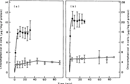
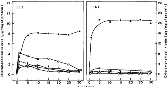
Figure 1 illustrates the time course of the uptake of chloramphenicol for E. coli and
P. aeruginosa in BYGNaCl medium at concentrations of 10 and 40 mg/1 respectively.
Uptake of the drug by either bacterium demonstrated biphasic kinetics: a rapid uptake, which ceased at the time the first sample was taken (1 min) and a slower uptake, which then continued for 5 min; thereafter, the uptake reached a plateau of a period of 5-30 min. With a value of 2-7 ml cell water per g dry weight of bacteria assumed (Winkler & Wilson, 1966), the concentration of cell-associated chloramphenicol after 1 min of incubation for E. coli was 689-7 mg/1 cell water at a chloramphenicol concentration in the medium of 10 mg/1. For P. aeruginosa, the concentration of cell-associated chloramphenicol was 2595-3 mg/1 cell water at a chloramphenicol concentration in the medium of 40 mg/1. Thus, after 1 min of incubation the concentration of chloramphenicol accumulated by E. coli and
P. aeruginosa reached about 69 and 65 times the concentration in the medium,
respectively. After 5 min of uptake the concentration of chloramphenicol respectively accumulated by E. coli and P. aeruginosa exceeded about 140 and 97 times the concentration in the medium. Lowering the reaction temperature to + 15°C produced a marked change in both rates of uptake by either type of bacterium. Furthermore, the slow uptake appeared to last longer than usually seen at 37°C (Figure 1).
•E 12
-o>
2
-2 0 40 6 0 20 4 0 6 0 80
Time (min)
Figure 1. Uptake of chloramphenicol with time in BYGNaCl medium at 37°C ( • ) , and 15°C (O), >n the presence of 10 mg chloramphenicol/1 for E. coli (a) and 40 mg chloramphenicol/1 for P. aeruginosa (b). Bars represent standard errors of the mean for four experiments.
Table I. Effect of unlabelled chloramphenicol and chloramphenicol isomers on
14C-chloramphenicol uptake by E. coli and P. aeruginosa
Bacteria E. coli P. aeruginosa Unlabelled substance Control CM L-threo-CM D-erythro-CM L-erthryo-CM Control CM L-threo-CM D-erythro-CM L-erythro-CM 14C-CM taken up" (/ig/mg protein) 13200 + 5400 0 4 5 0 0 + 1400 0 2 5 0 0 + 800 7 3 0 0 + 1 0 0 0 3-15+2-22 1-30+1-14 0 2 5 0 0 + 800 % inhibition of uptake 0 100 66 100 81 0 96 98 100 66
"CM, chloramphenicol; in the presence of 14C-CM and unlabelled substance at concentrations of
3xlO"6M and 3 X 1 0 "4M respectively; accumulation period 1 min in BYGNaCl medium. Values were
obtained from two experiments.
Measured after 1 min, the uptake of 14C-chloramphenicol by either type of bacterium was greatly reduced by the unlabelled chloramphenicol isomers: L-threo and L-erythro when each was added at 100 times the concentration of 14C-chloramphenicol (Table I). The unlabelled chloramphenicol and the D-erythro isomer completely inhibited the uptake of 14C-chloramphenicol. MgSO4 reduced the rate of uptake of chloramphenicol by both strains (Table II) and also elevated the MIC respectively two and four times for E. coli and P. aeruginosa, when added to a final concentration of 5 mM. Similar results were obtained with MgCl2 (Table II) indicating that the
Table II. Inhibition of chloramphenicol uptake by magnesium sulphate
Bacteria E. coif P. aeruginosa1' M g+ + added (mM) 0 0 0 0-50 100 1 50 2 0 0 2 0 0 0 0 0 0-50 100 1 50 2 0 0 2 0 0 Cell associated CM 0ig/mg protein) 7-85 ± 0 1 5 4-90 + 0-20 4 1 0 + 0-60 2-35 + 0-35 1-85 + 0-05 (1 10+014)' 21-40 + 3-00 15-45+1-25 14-70 + 3-10 13 65 + 0-25 11-15+1-55 (6-15±0-21) CM, chloramphenicol; 'chloramphenicol concentration, lOmg/1, accumu-lation period, 5 min in BYGNaCl medium; 'chloramphenicol concentration, 40mg/l; accumulation period, 5 min in BYGNaCl medium; cthe numbers in
parentheses are the uptake of CM in the presence of MgCl2; values were
inhibition of chloramphenicol uptake by MgSO4 was due to M g+ + effect and not to sulphate. Cells grown in minimal BM2 medium exhibited a reduced uptake of chloramphenicol compared with those grown in BYGNaCl medium, with values of 27% and 14% for E. coli and P. aeruginosa, respectively. Cells grown in BM2 medium were thus phenotypically more resistant. For example, the MIC for the P. aeruginosa strain harvested from BM2 medium was 100 mg/1, whether the test medium was BM2 or BYGNaCl. In contrast, cells grown in BYGNaCl and transferred into BM2 were not able to incorporate 14C-chloramphenicol.
Chloramphenicol extracted from cells
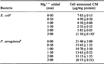
More than 90% of accumulated chloramphenicol was recovered from cells by ether extraction. Autoradiography of thin layer chromatograms demonstrated that the J?f value (0-68) of extracted chloramphenicol was identical to that of a control preparation of 14C-chloramphenicol (Figure 2). These results clearly indicated that the majority of accumulated chloramphenicol was recovered from cells in an unchanged form.
Figure 2. Autoradiographic demonstration of a thin layer chromatogram of chloramphenicol extracted from prelabelled cells of E. coli (a), P. aeruginosa (c), and of a control preparation of 14C-chloramphenicol
Equilibrium dialysis
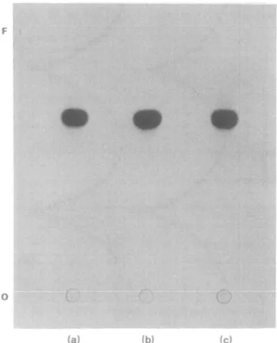
As shown by Figure 3, chloramphenicol passed through the dialysis membrane without being bound by either non-growing cells or isolated cell envelopes.
Uptake of amino-acids by cells preincubated in medium not supporting growth
Figure 4 illustrates the time-course of the uptake of labelled amino-acids for both strains in a mineral solution at a concentration of 8-8 nanoatom carbon/ml. After 60 s of incubation, the preincubated cells of E. coli and P. aeruginosa accumulated labelled amino-acids, respectively 49-5 and 191-8 times over the external concentration, as determined on the basis of cell water (Winkler & Wilson, 1966). Thereafter, the uptake reached a plateau over a period of measurements (60-120 s). The freshly washed cells of both strains accumulated amino-acids 1-3—1-5 times less than the preincubated cells. These results indicated that 2 h preincubation of cells in mineral solution did not significantly alter the active accumulation of amino-acids.
IO
o
-1 2 3 4 5 6 7 0 I Time (h)
3 4 5 6 7
Figure 3. Equilibrium dialysis of chloramphenicol against isolated envelope of E. coli (a), cell envelope of
P. aeruginosa (c), cells in non-growth conditions of E. coli (b), and P. aeruginosa (d). The dialysis chambers
contained as appropriate: 0-5 mg isolated envelope protein; 1 mg cell protein; 0-031 mM chloramphenicol against isolated envelope or cells of E. coli; and 0-124 mM chloramphenicol against isolated envelope or cells of P. aeruginosa. O, Radioactivity of samples taken from the compartments containing isolated envelope or cells; • , radioactivity of samples taken from the compartments containing '*C-chloramphenicol.
- 2-5 - - 12-5 T
6 0 120 0
Time (sec)
120
Figure 4. Uptake of [U-14C] protein hydrolysate by cells preincubated in medium not supporting growth
(O), ar|d freshly washed cells (#), of E. coli (a), and P. aeruginosa (b). Effect of energy inhibitors on chloramphenicol uptake
Uptake of chloramphenicol was decreased or inhibited by a variety of energy inhibitors when added to the cells 5 min before the addition of chloramphenicol. As shown by Figure 5(a), cyanide and azide, inhibitors of electron transport (Lehninger, 1976) and arsenate, an inhibitor of high energy phosphate synthesis (Lehninger, 1976), all reduced to various degrees the rapid uptake by E. coli; thereafter, the cells progressively released the accumulated chloramphenicol into the supernatant. Rapid uptake by E. coli was insensitive to 2,4-dinitrophenol, an uncoupler of oxidative phosphorylation (Lehninger, 1976), but the cells were unable to maintain the chloramphenicol gradient; about 81% of the rapid uptake was released into the
14 12 10 8 4 2 -0 -1 1 1 1 1 1 1 1 ( b) • 1 1 1 1 1 1 10 15 20 25 30 0 5 Time (min) 10 15 20 25 - 28 - 24 - 20 - 16 - 12 - 8 - 4 -02 30
Figure 5. Effect of energy inhibitors on chloramphenicol uptake by E. coli (a), and P. aeruginosa (b), in the presence of 10 mg and 40 mg chloramphenicol/1, respectively. Cells preincubated for 5 min without any inhibitor ( # ) , or individually with I mM DNP ( • ) , 2 HIM KCN (A), 5 mM azide (A), and 5 mM arsenate (O).
Table HI. Uptake of chloramphenicol by E. coli spheroplasts in sucrose phosphate buffer and sucrose
BYGNaCl medium
14C-CM taken up" Uptake medium (MS/nig protein) Sucrose phosphate buffer
Sucrose BYGNaCl medium
1-20 ±0-44 0-71 ±0-26
"After 5 min of incubation in the presence of 10 mg CM/1. Values were obtained from three experiments.
supernatant within 29 min. The inhibitors were more effective with P. aeruginosa than with E. coli. DNP and sodium azide completely inhibited the uptake of chloramphenicol by P. aeruginosa in both the rapid and the slow phases (Figure 5).
Uptake of chloramphenical by spheroplasts in phosphate buffer
Table III shows the uptake of 14C-chloramphenicol at 37°C by spheroplasts prepared from E. coli cells grown in BYGNaCl medium. After 5 min of incubation, the spheroplasts assayed in sucrose phosphate buffer incorporated chloramphenicol 1-6 times more than those assayed in sucrose BYGNaCl medium. These results clearly indicated, therefore, that the outer membrane and peptidoglycan deprived cells were not sensitive to the shift down of the environmental conditions and thus incorporated chloramphenicol even in phosphate buffer.
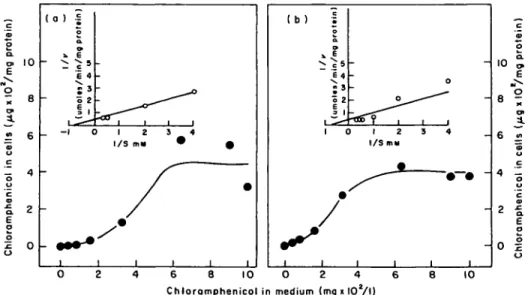
Uptake as a function of chloramphenicol concentration in medium
Measured after 1 min, the uptake of chloramphenicol by E. coli and P. aeruginosa increased as the concentration in medium was increased from 40 to 640 mg/1. Beyond
10 -o 2 6 4 -E o o o 2 0 -( 0 ) -S o a. 9 * E E 4 • o 2 ii -_ -1 0 1 2 3 4 I/S m» * 0 1 1 1 1 1 1 (b) I o a. :s o 2 E ^3 I 1 C / 1 1 0 ) 1 2 S 4 I/S my 1 1 1 1 - 10 6 8 10 O 2 4 Chloramphenicol in medium (mg i lOV
10 8 8 - 6 4 o - 2 0 I
Figure 6. Uptake of chloramphenicol by E. coli (a), and P. aeruginosa (b), in BYGNaCl medium at various chloramphenicol concentrations. The inset is a Lineweaver-Burk plot of the kinetic data.
I 2 r - 10 E 10 -Jt in 6 ^ 10 15 20 25 30 10 15 20 25 30 Time (min)
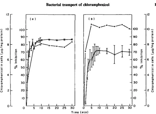
Figure 7. Relationship of chloramphenicol uptake (#), to inhibition of protein synthesis (O), in cells of £. coli at 10 mg chloramphenicol/1 (a), and P. aeruginosa at 40 mg chloramphenicol/1 (b). Bars represent standard errors of the mean for two experiments.
that range of concentration, the uptake by either strain of bacterium reached a plateau. A plot of the data in Lineweaver-Burk form revealed an apparent Km (Kt) of
1-8 mM and a Vmax of 2-5 mmoles/min/g protein, for both strains (Figure 6). Relationship of chloramphenicol uptake to inhibition of protein synthesis
As shown by Figure 7, when chloramphenicol was added simultaneously with 35S-methionine to growing cells of E. coli, 60% inhibition of protein synthesis occurred within 1 min; thereafter, an increasing inhibition occurred reaching 87% within 10 min. For the P. aeruginosa strain, 53% inhibition occurred within 3 min and thereafter, an increasing inhibition reached 72% within 10 min. Thus, it seems that the rapid phase of chloramphenicol uptake was more effective with E. coli than with
P. aeruginosa in inhibiting cellular protein synthesis.
Discussion
Our experimental results showed that chloramphenicol did not bind to either non-growing cells or to isolated cell envelopes of E. coli and P. aeruginosa. Therefore, we measured the uptake of chloramphenicol by exponentially growing cells as the depletion of 14C-chloramphenicol from the supernatant of centrifuged cultures, assuming the missing 14C-chloramphenicol to be found within the cells. Filtration could not be used to collect cells because of the retention of label by membrane filters. Hurwitz & Braun (1967) proposed that chloramphenicol crosses the cytoplasmic membrane of E. coli by passive diffusion. This proposal was supported by data
indicating that concentration of chloramphenicol by bacteria probably results from binding of the antibiotic to ribosomes (Harvey & Koch, 1980). The results of the present study show that accumulation of chloramphenicol after 5 min by sensitive strains of E. coli and P. aeruginosa reaches respectively about 140 and 97 times the concentration in the medium. When the amount of ribosome-bound chloramphenicol, calculated on the basis of 26,000 ribosomes per cell (Goldstein & Lowney, 1964) and one specific binding site per ribosome (Wolfe & Hahn, 1965; Das, Goldstein & Kenner, 1966; Hurwitz & Braun, 1967), was subtracted from the total amount of cell-associated chloramphenicol, it appeared that chloramphenicol concentrations in the cell "pool" of E. coli and P. aeruginosa were respectively 136-3 and 96-5 times the concentration in the medium. Thus, these results clearly indicate that concentration of chloramphenicol by bacteria does not result simply from binding of the antibiotic to ribosomes, but that it is accumulated against a concentration gradient. Results reported here showed that chloramphenicol accumulation by either strain required energy, presumably to transport chloramphenicol across the cytoplasmic membrane, since it is well recognized that ribosomal binding of chloramphenicol is not energy dependent (Vasquez, 1963, 1964).
Irvin & Ingram (1982) pointed out that acquisition of chloramphenicol resistance in
P. aeruginosa was a function of the concentration of free Mg+ + in the growth medium. The antagonism of chloramphenicol activity by Mg+ + was essentially immediate and was not a result of prolonged growth in Mg+ + limiting medium. More recently, however, Harvey & Koch (1980) demonstrated that Mg+ + enhances in-vitro binding of chloramphenicol to ribosomes up to a concentration of 10 mM. Our experimental results show that M g+ + reduces the uptake of chloramphenicol and raises the MICs of the antibiotic for both strains of E. coli and P. aeruginosa. Thus, the effect of Mg+ + is the reverse of that noted on ribosomal binding. It is plausible, therefore, that the interaction of M g+ + with the cell envelope of Gram-negative bacteria retards the movement of chloramphenicol across the cell envelope (diffusion through the outer membrane and subsequent transport across the cytoplasmic membrane). This in turn would permit bacterial growth in the presence of otherwise inhibitory concentrations of chloramphenicol.
The results reported here demonstrate that the accumulation kinetics of chloramphenicol are biphasic: an initial rapid phase and a second slower phase. In addition, uptake of the drug measured in minimal BM2 medium is much less than that rrteasured in enriched medium. These findings agree with those previously reported by Vasquez (1966) with Gram-positive bacteria. However, our results indicate that unlabelled chloramphenicol isomers effectively prevent the uptake of 14C-chloramphenicol (Table I); in other words, the isomers compete with chloramphenicol in cellular penetration. These findings do not agree with those previously reported by Vasquez (1966) with Gram-positive bacteria. He demonstrated that unlabelled chloramphenicol isomers: L-threo, D-erthryo, and L-erythro did not reduce the uptake of 14C-chloramphenicol by B. megaterium after 15 min of incubation with the drug; but the uptake of '^C-chloramphenicol was reduced by 77-9% when unlabelled chloramphenicol was added at ten times the concentration of 14C-chloramphenicol. However, it is doubtful that chloramphenicol uptake could be measured accurately by washing the cells twice with 15 ml of 10 mM Tris-HCl buffer. Most probably washing the cells removed most of the unbound chloramphenicol from the bacterial "pool" and the apparent reduction of uptake of 14C-chloramphenicol
could be explained by competition between the labelled and unlabelled molecules for binding to ribosomes. The lack of effect of chloramphenicol isomers was most probably due to inability of isomers to prevent stereospecific binding of chloramphenicol. Vasquez (1966) showed in his studies that all the compounds which inhibit uptake of chloramphenicol by intact bacteria, with the exception of ostreogrycins B group, also prevent binding of chloramphenicol to bacterial ribosomes in a cell-free system.
Cells grown in BYGNaCl and assayed in BM2 medium do not transport chloramphenicol. Such cells, when transferred to BM2 medium, exhibited a lag period over 6 h before the growth initiation. During the lag period the cells do not incorporate chloramphenicol. During growth in BM2 medium, they incorporate chloramphenicol, but however at a lower rate than in BYGNaCl medium. Cells transferred from BYGNaCl or from BM2 medium to phosphate buffer do not incorporate chloramphenicol. These results indicate that non-growing cells do not transport chloramphenicol. However, such cells actively accumulate amino-acids even after 2 h of preincubation in medium unable to support growth (Figure 4). Thus, the failure of non-growing cells to transport chloramphenicol cannot simply be attributed to loss of energy coupling for uptake. The data reported here demonstrate that spheroplasts prepared from cells grown in BYGNaCl medium and assayed in phosphate buffer transport chloramphenicol slightly more than those assayed in BYGNaCl medium. These findings clearly indicate that the permeability barrier to chloramphenicol in non-growing cells is located outside the cytoplasmic membrane.
Nakae & Nikaido (1975) demonstrated that the outer membrane does act as a limiting barrier for the penetration of hydrophilic substances, but not the peptidoglycan. Although chloramphenicol is relatively hydrophobic, it penetrates the outer membrane of Salmonella typhimurium and E. coli via aqueous (porin) pathways (Nikaido, 1976; Chopra & Eccles, 1978; Pugsley & Schnaitman, 1978). The data presented here suggest that diffusion of chloramphenicol across the outer bacterial membrane depends on active growth. It seems possibly a conformational change occurs in non-growing cells that prevents chloramphenicol recognizing the porin channels.
Acknowledgements
I am grateful to Professor Valentin Bonifas for discussions and for critical reading of this manuscript. The able technical assistance of Hong Fritschy and France Schmid is acknowledged.
References
Anraku, Y. (1978). Active transport of amino acids. In Bacterial transport (Rosen, B. P., Ed.), p. 171. Marcel Dekker, New York.
Chopra, I. & Eccles, S. J. (1978). Diffusion of tetracycline across the outer membrane of
Escherichia coli K-12: Involvement of protein Ia. Biochemical and Biophysical Research
Communications 83, 550-7.
Das, H. K.., Goldstein, A. & Kenner, L. C. (1966). Inhibition by chloramphenicol of the growth of nascent protein chains in Escherichia coli. Molecular Pharmacology 2, 158-70.
Foster, T. J. (1983). Plasmid-determined resistance to antimicrobial drugs and toxic metal in bacteria. Bacteriological Reviews 47, 361-409.
Foulds, J. & Chai, T. J. (1978). New major outer membrane protein found in an Escherichia coli to IF mutant resistant to bacteriophage Tulb. Journal of Bacteriology 133, 1478-83. Furlong, C. E., Morris, R. G., Kandrach, M. & Rosen, B. P. (1972). A multichamber
equilibrium dialysis apparatus. Analytical Biochemistry 47, 514-26.
Gilleland, H. E., Stinett, J. D. & Eagon, R. G. (1974). Ultrastructural and chemical alterations of the cell envelope of Pseudomonas aeruginosa associated with resistance to ethylenediamine-tetraacetate resulting from growth in Mg++-deficient medium. Journal of
Bacteriology 117, 302-11.
Goldstein, A. D. B. & Lowney, L. I. (1964). Protein synthesis at 0°C in Escherichia coli. Journal of Molecular Biology 9, 213-35.
Hancock, R. E. W. & Nikaido, H. (1978). Outer membranes of Gram-negative bacteria. XIX. Isolation from Pseudomonas aeruginosa PA01 and use in reconstitution and definition of the permeability barrier. Journal of Bacteriology 136, 381-90.
Harvey, R. J. & Koch, A. L. (1980). How partially inhibitory concentrations of chloramphenicol affect the growth of E. coli. Antimicrobial Agents and Chemotherapy 18, 323-37. Hurwitz, C. & Braun, C. B. (1967). Measurement of binding of chloramphenicol by intact cells.
Journal of Bacteriology 93, 1671-6.
Irvin, J. E. & Ingram, J. M. (1980). Chloramphenicol-resistant variants of Pseudomonas aeruginosa defective in amino acid transport. Canadian Journal of Biochemistry 58, 1165-71. Irvin, J. E. & Ingram, J. M. (1982). Divalent cation regulation of chloramphenicol resistance in
Pseudomonas aeruginosa. FEMS Microbiology Letters 13, 63-7. Lehninger, A. I. (1976). Biochemistry, p. 495. Worth Publishers, New York.
Lennox, E. S. (1955). Transduction of linked genetic characters of host by bacteriophage PI. Virology 1, 190-206.
Levy, S. B. (1971). Physical and functional characteristics of R-factor deoxyribonucleic acid segregated into Escherichia coli minicells. Journal of Bacteriology 108, 300-8.
Markwell, M. A. K., Haas, S. M., Birber, L. L. & Tolbert, H. E. (1978). A modification of the Lowry procedure to simplify protein determination in membrane lipoprotein samples. Analytical Biochemistry 87, 206-310.
Nakae, T. & Nikaido, H. (1975). Outer membrane as a diffusion barrier in Salmonella typhimurium. Journal of Biological Chemistry 250, 7359-65.
Nierhaus, D. & Nierhaus, K. H. (1973). Identification of the chloramphenicol-binding protein in Escherichia coli ribosomes by partial reconstitution. Proceedings of the National Academy of Sciences U.S.A. 70, 2224-8.
Nikaido, H. (1976). Outer membranes of Salmonella typhimurium: Transmembrane diffusion of some hydrophobic substances. Biochimica et Biophysica Ada 433, 118-32.
Pugsley, A. P. & Schnaitman, C. A. (1978). Outer membrane proteins of Escherichia coli: VII. Evidence that bacteriophage-directed protein 2 functions as a pore. Journal of Bacteriology 133, 1181-9.
Vasquez, D. (1963). Antibiotics which affect protein synthesis: The uptake of I4 C-chloramphenicol by bacteria. Biochemical and Biophysical Research Communications 12, 409-13.
Vasquez, D. (1964). Uptake and binding of chloramphenicol by sensitive and resistant organisms. Nature 203, 257-8.
Vasquez, D. (1966). Binding of chloramphenicol to ribosomes. The effect of a number of antibiotics. Biochimica et Biophysica Ada 114, 277-88.
Weiss, R. L. (1976). Protoplast formation in Escherichia coli. Journal of Bacteriology 128, 668-70.
Winkler, H. H. & Wilson, T. H. (1966). The role of energy coupling in the transport of /?-galactosides by Escherichia coli. Journal of Biological Chemistry 241, 2200-11.
Wolfe, A. D. & Hahn, F. E. (1965). Mode of action of chloramphenicol IX. Effects of chloramphenicol upon a ribosomal amino acid polymerization system and its binding to bacterial ribosome. Biochimica Biophysica Ada 95, 146-5.