Review
Maxime Bodak, Jian Yu and Constance Ciaudo*
Regulation of LINE-1 in mammals
Abstract: Transposable elements (TEs) are mobileDNA elements that represent almost half of the human genome. Transposition of TEs has been implicated as a source of genome evolution and acquisition of new traits but also as an origin of diseases. The activity of these ele-ments is therefore tightly regulated during the life cycle of each individual, and many recent discoveries involved the genetic and epigenetic mechanisms in their control. In this review, we present recent findings in this field of research, focusing on the case of one specific family of TEs: the long-interspersed nuclear elements-1 (LINE-1 or L1). LINE-1 elements are the most representative class of retrotransposons in mammalian genomes. We illustrate how these elements are conserved between mice and humans, and how they are regulated during the life cycle. Additionally, recent advances in genome-wide sequenc-ing approaches allow us not only to better understand the regulation of LINE-1 but also highlight new issues specifi-cally at the bioinformatics level. Therefore, we discuss the state of the art in analyzing such bioinformatics datasets to identify epigenetic regulators of repeated elements in the human genomes.
Keywords: bioinformatic challenges; LINE-1 regulation;
repeat elements; transposable elements.
DOI 10.1515/bmc-2014-0018
Received June 23, 2014; accepted August 19, 2014
List of abbreviations: 5caC, 5-carboxylcytosine; 5fC,
5-formylcytosine; 5hmC, 5 hydroxymethylcytosine; 5mC, 5-methylcytosine; AID, activation-induced deami-nase; APOBEC, apolipoprotein B mRNA-editing enzyme complex; ATPase, adenosine triphosphatase; ChIP-seq, chromatin immunoprecipitation followed by sequenc-ing; DNA, deoxyribonucleic acid; Dnmt, DNA meth-yltransferase; dpc, day postcoitum; ERV, endogenous retrovirus; FOA, fetal oocyte attrition; GSE, gonad-specific expression; HELP-seq, HpaII-tiny fragment enrichment by ligation-mediated PCR coupled to massively parallel DNA sequencing; HIV, human immunodeficiency virus; hnRNP, heterogeneous nuclear ribonucleoprotein; LINE-1 or L1, long-interspersed nuclear elements-1 or long inter-spersed element; Lsh, lymphoid-specific helicase; LTR, long terminal repeat; MbD, methyl-CpG-binding domain; MHR, multiple-hit read; miRNA, microRNA; mRNA, mes-senger RNA; NHP, non-human primate; NPC, neuronal progenitor cell; nt, nucleotide; ORF, open reading frame; PABP, poly(A) binding protein; PGC, primordial germ cell; piRNA, PIWI-interacting RNA; RDC, Rhino, Deadlock, and Cutoff; RISC, RNA-induced silencing complex; RNA, ribonucleic acid; RNAi, RNA interference; RNP, ribonu-cleoprotein; RSEM, RNA-Seq by expectation maximiza-tion; siRNA, small interfering RNA; shRNA, short hairpin RNA; TDRD, Tudor domain containing; TEs, transposable elements; TET, ten-eleven translocation; TF, transcription factor; TPRT, target primed reverse transcription; UHR, unique-hit read; UTR, untranslated region.
Introduction
Since their discovery by Barbara McClintock in the 1940s (1), transposable elements (TEs) still continue to enthuse and inspire researchers. Significant progress has been made in understanding the biology of TEs, which is reflected by the increasing number of published studies. The popularity of these classes of DNA stems from their ability to translocate to a new location in a genome, explaining why TEs are also called ‘jumping genes’ or ‘mobile elements’. Moreover, TEs account for 46% of the human genome (2) and about 39% of the mouse genome
*Corresponding author: Constance Ciaudo, Swiss Federal Institute
of Technology Zurich, Department of Biology, Institute of Molecular Health Sciences, HPL G32.1, Otto-Stern-Weg 7, CH-8093 Zurich, Switzerland, e-mail: cciaudo@ethz.ch
Maxime Bodak: Swiss Federal Institute of Technology Zurich,
Department of Biology, Institute of Molecular Health Sciences, Life Science Zurich Graduate School, Molecular Life Science Program, HPL G28, Otto-Stern-Weg 7, CH-8093 Zurich, Switzerland
Jian Yu: Swiss Federal Institute of Technology Zurich, Department
of Biology, Institute of Molecular Health Sciences, Life Science Zurich Graduate School, Molecular and Translational Biomedecine Program, HPL G28, Otto-Stern-Weg 7, CH-8093 Zurich, Switzerland
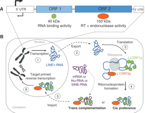
(3). They are divided into two classes, according to a clas-sification proposed by Finnegan in 1989 (4) and revisited by Wicker et al. in 2007 (5), based on their mechanism of transposition: class I retrotransposons require an RNA intermediate involving a ‘copy-and-paste’ mechanism (6) and class II DNA transposons move by a ‘cut-and-paste’ mechanism (7). Retrotransposons represent about 42% of the human and 38% of the mouse genome, whereas DNA transposons account for only 3% and 1%, respectively (2, 3). In this review, we will focus on retrotransposons and more specifically on the regulation of long-interspersed nuclear elements-1 (LINE-1 or L1), a subclass of this family. L1 is the most representative class of retrotrans-posons in the mammalian genome, representing 17% of the human (2) and almost 19% of the mouse genome (3). They belong to the autonomous non-LTR retrotranspo-son category, owing to their ability to encode the proteins required for their mobility. L1 elements are about 6–7 kb in length and contain a 5′ untranslated region (5′UTR) with an internal promoter activity, two open reading frames (ORF1 and ORF2), and a 3′UTR that ends with a poly(A) tail (Figure 1A). ORF1 encodes a 40 kDa protein with RNA binding activity (8), whereas ORF2 encodes a 150 kDa
protein with a reverse transcriptase (9) and an endonu-clease domain (10) (Figure 1A). Active L1 elements must be able to perform a complete retrotransposition cycle that includes the following steps in chronological order: tran-scription of L1 RNA, export into the cytoplasm, translation of ORF1 and ORF2, association of L1 RNA with ORF1 and ORF2 proteins to form ribonucleoprotein (RNP) particles (11), return to the nucleus, reverse transcription, and inte-gration at a new genomic location – a one-step process also called target primed reverse transcription (TPRT) (12) (Figure 1B). During this retrotransposition event, the 5′UTR is frequently truncated (2) and the L1 sequence is prone to inversion (13). These are the two main reasons for the accumulation of inactive L1 copies in the genome. Indeed, the number of L1 fragments in the human genome has been estimated at 516 000 elements (2); however, only 80–100 of them have kept their mobility capability (14). Similarly, the mouse genome contained almost 600 000 L1 fragments (3); however, previous studies showed that only 3000 full-length elements are potentially able to retrotranspose (15, 16). These active L1 copies can deeply influence the genome in numerous ways, beneficial and detrimental (17). L1 retrotransposition can act as a
5’ UTR ORF 1 ORF 2 3’ UTR
Translation Transcription LINE1-RNA Export Import L1ORF1p L1ORF2p Ribonucleoprotein formation 1 Cytoplasm Nucleus Ribosome 40 kDa
RNA binding activity RT + endonuclease activity150 kDa
2 3 4 6 5 Target primed reverse transcription or mRNA or Alu-RNA or SINE-RNA Cis preference Trans complementation A B
Figure 1 Schematic representation of L1 structure and retrotransposition cycle.
(A) L1 anatomy – an L1 is composed of a 5′UTR, two ORFs each coding for a protein [ORF1p (40 kDa) and ORF2p (150 kDa)], and a 3′UTR. (B) L1 retrotransposition cycle – during a complete retrotransposition cycle, an L1 element is transcribed (1) and the resulting L1 RNA is exported out of the nucleus into the cytoplasm (2). Then, the bicistronic RNA is translated (3) into ORF1 and ORF2 proteins, which bind the L1-RNA to form RNP particles (4): this is the cis preference. ORF1p and ORF2p can also bind other RNAs (such as cellular mRNA, Alu-RNA, or SINE-RNA): this is called the trans complementation phenomenon. Finally, RNP complexes are imported into the nucleus (5) where the RNA can be integrated at a new genomic location by the process of TPRT (6).
mutagen by inserting into exons, inducing aberrant splic-ing or exon skippsplic-ing by insertsplic-ing into introns, and less commonly, they can give rise to a gene-breaking phenom-enon (18). This last example could generate distinct tran-scription units (19), which can lead to sporadic cases of diseases (18). Thereby, since their discovery, almost 65 dis-eases-causing mutations in humans have been attributed to L1-mediated retrotransposition. Moreover, L1 elements are able to mediate the retrotransposition of other RNAs, a phenomenon called trans complementation (Figure 1B). In this case, L1 can mobilize non-autonomous retrotrans-posons such as short interspersed elements (SINEs) and Alu elements (20) or process pseudogenes from cellular mRNAs (21), and therefore participate in genome expan-sion (18). Here, however, we will focus on the regulation of L1 transcription and retrotransposition, as the impact of new L1 insertions on the genome has already been exten-sively reviewed (12, 17, 18). In humans, the current rate of L1 retrotransposition has been estimated between 1 inser-tion out of 20 and 1 out of 200 live births depending on the analysis method (18). L1 elements have been described to be expressed in the germ line, during early development and in some somatic tissues, as well as in tumor cells (22). In this review, we will focus on the regulatory mecha-nisms involved in the control of these TEs during the life cycle in mammals, from gamete precursors to somatic cells through the blastocyst stage. Our goal is to provide an overview of what is known about L1 regulation itself, the latest discoveries in the field, and the tools available to analyze such repeat elements. First, we will present a comparison of active L1 5′UTR from humans and mice, the two main mammalian systems used to study L1 regulation in mammals. Subsequently, we will describe the numer-ous regulatory mechanisms of L1 that are presently known and the latest hypothesis. Finally, we will present the bioinformatics tools used to study repeated elements as well as the problems related to the high number of copies present in the genomes. We will conclude our review with potential future research directions.
Comparison of human and mouse
active LINE-1 elements
Numerous studies have been conducted using mouse L1 constructions in human cell lines and vice versa, and have shown that human L1 are able to retrotranspose in the mouse, in vitro and in vivo (23, 24). At the same time, other studies employ artificial L1 constructions to better under-stand the mechanisms that can rule their regulation (25,
26). However, even if mouse and human L1 share common features concerning their biology, they also differ on many points, putting into question the conservation of the L1 regulatory mechanisms between the two mammalian systems used to study this class of TEs.
Differences in mobilization activity
First, mouse and human genomes differ considerably con-cerning the number of L1 active copies (27): the percent-age of active elements according to the number of copies in the genome is 25 times higher in mice (0.5%) than in humans (0.015–0.020%) (2, 3, 15, 16). This observation sug-gests a decline of L1 activity in humans. Comparative gene expression analysis performed on human and non-human primate (NHP) induced pluripotent stem cells revealed reg-ulation differences of L1 with an increase of their mobility in the NHP genomes (28). Regulation differences between human and NHPs support the idea that mouse and human L1 regulatory mechanisms can diverge as well.
Differences in structure
Furthermore, mouse and human L1 elements also differ in their structure: mouse L1 are approximately 7 kb in length (17), whereas human L1 are around 6 kb (12). Moreover, the two ORFs of human L1 are in frame and separated by a 67–70 bp non-coding spacer region containing a stop codon in the three reading frames. In mouse, the two ORFs are also separated by a spacer region (29–115 bp) (29) and they have different reading frames. Finally, unlike human L1, the 5′UTR of mouse L1 contains variable numbers of monomers, which are tandem repeat units of 205–210 bp with an intrinsic promoter activity (30). This allows the definition of several L1 subfamilies in the Repbase data-base (31), among them three are proved to be active in mice: A (32), Tf (15), and Gf (16).
Focus on the 5′UTR, a key region for L1
regulation
L1 5′UTR contains an internal promoter activity independ-ent of upstream sequences (33), as well as an antisense promoter (34–36), making the 5′UTR a key region in the regulation of these elements and in the definition of an active element. Moreover, human and mouse L1 5′UTR have different putative transcription factor (TF) binding sites (37). Considering these evidences, we chose to focus
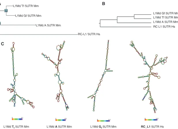
on the 5′UTR of active elements to gain deeper insight into the comparison between human and mouse L1. As we previously mentioned in the Introduction, the human genome contains around 80–100 active L1, called retro-transposition competent L1 (RC_L1) (14). In the literature, RC_L1 are also denoted as L1Hs or L1-PA1 (31). It has been recently shown that the 5′UTR of RC_L1 could adopt sec-ondary structures that can be recognized and processed by protein complexes (38). Here, we present a compari-son of the sequences and potential secondary structures of 5′UTR from active L1 subfamilies, including A, Tf, and Gf in mouse and RC_L1 (Figure 2). To investigate the dif-ference among these four regions, we considered the con-sensus sequence of each 5′ UTR element (31) and used two different approaches: (i) drawing the phylogenetic tree using a neighbor-joining algorithm to reveal the evolution-ary relation (39) (Figure 2A), and (ii) performing sequences and structure alignments by using LocARNA (40) (Figure 2B). As expected, mouse L1 5′UTR from the different sub-families clustered into the same group with both methods.
According to these trees, L1-Tf and L1-Gf 5′UTR appear to be the most similar regions, whereas RC_L1 5′UTR is, in both cases, the most divergent (Figure 2A,B). Then, we com-pared the predicted secondary structures of these four L1 5′UTR by RNAfold (41), and we found a substantial diver-gence not only between the human and mouse L1 5′UTR region but also among the mouse L1 5′UTR subfamilies (Figure 2C). These differences suggest that the mechanisms involved in the regulation of active mouse and human L1 can be potentially distinct, even between subfamilies.
L1 regulation during the mammalian
life cycle
It has been estimated that in humans, one new genomic L1 insertion appears in every 10–250 individuals and that the proportion of L1 retrotransposition causing disease could be around 0.07% (12). In mice, the number of active L1 has
L1Md Tf 5UTR Mm
L1Md Tf 5UTR Mm
A B
C
L1Md Gf 5UTR Mm L1Md Gf 5UTR MmL1Md Tf 5UTR Mm
L1Md A 5UTR Mm RC L1 5UTR Hs L1Md A 5UTR Mm RC-L1 5UTR Hs L1Md Gf 5UTR Mm RC_L1 5UTR Hs L1Md A 5UTR Mm
Figure 2 Sequences and structures comparison of human (RC_L1) and mouse (Gf, Tf, A) L1 5′UTRs.
(A) Phylogenetic tree generated with neighbor-joining algorithms using the Kimura model. The numbers on the nodes indicate the percent-age of the labeled node present in 93 bootstrap replicates. (B) Multiple sequence and secondary structure alignment by LocARNA. The height of the tree corresponds to the LocARNA scores, where lower scores/heights indicate a closer distance. (C) Secondary structures were predicted by RNAfold. The color represents the entropy for each base. L1Md Tf 5UTR Mm, Tf mouse L1 subfamily; L1Md A 5UTR Mm, a mouse L1 subfamily; L1Md Gf 5UTR Mm, Gf mouse L1 subfamily; RC_L1 5UTR Hs, human retrotransposition competent L1.
been estimated using cell culture assays to a total of 3100. In terms of ratio of active copies per total copies present in the genome, the proportion of operational L1 in mice is 25 times higher than in humans. Owing to their high muta-genic potential, these elements must be strongly regulated during the development and adult life of each individual. In this part of the review, we present the state of the art of the L1 regulatory mechanisms described in the literature. As a guideline, this section is divided according to the different steps of the mammalian life cycle; thus, we first describe the pathways regulating L1 in the germ line, after fertilization and to the blastocyst stage. Subsequently, we depict which mechanisms are involved in sustaining the control in somatic cells, and finally, we discuss about the particular cases of L1 reactivation in somatic cells.
Epigenetic regulation through DNA methylation is one the main mechanisms, which has evolved to play a role in the defense against TEs through transcriptional silencing
(42). During the mouse life cycle, L1 promoters undergo two waves of partial demethylation: one occurs during the specification and migration of primordial germ cells (PGCs) and the other during the preimplantation stage (43) (Figure 3A). However, DNA methylation is not the only L1 repressor system used to control their transcription and retrotransposition. Therefore, it has been proposed that overlapping epigenetic mechanisms evolved to control the expression of TEs in eukaryotic cells (44). Such example of multiple regulations of L1 subtypes has been recently demonstrated in human and mouse embryonic stem cells (mESCs) (45) (see ‘Regulation in the blastocyst’).
Regulation in the germ cell lineage
The majority of TEs spread in a population according to a vertical mode of inheritance (46). This transmission mode
A
B
gametes zygote 2 cells 4 cells 8 cells morula blastocyst fertilization
Relative 5hmC level
Low High
Developmental stage Day post coitum
v Global L1 meth ylation (%) 0 50 100 v fertilization . . .. ... . . .... . 0 0.5 3.5 pre-implantation 6.5 gametes zygote Somatic tissues 7.5 13.5 PGCs 0 Gametes NG oocyte 18.5 Birth First wave of demethylation Second wave of demethylation PGC precursors v epibast blastocyst
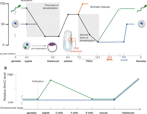
Figure 3 Global L1 methylation and 5hmC level during several steps of mammalian development and associated L1 regulatory mechanisms.
(A) Schematic representation of the global L1 methylation level of the mouse genome. During embryonic development, mouse L1 undergoes two main waves of demethylation. The first occurs after fertilization: from the zygote stage (0.5 dpc) until the blastocyst stage (3.5 dpc). The second occurs during the migration of the PGC precursors from the proximal epiblast to the gonad precursors (from 7.5 to 13.5 dpc). PGC, primordial germ cell; NG oocyte, non-growing oocyte. Adapted from ref. (43). (B) Schematic representation of 5-hydromethylcytosine dynamics in paternal and maternal genomes during preimplantation development. Adapted from ref. (47).
requires an activity in gamete precursors, making the germ line a pertinent developmental context to study L1 regula-tion. In mice, at 6.5 days postcoitum (dpc), cells from the proximal epiblast will differentiate into PGCs and migrate to reach the precursor of the gonads. These cells will form the germ line. During this process, the early developing germ line acquires pluripotency, an event coinciding with a relaxation of the epigenetic repression of the genome (43) (Figure 3A). This developmental window is indeed an opportunity for L1 invasion of the host genome.
Role of DNA methylation
DNA methylation is known to be involved in various biological processes as a regulator of gene expression, X-chromosome inactivation, genomic imprinting, and also in the silencing of TEs (47, 48). The reestablishment of the methylation status of L1 elements requires DNA meth-yltransferase proteins involved in the DNA methylation machinery. It has been shown that mutant mouse embryos for the maintenance DNA-methyltransferase DNMT1 lose the methylation of several types of TEs (49). A similar phe-notype has been observed for the double-mutant mouse for Dnmt3a and Dnmt3b genes encoding for two de novo DNA methyltransferases (50).
Studies from Bestor laboratory highlighted yet another player in this process: DNMT3L, the DNMT3A cofactor that is strictly required for L1 methylation in the male germ line (51). DNMT3L stabilizes the active confor-mation of DNMT3A, thereby increasing the efficiency of methyl group transfer onto target site and facilitating de
novo methylation (52). Problems in completing this
proce-dure involve the high expression level of L1 in germ cells, and also sterility, similar to the phenotype observed in
Dnmt3A and Dnmt3L male mutant mice (51). The Dnmt3L
gene is also evolutionarily correlated to the germ line pro-tection against TEs, as it emerged in eutherian mammals around 150 million years ago coinciding with an impor-tant TE expansion in mammalian genomes (53).
Furthermore, proteins assisting the DNA methylation process also play a role in TE regulation. For example, the protein UHRF1, for ubiquitin-like containing PHD and RING finger domains 1, is required by DNMT1 to load onto hemimethylated DNA strands. Uhrf1 inactiva-tion in mice results in decreased methylainactiva-tion of L1 (54). The protein lymphoid-specific helicase (LSH) should also be mentioned in this context. This member of the SNF2 family of chromatin remodeling ATPases supports the access of DNMTs to the DNA. Initially, LSH was described to be directly recruited to L1 elements in mice (55). More
recently, Meehan laboratory developed a new technology called HELP-seq (for HpaII tiny fragment enrichment by ligation-mediated PCR coupled to massively parallel DNA sequencing) to profile the methylation of TEs in Lsh and
Dnmt3b mutant fibroblasts (56). They observed
hypo-methylation of L1 in Lsh-/- and Dnmt3b-/- cells, which is consistent with previous investigations implicating LSH as a recruitment protein for de novo methylation. Nev-ertheless, they also observed differences of methylation between L1 subfamilies and a greater hypomethylation in the absence of LSH than DNMT3B, suggesting DNMT3B-independent roles for LSH in repeat methylation.
Finally, a recent study showed that in addition to DNA methylation, the PIWI-interacting RNA (piRNA) pathway is required to maintain a high level of repressive H3K9me3 on L1 elements in mouse germ cells (57).
Role of the piRNA pathway
Studies performed on germ cells from Drosophila depicted a new TE repression system based on the interaction of piRNA with PIWI proteins (P-element-induced wimpy testis) (58). Later, similar complexes have been described in the mouse and human germ lines where they play a role in the regulation of L1 elements (58).
PIWI proteins belong to the Argonaute superfamily (58). Three PIWI proteins have been identified in mice (four in humans), MILI, MIWI, and MIWI2, and all are mainly germ line restricted (59). In 2006, thanks to the develop-ment of deep-sequencing techniques, research groups identified specific populations of small RNAs binding to PIWI proteins: the piRNAs (58). These piRNAs are mainly generated from dedicated loci, called piRNA clusters (60), consisting of 20–90 kb genomic regions. Around 20% of them matched repetitive elements (61), thereby allowing to distinguish two kinds of piRNAs: repeat associated and non-repeat associated piRNAs. Among the repeat associ-ated piRNAs, which are mainly derived from retrotrans-posons, we can set apart primary and secondary piRNAs. The one belonging to the piRNA category is length and sequence specific: primary piRNAs are about 26 nucleo-tides (nt) in length, have a uracil nucleotide at position 1, and correspond to sense-strand transcripts, which predominantly associate with MILI protein. Secondary piRNAs are 31 nt in length, have an adenine nucleotide at position 10, and correspond to antisense strand, bound by MIWI2 protein (58). However, this pathway is still not entirely understood and several open questions need to be addressed. In particular, the initiation of the process is not yet fully understood as well as the loading of the
piRNAs into the PIWI proteins. However, recent studies performed in Drosophila have shown that a complex com-posed of Rhino, Deadlock, and Cutoff (RDC) suppresses piRNA cluster transcript splicing and drives piRNA bio-genesis (62, 63). Other evidences show that MIWI2 loading is strictly dependent of MILI SLICING activity (64), and that primary and secondary piRNAs are located in distinct cytoplasmic compartments. Primary piRNAs loaded on MILI supported by the Tudor domain-containing TDRD1 protein are located in specific organelles named pi-bodies (65), whereas secondary piRNAs loaded on MIWI2 sup-ported by TDRD9 protein are located in distinct compart-ments named piP-bodies (66). Both complexes have been described to be involved in L1 regulation in the germ line (67, 68). Mili and Miwi2 mouse mutant males are sterile, and L1 transcripts accumulate strongly in their germ line (61, 69), phenotypically mimicking the Dnmt3L mouse mutant phenotype (70). These similarities suggest that the piRNA pathway plays a role in the control of L1 tran-scriptional output by promoting de novo methylation (64). Finally, a study has shown that in Drosophila, Piwi exhibits a slicer activity: it can bind single-strand RNA and cleave the corresponding RNA target (71). This implies that the piRNA machinery could also be involved in the post-transcriptional silencing of L1.
Few reports have investigated the role of PIWI-piRNA pathways in L1 regulation in humans. The Larriba labora-tory showed that hypermethylation-associated silencing of PIWI2 and TDRD1 is correlated with the hypomethyla-tion of L1 and linked with spermatogenic disorders. This suggests that in humans, PIWI-piRNA pathways could also target L1 elements and that the reactivation of such TEs could contribute to spermatogenic failures (72).
Regulation in the mature gametes
From 13.5 to 16.5 dpc, the genome of male germ cells under-goes a wave of remethylation (Figure 3A) (43). Moreover, L1 elements of epididymal sperms from adult testis have been shown to be fully methylated (73). In contrast, female germ cells are never fully methylated (73, 74), whereas sec-ondary oocytes (ovulated but unfertilized) show an inter-mediate level of L1 methylation (around 42%) (75) (Figure 3A). Therefore, L1 are expressed in mature oocytes (76), which implies the presence of L1 regulatory mechanisms at the oocyte stage other than methylation.
A previous study revealed that exogenously introduced target RNAs containing L1 sequences were specifically degraded in oocytes by an RNA interference (RNAi)-dependent mechanism (77), suggesting that in mouse
oocytes, L1 could be repressed through the RNAi pathway (described in ‘RNAi pathways in mammals’). This idea was strengthened by the fact that oocytes produce small interfering RNAs (siRNAs) derived from L1 sequences (78). However, Dicer-deficient mutant oocytes did not show any L1 accumulation (78). This last observation could be explained by the identification of specific mouse oocyte piRNAs (78) (see ‘Role of the piRNA pathway’), which could also regulate L1 elements in oocytes.
Moreover, mammal oocytes are blocked at the diplo-tene stage of meiosis I from the fetal stage until sexual puberty. This long meiotic arrest could be an innate barrier to the accomplishment of the full L1 retrotrans-position cycle (Figure 1B). Interestingly, during develop-ment, oocytes also undergo extensive apoptosis, leading to a massive oocytes loss; this process is called fetal oocyte attrition (FOA) and is still poorly understood. A recent study, however, revealed that enhancement of L1 expression is involved in the reduction in the number of fetal oocytes at birth and that FOA can be modulated by controlling L1 activity (79). It is proposed that FOA corre-sponds to a select mechanism by which only oocytes with low L1 activity are conserved (79).
Regulation in preimplantation embryo
Shortly after implantation, at around 6.5 dpc, the mouse embryo undergoes a wave of de novo methylation, estab-lishing a genome-wide hypermethylation pattern (74, 75). However, during the development of preimplantation embryos, from the zygote to the blastocyst stage, a struc-ture formed around 2.5 days after fertilization, L1 elements are subject to a first wave of demethylation (Figure 3A). Indeed, a dramatic reduction of L1 methylation level is observed during the sperm-to-zygote transition and then methylation progressively decreases to reach 13–23% at the blastocyst stage (43, 74). This can lead to L1 transcript accumulation in the blastocyst and therefore to retro-transposition events (80). Moreover, other evidences have shown that L1 RNA transcribed in male or female germ line could remain competent for integration in the early mouse embryo after being carried over by the gametes through fertilization in mice (81) and in humans (82). These data imply that the preimplantation phase can be considered as an aperture favorable for L1 invasion.
DNA methylation during preimplantation
In mammals, DNA methylation occurs at the 5-posi-tion of cytosine (5mC) and is known to be essential for
development and many other biological processes, as a transcriptional regulation mechanism and for the mainte-nance of genome stability (47, 48). Until 5-hydroxymethyl-cytosine (5hmC) was discovered in mammals in 2009, it was the only known DNA epigenetic mark (83, 84). It has been shown that the ten-eleven translocation (TET) protein family is responsible for the conversion from 5mC into 5hmC through oxidation (85). This suggests that 5hmC may serve as an intermediate of DNA demethyla-tion. As 5hmC is not recognized by the maintenance DNA-methyltransferase DNMT1 during DNA replication (86), it is proposed that the conversion of 5mC to 5hmC leads to the restriction of DNA methylation patterns through passive DNA demethylation during cell division. Further studies showed that TET proteins could oxidize 5hmC to produce 5-formylsytosine (5fC) and 5-carboxylcytosine (5caC) that can be removed from the genome by a thymi-dine-DNA glycosylase (87, 88). Evidences suggest that 5fC and 5caC might serve as intermediates in an active DNA demethylation process, as in Tdg gene knockout mouse, leading to increased DNA methylation at certain genomic loci (89).
After fertilization, the level of 5hmC of the paternal genome increases strongly until the end of the zygote stage. Subsequently, it decreases to reach its basic level (Figure 3B). Later, during the blastocyst stage, the 5hmC level of both paternal and maternal genomes highly increases (Figure 3B) (47). Several lines of evidence support the notion that TET3 drives the 5mC oxidation during preimplantation and also DNA demethylation: (i) during the same time window (from zygote to blasto-cyst stage), an increase of the 5hmC level and a decrease of the global L1 methylation level are observed (Figure 3A); (ii) TET3 is highly expressed at the zygote stage (90); and (iii) Tet3 siRNA-mediated knockdown abolishes 5mC oxi-dation (91). Nonetheless, the role of 5hmC in the regula-tion of TEs still needs to be further investigated.
However, a recent report describes a gonad-specific expression gene (GSE) to be present specifically in germ cells and in preimplantation embryos. This protein is localized in the nuclei of cells from the zygote to the blastocyst stage (92). Zygote GSE knockdown mediated by antisense RNA results in an increase in LINE-1 meth-ylation level and reduction of 5mC to 5hmC on L1 (92), suggesting an active DNA demethylation role for GSE during zygote development and a role of 5hmC in LINE-1 regulation.
Finally, two recent articles described the methyla-tion profile during human and mouse early development (93, 94). These studies reveal that the global methylation reprogramming after fertilization is globally conserved.
Nonetheless, L1 with different evolutionary ages show different demethylation patterns: young L1 elements are more resistant to demethylation than their older counter-parts in human embryos (93).
RNAi pathways in mammals
In several organisms, RNAi pathways are used as a post-transcriptional mechanism for TE repression (95). Three major classes of endogenous small RNAs have been first identified in mammals: microRNAs (miRNAs), piRNAs, and endogenous siRNAs (endo-siRNAs) (58). While miRNAs and siRNAs are loaded into the AGO effector proteins, piRNAs interact only with the PIWI proteins, expressed mainly in germ cells (see ‘Role of the piRNA pathway’) (58).
Dicer promotes processive cleavage of double-stranded RNAs into endo-siRNAs (96) that are loaded into AGO2 to mediate endonucleolytic cleavage of their target transcript. miRNAs have been identified as important post-transcriptional regulators of gene expression (97). Most animal miRNAs are transcribed in the nucleus by RNA polymerase II as stem-loop long primary transcripts (pri-miRNA). Following transcription, they are processed sequentially in the nucleus and cytoplasm by a complex of RNAse-III endonucleases: DROSHA and DICER. Spe-cifically, DROSHA and its partner DGCR8, which repre-sent what we call ‘the microprocessor complex’, process the pri-miRNA transcript to a 70–93 nt stem-loop precur-sor RNA (pre-miRNA). Subsequently, they are delivered to the cytoplasm by Exportin 5, where they are cleaved by a non-processive DICER, which forms a complex with TRBP, to produce an ∼22 nt miRNA-3p:miRNA-5p duplex (canonical pathway). Once in the cytoplasm, one of the duplex strands (miRNA-3p or miRNA-5p) is preferentially incorporated into the RNA-induced silencing complex (RISC) in association with an AGO family member. Within the RISC-AGO entity, the miRNA guides the complex to its RNA target, thereby mediating its repression. In animals, miRNAs control gene expression by binding to the 3′UTR of their target genes through Watson-Crick base pairing between the target and the 5′-end of the miRNAs: the ‘seed’ sequence (nt 2–8). Furthermore, the Microproces-sor is directly involved in the regulation of L1 elements in humans (described in ‘Regulation in the blastocyst’) (38).
Finally, recent technical developments have enabled the identification of a multitude of novel types of small RNA molecules that do not fit into the well-established classes (35, 98). Moreover, RNAi pathways expand beyond the post-transcriptional regulation, by having a role in the
maintenance of cellular integrity (35) and by acting as an antiviral defense mechanism (99).
A shift of the small RNA balance during preimplantation
After fertilization, the first event of gene expression is called zygotic gene activation. It takes place at the two-cell stage in mice and at the four-two-cell stage in humans, and has been involved in qualitative and quantitative changes in coding and non-coding gene expression (100, 101). In zebrafish, an miRNA (miR-430) has been involved in the deadenylation and the clearance of mater-nal mRNAs (102). This result and the development of sequencing technologies have encouraged researchers to profile small RNAs during mouse early development from mouse unfertilized (metaphase II: MII) oocytes, 8–16-cell stage embryos to the blastocyst stage (103). This study revealed a switch in small RNA populations from siRNA and piRNA derived from retrotransposons to the tran-scription of miRNAs (103).
How and why this change in small RNA populations occurs still needs to be further investigated. Recently, it has been shown that the processivity of DICER, the key enzyme in siRNA and miRNA pathways, decreases during early development (104), which could explain the switch between siRNA to miRNA populations. The decrease of the piRNA population correlates with the first wave of demethylation of the genome (see ‘DNA methylation during preimplantation’), suggesting a cooperative role of piRNA and methylation pathways.
L1 mRNAs have been recently monitored by RNA-seq and RT-qPCR from the two-cell stage to the blastocyst stage (105). This work revealed that L1 are reactivated after fer-tilization and intensively transcribed at the two-cell stage embryo; however, their expression strongly decreases until the eight-cell stage and is maintained at a low level until the blastocyst stage, suggesting the existence of an active and fast repression of L1 elements during preimplanta-tion. However, little is known about the regulation of L1 during early development. The expression profiles of the
Piwi family genes indicated that MILI protein is transiently
expressed at the eight-cell stage (103), suggesting the pos-sibility of a regulation through the piRNA pathway. In con-trast, experiments performed on fertilized 1- and 8–16-cell embryos, involving the introduction of GFP RNAs carrying L1 sequences and the monitoring of the target RNA degra-dation, revealed a specific degradation of the target RNAs at both stages: 1-cell and 8–16-cell embryo (103). Finally, maternally derived L1 small RNAs also appeared to be active to at least the 8–16-cell stage (103).
Regulation in the blastocyst
The hypomethylated status of L1 elements (75), the lack of piRNAs (103), and the ability of engineered L1 to integrate during embryogenesis (81) turn the blastocyst stage into a critical window for L1 regulation. mESCs are pluripotent stem cells derived from the inner cell mass of blastocysts. They can be maintained undifferentiated under controlled culture conditions or induced into the three primary germ layers, therefore representing a useful model to mimic
in vitro early development.
Profiling of small RNAs from mESCs revealed the pres-ence of miRNAs and some endogenous siRNAs derived from repeated elements (106, 107), but not piRNAs. These observations bring them up as a suitable model to study the mechanism that might regulate L1 expression and ret-rotransposition during the last step of preimplantation development.
Both human and mouse L1 5′UTR contain sense and antisense promoters. This bidirectional transcription of L1 5′UTR has the potential to generate specific double-stranded RNAs, which could be a perfect substrate for Dicer (34–36). Moreover, experiments in human cultured cells have shown that the RNA transcribed from the 5′UTR antisense promoter induces post-transcriptional mRNA degradation of the 5′UTR sense derived transcript through RNAi, suggesting a role of DICER in the regulation of L1 elements in human cells (108). Experiments performed in
Dicer knockout mESCs showed an upregulation of mRNAs
and ORF1 protein derived from L1, hypomethylation of the 5′UTR, and a gain of L1 copy number in the genome (35). These observations, combined with the fact that
Ago knockout mESCs also shows an upregulation of L1
transcripts and an increase of L1 copy number (35), dem-onstrate that the siRNA pathway is involved in the L1 regu-lation in mESCs and probably also at the blastocyst stage of the mouse embryos.
Other evidences suggest the involvement of another actor of the RNAi pathway in the regulation of human L1 element: the microprocessor. It has been shown that the Microprocessor is able to bind L1-derived RNAs, to regulate the level of L1 mRNAs and ORF1 protein, and to cleave in vitro the 5′UTR of L1 mRNAs (38). In the proposed model, the microprocessor restricts L1 retrotransposition at a post-transcriptional level by binding L1 mRNAs within the nucleus and cleaving hairpin structures contained in the L1 5′UTR (Figure 2C). This process has the potential to destabilize L1 transcripts and decrease the produc-tion of L1 proteins, and finally to reduce retrotransposi-tion (38). A similar process has already been described for regulation at the human immunodeficiency virus (HIV)
type 1 promoter (109), where it was reported that the microprocessor can cleave the nascent RNA and gener-ate an uncapped transcript. This uncapped RNA serves as a signal for the recruitment of a termination factor that degrades the ongoing transcript, leading to the termina-tion of transcriptermina-tion (109).
Recently, a study from the Trono laboratory demon-strated that a specific subset of L1 elements is repressed by the KAP1 protein, a KRAB-containing zinc finger protein cofactor, in human ESCs. Moreover, the mutation of this gene in mESCs reactivates the expression of old L1 elements (45). The authors proposed an evolutionary model in which newly emerged L1 lineages are first repressed by DNA meth-ylation before being taken over by a KAP1 protein-mediated silencing process (45). This model is consistent with the observation made in human early embryos by the Qiao laboratory (93), which described that young L1 elements are more resistant to demethylation that their older coun-terparts (see ‘DNA methylation during preimplantation’).
Finally, a recent report from the Junewein laboratory demonstrated that heterochromatin is required to restrict the aberrant expression of TEs and that Suvar39 histone methyltransferase is important for the silencing of L1 type A in mESCs (110).
However, it must be remembered that mESCs and human ESCs are not equivalent: they use different signal-ing pathways to maintain their pluripotency and diverge at the epigenetic state (111). Data obtained on LINE-1 ele-ments in mESCs are not necessarily applicable to human L1 and vice versa. Moreover, several reports based on tran-scriptome analyses proposed that human ESCs are more closely related to post-implantation mouse epiblast stem cells than to mESCs (111, 112).
Regulation in somatic cells
DNA methylation patterns are established during embry-onic development by de novo methylated enzymes, DNMT3A and DNMT3B (see ‘Role of DNA methylation’). In mammals, this methylation at 5mC is mainly maintained by the maintenance DNA methyltransferase DNMT1 (48). Moreover, this epigenetic modification can inactivate genes by direct exclusion of the transcriptional machinery from methylated promoter DNA. It acts by directly imped-ing the bindimped-ing of transcriptional factors to their target sites, and altering chromatin structure through histone modification and nucleosome occupancy within the pro-moter regions of genes (48).
The implication of DNA methylation in regulating L1 expression originates from multiple studies. In various
cell lines, the expression of L1 full-length transcripts is correlated with the differential methylation level of the promoter region (47, 48). In somatic cells, few or no L1 pro-teins are expressed, and most L1 elements are fully meth-ylated. On the contrary, a full-length RNA signal becomes detectable if methylation is inhibited, and the level of 5′UTR-containing transcripts is increased in cultured cells (113, 114). However, DNA methylation is not the only L1 regulatory mechanism present in somatic cells, and in the following section we will also focus on the role of RNA editases in L1 element repression.
L1 methylation in somatic tissues
Cell-based reporter assays performed in human cells showed that L1 promoter activity is significantly inhibited by CpG methylation and that only a subset of these CpG sites affects L1 promoter activity in an in vitro transcription assay when mutated (115). Methylated CpG sites are rec-ognized by members of the methyl-CpG-binding domain (MBD) protein family, which in turn recruit histone de acetylases and generate transcriptionally inactive chro-matin structures (116). Reporter assays have shown that methyl-CpG-binding protein 2 (MeCP2), the founding member of the MBD family, can bind to methylated L1 5′UTR and represses its transcription (117, 118).
The characterization of L1 5′UTR made it possible to specifically investigate the methylation status of L1 pro-moters, and a large number of CpG sites in L1 5′UTR have been surveyed by bisulfite sequencing (119). In general, the methylation patterns of the 5′UTR during development are consistent with those of the body of L1. Finally, it has been shown that the body of endogenous L1 is fully meth-ylated in somatic tissues such as the brain, kidney, liver, and spleen (75).
Role of the RNA editases
RNA editing is a molecular process that allows the modi-fication of the information content in an RNA molecule by switching nucleotide sequences, usually cytidine into uridine (C to U) or adenosine into inosine (A to I) (120). RNA-dependent deaminases are the enzymes responsible for the base deamination process. By altering the RNA, and consequently the amino acid sequence of encoded proteins, RNA editing plays an important role in expand-ing the genome capacity. Mutant mouse models for such enzymes show immunity issues and susceptibility to viral infection (121), suggesting that RNA-editing proteins are
involved in innate immune responses against infectious RNA viruses. As RNA editases can act against invading RNA particles, they can also be potential inhibitors of TEs.
Among them, we can highlight the APOBEC protein family that catalyzes the deamination of cytosine resi-dues into uracils. It has been shown that one member, APOBEC3G, can reduce the replication of HIV by inducing uracil mutation accumulation on the retroviral comple-mentary strand, and thus inactivating the newly inte-grated copy (122). Additionally, retrotransposition assay experiments have highlighted APOBEC3A and APOBEC3B as potent inhibitors of retrotransposons (123). Recent work from the Moran laboratory demonstrated that APOBEC3A can inhibit L1 retrotransposition by deaminating tran-siently exposed single-stranded DNA that arises during new L1 integration events (124). Similarly, the activation-induced deaminase (AID) protein is also able to inhibit L1 retrotransposition, through a mechanism independent from deamination (125) and not yet understood. It has been hypothesized that this family mediates cytoplasmic sequestration of L1 RNA and/or L1-encoded proteins, or inhibits the activity of L1 ORFs (126).
The special case of the neuronal progenitor cells
The neural stem cells residing in the neurogenic regions of the brain have the abilities to remain multipotent, rep-licate, and differentiate into glial progenitors or neuronal progenitor cells (NPCs). L1 promoters are highly methyl-ated in neural stem cells, and MeCP2 is recruited for their repression (117, 118). However, it has been observed that not only L1 expression increases upon the NPC differen-tiation (127) but also that L1 can retrotranspose in human and mouse NPCs in vitro (127, 128). These L1 integration events lead to an alteration of the expression of neigh-boring genes by promoter enhancement and epigenetic silencing (127), which could affect neuronal fate and func-tion. The details of these retrotransposition events and their possible consequences have been recently reviewed (129). This unique ability of L1 to retrotranspose in NPCs could be explained by its repression mechanism involving the TF sex determining-region Y-box 2 (Sox2). It has been shown that the L1 promoter contains several TF binding sites, including a Sox2 site (130). This key factor is involved in the maintenance of the multipotency and the prolifera-tive state of neural stem cells (131). However, Sox2 is also involved in the inhibition of L1 expression in neural stem cells by forming a repressor complex and associating with the L1 promoter (127). When neural stem cells commit to the neuronal lineage, Sox2 expression decreases, repressor
complexes dissociate, and L1 promoter methylation level drops (127), thus explaining the high expression of L1 in NPCs. Interestingly, the promoter region of a major TF pro-moting neurogenesis, NeuroD1, harbors a Sox2 binding domain similar to the L1 5′UTR (129). Hence, expression of NeuroD1 and L1 could follow the same pattern during neurogenesis, explaining the high L1 expression during neuronal differentiation. Finally, a study has shown enrichment of 5hmC in Purkinje neurons (83). As we have previously explained (see ‘DNA methylation during pre-implantation’) that this epigenetic mark is related to DNA demethylation and therefore to the relaxation of L1 repres-sion, this observation could explain the higher sensitivity of neuronal precursors to L1 mobilization.
Other LINE-1 regulators described in somatic cells
Numerous studies performed on human cell lines have recently revealed new putative L1 regulators (132). Here, we present some newly described examples in the lit-erature. An RNA helicase, the MOV10 protein, has been shown to be able to inhibit retrotransposition (133). MOV10 is able to associate with the L1 RNP particle and reduces L1 retrotransposition activity when it is overexpressed in human cell lines (133). The role of MOV10 as a potential L1 inhibitor is strengthened by the significant increase of L1 retrotransposition events in MOV10-depleted cells. However, the inhibitory mechanism involving MOV10 is still unclear. MOV10 has been shown to interact with the RISC, indicating that RNAi pathways could be involved in L1 regulation in human cell lines. In mice, MOV10-like-1, the testis specific paralogue of MOV10, is able to bind the piRNA-associated proteins MILI and MIWI (134). Finally, in the testis of Mov10-like-1 knockout mice, an upregula-tion of L1 mRNA has been observed (134), suggesting a role of piRNA pathway in L1 repression.
The work of the Martin laboratory has highlighted other classes of L1 repressors such as the heterogene-ous nuclear RNPs (hnRNPs), which are composed of four members: R, Q, L, and nucleolin (NLC) (135). In their study, the authors performed an hnRNPL knockdown in human cell lines and observed a significant increase of ORF1p level, L1 mRNA level, and a 10-fold increase of L1 retrotransposition events, implying a role of hnRNPL as a potent negative regulator of L1 in human cell lines (135). On the contrary, depletion of NLC leads to a decrease of L1 retrotransposition rate by about 10-fold, designating NLC as a positive actor in the L1 retrotransposition process (135). As hnRNPL plays multiple roles in RNA metabolism and no effect on the L1 mRNA processing was observed
in the depleted cells, the research team hypothesized that hnRNPL acts as defense factor by decreasing the L1 mRNA and therefore the level of RNP available for the final step of the retrotransposition cycle.
Furthermore, in human cells, Boeke’s team showed that two poly(A) binding proteins (PABPs), PABPN1 and PABPC1, can affect the L1 retrotransposition activity (136). They proved that both of them are associated with L1 RNP complexes, probably through the ability of both proteins to bind the L1 mRNA. Moreover, knockdown experiments of the corresponding mRNA result in significant reduc-tion of L1 retrotransposireduc-tion rate. By opposireduc-tion, in PAIP2 knockdown cells, an inhibitor of PABPC1, a 2-fold L1 ret-rotransposition increase has been observed (136). Taken together, these results suggest that PABPC1 is essential for the formation of the RNP, and any depletion of PABPC1 can lead to defect in the RNP formation and in the achieve-ment of a retrotransposition cycle (136). As it is known that PABPC1 can shuttle between the cytoplasm and the nucleus, the authors assumed that PABPC1 might provide a direct trafficking function for the RNP nuclear translo-cation, an essential step to complete a retrotransposition cycle (136).
Finally, another study proposes the 3′ repair exonucle-ase Trex-1 as a potent regulator of L1 (137). Trex-1 is a nega-tive regulator of the interferon-stimulatory DNA pathway, a cell-intrinsic antiviral response activated by cytosolic DNA detection. An accumulation of single-stranded DNA fragment derived from endogenous retroelements is observed in Trex-1-deficient cells, and it has been shown that Trex-1 can metabolize specifically reverse-transcribed DNA of endogenous retroelements (137). These evidences suggest a connection between retroelements and the interference DNA signaling (IDS) pathway. However, the mechanism of metabolization remains unclear.
LINE-1 in cancers
The hypomethylation status of L1 5′UTR in malignant cells has been correlated with the L1 expression levels, and numerous cancers showed distinct L1 activation. What is known about L1 activity during oncogenesis and tumor progression has already been reviewed intensively recently (138, 139), and we refer to these articles for more details. However, even if a reactivation of L1 is observed in such tissues, a crucial question remains elusive: is L1 mobilization a cause or a consequence of cancer progression?
In this line of idea, a recent study analyzed 290 cancer samples from 244 patients across 12 tumor types and
revealed that most retrotransposition events are harm-less, arguing in favor of the fact that a huge amount of L1 retrotransposition will be a consequence of tumorigenesis (140).
Challenges in analyzing the
epigenetic status of repeat
elements
Chromatin immunoprecipitation followed by sequenc-ing (ChIP-seq) has become a gold standard technique to genome-wide profiling of chromatin signatures, including DNA-binding proteins and histone modifications. Up to date, vast amounts of ChIP-seq data have been generated, focusing mainly on the epigenetic patterns of genes, pro-moters, and enhancers, but not TEs (141), thereby leaving this area largely unexplored (142).
Usually, ChIP-seq data analysis can be divided into the following steps: quality control, read mapping, peak calling, peak annotation, and motif identification (143). For each step, algorithms have been well developed and a series of bioinformatic software are available (142). However, the main challenge in analyzing ChIP-seq data on TEs is still how to allocate multiple-hit reads (MHRs; i.e., reads that could be mapped to several equally good positions). The small size of next-generation sequencing reads (36–50 bp with the first generation of deep-sequenc-ing machines) and the huge amount of repeat elements in the genome are therefore the main issues. In this part, we discuss two different strategies already used to annotate reads on L1 and compare their pros and cons.
Mapping on consensus sequences
One solution to avoid, but not directly solve, this problem is to identify the peaks at the level of the con-sensus sequence for each repeat type or family, and not on the genome sequences (105, 144, 145). For example, instead of discarding all MHRs, Day et al. (144) kept the reads that could be mapped to the same or similar repeat type using a phylogenetic approach. By implementing this method with histone modification ChIP-seq dataset from mouse cell lines, they achieved a 10-fold increase of read usage for repeat elements. Indeed, the authors iden-tified distinct histone modification patterns between endogenous retrovirus (ERV)-K and ERV1 subfamilies, in a subtype of LTR retrotransposons. It will now be
interesting to implement this method for other kinds of repeats, to investigate their epigenetic pattern. However, as the enrichment estimation is based on a consensus repeat sequence, other approaches are still needed to investigate the epigenetic pattern in every individual repeat element in the genome.
Using computational approaches to rescue
MHRs
A more direct way to address MHRs is to allocate them into potential regions according to known information, e.g., the number of unique-hit reads (UHRs). A similar strategy is currently used for isoform quantification from RNA-seq data and could be addressed using similar algorithms implemented in CuffLink (146) and RSEM (147). This type of methods could rescue all MHRs, which are usually fil-tered out in ChIP-seq data analysis. For example, Chung et al. (148) conducted an expectation-maximization algorithm to allocate the MHRs to each potential region according to the count of UHRs. By implementing this algorithm for STAT1 and GATA1 ChIP-seq libraries in human and mouse cells, they identified up to 35% novel binding sites, most of them being located in repeat ele-ments and segmental duplications (148). More recently, Wang et al. (149) also developed an empirical formula to distribute MHRs according to two factors: the distance of MHR to its nearest peak identified by UHR and the enrich-ment score of the peak. They impleenrich-mented this algorithm to several datasets, including DNA methylation, histone modifications, and TF binding, and identified up to 60% new peaks (138).
Strategies remain imperfect
However, all these methods rely on assumptions, which could be not true. For example, the distribution of UHRs may not reflect the true binding preference of a TF and thus will mislead the allocation of MHRs. Also, an empirical formula derived from one experimental design may not fit another setup. The current approach in the field of TEs to address this problem is to increase the read size (100–200 bp) and use a paired-end library, or even develop new experimental protocols that could specifically capture repeat elements (110, 138). Besides, special attention should be paid when mapping reads to species with only draft genome (e.g., sloth and tarsier), where the annotation of repeat elements is far from comprehensive.
Expert opinion
In this review, we tried to provide an overview about what is known concerning the regulation of the most repre-sented TE in the mouse and human genomes. Taking into account the high mutagenic potential of L1, due to their ability to ‘jump’, specific repressive mechanisms have been established in mammals to prevent invasion by these mobile elements during the life cycle. First, we compared the L1 elements from the two major models used for their study: the mouse and the human L1. We showed that even if they share common features, they also differ in their mobilisation activity and at the structure level, par-ticularly of the 5′UTR, a key region for the expression and mobilization of L1. The differences observed between the predicted secondary structures of mouse and human L1 5′UTR (Figure 2C) could involve the existence of different L1 regulatory mechanisms between this two species. This implies that results derived from studies performed with human L1 constructs in mouse or vice versa should be con-sidered with more caution.
Subsequently, we presented the major L1 defense mechanisms currently described in the literature. We showed that over the life cycle of a mammalian indi-vidual, a range of mechanisms regulate the remaining L1 active copies present in the genome. In somatic cells, studies revealed a potential role of RNA editases in the repression of L1. However, DNA methylation seems to be the main regulatory mechanism in somatic cells to ensure transcriptional repression of these mobile elements. During embryonic development, mammals undergo two waves of demethylation, thus establishing windows of opportunity for L1 expression and retrotransposition. The first wave starts at the zygote stage and continues until the blastocyst stage (Figure 3A). It has been shown that L1 are reactivated and expressed just after fertilization; however, this expression rapidly decreases as develop-ment progresses. Little is known about the L1 regulation during early embryonic development. However, evi-dences indicate that RNAi pathways are involved. In addi-tion, further results suggest that piRNA could be involved before the blastocyst stage. During the blastocyst stage, owing to the lack of piRNAs, other models have been pro-posed to explain L1 regulation involving the Microproces-sor complex, which could directly bind and cleave human L1 transcripts. Alternatively, DICER protein and the siRNA pathway are involved in mice. The previous model is sup-ported by the presence of sense and antisense promoters in L1 5′UTR of both humans and mice, which have the potential to generate specific double-stranded RNAs that are suitable substrates for DICER.
Later during development, the second wave of demethylation occurs during the migration of the PGC precursor cells (Figure 3A). During this phase, the piRNA pathway seems to be the major L1 regulatory mechanism. It is suggested that the PIWI proteins, by loading L1 piRNAs, could not only promote specific de novo methylation but also cleave the corresponding target. This implicates that the piRNA pathway could regulate L1 at two levels: tran-scriptional and post-trantran-scriptional. However, how the piRNAs are loaded into the PIWI proteins or how the PIWI proteins could recruit the de novo methylation machin-ery are still not understood and remain the major ques-tions in this field. At the level of the mature gamete, the piRNA pathway has been proposed to maintain L1 repres-sion in oocytes considering the low level of methylation of its genome compared with the male gamete. Recently, a publication has highlighted the FOA as a possible process of selecting suitable oocytes by eliminating the female gamete possessing a high level of L1 activity.
Finally, we discussed the challenge in studying the epigenetic mechanisms of such repeat elements. Because of the small size of reads generated from ChIP-seq experi-ments, accurate mapping is not possible, although several state-of-art computational methods have been developed. Compromises have to be made in this situation, either by mapping reads to consensus sequence, or trying to esti-mate the MHRs under some assumptions.
Outlook
According to the remaining questions raised in the ‘Expert opinion’ section, future studies should focus on the mechanisms driving the piRNA pathway. In particular, the studies should investigate the generation of the piRNAs, the loading of these piRNAs into the PIWI proteins, and the possible recruitment of the de novo methylation machinery by the PIWI proteins.
The regulation of L1 during preimplantation ment should also be further investigated. The develop-ment of new genetic screens in mammals, thanks to the establishment of the haploid stem cell system, could be a system to discover new actors in L1 regulation. More-over, the revolution at the genome engineering level with the discovery and the development of the CRISPR/Cas9 system in mammalian cells should be used to manipulate the L1 expression level in all cell types, both in vitro and
in vivo.
Furthermore, it has also been shown that mobile elements are responsive to stress; however, this area is
understudied and should be addressed further in the future. A good example is the effects of hypoxic stress, which are frequently associated with cancer progression and have been recently linked to an overrepresentation of SINEs in blind mole rats (150).
Highlights
– L1 elements are regulated by a large range of mech-anisms during the mammalian life cycle. These mechanisms are supposed to overlap to reinforce L1 repression. However, the question of recognition still remains: how are L1 elements initially recognized and triggered?
– Similarly, the question concerning the origin of L1 remains elusive. Consequently, the questions linked to the establishment of host defense mechanisms, the evolutionary strategies used by mobile elements to get past these mechanisms, as well as the arm race between jumping elements and the host organism are still unsolved and need to be investigated.
– The question of the biological significance also sub-sists. Do L1 have a biological role or can we just con-sider them as parasites tempting to expand in the host genome? Some evidences suggest that L1 are required for cell differentiation and early development. – L1 reactivation has been observed in cancer cells or
tissues. However, it is still not possible to determine if this reactivation is the cause or a consequence of the cancer.
– Finally, the factors making some L1 prone to activation and reactivation need to be investigated more deeply. Stem cells and NPCs have specific epigenetic status that could explain L1 activation. Indeed, studies of different pluripotent cell lineages could be particu-larly interesting to better understand L1 regulation.
Acknowledgments: We thank Dr. Tobias Beyer and the
Ciaudo laboratory for the critical reading of the manuscript and for fruitful discussions. This project was supported by a core grant from ETH-Z (supported by Roche) and a grant from Novartis Stiftung für Biologisch-Medizinische Forschung (13B063). M.B. is supported by a PhD fellow-ship from the ETH-Z foundation (ETH-21 13-1). We apolo-gize if relevant publications were not cited due to space constraints.
Conflict of interest statement: The authors declare no
References
1. McClintock B. The origin and behavior of mutable loci in maize. Proc Natl Acad Sci U S A 1950; 36: 344–55.
2. Lander ES, Linton LM, Birren B, Nusbaum C, Zody MC, Baldwin J, Devon K, Dewar K, Doyle M, FitzHugh W, Funke R, Gage D, Harris K, Heaford A, Howland J, Kann L, Lehoczky J, LeVine R, McEwan P, McKernan K, Meldrim J, Mesirov JP, Miranda C, Morris W, Naylor J, Raymond C, Rosetti M, Santos R, Sheridan A, Sougnez C, Stange-Thomann N, Stojanovic N, Subramanian A, Wyman D, Rogers J, Sulston J, Ainscough R, Beck S, Bentley D, Burton J, Clee C, Carter N, Coulson A, Deadman R, Deloukas P, Dunham A, Dunham I, Durbin R, French L, Grafham D, Gregory S, Hubbard T, Humphray S, Hunt A, Jones M, Lloyd C, McMurray A, Matthews L, Mercer S, Milne S, Mullikin JC, Mungall A, Plumb R, Ross M, Shownkeen R, Sims S, Waterston RH, Wilson RK, Hillier LW, McPherson JD, Marra MA, Mardis ER, Fulton LA, Chinwalla AT, Pepin KH, Gish WR, Chissoe SL, Wendl MC, Delehaunty KD, Miner TL, Delehaunty A, Kramer JB, Cook LL, Fulton RS, Johnson DL, Minx PJ, Clifton SW, Hawkins T, Branscomb E, Predki P, Richardson P, Wenning S, Slezak T, Doggett N, Cheng JF, Olsen A, Lucas S, Elkin C, Uberbacher E, Frazier M, Gibbs RA, Muzny DM, Scherer SE, Bouck JB, Sodergren EJ, Worley KC, Rives CM, Gorrell JH, Metzker ML, Naylor SL, Kucherlapati RS, Nelson DL, Weinstock GM, Sakaki Y, Fujiyama A, Hattori M, Yada T, Toyoda A, Itoh T, Kawagoe C, Watanabe H, Totoki Y, Taylor T, Weissenbach J, Heilig R, Saurin W, Artiguenave F, Brottier P, Bruls T, Pelletier E, Robert C, Wincker P, Smith DR, Doucette-Stamm L, Rubenfield M, Weinstock K, Lee HM, Dubois J, Rosenthal A, Platzer M, Nyakatura G, Taudien S, Rump A, Yang H, Yu J, Wang J, Huang G, Gu J, Hood L, Rowen L, Madan A, Qin S, Davis RW, Federspiel NA, Abola AP, Proctor MJ, Myers RM, Schmutz J, Dickson M, Grimwood J, Cox DR, Olson MV, Kaul R, Shimizu N, Kawasaki K, Minoshima S, Evans GA, Athana-siou M, Schultz R, Roe BA, Chen F, Pan H, Ramser J, Lehrach H, Reinhardt R, McCombie WR, de la Bastide M, Dedhia N, Blöcker H, Hornischer K, Nordsiek G, Agarwala R, Aravind L, Bailey JA, Bateman A, Batzoglou S, Birney E, Bork P, Brown DG, Burge CB, Cerutti L, Chen HC, Church D, Clamp M, Copley RR, Doerks T, Eddy SR, Eichler EE, Furey TS, Galagan J, Gilbert JG, Harmon C, Hayashizaki Y, Haussler D, Hermjakob H, Hokamp K, Jang W, Johnson LS, Jones TA, Kasif S, Kaspryzk A, Kennedy S, Kent WJ, Kitts P, Koonin E V, Korf I, Kulp D, Lancet D, Lowe TM, McLysaght A, Mikkelsen T, Moran J V, Mulder N, Pollara VJ, Ponting CP, Schuler G, Schultz J, Slater G, Smit AF, Stupka E, Szustakowski J, Thierry-Mieg D, Thierry-Mieg J, Wagner L, Wallis J, Wheeler R, Wil-liams A, Wolf YI, Wolfe KH, Yang SP, Yeh RF, Collins F, Guyer MS, Peterson J, Felsenfeld A, Wetterstrand KA, Patrinos A, Morgan MJ, de Jong P, Catanese JJ, Osoegawa K, Shizuya H, Choi S, Chen YJ, Szustakowki J. Initial sequencing and analysis of the human genome. Nature 2001; 409: 860–921.
3. Waterston RH, Lindblad-Toh K, Birney E, Rogers J, Abril JF, Agarwal P, Agarwala R, Ainscough R, Alexandersson M, An P, Antonarakis SE, Attwood J, Baertsch R, Bailey J, Barlow K, Beck S, Berry E, Birren B, Bloom T, Bork P, Botcherby M, Bray N, Brent MR, Brown DG, Brown SD, Bult C, Burton J, Butler J, Campbell RD, Carninci P, Cawley S, Chiaromonte F, Chinwalla AT, Church DM, Clamp M, Clee C, Collins FS, Cook LL, Copley RR, Coulson A, Couronne O, Cuff J, Curwen V, Cutts T, Daly M, David R, Davies J, Delehaunty KD, Deri J, Dermitzakis ET, Dewey C, Dickens NJ,
Diekhans M, Dodge S, Dubchak I, Dunn DM, Eddy SR, Elnitski L, Emes RD, Eswara P, Eyras E, Felsenfeld A, Fewell GA, Flicek P, Foley K, Frankel WN, Fulton LA, Fulton RS, Furey TS, Gage D, Gibbs RA, Glusman G, Gnerre S, Goldman N, Goodstadt L, Grafham D, Graves TA, Green ED, Gregory S, Guigó R, Guyer M, Hardison RC, Haussler D, Hayashizaki Y, Hillier LW, Hinrichs A, Hlavina W, Holzer T, Hsu F, Hua A, Hubbard T, Hunt A, Jackson I, Jaffe DB, Johnson LS, Jones M, Jones TA, Joy A, Kamal M, Karlsson EK, Karolchik D, Kasprzyk A, Kawai J, Keibler E, Kells C, Kent WJ, Kirby A, Kolbe DL, Korf I, Kucherlapati RS, Kulbokas EJ, Kulp D, Landers T, Leger JP, Leonard S, Letunic I, Levine R, Li J, Li M, Lloyd C, Lucas S, Ma B, Maglott DR, Mardis ER, Matthews L, Mauceli E, Mayer JH, McCarthy M, McCombie WR, McLaren S, McLay K, McPherson JD, Meldrim J, Meredith B, Mesirov JP, Miller W, Miner TL, Mongin E, Montgomery KT, Morgan M, Mott R, Mullikin JC, Muzny DM, Nash WE, Nelson JO, Nhan MN, Nicol R, Ning Z, Nusbaum C, O’Connor MJ, Okazaki Y, Oliver K, Overton-Larty E, Pachter L, Parra G, Pepin KH, Peterson J, Pevzner P, Plumb R, Pohl CS, Poliakov A, Ponce TC, Ponting CP, Potter S, Quail M, Reymond A, Roe BA, Roskin KM, Rubin EM, Rust AG, Santos R, Sapojnikov V, Schultz B, Schultz J, Schwartz MS, Schwartz S, Scott C, Sea-man S, Searle S, Sharpe T, Sheridan A, Shownkeen R, Sims S, Singer JB, Slater G, Smit A, Smith DR, Spencer B, Stabenau A, Stange-Thomann N, Sugnet C, Suyama M, Tesler G, Thompson J, Torrents D, Trevaskis E, Tromp J, Ucla C, Ureta-Vidal A, Vinson JP, Von Niederhausern AC, Wade CM, Wall M, Weber RJ, Weiss RB, Wendl MC, West AP, Wetterstrand K, Wheeler R, Whelan S, Wierzbowski J, Willey D, Williams S, Wilson RK, Winter E, Worley KC, Wyman D, Yang S, Yang S-P, Zdobnov EM, Zody MC, Lander ES. Initial sequencing and comparative analysis of the mouse genome. Nature 2002; 420: 520–62.
4. Finnegan DJ. Eukaryotic transposable elements and genome evolution. Trends Genet 1989; 5: 103–7.
5. Wicker T, Sabot F, Hua-Van A, Bennetzen JL, Capy P, Chalhoub B, Flavell A, Leroy P, Morgante M, Panaud O, Paux E, SanMiguel P, Schulman AH. A unified classification system for eukaryotic transposable elements. Nat Rev Genet 2007; 8: 973–82. 6. Engels WR, Johnson-Schlitz DM, Eggleston WB, Svedt J.
High-frequency P element loss in Drosophila is homolog dependent. Cell 1990; 62: 515–25.
7. Kazazian Jr HH, Scott A. “Copy and paste” transposable ele-ments in the human genome. J Clin Invest 1993; 91: 1859–60. 8. Kolosha VO, Martin SL. In vitro properties of the first ORF
protein from mouse LINE-1 support its role in ribonucleoprotein particle formation during retrotransposition. Proc Natl Acad Sci U S A 1997; 94: 10155–60.
9. Mathias S, Scott A, Kazazian Jr HH, Boeke J, Gabriel A. Reverse transcriptase encoded by a human transposable element. Sci-ence 1991; 254: 1808–10.
10. Feng Q, Moran JV, Kazazian HH, Boeke JD. Human L1 retro-transposon encodes a conserved endonuclease required for retrotransposition. Cell 1996; 87: 905–16.
11. Doucet J, Hulme AE, Sahinovic E, Kulpa DA, Moldovan JB, Huira C. Characterization of LINE-1 ribonucleoprotein particles. PLoS Genet 2010; 6.
12. Ostertag EM, Kazazian HHJ. Biology of mammalian L1 retrotrans-posons. Annu Rev Genet 2001; 35: 501–38.
13. Ostertag EM, Kazazian HH. Twin priming: a proposed mecha-nism for the creation of inversions in L1 retrotransposition. Genome Res 2001; 11: 2059–65.