© The Author 2012. Published by Oxford University Press. All rights reserved. For Permissions, please email: journals.permissions@oup.com
doi:10.1093/carcin/bgs149
Advance Access publication April 16, 2012 Carcinogenesis 33 6 1203 1210 2012
The helicase domain and C-terminus of human RecQL4 facilitate replication elongation
on DNA templates damaged by ionizing radiation
Masaoki Kohzaki1, Maria Chiourea2, Gwennaelle Versini1,
Noritaka Adachi3, Shunichi Takeda4, Sarantis Gagos2and
Thanos D.Halazonetis1,5,�
1Department of Molecular Biology, University of Geneva, 1205 Geneva,
Switzerland,2Laboratory of Genetics and Gene Therapy, Center of Basic
Research II, Biomedical Research Foundation of the Academy of Athens, 11527 Athens, Greece,3Graduate School of Nanobioscience, Advanced
Medical Research Center, Yokohama City University, Yokohama 236-0027, Japan,4Department of Radiation Genetics, Graduate School of Medicine,
Kyoto University, Kyoto 606-8501, Japan and5Department of Biochemistry, University of Geneva, Sciences III, 1205 Geneva, Switzerland
�To whom correspondence should be addressed. Tel: þ41 22 379 6112;
Fax: þ41 22 379 6868;
Email: thanos.halazonetis@unige.ch
The vertebrate RECQL4 (RECQ4) gene is thought to be the or-tholog of budding yeast SLD2. However, RecQL4 contains within its C-terminus a RecQ-like helicase domain, which is absent in Sld2. We established human pre-B lymphocyte Nalm-6 cells, in which the endogenous RECQL4 gene was homozygously targeted such that the entire C-terminus would not be expressed. The RECQL4(DC/DC) cells behaved like the parental cells during un-perturbed DNA replication or after treatment with agents that induce stalling of DNA replication forks, such as hydroxyurea (HU). However, after exposure to ionizing radiation (IR), the RECQL4(DC/DC) cells exhibited hypersensitivity, inability to com-plete S phase and prematurely terminated or paused DNA repli-cation forks. Deletion of BLM, a gene that also encodes a RecQ helicase, had the opposite phenotype; an almost wild-type re-sponse to IR, but hypersensitivity to HU. Targeting both RECQL4 and BLM resulted in viable cells, which exhibited mostly additive phenotypes compared with those exhibited by the RECQL4(DC/DC) and the BLM(2/2) cells. We propose that RecQL4 facilitates DNA replication in cells that have been exposed to IR.
Introduction
Maintenance of genomic stability in eukaryotes involves a multitude of evolutionarily conserved proteins. Among these proteins, the RecQ DNA helicases are conserved from bacteria to mammals (1,2). There are five RecQ DNA helicase genes in humans: RECQL1, BLM (RECQL2), WRN (RECQL3), RECQL4 and RECQL5. Mutations in three of these genes, BLM, WRN and RECQL4, are linked to human syndromes characterized by genomic instability, cancer predisposi-tion and premature aging.
The RecQ DNA helicase proteins function in processes related to DNA replication, DNA recombination and DNA repair (1–4). BLM, a well-characterized member of the RecQ family, facilitates dissolu-tion of potential recombinogenic DNA substrates behind stalled rep-lication forks (5–8). Consequently, cells from patients with Bloom syndrome, in which the BLM gene is mutated, have an increased rate of sister chromatid exchanges (SCEs), particularly in response to agents, such as hydroxyurea (HU), that induce replication fork stall-ing. The WRN helicase, another well-characterized RecQ helicase, has been linked to DNA replication and branch migration of Holliday junctions, particularly at telomeres, suggesting functional similarities to BLM (9,10).
The RecQL4 helicase, which is the focus of this study, differs from all other RecQ helicases in that it contains an N-terminal domain with homology to the budding yeast Sld2 protein (11). When Sld2 is phosphorylated by cyclin-dependent kinases, it interacts with the BRCT domain-containing protein Dpb11; this interaction is essential for initiation of DNA replication (12–15). The role of Sld2 in repli-cation initiation is apparently conserved in all eukaryotes (11,16–19). Depletion of RecQL4 from Xenopus egg extracts inhibits loading of replication factors on chromatin and initiation of DNA replication and knockout of the entire RECQL4 gene or of the part of the gene encoding the Sld2-like N-terminus leads to embryonic lethality in mice and flies (20–24).
While the function of the N-terminal domain of RecQL4 is becom-ing more clear, the function of its C-terminus, which includes the RecQ helicase domain, is less well understood. Mutations targeting the C-terminus of RecQL4 in humans lead to the Rothmund–Thomson (RTS), RAPADILINO and Baller–Gerold syndromes, which are char-acterized by skin and skeletal abnormalities, juvenile cataracts, prema-ture aging and cancer predisposition, albeit with some variation in the extent to which these phenotypes are expressed (25–30). Interestingly, many of the mutations linked to RTS target the helicase domain itself, but many others target the protein sequences further C-terminal to the helicase domain (26,27). The common phenotype, in RTS patients, elicited by the mutations targeting the helicase domain itself or the region C-terminal to the helicase domain suggests that the entire C-terminus acts as one functional unit.
Experimental systems designed to address the role of the RecQL4 C-terminus, including its helicase domain, on cell proliferation have not converged on a clear answer. DT40 cells with homozygous dele-tions targeting the endogenous RECQL4 gene fail to proliferate, but the proliferation defect is rescued by ectopic expression of only the N-terminal Sld2-like domain, suggesting that the C-terminus is not required for cell proliferation (31). However, other experiments sug-gest that the C-terminus and its helicase domain are required for DNA replication. In frog extracts depleted of their endogenous RecQL4 protein, the DNA replication defect cannot be rescued by adding a recombinant RecQL4 protein carrying an amino acid substitution that inactivates the helicase domain (20). Furthermore, a RecQL4 mutant that lacks helicase activity cannot rescue a null mutant in Drosophila (32). Nevertheless, knockout mice lacking one of the exons that encode the helicase domain (exon 13) are viable but exhibit severe growth retardation, perinatal lethality (at a frequency of 95%) and tissue atrophy, particularly of tissues that normally have high proliferation rates (24).
Cells isolated from patients with RTS typically exhibit sensitivity to DNA damaging agents, implicating RecQL4 in the DNA damage re-sponse. However, the spectrum of agents to which RTS cells are sensitive differs from study to study. Some studies report sensitivity to agents that stall DNA replication forks, but not to agents that induce DNA double-strand breaks (DSBs), whereas other studies report the exact opposite spectrum (29,33–36). Furthermore, surprisingly, mouse embryo fibroblasts with a homozygous deletion of exon 13 are not hypersensitive to DNA damaging agents (24).
In an effort to better understand the function of the C-terminus of RecQL4, we generated human NALM-6 cells, in which a termination codon was introduced in both RECQL4 alleles just upstream of the exons encoding the helicase domain. These cells express the N-terminal Sld2-like domain of RecQL4 under the control of the endogenous promoter, thus allowing us to study the consequences of deleting the C-terminus of RecQL4. Furthermore, to explore the possibility of functional redundancy between RecQL4 and BLM, we also generated NALM-6 cells in which both the RECQL4 and BLM genes were homozygously targeted.
Abbreviations: 53BP1, p53-binding protein 1; BLM, Bloom syndrome pro-tein; HU, hydroxyurea; RecQL4, RecQ protein-like 4; UV, ultraviolet; WRN, Werner syndrome protein.
M.Kohzaki et al.
Materials and methods
Vector construction
The human RECQL4 genomic DNA sequence was amplified with primers: 5#-ATGGAGCGGCTGCGGGACGT-3# and 5#-TGGTCCTCCCAATCCTG-GAG-3#. The 6-kb PCR product was cloned into the pCR2.1-TOPO vector (Invitrogen). The RECQL4 sequence was subsequently inserted into the EcoRI site of the pCR2.1 vector and then the blasticidin (bsr) and puromycin (puro) selection cassettes were cloned into the BamHI site of the RECQL4 insert to generate the RECQL4-bsr and RECQL4-puro gene disruption constructs, re-spectively. The BamHI site in the RECQL4 gene is present within exon 8 (RefSeq file: NG_016430) and the bsr and puro cassettes had termination codons in all three frames, immediately downstream of their BamHI site. Thus, the RECQL4 targeting constructs directed the expression of the N-terminal 493 amino acids of the human RecQL4 protein. After linearization with PvuI, the targeting constructs were transfected into Nalm-6 cells. The 0.9-kb fragment generated by PCR amplification of genomic DNA using the primers 5#-CAGCGAGCCTTCATGCAGGG-3# and 5#-GTCCTCAGG-CAGCTGCTGGG-3# was used as a probe for Southern blot analysis to screen gene-targeting events.
Cell culture and DNA transfection
The human pre-B cell line Nalm-6 was maintained in ES medium (Nissui Seiyaku Co., Tokyo) supplemented with 50lM 2-mercaptoethanol, 10% fetal bovine serum (Gibco) at 37 C. Cells (2 106) were resuspended with 2lg
linearized DNA and electoroporated using an Amaxa Nucleofector II set at program C-05. Following electroporation, the cells were transferred into 5 ml fresh medium; 24 h later, the cells were resuspended in 80 ml medium con-taining 0.5 lg/ml puromycin (Sigma) or 6 lg/ml blasticidin (Sigma) and aliquoted into four 96-well plates. After a 2- to 4-week incubation, the drug-resistant colonies were isolated and screened for gene targeting by Southern blot analysis.
Flow cytometry analysis, immunofluorescence and western blot analysis Flow cytometry analysis, immunofluorescence and western blot analysis were performed as described previously (37) using antibodies against human RecQL4 (25470002; Novus), Chk1 (G-4; Santa Cruz), Chk1 phosphorylated on Ser317 (2344; Cell Signaling), Chk2 phosphorylated on Ser33/35 (2665; Cell Signaling) or on Thr68 (2661; Cell Signaling),c-H2AX (JBW301; Milli-pore),a-actinin (AT6172; Millipore) and hybridoma supernatants recognizing Chk2 or p53-binding protein 1 (53BP1) (38).
Proliferation assay
Cells were cultured at a starting concentration of 2 106cells/ml in a total
volume of 5 ml. The cell number was determined every 24 h using a hemato-cytometer. After cell counting, the cells were diluted to their original concen-tration of 2 106cells/ml to maintain exponential growth.
Clonogenic assay
To determine sensitivity to genotoxic agents, 1 102to 2 105cells were
plated in 6-well plates containing 4 ml of ES medium supplemented with 50lM 2-mercaptoethanol, 20% fetal bovine serum and 0.15% agarose (agarose medium) with various concentrations of etoposide/VP16 (100–400 nM), 4-nitroquinoline 1-oxide/4NQO (50–200 nM), methyl methanesulfonate/MMS (50–125lM), camptothecin/CPT (3–5 nM), cisplatin/CDDP (1.2–4.5 lM) or hydroxyurea/HU (50–200lM). To determine sensitivity to IR, the cells, al-ready plated in 6-well dishes containing 4 ml agarose medium, were exposed to IR using an X-Rad 320 irradiator (Precision X-Ray, North Branford, CT) operating at 320 kV and 12.5 mA. To determine sensitivity to ultraviolet (UV) light, the cells were exposed to different doses of UVB irradiation, in-oculated in agarose medium and cultured as above. To determine sensitivity to H2O2, the cells were exposed to different H2O2concentrations for 1 h at 37 C
and then plated into 6-well dishes containing 4 ml agarose medium. Colonies were counted 2–3 weeks later. For each assay, at least three independent experi-ments were performed and results are presented as means ± 1 SE.
Analysis of SCEs
The frequency of SCEs was performed as described previously (39) with slight modifications. Briefly, Nalm-6 cells were cultured for 34–48 h in medium containing 5’-bromo-2’-deoxyuridine (BrdU; Sigma) at a final concentration of 3lg/ml. Colcemid (0.1 lg/ml; Gibco) was added for 1 h prior to cell harvest. Metaphase spreads were prepared by conventional cytogenetic methods; chro-mosome preparations were stained with Hoechst 33258 (0.5lg/ml; Sigma), incubated in 2 standard saline citrate (Invitrogen) for 20 min at room tem-perature and exposed to 365-nm UV light (Stratalinker 1800 UV irradiator) for 40 min. The slides were then dehydrated through a cold ethanol series (70, 85 and 100%) and air-dried. All preparations were mounted and
counter-stained with VectaShield antifade medium (Vector), containing 0.1 lg/ml 4#,6-diamidino-2-phenylindole (Sigma).
Radio-resistant DNA synthesis assay
To monitor inhibition of DNA synthesis after exposure to IR, 5 104cells
were plated in 96-well tissue culture plates with 200ll medium and exposed to either no IR or 8 Gy IR. [3H]thymidine (2.5lCi/ml) was added 15 min
after irradiation. The cells were fixed in 70% methanol 15 min later and in-corporated [3H]thymidine was determined by liquid scintillation counting. The
experiment was performed in quadriplicate. DNA molecular combing
Genomic DNA was prepared and combed onto silanized coverslips (Matsunami Glass), as described previously with modifications (40). Briefly, 2 106
Nalm-6 cells were pulse labeled for 20 min with 20lM IdU (Sigma), washed with medium twice and then pulse labeled for 40 min with 100lM CldU (Sigma). To remove the mitochondrial genome, the nuclei were extracted with buffer A [250 mM sucrose, 20 mM N-2-hydroxyethylpiperazine-N#-2-ethanesulfonic acid (pH 7.5), 10 mM KCl, 1.5 mM MgCl2, 1 mM
ethylenediaminetetraacetic acid, 1 mM ethyleneglycol-bis(aminoethylether)-tetraacetic acid, 1 mM dithiothreitol and 0.1 mM phenylmethylsulfonyl fluoride] before resuspension into low melting point agarose (InCert agarose; Lonza). DNA molecules were denatured in 2.5 N HCl for 45 min, combed on coverslips under constant speed using an apparatus custom-made for this purpose and then incubated with mouse anti-BrdU monoclonal antibody (Beckton Dickinson) to detect DNA that had incorporated IdU, rat anti-BrdU monoclonal antibody [BU1/75(ICR1) Abcam] to detect DNA that had incorporated CldU and rabbit anti-single strand DNA antibody (JP18731, IBL) to detect the DNA fibers that had not incorporated IdU or CldU. After incubation at 37 C for 1 h, the fibers were washed three times with phosphate-buffered saline and 0.05% Tween-20. After washing, the fibers were incubated with Alexa Fluor 555-conjugated goat anti-mouse IgG, Alexa Fluor 488-conjugated rabbit anti-rat IgG and Alexa Fluor 647-conjugated goat anti-rabbit IgG (Invitrogen) at 37 C for 30 min. All antibodies were diluted in blocking buffer (BlockAid, Invitrogen). After washing with phosphate-buffered saline and 0.05% Tween-20, coverslips were mounted in Fluoromount-G (Southern Biotech). To calibrate the lengths of the measured DNA molecules, coverslips were prepared with lambda-DNA (N3011; New England Biolabs).
Results
Establishment of RECQL4 DC-terminus/DC-terminus (DC/DC) Nalm-6 cells
To examine the function of the C-terminus of RecQL4, we designed a knock-in construct that introduces a termination codon after exon 8 of the endogenous human RECQL4 gene. This termination codon leads to expression of a truncated protein that includes the N-terminal Sld2-like domain and the nuclear localization signal, but which lacks the entire C-terminus, including the helicase domain and the sequen-ces further C-terminal to the helicase domain (41–44). To disrupt both alleles of the RECQL4 gene, we generated two targeting constructs that contain blasticidin- or puromycin-resistance genes, respectively (Figure 1A). First, the blasticidin-resistance construct was transfected into Nalm-6 cells, a diploid pre-B cell acute lymphoblastic leukemia cell line that is highly proficient for homologous recombination-mediated gene targeting (45). Analysis of blasticidin-resistant clones by Southern blot analysis led to the identification of two targeted heterozygous clones (Figure 1B). The puromycin-resistant construct was subsequently transfected into these two clones, leading to the generation of several homozygously targeted clones (Figure 1C). The successful disruption of both RECQL4 alleles was further con-firmed by western blot analysis, which showed expression of a trun-cated RecQL4 protein, in place of the full-length protein (Figure 1D). All together, several independently derived clones were obtained, allowing us to verify that any observed effects were due to deletion of the RecQL4 C-terminus rather than to clonal variations.
Proliferation and DNA damage sensitivity profile of RECQL4(DC/ DC) cells
Deletion of the RECQL4 gene or depletion of the RecQL4 protein is incompatible with cell proliferation, reflecting the requirement of the N-terminal Sld2-like domain for initiation of DNA replication
M.Kohzaki et al.
(20–24). However, the RECQL4(DC/DC) cells proliferated and, in fact, did so at rates that approached those of the parental wild-type cells (Figure 2A). For comparison, we also examined BLM( / ) Nalm-6 cells (46) since both RecQL4 and BLM are RecQ helicases. The BLM( / ) cells proliferated at a considerable lower rate than the wild-type and RECQL4(DC/DC) cells (Figure 2A). These results sug-gest that the C-terminus of RecQL4 is not critical for unperturbed DNA replication.
To further explore the function of the C-terminus of RecQL4, we compared the DNA damage sensitivity profiles of wild-type, RE-CQL4(DC/DC) and BLM( / ) cells. RECQL4(DC/DC) cells were hypersensitive to ionizing radiation (IR), whereas the IR sensitivity of BLM( / ) cells was similar to that of the wild-type cells (Figure 2B). Remarkably, the opposite pattern was observed with agents that stall DNA replication forks, such as HU and UV light: RECQL4(DC/DC) cells had the same sensitivity as wild-type cells, whereas BLM( / ) cells were hypersensitive (Figure 2B, Supple-mentary Figure 1, available at Carcinogenesis Online). Finally, both the RECQL4(DC/DC) and BLM( / ) cells were sensitive to agents that induce both fork stalling and DNA DSBs, such as cisplatin (Sup-plementary Figure 1, available at Carcinogenesis Online).
Consistent with the C-terminus of RecQL4 not being involved in the response of cells to agents that induce stalling of DNA replication forks, RECQL4(DC/DC) cells did not have an elevated frequency of spontaneous or aphidicolin-induced SCEs, in agreement with a previous report (47), whereas the frequency of SCEs in Nalm-6 BLM( / ) cells was elevated (Figure 2C, Supplementary Figure 2, available at Carcinogenesis Online).
All the effects described above were reproduced with several in-dependently derived RECQL4(DC/DC) clones, suggesting that the
effects were due to deletion of the C-terminus of RecQL4 and not due to clonal variations or other mutations that the cells might have acquired during the process of targeting the endogenous RECQL4 alleles. Nevertheless, to further support this conclusion, we transiently transfected RECQL4(DC/DC) cells with plasmids expressing full-length wild-type RecQL4 or a mutant RecQL4 protein containing an amino acid substitution of Lys508 with Asn that disrupts the func-tion of the helicase domain (Figure 2D). The cells expressing full-length wild-type RecQL4 were no longer hypersensitive to IR, whereas the cells expressing the helicase domain mutant retained IR sensitivity (Figure 2E). We conclude that the IR sensitivity of RECQL4(DC/DC) cells is due to loss of the C-terminus of RecQL4 and more specifically to the loss of helicase activity. Of course, we cannot rule out the possibility that the sequences C-terminal to the helicase domain also contribute to the function of RecQL4 in the response to IR. In fact, the mutation spectrum of RECQL4 in RTS patients suggests that the helicase domain and the sequences C-terminal to the helicase domain constitute one functional unit. Absence of a DNA replication checkpoint defect in RECQL4(DC/DC) cells
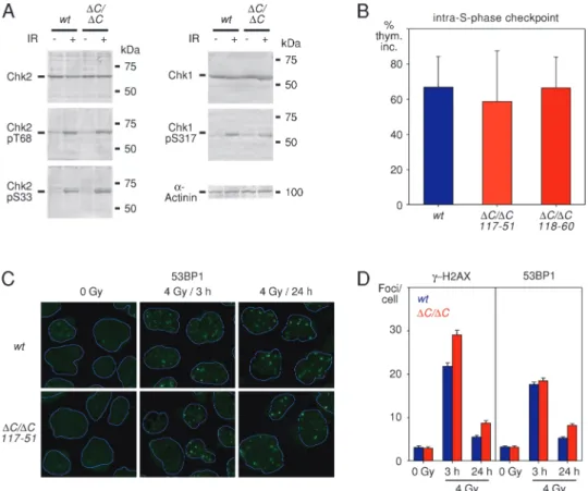
The sensitivity of RECQL4(DC/DC) cells to IR could reflect check-point and/or DNA repair defects. To explore for a possible checkcheck-point deficit, we exposed wild-type and RECQL4(DC/DC) cells to IR and monitored phosphorylation of the checkpoint kinases Chk1 and Chk2. These two kinases are downstream of ATR and ATM, respec-tively, and are good molecular indicators of activated DNA damage checkpoint pathways (48). There was no difference in Chk1 or Chk2 phosphorylation after irradiation between the wild-type and
Fig. 1. Establishment of RECQL4(DC/DC) Nalm-6 cells. (A) Diagram of the wild-type RECQL4 genomic locus (top) and of the same locus after integration of the knock-in constructs that introduce a termination codon, indicated by the red asterisk, after exon 8. The nuclear localization signal (Nls) spans exons 5–8; the helicase domain (Hel) spans exons 8–14. Puro and Bsr indicate the puromycin- and blasticidin-resistance genes, respectively. Nh and Xm indicate the NheI and XmnI restriction enzyme cleavage sites, respectively. (B) Southern blot analysis showing targeting of one of the RECQL4 alleles by the blasticidin-resistance construct in the independently derived clones 117 and 118. Horizontal lines indicate the bands corresponding to the targeted alleles. (C) Southern blot analysis showing targeting of the second RECQL4 allele by the puromycin-resistance construct in several independently derived clones. Horizontal lines indicate the bands corresponding to the targeted alleles. (D) Western blot analysis showing expression of N-terminally truncated RecQL4 protein in Nalm-6 cells with targeting of both RECQL4 alleles (DC/DC). Parental Nalm-6 cells (wt) and Nalm-6 cells with knockout of both BLM alleles (BLM / ) serve as controls and reveal the band corresponding to full-length RecQL4, as well as bands corresponding to lower molecular weight degradation products of full-length RecQL4.a-Actinin serves as a loading control.
M.Kohzaki et al.
RECQL4(DC/DC) cells (Figure 3A). Subsequently, we examined whether deletion of the C-terminus of RecQL4 compromised the in-tra-S-phase checkpoint. In mammals, this checkpoint inhibits both DNA replication initiation and elongation and is dependent on acti-vation of the Chk1 kinase (49). We ascertained intra-S-phase check-point integrity by monitoring tritiated thymidine incorporation either before or 30 min after exposure to 8 Gy IR. In both wild-type and RECQL4(DC/DC) cells, tritiated thymidine incorporation was reduced in response to IR suggesting that the checkpoint was intact (Figure 3B). Analysis of the G2 DNA damage checkpoint by monitoring mitotic entry at several time points after irradiation also revealed no checkpoint defect in RECQL4(DC/DC) cells (data not shown).
The absence of an apparent checkpoint defect prompted us to con-sider a possible role of RecQL4 in DNA DSB repair. Analysis of the function of RecQL4 in human cells and in frog extracts has already revealed a role of this protein in DNA repair (36,50,51). We per-formed a DNA repair analysis in the wild-type and RECQL4(DC/ DC) cells, monitoring DNA DSB repair indirectly by counting IR-induced 53BP1 andc-H2AX foci at 3 and 24 h after irradiation, as described previously (36). Consistent with the previous analyses of RTS cells, we observed a DNA repair defect, which was statistically significant. However, the magnitude of the defect, as revealed by comparing the mean values, was very small (Figure 3C and D). S phase progression defect in irradiated RECQL4(DC/DC) cells Since RecQL4 has a clear role in DNA replication, via its N-terminal Sld2-like domain, we reasoned that we might obtain more clear phe-notypes, if we examined specifically cells that were in S phase. For
this purpose, wild-type and RECQL4(DC/DC) cells were pulse la-beled with EdU, a thymidine analog, to allow cells in S phase to be distinguished from G1/G2cells. Then, the cells were irradiated (4 Gy
IR) and progression through S phase was monitored 8 and 12 h later by flow cytometry (Figure 4A). EdU incorporation could readily dis-tinguish S phase cells from those in G1/G2(Figure 4B, Supplementary
Figure 3, available at Carcinogenesis Online), thus allowing us to monitor whether cells that were in S phase at the time of irradiation progressed through S to G2/M. A significant fraction of wild-type and
BLM(�/�) cells that had been irradiated during S phase completed DNA replication over the study period and accumulated in G2/M. In
contrast, practically all the RECQL4(DC/DC) cells irradiated during S phase remained in S phase during the study period (Figure 4C).
To better understand the nature of the S phase progression defect, we performed a DNA combing assay to monitor the dynamics of DNA replication at the level of single molecules. Irradiated and non-irradiated wild-type, RECQL4(DC/DC) and BLM(�/�) cells were pulse-labeled with IdU for 20 min and then with CldU for 40 min (Figure 4D). DNA fibers were prepared from these cells and incubated with antibodies that recognize single-stranded DNA, IdU and CldU (Figure 4E). We focused our analysis on DNA molecules that had incorporated sequentially both IdU and CldU and tabulated the ratios of the lengths of the fibers labeled with each nucleoside analog. In the absence of irradiation, the ratios of the lengths of the fibers incorpo-rating IdU and CldU were similar in the three cell types, suggesting that deletion of the RecQL4 C-terminus does not affect DNA repli-cation elongation rates in the absence of exogenous DNA damaging agents (Figure 4F). However, after irradiation, the CldU/IdU length ratios for the RECQL4(DC/DC) cells decreased, suggesting stochastic
Fig. 2. Growth kinetics and DNA damage sensitivity of RECQL4(DC/DC) Nalm-6 cells. (A) Cell proliferation of wild-type (wt), RECQL4(DC/DC) and BLM(�/�) Nalm-6 cells. Results from three different RECQL4(DC/DC) clones are shown. (B) Sensitivity of wild-type (wt), RECQL4(DC/DC) and BLM(�/�) Nalm-6 cells to IR and HU. The surviving fraction (Surv. Fr.) is plotted relative to the dose of the DNA damaging agent. The RecQL4 curves were generated by averaging the data from four independently derived RECQL4(DC/DC) clones. (C) Frequencies (means and standard deviations) of spontaneous and aphidicolin-induced SCEs in wild-type (wt), RECQL4(DC/DC) and BLM(�/�) Nalm-6 cells. Data from two different RECQL4(DC/DC) clones (117–51 and 118–60) are shown. For each condition, 47 metaphase spreads, corresponding to 2002–2096 chromosomes, were examined. (D) Expression of full-length wild-type (þwt) and K508N mutant (þ508) RecQL4 in transiently transfected RECQL4(DC/DC) cells compared with non-transfected (�) RECQL4(DC/DC) cells and to wild-type (wt) Nalm-6 cells, as determined by immunoblotting with an anti-RecQL4 antibody. The transiently expressed RecQL4 proteins contain two N-terminal FLAG tags, which explain their slow migration compared with endogenous RecQL4.a-Actinin serves as a loading control. (E) Rescue of the IR sensitivity of RECQL4(DC/DC) cells by transient expression of wild-type (þwt), but not mutant (þ508), full-length RecQL4. Parental Nalm-6 cells (wt) serve as controls. M.Kohzaki et al.
pausing and/or collapse of the replication forks, whereas no such decrease was observed with the wild-type and BLM( / ) cells (Figure 4F). We conclude that in irradiated cells, the RecQL4 C-terminus allows replication forks to negotiate DNA templates that have been damaged by irradiation.
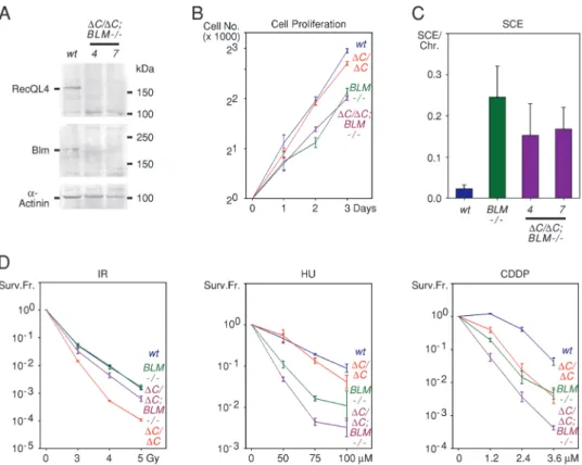
Phenotype of RECQL4(DC/DC);BLM( / ) cells
The distinct phenotypes of the RECQL4(DC/DC) and BLM( / ) cells suggest that these genes function in distinct DNA damage response pathways. To examine the validity of this prediction, we generated human NALM-6 cells, in which both alleles of the RECQL4 and BLM genes were targeted by homologous recombination. To our knowledge, targeting both these genes has not been examined in any system. Several independently derived clones of RECQL4(DC/DC); BLM( / ) cells were obtained by targeting both RECQL4 alleles in BLM( / ) cells (Figure 5A). This indicates that concurrent inactiva-tion of the funcinactiva-tion of the C-terminus of RecQL4 and of BLM is compatible with cell viability. The doubly targeted cells exhibited pro-liferation rates that were reduced compared with those of wild-type and RECQL4(DC/DC) cells but similar to those of BLM( / ) cells (Figure 5B). The number of SCEs in RECQL4(DC/DC);BLM( / ) cells was elevated compared with wild-type and RECQL4(DC/DC) cells; however, it was somewhat lower than the number of SCEs in BLM( / ) cells (Figure 5C).
In terms of sensitivity to DNA damaging agents, the RECQL4(DC/ DC) and BLM( / ) cells showed a somewhat complex profiles
consistent with RecQL4 and BLM functioning in distinct, but related, pathways. Thus, the RECQL4(DC/DC);BLM(-/-) cells were less sen-sitive to IR than the RECQL4(DC/DC) cells, although more sensitive than the wild-type and BLM( / ) cells (Figure 5D). Thus, deletion of BLM rescues in part the IR sensitivity of RECQL4(DC/DC) cells. The opposite effect was observed with HU and other DNA damaging agents, such as cisplatin (Figure 5D and data not shown). In this case, deletion of BLM enhanced the sensitivity of the RECQL4(DC/DC) cells. In these cases, the effects were mostly additive, with the RE-CQL4(DC/DC);BLM( / ) cells exhibiting the sensitivity expected by adding the sensitivites of the RECQL4(DC/DC) and BLM( / ) cells.
Discussion
Prior studies have established a role for RecQL4 in initiation of DNA replication (17–24). This function is mediated by the N-terminal Sld2-like domain. Here, we addressed the function of the C-terminus by targeted mutagenesis of the endogenous RECQL4 alleles in human Nalm-6 cells. A significant body of evidence already indicated that the C-terminus plays a role in the response to DNA damage (25–31,33– 36,50,51). However, some studies suggested a function in the re-sponse to agents that induce replication fork stalling, whereas others suggested a role in the response to agents that induce DNA DSBs. There are many ways to explain these apparent discrepancies. First, most studies focused on analysis of cells obtained from RTS patients;
Fig. 3. IR-induced DNA damage responses of RECQL4(DC/DC) Nalm-6 cells. (A) Phosphorylation of Chk1 and Chk2 protein kinases in wild-type (wt) and RECQL4(DC/DC) Nalm-6 cells 1 h after exposure to 6 Gy IR. pT68, phospho-Thr68; pS33, phospho-Ser33; pS317; phospho-Ser317. a-Actinin serves as a loading control. (B) Intact intra-S-phase checkpoint in RECQL4(DC/DC) Nalm-6 cells. Thymidine incorporation (thym. inc.) 30 min after exposure to 8 Gy IR is expressed as a percentage relative to thymidine incorporation in the absence of exogenous DNA damage. Means and standard deviations are derived from four independent experiments. (C) IR-induced 53BP1 focus formation in wild-type (wt) and RECQL4(DC/DC) Nalm-6 cells, as ascertained by immunofluorescence analysis. The peripheries of the nuclei are indicated by blue lines. (D) DNA DSB repair defect in RECQL4(DC/DC) Nalm-6 cells (clone 117–51). The average number of c-H2AX and 53BP1 foci per cell was determined in untreated cells and at the indicated timepoints after exposure to 4 Gy IR. For each condition, the number of foci in.200 cells was counted. Results are shown as means and standard errors. The differences between the means of wild-type and RECQL4(DC/DC) Nalm-6 cells were statistically significant for the number ofc-H2AX foci at 3 h (P , 0.05) and 24 h (P , 0.01) and for the number of 53BP1 foci at 24 h (P , 0.05), as determined by the Student’s t-test.
M.Kohzaki et al.
the mutations present in these cells differ from patient to patient and may not fully inactivate function, making comparisons among studies difficult (25–30). Second, some mutations may facilitate protein un-folding, thereby targeting the mutant RecQL4 protein for degradation or sequestering it in inactive complexes with protein chaperones. As a result, these mutations have the potential to interfere with the func-tion of the N-terminal domain.
To study the function of the C-terminus of RecQL4 in a well-defined system, we introduced in human Nalm-6 cells a termination codon in the endogenous RECQL4 gene just upstream of the coding sequence for the helicase domain. This mutation resulted in expres-sion of a truncated RecQL4 protein that contained the essential N-terminal Sld2-like domain and the nuclear localization signal, but which lacked the entire C-terminus, including the helicase domain. Thus, the deleted segment corresponds to the part of the human RecQL4 protein that is missing in budding yeast Sld2. Several in-dependently derived Nalm-6 clones expressing the truncated RecQL4 protein were analyzed.
Deletion of the RecQL4 C-terminus did not have a major effect on unperturbed DNA replication, as ascertained by cell proliferation rates and by analysis of DNA replication at the single-molecule level. This conclusion appears to contradict some previously published re-sults. For example, knockout mice lacking exon 13 of RECQL4 ex-hibit severe growth retardation, tissue atrophy and perinatal lethality and mouse embryo fibroblasts prepared from these mice have a pro-liferation defect in vitro (24). However, because the helicase domain is encoded by exons 8–14, deletion of exon 13 will almost certainly result in expression of an unfolded protein, thereby potentially also
compromising the function of the N-terminal Sld-2 like domain. A similar rationale may also explain why RecQL4 proteins with muta-tions in the helicase domain cannot rescue a null mutant in Drosophila and the apparent conflicting results in Xenopus regarding whether the helicase domain of RecQL4 is needed for unperturbed DNA replication (20,21,32). Consistent with our conclusion that the RecQL4 C-terminus is not critical for unperturbed DNA replication, the replication defect of chicken DT40 cells with homozygous RECQL4 deletions is rescued by ectopic expression of the RecQL4 N-terminus (31).
Even though the C-terminus of RecQL4 appears dispensable for unperturbed DNA replication, it is apparently required for the cellular response to IR. Previously, other groups have reported conflicting re-sults regarding the sensitivity of RTS cells to agents that induce stalling of DNA replication forks, such as UV light and HU, and to agents that induce DNA DSBs (25–31,33–36,50,51). These conflicting results could be explained by the fact that the mutant RecQL4 proteins ex-pressed in RTS cells may retain partial activity and/or be defective in the activity of their N-terminal Sld2-like domain. However, our analysis of the sensitivity profiles of wild-type, RECQL4(DC/DC) and BLM( / ) Nalm-6 cells to IR, UV light and HU provides a very clear result. The RECQL4(DC/DC) cells were hypersensitive to IR, but their response to UV light and HU was not different from that of wild-type cells. The wild-type response cannot be attributed to an in-herent resistance of Nalm-6 cells to these agents since deletion of the BLM gene led to sensitivity to UV and HU, as expected.
The sensitivity of RECQL4(DC/DC) cells to IR could be due to a checkpoint defect, a general DNA DSB repair defect and/or a defect of replication forks in negotiating IR-induced DNA damage. Previous
Fig. 4. S phase progression defect in irradiated RECQL4(DC/DC) cells. (A) Outline of the experiment to monitor cell cycle progression of wild-type and RECQL4(DC/DC) cells irradiated in S phase. Cells were exposed to EdU (E) for 1 h and then irradiated (4 Gy). (B) Efficiency of gating of S phase versus G1/G2
cells by EdU incorporation. The propidium iodide (PI) profiles of the entire cell population (all) and of the EdU-negative and EdU-positive subpopulations are shown for wild-type (wt) Nalm-6 cells at the 0-h timepoint. (C) Cell cycle progression of wild-type (wt), RECQL4(DC/DC) and BLM( / ) Nalm-6 cells after exposure to 4 Gy IR. The PI profiles of the EdU-positive cells are shown. (D) Outline of the experiment to monitor DNA replication at the single-molecule level by DNA combing. Cells were untreated or exposed to 4 Gy IR and 4 h later were incubated sequentially with IdU (I; for 20 min) and CldU (Cl; for 40 min). (E) Images of DNA replication fibers from wild-type (wt) and RECQL4(DC/DC) cells exposed to 4 Gy IR. IdU incorporation, red; CldU incorporation, green; single-stranded DNA, blue. (F) Ratios of lengths of DNA fibers incorporating CldU over IdU from wild-type (wt), RECQL4(DC/DC) (clone 118–60) and BLM( / ) cells. For each condition, 150–200 DNA replication forks were counted. The results are expressed as percentages. The profile of the irradiated RECQL4(DC/DC) cells is statistically significantly different from the profiles of the irradiated wild-type and BLM( / ) cells (P , 0.01), as determined by the Mann–Whitney U-test. M.Kohzaki et al.
studies have reported checkpoint defects in RTS cells or in Drosophila cells in which the endogenous RecQL4 protein was depleted by small interfering RNA (52,53). We did not observe a checkpoint defect in the RECQL4(DC/DC) cells when we monitored Chk1 and Chk2 phos-phorylation, the intra-S-phase checkpoint or the G2/M checkpoint. We
attribute the previously observed checkpoint defects to suppression of DNA replication resulting from compromised RecQL4 function since suppression of DNA replication compromises ATR activation (54).
A general defect in DNA DSB repair could also have explained the sensitivity of RECQL4(DC/DC) cells to IR. Previous studies have reported a DNA DSB repair defect in RTS cells, as ascertained by monitoring the number of 53BP1 foci at various timepoints after irradiation (36). We also observed a subtle defect in resolution of c-H2AX and 53BP1 foci in irradiated RECQL4(DC/DC) cells over time. However, the magnitude of the defect, although statistically significant, appears insufficient to explain the profound sensitivity of these cells to IR. Thus, instead of a general defect in DNA DSB repair, we propose a DNA replication defect in cells that have been exposed to IR. More specifically, one could envision a defect in the ability of DNA replication forks to negotiate IR-induced DNA dam-age. Consistent with this proposal, RECQL4(DC/DC) cells, that were irradiated while in S phase, failed to complete DNA replication. In addition, by DNA combing analysis, these cells displayed stochastic premature termination of DNA replication forks. It has been proposed that RecQL4 travels with the DNA replication fork (19,22), which would be consistent with a role of its helicase domain in facilitating the ability of forks to negotiate IR-induced DNA damage. It is note-worthy that, compared with the other RecQ helicases, RecQL4 ap-pears to stand out in its role in the response to IR, whereas all other RecQ helicases appear to be more important for the response to agents that stall DNA replication forks, such as HU and UV light (1,2,55,56).
Prokaryotes and budding yeast have only one RecQ helicase, whereas mammals have five such helicases, including RecQL4 and BLM (1,2). The presence of multiple RecQ helicases in higher eukar-yotes raises the question whether these helicases have distinct or overlapping functions. In DT40 chicken cells, double knockout mu-tants, such as BLM( / );WRN( / ), BLM( / );RECQL1( / ), BLM( / );RECQL5( / ) and RECQL1( / );RECQL5( / ), have been generated and, in general, show more severe phenotypes after induction of DNA damage, as compared with the single mutants (55,56). However, to our knowledge, no double mutant of RECQL4 with any other RecQ helicase gene has been described. We were surprised to see that RECQL4(DC/DC);BLM( / ) cells were viable, given that both the single RECQL4(DC/DC) and BLM( / ) mutants had strong phenotypes. The epistasis analysis of the RECQL4(DC/ DC) and BLM( / ) mutants described here is consistent with RecQL4 and BLM having distinct functions. Thus, we conclude that the function of the RecQ helicases has diverged during evolution, with RecQL4 acquiring a function that allows cells to negotiate DNA replication templates that have been damaged by IR.
Supplementary material
Supplementary Figures 1–3 can be found at http://carcin. oxfordjournals.org/
Funding
Swiss National Foundation (to T.D.H.), BRFAA intramural funds (to S.G.), a long-term post-doctoral fellowship from the Human Frontier Science Program (to G.V.) and the CANGENIN COST network (for scientific exchanges between the laboratories of T.D.H. and S.G.).
Fig. 5. Phenotype of RECQL4(DC/DC);BLM( / ) cells. (A) Lack of expression of full-length RecQL4 and BLM proteins in two independently derived RECQL4(DC/DC);BLM( / ) clones, as determined by immunoblotting. Wild-type (wt) Nalm-6 cells serve as controls. a-Actinin serves as a loading control. (B) Cell proliferation of wild-type (wt), RECQL4(DC/DC), BLM( / ) and RECQL4(DC/DC);BLM( / ) Nalm-6 cells. Results from three different RECQL4(DC/DC);BLM( / ) clones are shown. (C) Frequencies (means and standard deviations) of spontaneous SCEs in wild-type (wt), BLM( / ) and RECQL4(DC/DC);BLM( / ) Nalm-6 cells. Data from two different RECQL4(DC/DC);BLM( / ) clones (4 and 7) are shown. For each condition, 25 metaphase spreads were examined. (D) Sensitivity of wild-type (wt), RECQL4(DC/DC), BLM( / ) and RECQL4(DC/DC);BLM( / ) Nalm-6 cells to IR, HU and cisplatin (CDDP). The surviving fraction (Surv. Fr.) is plotted relative to the dose of the DNA damaging agent. Data from three independently derived RECQL4(DC/DC); BLM( / ) clones were averaged.
M.Kohzaki et al.
Acknowledgements
The authors thank U. Schibler (University of Geneva), M. Seki, T. Abe (Tohoku University), A. Carr (Sussex University) and K. Nishihara (Kyoto University) for helpful discussions.
Conflict of Interest Statement: None declared.
References
1. Bohr,V.A. (2008) Rising from the RecQ-age: the role of human RecQ helicases in genome maintenance. Trends Biochem. Sci., 33, 609–620. 2. Chu,W.K. et al. (2009) RecQ helicases: multifunctional genome caretakers.
Nat. Rev. Cancer, 9, 644–654.
3. Petermann,E. et al. (2010) Pathways of mammalian replication fork restart. Nat. Rev. Mol. Cell Biol., 11, 683–687.
4. Branzei,D. et al. (2010) Maintaining genome stability at the replication fork. Nat. Rev. Mol. Cell Biol., 11, 208–219.
5. Wu,L. et al. (2003) The Bloom’s syndrome helicase suppresses crossing over during homologous recombination. Nature, 426, 870–874.
6. Raynard,S. et al. (2006) A double Holliday junction dissolvasome com-prising BLM, topoisomerase IIIalpha, and BLAP75. J. Biol. Chem., 281, 13861–13864.
7. Seki,M. et al. (2006) Bloom helicase and DNA topoisomerase IIIalpha are involved in the dissolution of sister chromatids. Mol. Cell. Biol., 26, 6299–6307. 8. Liberi,G. et al. (2005) Rad51-dependent DNA structures accumulate at damaged replication forks in sgs1 mutants defective in the yeast ortholog of BLM RecQ helicase. Genes Dev., 19, 339–350.
9. Crabbe,L. et al. (2004) Defective telomere lagging strand synthesis in cells lacking WRN helicase activity. Science, 306, 1951–1953.
10. Laud,P.R. et al. (2005) Elevated telomere-telomere recombination in WRN-deficient, telomere dysfunctional cells promotes escape from senes-cence and engagement of the ALT pathway. Genes Dev., 19, 2560–2570. 11. Masai,H. (2011) RecQL4: a helicase linking formation and maintenance of
a replication fork. J. Biochem., 149, 629–631.
12. Kamimura,Y. et al. (1998) Sld2, which interacts with Dpb11 in Saccharo-myces cerevisiae, is required for chromosomal DNA replication. Mol. Cell. Biol., 18, 6102–6109.
13. Masumoto,H. et al. (2002) S-Cdk-dependent phosphorylation of Sld2 essential for chromosomal DNA replication in budding yeast. Nature, 415, 651–655. 14. Zegerman,P. et al. (2007) Phosphorylation of Sld2 and Sld3 by
cyclin-dependent kinases promotes DNA replication in budding yeast. Nature, 445, 281–285.
15. Tanaka,S. et al. (2007) CDK-dependent phosphorylation of Sld2 and Sld3 initiates DNA replication in budding yeast. Nature, 445, 328–332. 16. Noguchi,E. et al. (2002) CDK phosphorylation of Drc1 regulates DNA
replication in fission yeast. Curr. Biol., 12, 599–605.
17. Xu,X. et al. (2009) MCM10 mediates RECQ4 association with MCM2-7 helicase complex during DNA replication. EMBO J., 28, 3005–3014. 18. Im,J.S. et al. (2009) Assembly of the Cdc45-Mcm2-7-GINS complex in
human cells requires the Ctf4/And-1, RecQL4, and Mcm10 proteins. Proc. Natl Acad. Sci. USA, 106, 15628–15632.
19. Thangavel,S. et al. (2010) Human RECQ1 and RECQ4 helicases play distinct roles in DNA replication initiation. Mol. Cell. Biol., 30, 1382–1396. 20. Sangrithi,M.N. et al. (2005) Initiation of DNA replication requires the RECQL4
protein mutated in Rothmund-Thomson syndrome. Cell, 121, 887–898. 21. Matsuno,K. et al. (2006) The N-terminal noncatalytic region of Xenopus
RecQ4 is required for chromatin binding of DNA polymerase alpha in the initiation of DNA replication. Mol. Cell. Biol., 26, 4843–4852.
22. Wu,J. et al. (2008) Drosophila homologue of the Rothmund-Thomson syn-drome gene: essential function in DNA replication during development. Dev. Biol., 323, 130–142.
23. Xu,Y. et al. (2009) dRecQ4 is required for DNA synthesis and is essential for cell proliferation in Drosophila. PLoS One, 4, e6107.
24. Hoki,Y. et al. (2003) Growth retardation and skin abnormalities of the Recql4-deficient mouse. Hum. Mol. Genet., 12, 2293–2299.
25. Kitao,S. et al. (1999) Mutations in RECQL4 cause a subset of cases of Rothmund-Thomson syndrome. Nat. Genet., 22, 82–84.
26. Wang,L.L. et al. (2003) Association between osteosarcoma and deleterious mutations in the RECQL4 gene in Rothmund-Thomson syndrome. J. Natl. Cancer Inst., 95, 669–674.
27. Siitonen,H.A. et al. (2003) Molecular defect of RAPADILINO syndrome expands the phenotype spectrum of RECQL diseases. Hum. Mol. Genet., 12, 2837–2844.
28. Siitonen,H.A. et al. (2009) The mutation spectrum in RECQL4 diseases. Eur. J. Hum. Genet., 17, 151–158.
29. Cabral,R.E. et al. (2008) Identification of new RECQL4 mutations in Cau-casian Rothmund-Thomson patients and analysis of sensitivity to a wide range of genotoxic agents. Mutat. Res., 643, 41–47.
30. Van Maldergem,L. et al. (2006) Revisiting the craniosynostosis-radial ray hypoplasia association: Baller-Gerold syndrome caused by mutations in the RECQL4 gene. J. Med. Genet., 43, 148–152.
31. Abe,T. et al. (2011) The N-terminal region of RECQL4 lacking the helicase domain is both essential and sufficient for the viability of vertebrate cells. Role of the N-terminal region of RECQL4 in cells. Biochim. Biophys. Acta, 1813, 473–479.
32. Capp,C. et al. (2009) Drosophila RecQ4 has a 3’-5’ DNA helicase activity that is essential for viability. J. Biol. Chem., 284, 30845–30852. 33. Jin,W. et al. (2008) Sensitivity of RECQL4-deficient fibroblasts from
Rothmund-Thomson syndrome patients to genotoxic agents. Hum. Genet., 123, 643–653.
34. Werner,S.R. et al. (2006) RECQL4-deficient cells are hypersensitive to oxidative stress/damage: insights for osteosarcoma prevalence and hetero-geneity in Rothmund-Thomson syndrome. Biochem. Biophys. Res. Commun., 345, 403–409.
35. Fan,W. et al. (2008) RecQ4 facilitates UV light-induced DNA damage repair through interaction with nucleotide excision repair factor xeroderma pigmentosum group A (XPA). J. Biol. Chem., 283, 29037–29044. 36. Singh,D.K. et al. (2010) The involvement of human RECQL4 in DNA
double-strand break repair. Aging Cell, 9, 358–371.
37. Cescutti,R. et al. (2010) TopBP1 functions with 53BP1 in the G1 DNA damage checkpoint. EMBO J., 29, 3723–3732.
38. Mochan,T.A. et al. (2003) 53BP1 and NFBD1/MDC1-Nbs1 function in parallel interacting pathways activating ataxia-telangiectasia mutated (ATM) in response to DNA damage. Cancer Res., 63, 8586–8591. 39. Bailey,S.M. et al. (2004) Frequent recombination in telomeric DNA may
extend the proliferative life of telomerase-negative cells. Nucleic Acids Res., 32, 3743–3751.
40. Michalet,X. et al. (1997) Dynamic molecular combing: stretching the whole human genome for high-resolution studies. Science, 277, 1518–1523. 41. Burks,L.M. et al. (2007) Nuclear import and retention domains in the
amino terminus of RECQL4. Gene, 391, 26–38.
42. Suzuki,T. et al. (2009) DNA helicase activity in purified human RECQL4 protein. J. Biochem., 146, 327–335.
43. Xu,X. et al. (2009) Dual DNA unwinding activities of the Rothmund-Thomson syndrome protein, RECQ4. EMBO J., 28, 568–577.
44. Rossi,M.L. et al. (2010) Conserved helicase domain of human RecQ4 is required for strand annealing-independent DNA unwinding. DNA Repair (Amst), 9, 796–804.
45. Adachi,N. et al. (2006) The human pre-B cell line Nalm-6 is highly pro-ficient in gene targeting by homologous recombination. DNA Cell Biol., 25, 19–24.
46. So,S. et al. (2004) Genetic interactions between BLM and DNA ligase IV in human cells. J. Biol. Chem., 279, 55433–55442.
47. Mann,M.B. et al. (2005) Defective sister-chromatid cohesion, aneuploidy and cancer predisposition in a mouse model of type II Rothmund-Thomson syndrome. Hum. Mol. Genet., 14, 813–825.
48. Jackson,S.P. et al. (2009) The DNA-damage response in human biology and disease. Nature, 461, 1071–1078.
49. Seiler,J.A. et al. (2007) The intra-S-phase checkpoint affects both DNA replication initiation and elongation: single-cell and -DNA fiber analyses. Mol. Cell. Biol., 27, 5806–5818.
50. Petkovic,M. et al. (2005) The human Rothmund-Thomson syndrome gene product, RECQL4, localizes to distinct nuclear foci that coincide with proteins involved in the maintenance of genome stability. J. Cell Sci., 118, 4261–4269.
51. Kumata,Y. et al. (2007) Possible involvement of RecQL4 in the repair of double-strand DNA breaks in Xenopus egg extracts. Biochim. Biophys. Acta, 1773, 556–564.
52. Park,S.J. et al. (2006) A positive involvement of RecQL4 in UV-induced S-phase arrest. DNA Cell Biol., 25, 696–703.
53. Kondo,S. et al. (2011) A genome-wide RNAi screen identifies core com-ponents of the G2-M DNA damage checkpoint. Sci. Signal., 4, rs1. 54. Zou,L. et al. (2003) Sensing DNA damage through ATRIP recognition of
RPA-ssDNA complexes. Science, 300, 1542–1548.
55. Imamura,O. et al. (2002) Werner and Bloom helicases are involved in DNA repair in a complementary fashion. Oncogene, 21, 954–963.
56. Wang,W. et al. (2003) Functional relation among RecQ family helicases RecQL1, RecQL5, and BLM in cell growth and sister chromatid exchange formation. Mol. Cell. Biol., 23, 3527–3535.
Received February 23, 2012; revised April 3, 2012; accepted April 6, 2012 M.Kohzaki et al.