HAL Id: hal-00258941
https://hal.archives-ouvertes.fr/hal-00258941
Submitted on 30 May 2020
HAL is a multi-disciplinary open access
archive for the deposit and dissemination of
sci-entific research documents, whether they are
pub-lished or not. The documents may come from
teaching and research institutions in France or
abroad, or from public or private research centers.
L’archive ouverte pluridisciplinaire HAL, est
destinée au dépôt et à la diffusion de documents
scientifiques de niveau recherche, publiés ou non,
émanant des établissements d’enseignement et de
recherche français ou étrangers, des laboratoires
publics ou privés.
Distributed under a Creative Commons Attribution| 4.0 International License
Boyan Grigorov, Didier Decimo, Fatima Smagulova, Christine Péchoux,
Marylène Mougel, Delphine Muriaux, Jean-Luc Darlix
To cite this version:
Boyan Grigorov, Didier Decimo, Fatima Smagulova, Christine Péchoux, Marylène Mougel, et al..
Intracellular HIV-1 Gag localization is impaired by mutations in the nucleocapsid zinc fingers..
Retro-virology, BioMed Central, 2007, 4, pp.54. �10.1186/1742-4690-4-54�. �hal-00258941�
Open Access
Research
Intracellular HIV-1 Gag localization is impaired by mutations in the
nucleocapsid zinc fingers
Boyan Grigorov
†1, Didier Décimo
†1, Fatima Smagulova
2, Christine Péchoux
1,
Marylène Mougel
2, Delphine Muriaux*
1and Jean-Luc Darlix
1Address: 1LaboRetro, Unité de virologie humaine INSERM U758, IFR128, ENS, 46 allée d'Italie, 69 364 Lyon, France and 2CPBS, UMI, CNRS, 4 bd Henri IV, 34000 Montpellier, France
Email: Boyan Grigorov - [email protected]; Didier Décimo - [email protected]; Fatima Smagulova - [email protected]; Christine Péchoux - [email protected]; Marylène Mougel - [email protected]@univ-montp1.fr;
Delphine Muriaux* - [email protected]; Jean-Luc Darlix - [email protected] * Corresponding author †Equal contributors
Abstract
Background: The HIV-1 nucleocapsid protein (NC) is formed of two CCHC zinc fingers flanked
by highly basic regions. HIV-1 NC plays key roles in virus structure and replication via its nucleic acid binding and chaperoning properties. In fact, NC controls proviral DNA synthesis by reverse transcriptase (RT), gRNA dimerization and packaging, and virion assembly.
Results: We previously reported a role for the first NC zinc finger in virion structure and
replication [1]. To investigate the role of both NC zinc fingers in intracellular Gag trafficking, and in virion assembly, we generated series of NC zinc fingers mutations. Results show that all Zinc finger mutations have a negative impact on virion biogenesis and maturation and rendered defective the mutant viruses. The NC zinc finger mutations caused an intracellular accumulation of Gag, which was found either diffuse in the cytoplasm or at the plasma membrane but not associated with endosomal membranes as for wild type Gag. Evidences are also provided showing that the intracellular interactions between NC-mutated Gag and the gRNA were impaired.
Conclusion: These results show that Gag oligomerization mediated by gRNA-NC interactions is
required for correct Gag trafficking, and assembly in HIV-1 producing cells and the release of infectious viruses.
Background
The retroviral Gag polyprotein precursor is formed of three essential domains, namely the matrix (MA), the cap-sid (CA) and the nucleocapcap-sid (NC), which upon protease mediated processing of Gag constitute the architecture of the infectious mature viral particle. The three Gag domains contain the critical determinants that orchestrate virus assembly in the infected cell, via membrane-MA, CA-CA and NC-gRNA interactions [2-8]. In the mature virus,
the MA protein is located under the virion envelope, which derives from the infected cell membrane. In the case of HIV-1, MA is myristoylated and contains basic amino acids within its N-terminus required for Gag-mem-brane binding and determinants that specifically interact with the cellular adaptator proteins AP-3 and AP-2. These AP proteins contribute to the intracellular transport of Gag to endosomal compartments and retroviral budding [9-11]. The CA molecules form the outer shell of the viral
Published: 3 August 2007
Retrovirology 2007, 4:54 doi:10.1186/1742-4690-4-54
Received: 4 May 2007 Accepted: 3 August 2007 This article is available from: http://www.retrovirology.com/content/4/1/54
© 2007 Grigorov et al; licensee BioMed Central Ltd.
This is an Open Access article distributed under the terms of the Creative Commons Attribution License (http://creativecommons.org/licenses/by/2.0), which permits unrestricted use, distribution, and reproduction in any medium, provided the original work is properly cited.
core while NC molecules extensively coat and condense the gRNA in the interior of the virion core [2]. HIV-1 NC contains two zinc fingers flanked by basic regions and is located at the C-terminus of Gag, followed by the p6 domain. This later p6 domain is required for particle bud-ding during which the viral particles pinch-off from the cellular membrane (reviewed in [5]. The p6 domain con-tains a Proline-rich and a di-Leucine domains, which are the target of the cellular proteins Tsg101 and Alix, respec-tively, involved in the cellular class E protein sorting path-way and the HIV-1 budding machinery [5,12,13]. HIV-1 NC has been extensively studied during the past 15 years and was shown to be implicated in virus structure, gRNA dimerization and proviral DNA synthesis [3,4,7]. The highly basic nature of NC makes it a partner of choice of RNA while the zinc fingers appear to provide specific recognition of the HIV-1 Psi packaging signal necessary for gRNA packaging [14]. Furthermore, specific RNA-NC interactions promote Gag-Gag oligomerization which turns out to be a prerequisite for assembly and virus bio-genesis [15-18]. Both NC zinc fingers and basic domains are essential for virus formation and infectivity [1,16,17,19-21]. Mutations in NC basic residues cause defects in Gag-viral RNA interactions and thus in HIV-1 assembly and budding [15,16,22]. More recently, new insights into the role of NC in Gag assembly show that mutations and deletions in the basic residues of NC pre-vent Gag-Gag multimerization but not Gag association with cellular membranes [23].
In the present study, we explored the influence of the NC zinc fingers in HIV-1 assembly by analyzing intracellular Gag and gRNA localization, Gag/membrane association and virion morphogenesis.
Methods
Plasmid DNA
HIV-1 pNL4-3 DNA was provided by the National Insti-tute of Health, USA. The HIV-1 ΔZF1 and H23C Gag mutant DNA constructs were described elsewhere [1]. The HIV-1 GagΔNC proviral DNA construct [24] was provided by A.Cimarelli. The HIV-1 ΔZF2 and H44C Gag mutants were obtained by site directed mutagenesis on the pNL4.3 HIV-1 molecular clone as described [1] using the follow-ing oligonucleotides 5'CCTGTCTCTCAGTACCGCCCTTTTTCCTAG3' and 5'CTTTCATTTGGCATCCTTCC3', respectively. The double ΔZF1ZF2 and H23C/H44C Gag mutants were obtained by cloning the ApaI-AgeI fragments of H44C and ΔZF2 into the H23C and ΔZF1 pNL4.3 mutant clone, respectively. The pcDNA3.1 plasmid (Clonetech) was used as a control DNA vector.
Mammalian cell culture, DNA transfection and virus production
The human 293T cell line, HeLa P4 cells expressing the CD4 receptor and the LacZ gene under the control of the HIV-1 LTR and HeLa cells used were grown in Dulbecco's modified essential medium (DMEM), all supplemented with 10% fetal calf serum and antibiotics. 293T were transfected using the calcium phosphate method [18]. For immunofluorescence staining, HeLa cells were transfected with DNA using the Fugene® transfection method (Invitro-gen). To analyse virus production, cells were washed with PBS and medium was changed 5 h post-transfection. Cul-ture supernatants containing virus particles were har-vested 24 hours later and clarified by filtration (0.45 μm, Nalgen). The cells were then washed and lysed with 0,5% Triton-PBS.
Virus preparation
Virions were purified from filtered culture supernatants by pelleting them through a cushion of 20% sucrose in TNE (100 mM NaCl, 10 mM Tris HCl, pH 7.4 and 1 mM EDTA) at 35 K rpm for 1 h in a Beckman SW41 rotor.
CAp24 antigen ELISA
To measure viral production, a CAp24 ELISA test was used. Aliquots of the same volume of viral supernatants (free CAp24 + virion associated = S) and pellet virions by ultracentrifugation (V) were resuspended in cell media with 0.5% Triton, and administered on 96 well plates coated with 10 μg/ml anti-CAp24 antibodies (23A5G and 3D10G9B8, BioMérieux) and then blocked with 10% horse serum in PBS-0.05% Tween-20. A biotinylated anti-CAp24 antibody (bioMérieux) was added and the ELISA was revealed with streptavidin and orthophenylene-diamine (OPD)-H2O2 (Sigma). The plate was read on ELISA-reader at 490 and 630 nm.
HIV Infectivity assays
Virus infectivity was assessed on HeLaP4 cells as described in [25]. The infectivity was determined by counting the number of blue cells.
Genomic RNA analysis by Dot-Blot
For viral RNA analysis, virus pellets were resuspended in TNE buffer and lysed in 1% SDS, 100 μg of proteinase K per ml. Nucleic acids were extracted twice with phenol-chloroform and ethanol precipitated. Pellets were resus-pended in DNase buffer (40 mM Tris-HCl, pH 7.5, 6 mM MgCl2, 10 mM NaCl, 10 mM dithiotreitol, 200 U of RNa-sin per ml) and contaminant plasmid DNA was digested with RQ1 DNase (100 U/ml) for 20 min at 37°C. RNA was purified by phenol-chloroform extraction, ethanol precipitated and resuspended in water. Hybridization with a random 32P-labeled 5.3 kb SacI-SalI fragment of the pNL4-3 plasmid corresponding to gag and pol
sequences and quantitative analyses were done as previ-ously described [19].
Subcellular fractionation
Twenty-four hours post transfection, 293T cells were washed with PBS and removed from the plate in PBS-1 mM EDTA, pelleted by centrifugation at 600 × g, resus-pended in 1 ml of a homogenization buffer containing 10 mM Tris-HCl, pH 7.5, 0.25 M sucrose, 1 mM EDTA and protease inhibitors (Complete Mini EDTA-free from Roche), and then fragmented using a glass homogenizer. Nuclei were eliminated by centrifugation at 600 × g for 10 min at 4°C. The resulting post-nuclear supernatant (PNS) was subjected to subcellular fractionation on OptiPrep® gradient for the separation of different membrane com-partments as described elsewhere [25]. Fractions were col-lected and proteins were analyzed by SDS-PAGE and immunoblotting.
Immunoblotting
Viral proteins were separated on 10% SDS-PAGE and detected by immunoblotting with a mouse anti-CAp24 (P3D10G9B8, BioMérieux), and the cellular protein in the gradient was detected with the mouse anti-Lamp2 (Santa Cruz Biotechnology Inc.). The corresponding immunoglobulins conjugated with horse radish peroxi-dase (HRP) (DakoCytomation) were used and the signal was detected using SuperSignal® West Pico Chemilumi-nescent Substrate (Pierce).
RT-PCR
Fractions from Optiprep® gradients were resuspended in equal volumes of a lysis buffer containing 100 mM Tris-HCl, pH 7.4, 20 mM EDTA, 2% SDS, 200 mM NaCl and 200 μg/ml proteinase K and incubated at 37°C for 30 min. RNA was purified by phenol/chloroform extraction, and precipitated with ethanol. RNA samples were pelleted by centrifugation at 4°C, 14 000 rpm for 30 min, and resuspended in RNAse-free water. Contaminant DNA was eliminated by digestion with RQ1 DNAse. RNA aliquots were reverse transcribed using the Invitrogene RT assay. The RT reaction was followed by PCR of the cDNA using primers for the cPPT as follows: up cPPT- nt. 4775
GCGCGATCGATCCACAATTTTAAAAGAAAAG-GGGGGATTG, and down cPPT- nt. 4907 GCGCGATC-GATTGTAATAAACCCGAAAATTTTG. The PCR DNA product of 132 bp was separated on a 2% agarose gel and visualized by ethidium bromide staining. The gel images were quantified by Metamorph software and semi-quanti-tave analysis of the gRNA was evaluated.
Immunofluorescence staining and confocal microscopy imaging
293T cells grown on poly-lysine coated coverslips and HeLa cells were transfected and, 24 h later, fixed in 3%
paraformaldehyde-PBS for 20 min. After fixation, cells were permeabilized using 0.2 % Triton, and then incu-bated in 1% BSA-PBS with primary antibodies: mouse anti-CAp24 (BioMérieux or NIH), rabbit anti-MAp17 (NIH, USA), mouse anti-Lamp1 and anti-Lamp3 (Santa Cruz Biotechnology Inc.). The corresponding fluorescent Alexa® 488 and 546-conjugated secondary antibodies were used (Molecular probes). Coverslips were washed and mounted on microscope slides with Mowiol (Sigma). Images were acquired on Axioplan 2 Zeiss CLSM 510 con-focal microscope with Argon 488/458, HeNe 543 lasers and plan apochromat 63× 1.4 oil objective, supplied with LSM 510 software. The percentage of colocalization (merge signals) was evaluated by the Metamorph software (UIC).
Fluorescent in situ hybridization (FISH)
Transfected 293T or HeLa cells were grown on poly-lysine treated coverslips and 24 h post-transfection were washed with PBS and fixed as described before [26] and stored at 4°C in 70% ethanol. Detection of the HIV-1 genomic RNA was performed by FISH [26] with a Cy3-conjugated oligonucleotide (GagHIVCy3), corresponding to position 1524 to 1563 of the HIV-1 sequence (MWG-Biotech). The probe was adjusted to 1 ng/ul in 66% formamide, 0.2× SSC, 2 μg/μl tRNA and 2 μg/μl of sheared salmon sperm DNA. After a denaturation step of 5 min at 95°c, the probe was mixed V/V with a solution containing 20% sul-fate dextran, 4 × SSC, 0,04% RNase-free BSA and 4 mM Vanadyl-ribonucleoside complex, and applied to each coverslip. Hybridization was performed at 37°C over-night in a humid chamber. 24 hs later, the coverslips were washed twice with 50% formamide, 2× SSC at 37°c, fol-lowed by 3 washes with 50% formamide, 1× SSC at 37°c, and mounted on a slide in Vectashield with DAPI (Vector Laboratories Inc.). Image acquisition and analysis were performed in the Montpellier RIO Imaging microscopy facility. Images were taken with a Leica DMRA wide-field microscope and acquisition was performed with a Cool-snap HQ camera driven by Metamorph software.
Transmission electron microscopy
HeLaP4 cells expressing HIV-1 or either one of the NC zinc finger mutants were fixed in 2% glutaraldehyde, 0.1 M Sörensen phosphate buffer, pH 7.4 for 30 min at 4°C. Then, cells were washed 3 × 10 min with phosphate buffer containing 0.2 M sucrose and post-fixed in 1% OsO4 that was 1.5% with respect to potassium ferrocyanide for 1 hr at room temperature. Cells were dehydrated through graded ethanol and embedded in Epon 812. Thin sections were cut and picked up on 200 mesh copper grids, stained with uranyl acetate and counter-stained with lead citrate. Specimens were analyzed with a Philips CM120 electron microscope (CMEABG – Villeurbanne – France).
Results
1- Mutations in NC zinc fingers impair virus production, maturation, gRNA packaging and infectivity
Mutations were generated in the HIV-1 NC zinc fingers (ZF), namely ZF1 and ZF2, so as to change or impair Zn2+ coordination known to modify the central globular domain of NC formed by the ZFs [1,19,21,27,28]. Four HIV-1 constructs containing ZF mutations were analyzed, namely H44C, H23H44C, ΔZF2 and ΔZF1ZF2 (Fig. 1A), while two additional mutants, namely H23C and ΔZF1 have previously been characterized [1]. HIV-1 wild-type and ZF mutant virions (V) and cell lysate (Cell) were recovered and analyzed by immunoblotting (Fig. 1B). To monitor the impact of the ZF mutations on virus pro-duction, the levels of CAp24 present in the supernatant (S) or as viral particles (V) were determined by ELISA (Table 1). In comparison with wild-type, all the ZF mutants were impaired for CAp24 release, reducing parti-cle production by more than two fold, with a 3 to 5-fold reduction for the mutations affecting both zinc fingers in comparison with wild-type (Table 1). Furthermore, when the amounts of virus (V) were determined and compare to
the total CAp24 production, the impact of the ZF muta-tions was found to be even more pronounced since delet-ing or mutatdelet-ing the second ZF extensively decreased particle production found to be below 10% of the wild-type level (Table 1, column V). The most drastic effect was observed when both ZFs were deleted because less than 1% of virus was released in particular for ΔZF1ZF2 mutant, just as for an HIV-1 mutant carrying the complete deletion of NC (Table 1, column V). Very similar results were obtained for the HIV-1 NC-ZF mutants expressed in 293T cells (data not shown). In conclusion, in both 293T and HeLaP4 cells mutating or deleting both ZFs prevented the proper assembly and efficient production of HIV-1. Viral proteins present in cells and viral particles were ana-lyzed by immunoblotting using anti-CAp24 (Fig. 1B). In wild-type virions, the vast majority of Gag has been proc-essed (Fig. 1B, lane wt, V) while large amounts of unproc-essed Gag and p41/p49 were found in the ZF mutant virions (Fig. 1B, lanes V). In comparison with the wild-type HIV-1, processing of Gag in viral particles was par-tially changed by mutating the NC zinc fingers, as evi-denced by an accumulation of the MA-CA precursor (p41)
A. The HIV-1 NC Zinc finger mutants
Figure 1
A. The HIV-1 NC Zinc finger mutants. The sequence of HIV-1 NCp7 (1–55) is shown. Mutations H23C and H44C are
indicated. Deletions ΔZF1 and ΔZF2 correspond to a complete deletion of the zinc fingers (ZF). ΔNC was described else-where [24]. B. Gag expression and maturation in HeLaP4 cells. HelaP4 cells were transfected with the pNL4.3 DNA (wild-type or either one of the NC mutants) and subsequently harvested and lysed. Viral proteins were analyzed by SDS-PAGE and revealed by immunoblotting with anti-CAp24. Immunodetection of the Gag maturation products. Wild-type HIV-1 and NC mutants are indicated. Lanes "V" and "Cell" representing pelletable virions from culture medium and cell lysates, respectively. Pr55Gag, p41(MA-CA), p49(MA-CA-p2-NC) and CAp24/p25 are indicated by arrows.
and probably MA-CA-p2-NC (p49) (Fig. 1B, lanes V). The same defect in Gag processing was observed following mutating or deleting the first ZF [1]. Processing of Gag was modified by the H23H44C mutations and ΔZF1ZF2 dele-tions, as indicated by the accumulation of partially cleaved Gag products, in particular p41 in virions (Fig. 1B, see arrows; compare lane "V" of these ZF mutants with lane "V" for the WT). In particular, these two NC mutants lack p49; thus the absence of p49 could be due to an undetectable level of protein or that mutations in both ZFs still permit maturation of p49. All the other NC mutants show p41 and p49 accumulation in the virions (Fig. 1B, lanes V), indicating a defect in maturation cleav-age between NC-p1 or p1-p6 (that can be due to a confor-mational change of NC induced by ZF mutation, as previously reported [27]. All NC mutants show intracellu-lar Gag and p41/49 accumulation, except for limited amounts of CAp24/25, suggesting some defect in the bud-ding process (Fig 1B, lanes Cell). These results suggest that ZF mutants have a negative impact on virus budding and Gag processing.
Although the viral production was low, enough virus could be recovered to monitor the relative level of genomic RNA (gRNA) in virions. The level of gRNA in vir-ions was analyzed by dot blot hybridization (not shown) and, as reported in Table 1, none of the NC mutants har-bored a wt gRNA level. In fact, mutations in the ZF reduced gRNA levels by 10 to 20 fold in comparison with wild-type HIV-1. Finally, the infectivity of HIV-1 ZF mutants was assessed and none of the mutants were infec-tious (Table 1).
2- Intracellular accumulation and localization of Gag proteins with NC ZF mutations
Since Gag with ZF mutations accumulated in cells, we examined the intracellular Gag localization in HeLa cells by immunofluorescence and confocal microscopy using
an anti-CAp24 antibody (Fig. 2). Wild-type HIV-1 Gag displayed a punctuate pattern in the cytoplasm and was found in patches at or near the plasma membrane (PM) (Fig. 2), in agreement with the data of [29,30]. As shown in Fig. 2, NC mutated Gag accumulated at the PM and in the cytoplasm. More precisely, Gag with a mutation or a deletion of the first ZF (ΔZF1 and H23C) preferentially accumulated in patches at the PM while Gag carrying mutations in the second ZF (ΔZF2 and H44C) accumu-lated at the PM and in intracellular vesicles (Fig. 2). Simi-lar results were observed with 293T cells (data not shown). The most dramatic effect was observed with Gag carrying mutations in both ZFs, namely Gag-ΔZF1ZF2 and Gag-H23H44C, which strongly accumulated at the PM and sometimes in intracellular membranes with
"rings"-like structures (~1% of the cells) (Fig. 2, ΔZF1ZF2 zoomed
picture). In addition, these NC-Gag mutants also showed a very diffuse pattern in the cytoplasm, as if mutated Gag
Table 1: Properties of HIV-1 NC zinc fingers mutants produced by HeLa-P4 cells Virus total p24 release (S) % of p24 in virions (V) relative levels of gRNA in virions Infectivity wt 100% 100% 100% + H23C 37 ± 9 31.5 ± 4.5 10 ± 3 -b H44C 18.5 ± 9.5 3 ± 1 3.5 ± 1.5 -ΔZF2 43 ± 6 8.3 ± 0.5 5 ± 1 -H23CH44C 24.7 ± 12 17 ± 7 4 ± 0 -ΔZF1ZF2 23.7 ± 10 1 ± 0.1 3.5 ± 0.5 -ΔNC ~10 0.5 ± 0.1 nd -a
Viral production was assessed by Elisa test: The percentage of CAp24 found in filtered viral supernatant and in virions after
ultracentrifugation (V) in comparison to wt (the numbers are representative of at least 2 experiments). The relative level of gRNA in virions was assessed by dot blot. The infectivity was assessed on HeLaP4 relative to the same amount of gRNA. References: (a) by [24]; (b) by [1]. Note that ΔZF1 is described in [1].
Localization of HIV-1 Gag carrying NC zinc finger mutations by immunofluorescence microscopy
Figure 2
Localization of HIV-1 Gag carrying NC zinc finger mutations by immunofluorescence microscopy. Cells
were transfected with the indicated viral DNA and then fixed and stained with an anti-CAp24 antibody, as described in material and methods. In addition, cytoplasmic ring-like membranes were found labeled with these two latter Gag mutants (zoomed picture) in less than 1% of the cells. Note that the images obtained for NC(ΔZF1ZF2) was also found for NC(H23H44C).
had lost membrane association (Fig. 2, see H23H44C). The complete deletion of NC domain of Gag caused an overall accumulation, where Gag-ΔNC was found essen-tially at the PM, in the cytoplasm and even sometimes in the nucleus (Fig. 2, ΔNC).
In order to assess the nature of the membranes where the ZF mutant Gag was found, cells expressing either Gag-ΔZF1ZF2 or Gag-H23H44C were analyzed by immunoflu-orescence staining using late endosomal (Lamp3) and lys-osomal (Lamp1) markers (Fig. 3). Wild-type HIV-1 Gag colocalized with Lamp3-containing vesicles (~30%), i.e. late endosomes, but very few with Lamp1 (less than 10%), i.e. lysosomes (Fig. 3A). For the ΔZF1ZF2 NC mutant, Gag was found either in the cytoplasm, poorly associated with the Lamp1 marker (Fig. 3B), or at the PM. Similar observations were made with the H23H44C NC mutant (data not shown). This latter Gag mutant weakly localized with Lamp3(+) vesicles (7 ± 4%) and with Lamp1(+) vesicles (5 ± 2%). Thus, it appears that the dou-ble ZF-mutated Gag accumulated in the cytosol and strongly at the PM, and is delocalized from endosomal membranes in comparison with the wild-type Gag. Taken together these results suggest that upon synthesis NC-mutated Gag molecules are targeted mainly to the PM (or to intracellular ring-shape membranes, that can derived from PM invaginations) where they concentrate, resulting in a strong intracellular Gag retention and a decrease in virus production.
3- Influence of ZF mutations on intracellular genomic RNA localization
Intracellular localization of the gRNA was assessed by FISH analysis in HeLa cells expressing either HIV-1 wild-type, ΔZF1ZF2- or H23H44C-NC mutant (Fig. 4). For wild-type HIV-1, the gRNA labeling was located in the nucleus, in the cytoplasm and at distinct PM locations (Fig. 4A, see arrows). With the NC mutants, signals were found in the nucleus and in the form of patches in the cytoplasm but not at the PM (Fig. 4B and 4D). Thus, the main difference between the wild-type and the ZF mutants was that the gRNA of the ZF mutants accumu-lated in the cytoplasm.
These results suggest that ZF-mutated Gag is poorly asso-ciated with the gRNA at the cell surface and that the ΔZF1ZF2 and H23H44C NC mutations alter intracellular Gag-gRNA interactions.
4- NC ZF mutations prevent intracellular Gag-RNA localization in late endosomes
The fact that intracellular HIV-1 Gag molecules co-frac-tionate with late endosomal markers [25] prompted us to examine the localization of ZF-mutated Gag and the gRNA using the same subcellular fractionation and gradi-ent protocols as before (see methods). Post-nuclear
super-natants from 293T cells expressing either wild-type HIV-1, the ΔZF1ZF2-NC or ΔNC mutant were fractionated and each fraction was analyzed for its content in viral proteins and gRNA (Fig. 5A, 5B and 5C, respectively). In agreement with our previous findings [25], wild-type Gag was found at the bottom of the gradient together with the gRNA (Fig. 5A, fractions 18–21) and associated with small vesicles or in dense complexes with very few gRNA (fractions 14– 16). In the late endosomal/lysosomal fractions, Gag and processed proteins were found together with the gRNA (fractions 8–12), indicating that Gag and the viral RNA are most probably associated in the form of viral ribonu-cleoprotein complexes. Only small amounts of Gag were present at the PM together with the gRNA (fraction 1). By immunofluorescence microscopy, Gag was present in patches at the PM and in the cytoplasm, ressembling the gRNA pattern by FISH (see IF and FISH pictures, Fig. 5A). In the case of the ΔZF1ZF2-NC mutant, the subcellular fractionation only reveals the mutated Gag and the viral RNA located at the bottom of the gradient (Fig. 5B, frac-tions 18–20), probably associated with active ribosomes. Semi-quantitative analysis of the gRNA level by RT-PCR show that 70% of the gRNA for ΔZF1ZF2-NC mutant and 15% for ΔNC-Gag mutant is remaining in comparison with wild-type gRNA (100% in the whole gradient), indi-cating that deletion of the NC domain results in gRNA instability, possibly due to impaired interactions between Gag and the gRNA. In addition, colocalization of the mutated Gag and the gRNA disappeared at the level of late endosomes and at the PM (Fig. 5B, fractions 7–10, and fraction 1, respectively). Abnormal processed mutated Gag was found in fractions 14–15 in comparison with wild-type Gag, suggesting a defect in Gag targeting and/or budding. As in HeLa cells, we observed by immunofluo-rescence microscopy of 293T cells that mutated Gag accu-mulated in endosomal membranes and in discrete domains at the PM (Fig. 5B, see IF). By FISH, the gRNA accumulated in the cytoplasm, and again the PM labeling was lost (Fig. 5B, see FISH). Similar results were obtained upon deletion of NC (Fig. 5C) since GagΔNC was found all over the gradient, in agreement with the immunofluo-rescence analysis where Gag was evenly distributed within the cell (Fig. 5C, see IF) as well as the gRNA (see FISH). Similar results were obtained with the H23H44C-Gag mutant (data not shown).
Taken together, these results indicate that the ZF muta-tions impair intracellular Gag/gRNA association, most probably due to an alteration of their interactions.
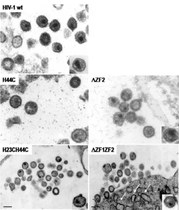
5- Impact of the NC zinc finger mutations on the structure of the viral particles as seen by electron microscopy
To analyze the influence of ZF-mutations on virus assem-bly, virions produced by HeLa cells expressing either one of the HIV-1 ZF-mutants were collected and processed for
electron microscopy as described before [1] (Fig. 6). Most HIV-1 wild-type virions show a mature morphology with either a conical or central globular nucleocore (Fig. 6, wt). NC mutants H44C and ΔZF2 exhibited either an imma-ture morphology or a poorly defined core strucimma-ture (Fig. 6, H44C; ΔZF2). NC mutant H23H44C and ΔZF1ZF2 had often an immature morphology or contained a small core-like structure located close to the viral envelope (Fig. 6, H23H44C and ΔZF1ZF2). The arrows indicate the elec-tron-dense structure at the PM of ΔZF1ZF2-NC mutant,
showing an accumulation of mutated Gag unable to com-plete particle assembly and release.
Data presented in Table 2 show that HIV-1 particles had a canonical morphology with either a conical or a rod-like core with with a mean particle diameter of about 110 nm (Table 2). All the ZF NC mutant particles displayed drastic changes in the core structure with sometimes an imma-ture-like morphology (Fig. 6, see H44C, ΔZF2, H23H44C and ΔZF1ZF2) and displaced or poorly defined cores or Plasma membrane accumulation of the HIV-1 NC(ΔZF1ZF2) Gag
Figure 3
Plasma membrane accumulation of the HIV-1 NC(ΔZF1ZF2) Gag. HeLa cells were transfected with wild-type HIV-1
(A) or NC(ΔZF1ZF2) (B) DNA, then fixed and stained for the detection of Gag with an anti-MAp17; with an anti-CD63/Lamp3 for late endosomes, and with an anti-Lamp1 for lysosomes, as indicated. Zoomed-1 picture shows wild-type Gag colocalization with CD63/Lamp3 late endosomal marker (26 ± 6%) and zoomed-2 picture with the Lamp1 marker (6 ± 2%). In contrast, zoomed-3 picture shows less colocalization of this mutant with Lamp3 in comparison to wt (6 ± 4%). Zoomed-4 picture shows an accumulation of NC(ΔZF1ZF2)-Gag mutant at the PM, and less or equal with Lamp1(+) intracytoplasmic vesicles (3 ± 1%).
even two core structures (Table 2). All mutant virions har-bored a defect in the conical shape of the core (Table 2) that can be correlated with the defect in Gag maturation (Fig. 1B, lanes V). In the case of HIV-1 ΔZF1ZF2, the mutant has no distinct core and seemed to accumulate at the budding site but unable to complete the process since we observed electron-dense curvatures of the PM reminis-cent of an accumulation of Gag at the PM (Fig. 6, see white arrows).
Taken together, these results show that HIV-1 NC zinc fin-gers play an important role in viral core structure and sug-gest that NC-NC and/or NC-gRNA interactions are essential for HIV-1 Gag assembly and particle release.
Discussion
The retroviral Gag polyprotein orchestrates retrovirus assembly in the infected cell via two platforms, which are a cellular membrane and a RNA (reviewed in [2,6]. The current view of the assembly process implies that the newly made Gag binds to the cellular membrane by the N-terminal myristoylated domain and stretches of basic res-idues of the matrix domain (reviewed in [31,32], also
involving inositol phosphates/Gag interactions [33-36]. At the same time, the NC domain selectively binds the gRNA via specific interactions with the packaging Psi sig-nal [4], which in turn promote Gag oligomerization [15]. Although, a leucine zipper motif could functionally, at least in part, replace NC to drive the assembly of a "mini-mal" Gag [37]. We propose that the interactions between Gag-NC and the genomic Psi signal will ensure both the formation of Gag oligomers and the selective recruitment of the gRNA. Consistent with this view, mutations in the first NC zinc finger or the flanking basic residues result in a strong decrease of viral particle production and infectiv-ity [1,16,17,19,38]. Thus, it has been proposed that the NC domain of Gag is required for the proper assembly and release of infectious virions.
To confirm the multiple roles played by NC in HIV-1 assembly, we have examined the role of the NC zinc fin-gers (ZF) in Gag trafficking. Taken together, our results show that both NC zinc fingers play critical roles in the ability of Gag to properly assemble and ultimately to bud. In fact, all HIV-1 ZF mutants examined so far produced particles at levels five to ten fold, or more, lower than that of wild-type HIV-1 and were not infectious (Table 1). In model cell lines, the wild-type HIV-1 Gag was found to accumulate either at the PM or on intracellular tet-raspanin-rich endosomal membranes as recently reported [8,25,30,39-43]. Mutating the NC Zinc fingers caused Gag to accumulate within the cell, in a diffuse manner and at the PM (Fig. 2). Thus, mutating the NC ZF appears to pre-vent Gag targeting to and accumulation in endosomes (Fig. 3). This favors the view that endosomes are an important site for virus formation and release (Fig. 3), and also maturation since intracellular mature CAp24 was drastically reduced in the case of the ZF mutants (Fig. 1B). It also indicates that NC is not the major determinant for Gag targeting to the PM, such as the basic MA domain of Gag and phosphatidyl inositol phosphate lipids are [8,30,34,44,45]. However, NC contributes to the localiza-tion of Gag in late endosomes (Fig. 3 and 5).
As already stated, the other platform necessary for virus assembly is the gRNA since it directs the oligomerization of the newly made Gag upon binding [15,46,47]. As pre-viously shown for mutations in the first ZF [1], we found that mutating the second ZF and both of them strongly impaired gRNA content in mutant virions as well as parti-cle release (Table 1), and resulted in the production of replication defective viruses. Staufen, a cellular RNA-bind-ing protein involved in RNA transport and metabolism, was reported to interact with both the gRNA and the NC domain of HIV-1 Gag, and to have a role in HIV-1 gRNA encapsidation and Gag assembly [48,49]. Mapping the interaction domain between Staufen and Gag reveals the Intracellular localization of the gRNA in cells expressing the
NC zinc finger-mutant Gag
Figure 4
Intracellular localization of the gRNA in cells
expressing the NC zinc finger-mutant Gag. HeLa cells
were transfected with wild-type HIV-1 (A), or NC(ΔZF1ZF2) (B), or NC(H23H44C) (D) DNA, then fixed and stained for the detection of the gRNA by FISH, as described in material and methods. The fluorescent Cy3-labelled oligonucleotide probe hybridized to the HIV-1gag gene (in red). The nucleus was stained with Dapi in the "mock" HIV-negative cells (C). The arrows indicate the accumulation of wt gRNA at the PM.
Subcellular localization of Gag and the gRNA
Figure 5
Subcellular localization of Gag and the gRNA. Subcellular fractionations of 293T cells expressing wild-type HIV-1 (A, as
in (21)) or NC(ΔZF1ZF2) (B) or ΔNC (C) were analyzed by OptiPrep gradient centrifugation. Cells were broken as described in materials and methods and the post-nuclear supernatant (PNS) was fractionated by Optiprep gradient. 20 μl of each fraction were loaded on SDS-PAGE, and Gag and Lamp2 were analyzed by immunoblotting using anti-Cap24 and anti-Lamp2 antibodies. Each fraction of the gradient was tested for the presence of the gRNA by RT-PCR as described in materials and methods. The expected 132 bp DNA fragment was detected on 1% agarose gel. In addition to the gradient analyses, the immunofluorescence (IF) detections are shown, representing the cells stained with an anti-CAp24 (in green) for Gag (A) or mutated Gag (B and C), and the FISH treatment of the 293T cells expressing HIV-1 (A) or the NC Gag mutants (B and C) for the gRNA using the Gag-oligo-Cy3 probe (in red).
importance of the NC domain, in particular the second ZF [48,49]. This could explain, at least in part, the role played by the second ZF on gRNA encapsidation if Staufen is responsible for gRNA trafficking to the assembly site and consequently RNA-dependent Gag oligomerization and assembly.
However, Ott and colleagues reported that the decrease in viral production due to NC deletion can be compensated by an RNA binding site in HIV-1 MA domain of Gag [50]. It is very difficult to dissociate the role of NC in virus assembly from NC-RNA interactions, which are critical for the structure of the viral particle [22,51], rather than assembly or budding [16,17]. Our data favor a model in wich all events are linked and dependent upon Gag-RNA interactions via the NC domain, required to achieve a proper viral assembly, i.e. multimerization of Gag, Gag oligomer targeting and trafficking, and ultimately particle assembly, budding and release.
To better examine the role of Gag-RNA interactions in assembly, we examined gRNA localization by FISH (Fig. 4) and by subcellular fractionation (Fig 5). The gRNA was found in the nucleus, most probably due to provirus tran-scription, in the cytoplasm and at the PM of cells express-ing wild-type HIV-1 (Fig. 4), in agreement with Berthold and Mandarelli [52]. Wild-type Gag and the gRNA were also found at the level of late endosomes [25,53,54]. Mutating the NC ZF motifs drastically altered the cellular distribution of the gRNA, because it was found evenly dis-tributed within the cell and no longer associated with the PM (Fig. 4, by FISH) or the late endosomes (Fig. 5, by gra-dient), while the NC-mutated Gag accumulated mainly at the PM (Fig. 3 and 5). Thus, specific Gag-gRNA interac-tions via the NC-ZF are most probably required for proper Gag trafficking through Gag-Gag multimer complexes. In agreement with this view, it was reported that Gag expressed from Psi(-) RNA diffuses throughout the cell and shows delayed cytoplasmic colocalisation with the gRNA [54]. The authors propose that the packaging signal may coordinate capture of the genomic Psi(+) RNA by
Gag, followed by assembly and transport to the budding site. This data indeed mirrors the results obtained with the NC mutants, in particular the Gag-ΔNC mutant, for which both the gRNA and mutated Gag were found diffuse throughout the cell and had probably lost Gag-gRNA association (as seen in Fig. 4 and 5). Taken together, these data strongly suggest that the specific Gag-gRNA interac-tions via the NC domain are necessary for proper Gag traf-ficking and assembly, for Gag oligomers to be targeted to
Table 2: Quantitative analysis of virus core morphology of NC mutant HIV-1 particles produced by HeLaP4 cells
Dense cone shape core Dense material in base of cone Round centered core Round displaced core Tubular core structure Two core structures or membrane* No defined core Diameter of particles wt 19 38 21 1 0 7 113 ± 4 H23C 7 16 16 40,5 6 0 14,5 134 ± 14 H44C 1 7 5 45 13 1 28 129 ± 14 H23-H44C 0 0 13 16 23 15* 33 96 ± 12 ΔZF1 6 16 15 39 4 0 20 131 ± 12 ΔZF2 1 8 3 62 6 2 17 ND ΔZF1-ZF2 1 0 11 23,5 13 22,5* 29 101 ± 13
Number of particles observed: 80 to 200. Numbers are expressed in % of total observed particles.
Electron microscopy analysis of HIV-1 NC mutant virions
Figure 6
Electron microscopy analysis of HIV-1 NC mutant virions. Virions were produced by DNA transfected HeLaP4
cells and further processed as indicated in materials and methods. Bar is 100 nm.
either specific domains at the PM or to the late endo-somes.
Finally, the structure of the HIV-1 virions carrying one or several mutations in the NC ZF was determined by elec-tron microscopy (Fig. 6). Results show that the NC mutated virions have lost their conical core shape and are immature, which confirm the role of NC in HIV-1 virion structure [1,16], as well as for other retroviruses, such as SIV [55] and MLV [17]. Our results correlate with previous studies on the assembly of several retroviruses, namely HIV, MLV, RSV that have revealed a critical role of NC in virus assembly and release [1,16,17,21,56,57].
In conclusion, the HIV-1 NC protein appears to be a major actor in the late steps of the virus replication in addition to its roles in proviral DNA synthesis and varia-bility [4,58-60].
Acknowledgements
Thanks are due to Fabienne Simian-Lermé and Claire Lionnet (PLATIM, ENS Lyon, France) for their helpful assistance in CSLM. We thank Pr Radka Argirova (National Institute of Infectious and Parasitic Diseases, Bulgaria) for the support of B.G. Work supported by INSERM, CNRS, ANRS, French Embassy in Bulgaria, FRM, the european TRIoH consortium, and by the Ministère de la Recherche (ACI) and SIDACTION (to MM). D.M. was sup-ported by FRM, TRIoH and ANRS (French agency against AIDS) and FS by the CNRS.
References
1. Tanchou V, Decimo D, Pechoux C, Lener D, Rogemond V, Berthoux L, Ottmann M, Darlix JL: Role of the N-terminal zinc finger of
human immunodeficiency virus type 1 nucleocapsid protein in virus structure and replication. Journal of virology 1998, 72(5):4442-4447.
2. Cimarelli A, Darlix JL: Assembling the human
immunodefi-ciency virus type 1. Cell Mol Life Sci 2002, 59(7):1166-1184.
3. Darlix JL, Cristofari G, Rau M, Pechoux C, Berthoux L, Roques B:
Nucleocapsid protein of human immunodeficiency virus as a model protein with chaperoning functions and as a target for antiviral drugs. Advances in pharmacology (San Diego, Calif 2000, 48:345-372.
4. Darlix JL, Lapadat-Tapolsky M, de Rocquigny H, Roques BP: First
glimpses at structure-function relationships of the nucleo-capsid protein of retroviruses. Journal of molecular biology 1995, 254(4):523-537.
5. Demirov DG, Freed EO: Retrovirus budding. Virus research 2004,
106(2):87-102.
6. Muriaux D, Darlix JL, Cimarelli A: Targeting the assembly of the
human immunodeficiency virus type I. Current pharmaceutical
design 2004, 10(30):3725-3739.
7. Rein A, Henderson LE, Levin JG: Nucleic-acid-chaperone activity
of retroviral nucleocapsid proteins: significance for viral rep-lication. Trends in biochemical sciences 1998, 23(8):297-301.
8. Resh MD: Intracellular trafficking of HIV-1 Gag: how Gag
interacts with cell membranes and makes viral particles.
AIDS reviews 2005, 7(2):84-91.
9. Batonick M, Favre M, Boge M, Spearman P, Honing S, Thali M:
Inter-action of HIV-1 Gag with the clathrin-associated adaptor AP-2. Virology 2005, 342(2):190-200.
10. Dong X, Li H, Derdowski A, Ding L, Burnett A, Chen X, Peters TR, Dermody TS, Woodruff E, Wang JJ, Spearman P: AP-3 directs the
intracellular trafficking of HIV-1 Gag and plays a key role in particle assembly. Cell 2005, 120(5):663-674.
11. Camus G, Segura-Morales C, Molle D, Lopez-Verges S, Begon-Pescia C, Cazevieille C, Schu P, Bertrand E, Berlioz-Torrent C, Basyuk E:
The Clathrin Adaptor Complex AP-1 Binds HIV-1 and MLV Gag and Facilitates Their Budding. Mol Biol Cell 2007.
12. Strack B, Calistri A, Craig S, Popova E, Gottlinger HG: AIP1/ALIX
is a binding partner for HIV-1 p6 and EIAV p9 functioning in virus budding. Cell 2003, 114(6):689-699.
13. von Schwedler UK, Stuchell M, Muller B, Ward DM, Chung HY, Morita E, Wang HE, Davis T, He GP, Cimbora DM, Scott A, Krauss-lich HG, Kaplan J, Morham SG, Sundquist WI: The protein network
of HIV budding. Cell 2003, 114(6):701-713.
14. De Guzman RN, Wu ZR, Stalling CC, Pappalardo L, Borer PN, Sum-mers MF: Structure of the HIV-1 nucleocapsid protein bound
to the SL3 psi-RNA recognition element. Science (New York, NY
1998, 279(5349):384-388.
15. Burniston MT, Cimarelli A, Colgan J, Curtis SP, Luban J: Human
immunodeficiency virus type 1 Gag polyprotein multimeri-zation requires the nucleocapsid domain and RNA and is promoted by the capsid-dimer interface and the basic region of matrix protein. Journal of virology 1999, 73(10):8527-8540.
16. Cimarelli A, Sandin S, Hoglund S, Luban J: Basic residues in human
immunodeficiency virus type 1 nucleocapsid promote virion assembly via interaction with RNA. Journal of virology 2000, 74(7):3046-3057.
17. Muriaux D, Costes S, Nagashima K, Mirro J, Cho E, Lockett S, Rein A:
Role of murine leukemia virus nucleocapsid protein in virus assembly. Journal of virology 2004, 78(22):12378-12385.
18. Muriaux D, Mirro J, Harvin D, Rein A: RNA is a structural
ele-ment in retrovirus particles. Proceedings of the National Academy
of Sciences of the United States of America 2001, 98(9):5246-5251.
19. Berthoux L, Pechoux C, Darlix JL: Multiple effects of an
anti-human immunodeficiency virus nucleocapsid inhibitor on virus morphology and replication. Journal of virology 1999, 73(12):10000-10009.
20. Buckman JS, Bosche WJ, Gorelick RJ: Human immunodeficiency
virus type 1 nucleocapsid zn(2+) fingers are required for effi-cient reverse transcription, initial integration processes, and protection of newly synthesized viral DNA. Journal of virology
2003, 77(2):1469-1480.
21. Gorelick RJ, Gagliardi TD, Bosche WJ, Wiltrout TA, Coren LV, Chabot DJ, Lifson JD, Henderson LE, Arthur LO: Strict
conserva-tion of the retroviral nucleocapsid protein zinc finger is strongly influenced by its role in viral infection processes: characterization of HIV-1 particles containing mutant nucle-ocapsid zinc-coordinating sequences. Virology 1999,
256(1):92-104.
22. Wang SW, Noonan K, Aldovini A: Nucleocapsid-RNA
interac-tions are essential to structural stability but not to assembly of retroviruses. Journal of virology 2004, 78(2):716-723.
23. Ono A, Waheed AA, Joshi A, Freed EO: Association of human
immunodeficiency virus type 1 gag with membrane does not require highly basic sequences in the nucleocapsid: use of a novel Gag multimerization assay. Journal of virology 2005, 79(22):14131-14140.
24. Cimarelli A, Luban J: Human immunodeficiency virus type 1
vir-ion density is not determined by nucleocapsid basic residues.
Journal of virology 2000, 74(15):6734-6740.
25. Grigorov B, Arcanger F, Roingeard P, Darlix JL, Muriaux D:
Assem-bly of infectious HIV-1 in human epithelial and T-lymphob-lastic cell lines. Journal of molecular biology 2006, 359(4):848-862.
26. Smagulova F, Maurel S, Morichaud Z, Devaux C, Mougel M, Houzet L:
The highly structured encapsidation signal of MuLV RNA is involved in the nuclear export of its unspliced RNA. Journal of
molecular biology 2005, 354(5):1118-1128.
27. Demene H, Dong CZ, Ottmann M, Rouyez MC, Jullian N, Morellet N, Mely Y, Darlix JL, Fournie-Zaluski MC, Saragosti S, et al.: 1H NMR
structure and biological studies of the His23-->Cys mutant nucleocapsid protein of HIV-1 indicate that the conforma-tion of the first zinc finger is critical for virus infectivity.
Bio-chemistry 1994, 33(39):11707-11716.
28. Ottmann M, Gabus C, Darlix JL: The central globular domain of
the nucleocapsid protein of human immunodeficiency virus type 1 is critical for virion structure and infectivity. Journal of
virology 1995, 69(3):1778-1784.
29. Hermida-Matsumoto L, Resh MD: Localization of human
immu-nodeficiency virus type 1 Gag and Env at the plasma mem-brane by confocal imaging. Journal of virology 2000, 74(18):8670-8679.
Publish with BioMed Central and every scientist can read your work free of charge "BioMed Central will be the most significant development for disseminating the results of biomedical researc h in our lifetime."
Sir Paul Nurse, Cancer Research UK Your research papers will be:
available free of charge to the entire biomedical community peer reviewed and published immediately upon acceptance cited in PubMed and archived on PubMed Central yours — you keep the copyright
Submit your manuscript here:
http://www.biomedcentral.com/info/publishing_adv.asp
BioMedcentral
30. Ono A, Freed EO: Cell-type-dependent targeting of human
immunodeficiency virus type 1 assembly to the plasma membrane and the multivesicular body. Journal of virology 2004, 78(3):1552-1563.
31. Conte MR, Matthews S: Retroviral matrix proteins: a structural
perspective. Virology 1998, 246(2):191-198.
32. Resh MD: A myristoyl switch regulates membrane binding of
HIV-1 Gag. Proceedings of the National Academy of Sciences of the
United States of America 2004, 101(2):417-418.
33. Campbell S, Fisher RJ, Towler EM, Fox S, Issaq HJ, Wolfe T, Phillips LR, Rein A: Modulation of HIV-like particle assembly in vitro
by inositol phosphates. Proceedings of the National Academy of
Sci-ences of the United States of America 2001, 98(19):10875-10879.
34. Ono A, Ablan SD, Lockett SJ, Nagashima K, Freed EO:
Phosphati-dylinositol (4,5) bisphosphate regulates HIV-1 Gag targeting to the plasma membrane. Proceedings of the National Academy of
Sciences of the United States of America 2004, 101(41):14889-14894.
35. Saad JS, Miller J, Tai J, Kim A, Ghanam RH, Summers MF: Structural
basis for targeting HIV-1 Gag proteins to the plasma mem-brane for virus assembly. Proceedings of the National Academy of
Sciences of the United States of America 2006, 103(30):11364-11369.
36. Datta SA, Zhao Z, Clark PK, Tarasov S, Alexandratos JN, Campbell SJ, Kvaratskhelia M, Lebowitz J, Rein A: Interactions between
HIV-1 Gag molecules in solution: an inositol phosphate-mediated switch. Journal of molecular biology 2007, 365(3):799-811.
37. Accola MA, Strack B, Gottlinger HG: Efficient particle production
by minimal Gag constructs which retain the carboxy-termi-nal domain of human immunodeficiency virus type 1 capsid-p2 and a late assembly domain. Journal of virology 2000, 74(12):5395-5402.
38. Gorelick RJ, Benveniste RE, Gagliardi TD, Wiltrout TA, Busch LK, Bosche WJ, Coren LV, Lifson JD, Bradley PJ, Henderson LE, Arthur LO: Nucleocapsid protein zinc-finger mutants of simian
immunodeficiency virus strain mne produce virions that are replication defective in vitro and in vivo. Virology 1999, 253(2):259-270.
39. Jouvenet N, Neil SJ, Bess C, Johnson MC, Virgen CA, Simon SM, Bieniasz PD: Plasma membrane is the site of productive
HIV-1 particle assembly. PLoS biology 2006, 4(HIV-12):e435.
40. Nydegger S, Foti M, Derdowski A, Spearman P, Thali M: HIV-1
egress is gated through late endosomal membranes. Traffic
(Copenhagen, Denmark) 2003, 4(12):902-910.
41. Perlman M, Resh MD: Identification of an intracellular
traffick-ing and assembly pathway for HIV-1 gag. Traffic (Copenhagen,
Denmark) 2006, 7(6):731-745.
42. Rudner L, Nydegger S, Coren LV, Nagashima K, Thali M, Ott DE:
Dynamic fluorescent imaging of human immunodeficiency virus type 1 gag in live cells by biarsenical labeling. Journal of
virology 2005, 79(7):4055-4065.
43. Sherer NM, Lehmann MJ, Jimenez-Soto LF, Ingmundson A, Horner SM, Cicchetti G, Allen PG, Pypaert M, Cunningham JM, Mothes W:
Visualization of retroviral replication in living cells reveals budding into multivesicular bodies. Traffic (Copenhagen,
Den-mark) 2003, 4(11):785-801.
44. Freed EO, Orenstein JM, Buckler-White AJ, Martin MA: Single
amino acid changes in the human immunodeficiency virus type 1 matrix protein block virus particle production. Journal
of virology 1994, 68(8):5311-5320.
45. Zhou W, Parent LJ, Wills JW, Resh MD: Identification of a
mem-brane-binding domain within the amino-terminal region of human immunodeficiency virus type 1 Gag protein which interacts with acidic phospholipids. Journal of virology 1994, 68(4):2556-2569.
46. Campbell S, Rein A: In vitro assembly properties of human
immunodeficiency virus type 1 Gag protein lacking the p6 domain. Journal of virology 1999, 73(3):2270-2279.
47. Roldan A, Russell RS, Marchand B, Gotte M, Liang C, Wainberg MA:
In vitro identification and characterization of an early com-plex linking HIV-1 genomic RNA recognition and Pr55Gag multimerization. The Journal of biological chemistry 2004, 279(38):39886-39894.
48. Chatel-Chaix L, Abrahamyan L, Frechina C, Mouland AJ, DesGroseill-ers L: The host protein Staufen1 participates in human
immunodeficiency virus type 1 assembly in live cells by influ-encing pr55Gag multimerization. Journal of virology 2007, 81(12):6216-6230.
49. Chatel-Chaix L, Clement JF, Martel C, Beriault V, Gatignol A, Des-Groseillers L, Mouland AJ: Identification of Staufen in the
human immunodeficiency virus type 1 Gag ribonucleopro-tein complex and a role in generating infectious viral parti-cles. Molecular and cellular biology 2004, 24(7):2637-2648.
50. Ott DE, Coren LV, Gagliardi TD: Redundant roles for
nucleocap-sid and matrix RNA-binding sequences in human immuno-deficiency virus type 1 assembly. Journal of virology 2005, 79(22):13839-13847.
51. Wang SW, Aldovini A: RNA incorporation is critical for
retro-viral particle integrity after cell membrane assembly of Gag complexes. Journal of virology 2002, 76(23):11853-11865.
52. Berthold E, Maldarelli F: cis-acting elements in human
immuno-deficiency virus type 1 RNAs direct viral transcripts to dis-tinct intranuclear locations. Journal of virology 1996, 70(7):4667-4682.
53. Houzet L, Gay B, Morichaud Z, Briant L, Mougel M: Intracellular
assembly and budding of the Murine Leukemia Virus in infected cells. Retrovirology 2006, 3:12.
54. Poole E, Strappe P, Mok HP, Hicks R, Lever AM: HIV-1 Gag-RNA
interaction occurs at a perinuclear/centrosomal site; analysis by confocal microscopy and FRET. Traffic (Copenhagen,
Den-mark) 2005, 6(9):741-755.
55. Yovandich JL, Chertova EN, Kane BP, Gagliardi TD, Bess JW Jr., Sow-der RC 2nd, HenSow-derson LE, Gorelick RJ: Alteration of
zinc-bind-ing residues of simian immunodeficiency virus p8(NC) results in subtle differences in gag processing and virion mat-uration associated with degradative loss of mutant NC.
Jour-nal of virology 2001, 75(1):115-124.
56. Dawson L, Yu XF: The role of nucleocapsid of HIV-1 in virus
assembly. Virology 1998, 251(1):141-157.
57. Lee EG, Linial ML: Deletion of a Cys-His motif from the
Alpharetrovirus nucleocapsid domain reveals late domain mutant-like budding defects. Virology 2006, 347(1):226-233.
58. Bampi C, Bibillo A, Wendeler M, Divita G, Gorelick RJ, Le Grice SF, Darlix JL: Nucleotide excision repair and
template-independ-ent addition by HIV-1 reverse transcriptase in the presence of nucleocapsid protein. The Journal of biological chemistry 2006, 281(17):11736-11743.
59. Levin JG, Guo J, Rouzina I, Musier-Forsyth K: Nucleic acid
chaper-one activity of HIV-1 nucleocapsid protein: critical role in reverse transcription and molecular mechanism. Progress in
nucleic acid research and molecular biology 2005, 80:217-286.
60. Hu WS, Rhodes T, Dang Q, Pathak V: Retroviral recombination: