HAL Id: tel-00622550
https://tel.archives-ouvertes.fr/tel-00622550
Submitted on 12 Sep 2011HAL is a multi-disciplinary open access archive for the deposit and dissemination of sci-entific research documents, whether they are pub-lished or not. The documents may come from teaching and research institutions in France or abroad, or from public or private research centers.
L’archive ouverte pluridisciplinaire HAL, est destinée au dépôt et à la diffusion de documents scientifiques de niveau recherche, publiés ou non, émanant des établissements d’enseignement et de recherche français ou étrangers, des laboratoires publics ou privés.
NADPH oxydase Nox4 : structure/fonction protéomique
recombinante et approche immunologique
Leilei Zhang
To cite this version:
Leilei Zhang. NADPH oxydase Nox4 : structure/fonction protéomique recombinante et approche immunologique. Autre. Université de Grenoble, 2011. Français. �NNT : 2011GRENV029�. �tel-00622550�
THÈSE
Pour obtenir le grade de
DOCTEUR DE L’UNIVERSITÉ DE GRENOBLE
Spécialité : Biologie cellulaireArrêté ministériel : 7 août 2006
Présentée par
LEILEI ZHANG
Thèse dirigée par Françoise MOREL
préparée au sein du Laboratoire d'Enzymologie
dans l'École Doctorale CHIMIE ET SCIENCES DU VIVANT
NADPH oxydase Nox4:
structure/fonction
Protéomique recombinante et
approche immunologique
Thèse soutenue publiquement le 30 Mai 2011 devant le jury composé de :
Mme, Corinne, DUPUY
Fonction et lieu de la fonction, Rapporteur Mr, Yves, GORIN
Fonction et lieu de la fonction, Rapporteur Mr, Bernard, Lassegue
Fonction et lieu de la fonction, Membre Mr, Bernard, LARDY
Fonction et lieu de la fonction, Membre Mr, Christian, DROUET
Fonction et lieu de la fonction, Président Mme, Françoise, MOREL
Remerciements
I would like to thank Pr. Françoise Morel, my dissertation advisor, for directing me through my Ph.D study. She spent a lot of time and energy on educating me, modifying the papers and the dissertation for me, helping me with my life in France. I am very grateful for having the opportunity to work with her and learn from her.
Special thanks go to the committee members Dr. Yves, Gorin, Dr. Bernard, Lassegue, Dr. Corinne, Dupuy, Dr. Christian, Drouet for serving on the committee and giving me good suggestions and comments on the dissertation.
I would like to thank Dr. Bernard Lardy, Dr. Chuong Nguyen, Dr. Marie-Hélène Paclet and sylvie Berthier for the encouragement and helpful advice through all of the technology challenges. They always give me a hand when I have difficulties and need help in daily life.
I thank Candice Trocme, Francis Rousset, Adam Baillet, Marie-claire Dagher for the help in learning the details of lab’s life and French.
I thank all the members in the Laboratoire d'Enzymologie for everything. I do enjoy a memorial time staying with you guys in the past 3 years.
Thanks also go out to Alexei Grichine for the technique support in the use of confocal microscopy, TIRFM and to Pr. Algirdas J. Jesaitis for the phage display technique.
I am grateful to Prof. Guanxiang Qian and Prof. Shengfang Ge. They always give me a huge support and encouragement.
I am also thankful to Prof. Guy Vincendon for the help in scholarship application.
A special thanks to all my friends in China. And also friends in France, Samuel Degoul, Kelly Dilworth, Hai Huang, Rang Xu, Zhen Jiang, Yan Wang. They help me to adapt my life and study in France.
My very special thanks will also give to my parents Xudong Zhang and Chaofen Qian, my husband Aiping Ding. They use their broad mind and deep love to accept all my strengths and weaknesses. No matter what happens, They will always in there supportting me, encouraging me, helping me, and be my powerful support.
I appreciate the financial support from “the Region Rhône-Alpes, programme ARCUS”, “the National Key Program for Basic Research of China (2010CB529902)”, “the Science and Technology Commission of Shanghai (S30205, 06SR07110)”, “the National Natural Science Foundation of China (30973663)”, “L’Ambassade de France en Chine”.
Table of Content
1Table of Figures and Tables
4List of the abbreviations
5Summary in English
7Part 1 Introduction
8-41I. Reactive Oxygen Species 8
1 Introduction 8
2 Sources of the Generation of ROS 9
2.1 Mitochondria 9
2.2 5-Lipoxygenase 10
2.3 NADPH oxidase 11
2.3.1. Activation of the Phagocyte NADPH Oxidase 12
II. A Historical Overview of ROS-Generating NADPH Oxidases 13
III. Structure, activity and function of NADPH oxidases 15
1 The Family of NADPH Oxidases 15
2 Structure and Topology of NADPH Oxidases 18
2.1 Conserved Structural Properties of NADPH Oxidases 18
2.2 Topology of NADPH oxidases 21
2.3 Post-translational Modification of NADPH Oxidases 22
2.4 Isoforms of NADPH oxidases 23
3 Regulation of the NADPH Oxidase Activity 25
3.1 Electron Transfer Mechanism of NADPH Oxidase Nox2 25
3.2 Nox Subunits and Regulation Proteins 26
3.2.1. p22phox: Indispensible Partner of NADPH Oxidases 26 3.2.2. p47phox, p67phox, p40phox and Rac: Cytosolic Regulatory Protein of Phagocyte NADPH
Oxidase 28
3.3 Assembly of the Phagocyte NADPH oxidase Nox2 30
3.4 Characteristic of NADPH Oxidases Dependent ROS Generation 35
IV. Physiological Function of NADPH Oxidases 36
1 HEK293 cells 39
2 HEK293 T-RExTM Nox4 cells 39
3 C-20/A4 chondrocyte cell lines 40
VI. The Objectives of Our Work 41
Part 2 Research Work
42-125Chapter 1: Validation and characterization of the first monoclonal antibodies against NADPH oxidase Nox4: essential tools for the structural and immunochemical investigations. 42
Résumé en français 42
Summary in English 45
1 Background 45
2 Generation and validation of monoclonal antibodies raised against recombinant Nox4 45
3 Subcellular localization of NADPH oxidase 4 46
4 Characterization of monoclonal antibodies raised against recombinant Nox4 46
Article 1: 47
Chapter 2: The E-loop is involved in the hydrogen peroxide formation of Nox4 69
Résumé en français 69
Summary in English 72
1 Background 72
2 H2O2 production is an intrinsic feature of Nox4 72
3 Structural basis for H2O2 formation of Nox4 and its biological consequence 73
4 Identification of a molecular explanation for H2O2 formation by Nox4 73
Article 2: 74
Chapter 3: The study of constitutive diaphorase activity of Nox4 and topological study of the
transmembrane heterodimer Nox4/p22phox. 85
Résumé en français 85
Summary in English 87
1 Background 87
2 In vitro expression of Nox4 truncated proteins by RTS 87
3 Expression of Nox4 truncated proteins by bacterial induction 87
4 Diaphorase activity of recombinant truncated Nox4 truncated constructions 88
Article 4: 109
1 Introduction 110
2 Materials and methods 111
2.1 Materials 111
2.2 Cell culture 111
2.3 Stable transfection of mammalian expression plasmids 111
2.4 Flow cytometry 111
2.5 SDS/PAGE and Western Blotting 113
3 Results 113
3.1 Establishment of the TDUFA technique 113
3.2 TDUFA reveal the topology of the protein IL1R1 and Nox2N131 114
3.3 Membrane topology of Nox4 by the TDUFA approach 116
3.4 Membrane topology of p22phox by the TDUFA approach 120
4 Discussion 123
5 References 124
Part 3: Discussion and perspectives
126-140En Français
126
In English
133
Reference
141
Annex
164
Publication list 164
Conferences & presentations 164
FIGURES
Figure 1. Reactive Oxygen Species. 8
Figure 2. Mitochondria as Source of Reactive Oxygen Species. 10
Figure 3. 5-Lipoxygenase as Source of Reactive Oxygen Species. 11
Figure 4. NADPH Oxidase as Source of Reactive Oxygen Species. 11
Figure 5. Signaling Pathway to Phagocyte NADPH Oxidase Activation. 12
Figure 6. Linear Representation of the Protein Sequence of NADPH Oxidases. 20
Figure 7. Topology of Nox2. 21
Figure 8. Schematic representastion of Nox4 isoforms 24
Figure 9. Electron transfer pathways within flavocytochrome b558. 25
Figure 10. The two organizer homologs, p47phox and NoxO1, share a similar set of motifs. 29
Figure 11. The two activator homologs, p67phox and NoxA1, share a similar overall domain structure 29 Figure 12. Regions of p40phox involved in protein/protein interactions. 30
Figure 13. Assembly of the phagocyte NADPH oxidase Nox2. 33
Figure 14. Activation of the NADPH oxidase isoforms. 33
Figure 15. Activation of Nox4. 35
Figure 16. Principle of T-RExTM system. 40
Figure 17. Cartoon of TDUFA (Topological Determination by Ubiquitin Fusion Assay) technique 114
Figure 18. TDUFA reveal the topology of the protein IL1R1 and Nox2N131. 116
Figure 19. Schematic of the truncated protein Nox4 117
Figure 20. Restriction enzyme digestion analysis of full length and C-terminal-deleted derivatives Nox4
recombinant plasmids (double digested by KpnI/ApaI) 117
Figure 21. TDUFA reveal the topology of Nox4. 119
Figure 22. Schematic of the truncated protein p22phox 120
Figure 23. Restriction enzyme digestion analysis of full length and C-terminal-deleted derivatives p22phox recombinant plasmids (double digested by KpnI/ApaI) 121
Figure 24. TDUFA reveal the topology of p22phox. 122
TABLES
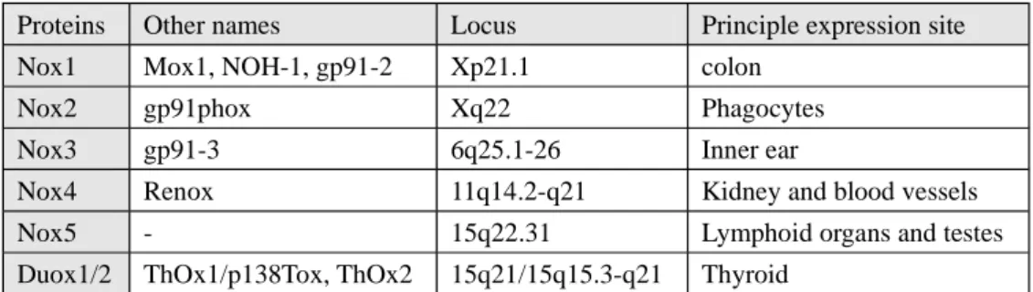
Table 1. Tissue distribution and main locus of NADPH oxidases 16
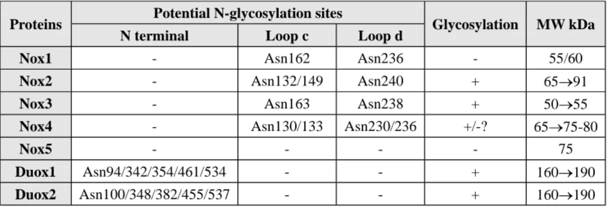
Table 2. NADPH oxidases and glycosylation. 23
List of the abbreviations
AA: Arachidonic acidAD: Activation domain AIR: Autoinhibitory region AFM: Atomic Force Microscopy CGD: Chronic Granulomatous Disease CDHD: Catalytic dehydrogenase domain CMV: Cytomegalovirus
DAG: Diacylglycerol DH: Dehydrogenase
DMEM: Dulbecco's Modification of Eagles Medium DPI: Diphenyliodonium
EDTA: Ethylenediamine tetraacetic acid ER: Endoplasmic reticulum
FAD: Flavin adenine dinucleotide FNR: Ferredoxin NADP+ reductase GFP: Green Fluorescent Protein
GSH/GSSG: Glutathione reduced/oxidized HEK: Human embryonic kidney
HUVECs: Human umbilical endothelial cells InsP3: Inositol 1,4,5-triphosphate
INT: Iodonitrotetrazolium JNK: c-jun N-terminal kinase LDL: Low density lipoproteins
MAPK: Mitogen activated protein kinase MKP-1: MAPK phosphatase 1
NADPH: Nicotinamide adenine dinucleotide phosphate NO: Nitric Oxide
8-OHdG: 8-hydroy-deoxyguanosine PA: Phosphatidic acid
PAI-1: Plasminogen activator inhibitor 1 PB1: Phox and Bem 1
PC: Phosphatidylcholine PhosR: Phosphorylation region PKC: Protein kinase C
PLA2: Phospholipase A2 PLC: Phospholipase C PLD: Phospholipase D
PMA: Phorbol-myristate acetate
Poldip2: Polymerase delta-interacting protein 2 PRR: Proline rich region
PX: Phagocyte oxidase (Phox)
ROS: Reactive oxygen species RTS: Rapid translation system SDS: Sodium dodecyl sulfate SH3: Src homology 3 SMC: Smooth muscle cells SOD: Superoxide dismutase
TGF : Transforming growth factor beta TIRF: Total Internal Reflection Fluorescence
TLCK: N-alpha-p-tosyl-L-lysine chloromethyl ketone TPR: Tetratricopeptide repeat
Ub: Ubiquitin
Summary in English
NADPH oxidase, Nox4, belongs to the Nox family which could generate reactive oxygen species by transferring an electron to molecular oxygen. Despite its wide distribution in tissues, Nox4 is still poorly understood. Unlike the other Noxes, Nox4 shows some unique characters: the constitutive activity, H2O2 formation. Nox4 involved ROS has been
proposed to be implicated in several pathologies. Thus, to study the structure/function and the regulation of the activity of Nox4 will provide new ideas and new drug targets for the effective prevention and treatment of clinical diseases related with ROS.
To know more about Nox4, in this study, 5 novel monoclonal antibodies were raised against a truncated recombinant protein (AA: 206-578) of Nox4. The specificity of 3 mAbs (8E9, 5F9, 6B11) was confirmed by western blot analysis in HEK293 transfected cells and human kidney cortex. In FACS studies, only mAb 8E9 could react with intact tet-induced T-RExTM Nox4 cells. Immunofluorescence confocal microscopy showed that Nox4 localized not only in the perinuclear and endoplasmic reticulum regions but also at the plasma membrane of the cells which was further confirmed by TIRF-microscopy. An interesting phenomena is that mAb 5F9 failed to detect Nox4 at the plasma membrane. Epitope determination showed that mAb 8E9 recognizes a region on the last extracellular loop of Nox4 (222H-E241), while mAb 6B11 (389S-P416) and 5F9 (392D-F398) are directed to its cytosolic tail. Cell-free oxidase assays showed a moderate but significant inhibition of constitutive Nox4 activity by mAb 5F9 and 6B11.
To study the protein region which is responsible for the unique ability of Nox4 of releasing H2O2 rather than O2-,
chimeric proteins and mutants were used. E-loop of Nox4 is 28 amino acid longer than that of Nox1 or Nox2. Deletion of E-loop amino acids only present in Nox4 or change of the two cysteines in the E-loop switch Nox4 from H2O2 to O2-
generation. In the presence of a NO donor, the O2--producing Nox4 mutants, but not widetype Nox4, generated
peroxynitrite, excluding artifacts of the detection systems as the apparent origin of O2-. A second approach was used to
confirm the responsibility of E-loop for the H2O2 formation. In Cos7 cells, which exhibit some plasma membrane
expression of Nox4, addition of the mAb 8E9 decreased H2O2 production but increased O2- formation. Unlike Nox1 or
Nox2, the E-loop of Nox4 contains a highly conserved histidine H222. Mutation of H222 also switched Nox4 from H2O2
to O2- formation. The structure of the E-loop might hinder O2- egress and/or provide a source for protons to accelerate
dismutation to form H2O2.
Two bacterial protein expression approaches (in vitro RTS and bacterial induction) were used to produce Nox4 cytosolic tail for characterizing the electronic transfer property of Nox4. The presence of rare codons (1363AGA AGA CUA1371) and high level of hydrophobicity affects the production of soluble and active recombinant Nox4Aqc and Nox4Bqc. After optimization of the conditions, soluble and active recombinant proteins were obtained by RTS or by bacteria induction. The soluble proteins were produced in large scale, purified onto affinity chromatography and were tested for the diaphorase activity (INT and cytochrome c). Results showed that electronic acceptor cytochrome c gives a higher rate than INT. Nox4Aqc produced a lower specific activity by a cell-based system compared to the protein synthesized in cell-free technology. This activity is not stimulated by the addition of cytosolic factors.
A new method, topological determination by ubiquitin fusion assay (TDUFA), was used to investigate the topology of Nox4 and p22phox. ubGFP fusion proteins are used as tools to obtain details of membrane protein topology. This method was first validated by using two membrane proteins with known topology and then should get more topology information of Nox4 and p22phox further.
Part 1 Introduction
I. Reactive Oxygen Species
1 Introduction
Reactive oxygen species (ROS) is a generic collective term indicating a number of active and reactive
partially reduced O2 metabolites, including superoxide (O2°-), hydroxyl (°OH), peroxyl (RO2°), alkoxyl (RO°)
and certain nonradicals that are either oxidizing agents and/or are easily converted into radicals, such as
hypochlorous acid (HOCl), ozone (O3), singlet oxygen (1O2), and hydrogen peroxide (H2O2) (Genestra, 2007).
ROS generation is generally a cascade of reactions that starts with the production of superoxide. Two
molecules of superoxide can react to generate hydrogen peroxide (H2O2) in a reaction known as dismutation,
which is accelerated by the enzyme superoxide dismutase. In the presence of iron, superoxide and H2O2 react
to generate hydroxyl radicals. In addition to superoxide, H2O2 and hydroxyl radical, other reactive oxygen
species (ROS) occur in biological systems. In inflamed areas, these include hypochlorous acid (HOCl),
formed in neutrophils from H2O2 and chloride by the phagocyte enzyme myeloperoxidase (MPO); singlet
oxygen, which might be formed from oxygen in areas of inflammation through the action of Phox and
MPO-catalysed oxidation of halide ions (Kanofsky, 1989); and ozone, which can be generated from singlet
oxygen by antibody molecules (Nathan, 2002; Wentworth et al., 2002) (Fig. 1).
Figure 1. Reactive Oxygen Species. Superoxide is generated from various sources, which include the NADPH
oxidase enzymes such as the phagocyte Nox (Phox).
lipids, carbohydrates, and nucleic acids. Various investigations have elucidated roles for ROS in causing
molecular damage such as DNA mutations, lipid peroxidation and protein oxidations. As a result of these
investigations, ROS has historically been viewed as a harmful but unavoidable consequence of an aerobic
lifestyle as major contributors to damage in biological organisms. In the first place, free radicals are present in
living cells which was demonstrated in vivo by a paramagnetic resonance absorption method (Commoner et
al., 1954). In 1956, Harmann observed that aging and degenerative diseases associated with it were attributed
basically to the deleterious side attacks of free radicals on cell constituents and on the connective tissues
(Harman, 1956). Since then, the free radical theory of aging has increasingly and widely been accepted
(Beckman et al., 1998). However, it is better not to think of ROS as bad . They are generated in a number of
reactions essential to life, for example, phagocytic cells generate radicals to kill invading pathogens. Also,
there are a large number of evidences indicating that oxygen radicals are involved in intercellular and
intracellular signaling.
2 Sources of the Generation of ROS
There are many sources of the generation of ROS. ROS can be produced as a byproduct by mitochondria,
peroxisomes, cytochrome P-450, lipoxygenase, xanthine oxidoreductase, etc (Gottlieb, 2003; Harrison, 2004;
Schrader et al., 2004; Balaban et al., 2005; Gonzalez, 2005). However, the NADPH oxidase was the only
enzyme whose primary function is to produce ROS. In living cells, without any doubt, the most relevant are
those described as following (Novo et al., 2008).
2.1 Mitochondria
Mitochondrial electron generates superoxide as an inevitable by-product and primary ROS at two
complexes, complexes I and III (Genova et al., 2003; Brand et al., 2004). Approximately 5% of electrons
flowing through the electron transport chain can be delivered to form O2 °-
at the levels of complex I
(NADPH/ubiquinone oxidoreductase) and complex III (ubiquinol/cytochrome c oxidoreductase). O2
is then
usually converted by mitochondrial SOD into H2O2, which can cross mitochondrial membranes to reach the
cytoplasm (Cadenas et al., 2000) (Figure 2). In mitochondria, Complex I releases superoxide into the matrix
(also at hypoxia when superoxide is produced due to the reverse electron transport), whereas Complex III
releases superoxide to both sides of the inner membrane. The sites of Complex I generating O2
are less
complexity of this complex in mammalian mitochondria. The Complex III contribution to O2°- generation by
autooxidation of the ubisemiquinone anion radical (UQ°-) is best understood. Within Complex III, the transfer
of electrons from ubiquinol (UQH2) to cytochrome c is catalyzed in the so- (Jezek et al.,
2005).
Figure 2. Mitochondria as Source of Reactive Oxygen Species. Major mitochondrial ROS sources and ROS
fluxes from mitochondria.Mitochondrial electron transport generates superoxide as an inevitable by-product and primary ROS at two complexes, Complexes I and III.Complex I releases superoxide into the matrix, whereas Complex III releases superoxide to both sides of the inner membrane.
2.2 5-Lipoxygenase
5-Lipoxygenase (5-LOX) is a mixed function oxidase involved in the synthesis of leukotrienes from
arachidonic acid in response to essentially the same stimuli that are able to stimulate Nox, particularly growth
factors and cytokines. The latter mediators lead to membrane ruffling and the generation of superoxide, and
then H2O2, through the intervention of the small GTPase Rac1 and a SOD isoform (Thannickal et al., 2000;
Droge, 2002; Chiarugi et al., 2003; Soberman, 2003; Chiarugi et al., 2007; D'Autreaux et al., 2007) (Figure
Figure 3. 5-Lipoxygenase as Source of Reactive Oxygen Species. 5-HPETE, acide
5-hydroperoxy-6-trans-8,11,14-cis-eicosatetraenoique.
2.3 NADPH oxidase
Figure 4. NADPH Oxidase as Source of Reactive Oxygen Species. The phagocyte oxidase is agonist-dependent,
exhibiting no detectable constitutive activity, and transfers electrons and consumes oxygen nearly instantaneously when stimulated. The regulation of its activity relies on p47phox, p67phox, p40phox and Rac. Regulation of other Nox family members in non-phagocytic cells is different, depending on the requirement of p22phox, cytosolic cofactors, the presence of EF-hands, and dependence on calcium. (see detail in 3.3)
The phagocyte NADPH oxidase was the first identified example of a system that generates ROS not as a
byproduct, but rather as the primary function of the enzyme system. The discovery of other members of the
Nox family of NADPH oxidases demonstrated that enzymes with the primary function of ROS generation are
not limited to phagocytes (Figure 4). Among the members of this family, gp91phox is best characterized. It is
functional as an assembled complex following a stimulus. The complex includes a catalytic core (gp91phox
and p22phox) and cytosolic regulatory factors (p40phox, p47phox, p67phox and Rac). The enzymes NADPH
oxidase (Nox) and dual oxidase (Duox) are present in both professional phagocytic cells (macrophages,
reactive oxygen in various cells and tissues in reponse to growth factors, cytokines and calcium signals. They
play a crucial role in different diseases (Babior, 1999; Vignais, 2002; Lambeth, 2007).
2.3.1. Activation of the Phagocyte NADPH Oxidase (Morel et al., 1991)
Figure 5. Signaling Pathway to Phagocyte NADPH Oxidase Activation. A, agonists; R, receptors; G, G protein;
PLC, phospholipase C; DAG, diacylglycerol; PA,phosphatidic acid; PLD, phospholipase D; PKC, protein kinase C; InsP3, inositol 1,4,5-triphosphate; MAPK, mitogen activated protein kinase; PLA2, phospholipase A2, AA,
arachidonic acid; P, phosphate. See text for detail explaination (2.3.1)
Most studies on NADPH oxidase and the mechanism of its activation have been performed with
neutrophils, which represent the most abundant cell type amongst the so-call professional phagocytes. There
are several steps in the signaling pathway to oxidase activation in neutrophils (Figure 5). Upon binding of
different agonists to specific receptors, agonists of the respiratory burst stimulate a
phosphatidylinositol-4,5-bis phosphate-(PtdInsP2)-specific phospholipase C through a
pertussis-toxin-sensitive G protein 1). Breakdown of PtdInsP2 results in the rapid generation of diacylglycerol
(DAG) and InsP3, which are considered as second messengers 2). In addition to the phospholipase C signaling
pathway, Hydrolysis of activated phospholipase D can produce phosphatidic acid, which is further degraded to
diacylglycerol by a phosphatidic acid hydrolase 3). DAG can directly activate protein kinase C (PKC) while
InsP3 indirectly activates PKC by binding to Ca2+ stores to induce the release of Ca2+ to the cytosol 4).
Activated PKC catalyses the phosphorylation of specific cytosolic proteins including p47phox 5), which is
translocated to the membrane and there, interacts with the cytochrome b component of the oxidase 6).
phosphorylates phospholipase A2 8). The effect of phospholipase A2 is directed towards membrane
phospholipids, it can release free fatty acids and most particularly arachidonic acid 9) which can interact with
p47phox and activate oxidase. All these phenomena led to the activation of NADPH oxidase complex and the
formation of superoxide ions.
II. A Historical Overview of ROS-Generating NADPH Oxidases
(Bedard et al., 2007)
Before the NADPH oxidase was identified, a respiratory burst by cells had already been described. The
first description of a so-called respiratory burst dates back to 1908, when fertilization in sea urchin eggs was
studied (Warburg, 1908). They found that a respiratory burst occurred after the fusion of spermatocytes with
the egg. Then the extra respiration of phagocytosis was first described in 1932 (Baldridge et al., 1932).
They noticed that neutrophils demonstrated a dramatic increase in oxygen uptake when phagocytosing
bacteria. It was assumed for many years that this respiratory burst was a response of the cell s mitochondria
to provide the extra energy required to engulf the particles. It was only when the respiratory burst was shown
to be insensitive to classic inhibitors of mitochondrial oxidative metabolism such as cyanide and azide that the
unusual nature of this process was realized (Sbarra et al., 1959). They demonstrated that the phagocyte
respiratory burst was an energy-requiring process that depended on glucose metabolism. In 1961, Iyer et al.
showed that the phagocyte respiratory burst results in the generation of hydrogen peroxide (Iyer et al., 1961).
There has been some controversy over the substrate specificity of this enzyme system, NADPH or NADH. In
1964, Rossi and Zatti correctly proposed that an NADPH oxidase was responsible for the respiratory burst
(Rossi et al., 1964). In 1967, it was found to be essential for the efficient killing of microbes that were
adequately engulfed, but not killed, in the absence of oxygen (Selvaraj et al., 1967). It gave a pointer as to the
possible role of the respiratory burst. In 1970, Klebanoff found that azid and cyanide inhibit the microbicidal
activity of myeloperoxidase and of intact normal leukocytes, but they have little or no effect on
peroxidase-negative leukocytes. This demonstrated a contribution of myeloperoxidase to the respiratory
burst-dependent antimicrobial activity of phagocytes (Klebanoff, 1970). In 1973, the circumstances under
which O2
is produced in leukocytes suggest that superoxide as well as H2O2 may participate in bacterial
killing. Superoxide instead of hydrogen peroxide was the initial product of the respiratory burst oxidase
Another important line of study that led to the discovery of the phagocyte NADPH oxidase came from
clinical research. Chronic granulomatous disease (CGD) was first described in 1957 (Berendes et al., 1957),
but was not well-characterized until 1959 (Bridges et al., 1959), when it was initially termed fatal
granulomatous disease of childhood. In 1957, Berendes et al. recognized a new and relatively rare syndrome
in young boys who suffered from recurrent pyogenic infections that was accompanied with granulomatous
reaction, lymphadenopathy, and hypergammaglobulinemia. It is now simply referred to as CGD. Although
originally thought to be only an X-linked disease that appeared exclusively in males, its recognition in girls in
1968 led to the determination of autosomal recessive forms as well (Azimi et al., 1968). It was recognized that
the respiratory burst was absent in the phagocytes of CGD patients. showed that diminished bactericidal
capacity was a characteristic of CGD phagocytes, although they demonstrated nearly normal phagocytic
capacity, such as chemotaxis, phagocytosis, and degranulation (Quie et al., 1967).
Further characterization of ROS generation by phagocytes revealed that this enzyme system 1) produced
superoxide and its downstream metabolite hydrogen peroxide; 2) was insensitive to cyanide, distinguishing it
from mitochondria and myeloperoxidase; 3) was present in phagocytes from MPO-deficient patients, but
absent in those of CGD patient; and 4) was selective for NADPH over NADH (Babior et al., 1975).
A great deal of frustration could have been avoided if the observation in the early 1960s of a b-type
cytochrome in neutrophils had been more widely recognized (Hattori, 1961; Shinagawa et al., 1966). Until
1978 there was a breakthrough in the identification of proteins responsible for ROS production in phagocytes.
The b-type cytochrome was rediscovered in human neutrophils and shown to be missing in the commonest
(X-linked) form of CGD patients (Segal et al., 1978; Segal et al., 1978). When it was initially purified (Harper
et al., 1984), only a single protein that ran on SDS gels with a molecular mass of about 60-100 kDa (because
of its heavy glycosylation (Harper et al., 1985)) co-purified with the haem. This protein was initially called
cytochrome b558 and its gene, commonly referred to as gp91phox, was found by positional cloning and
shown to be abnormal in patients with X-GCD (Royer-Pokora et al., 1986; Teahan et al., 1987). In the novel
Nox terminology, gp91phox is called Nox2.
However, it was rapidly understood that Nox2 was not the only component of the phagocyte enzyme.
First, the cytochrome was shown to be a heterodimer, when a 22-kDa protein co-purified with the haem and
the larger protein. Both subunits were shown to be missing in X-CGD (Segal, 1987; Parkos et al., 1988). The
smaller one was then called p22phox. Then the development of a cell-free system allowed activation of the
et al., 1985). This system provided the tools to discover the cytosolic subunits p47phox and p67phox (Nunoi et al., 1988; Volpp et al., 1988) and to define the roles of the small GTP-binding proteins Rac1 and Rac2
(Knaus et al., 1991).
Development of more sensitive analytical systems for ROS detection revealed that some physiological
and pathophysiological events in non-phagocytic cells were associated with ROS generation, although the
subcellular source of oxidants remained uncertain. A series of observations suggested that similar enzyme
systems exist in many other cell types, including fibroblasts (Meier et al., 1991), various tumor cells
(Szatrowski et al., 1991), and vascular smooth muscle (Griendling et al., 2000). The family of NADPH
oxidase now consists of Nox1, Nox2, Nox3, Nox4, Nox5 as well as Duox1 and Duox2.
The poor production of ROS in cells that were transfected with Nox1 alone led to a search for
homologues of p47phox and p67phox, and these have been recently cloned from colon epithelial cells, namely
NoxO1 and NoxA1 (Banfi et al., 2003; Geiszt et al., 2003; Takeya et al., 2003; Cheng et al., 2004). Similarly,
heterologous expression of Duox enzymes is only successfully achieved since the identification of the Duox
maturation factors DuoxA1 and DuoxA2 (Grasberger et al., 2006).
III. Structure, activity and function of NADPH oxidases
1 The Family of NADPH Oxidases
NADPH oxidases are membrane-bound enzymes that transport electrons across biological membranes to
reduce oxygen to superoxide, and this is further converted to various reactive oxygen species. They exist in
various supergroups of eukaryotes but not in prokaryotes, and play crucial roles in a variety of biological
processes, such as host defense, signal transduction, and hormone synthesis. The first example of such
enzymes is an NADPH oxidase expressed in mammalian professional phagocytes. In the mid-1990s,
homologs of the flavocytochrome gp91phox were discovered in land plants; these have been designated
respiratory burst oxidase homolog (Rboh) (Overmyer et al., 2003; Torres et al., 2005; Sagi et al., 2006).
Subsequent searches in genome databases led to the identification of novel homologs of gp91phox in animals,
which are presently known as Nox (NADPH oxidase) or Duox (dual oxidase) (Geiszt et al., 2004; Quinn et al.,
2004). The first one was identified by three separate groups. It is called Mox1 (mitogenic oxidase 1) because
its overexpression in NIH3T3 cells leads to increased growth and cell transformation (Suh et al., 1999). It is
was quickly followed by the cloning of gp91-3in fetal kidney tissue (Kikuchi et al., 2000; Cheng et al., 2001),
Renox in kidney cells (Geiszt et al., 2000; Shiose et al., 2001), and p138Tox (Dupuy et al., 1999) or ThOx2
and ThOX1 (De Deken et al., 2000) in the thyroid. Cloning of Nox5 (Banfi et al., 2001; Cheng et al., 2001)
completes the family. In the new terminology, NADPH oxidases are known as Nox (NADPH oxidase) which
Mox1 (NOH-1 or gp91-2) becomes Nox1; gp91phox, Nox2; gp91-3, Nox3; Renox, Nox4; p138Tox (ThOx2),
Duox2 and ThOx1, Duox1 (Cheng et al., 2001). There are two levels of tissue expression of these proteins. In
most cases, these proteins were cloned from the type of tissue where they are predominantly expressed except
Nox3 (Krause, 2004) (Table 1).
Proteins Other names Locus Principle expression site
Nox1 Mox1, NOH-1, gp91-2 Xp21.1 colon
Nox2 gp91phox Xq22 Phagocytes
Nox3 gp91-3 6q25.1-26 Inner ear
Nox4 Renox 11q14.2-q21 Kidney and blood vessels
Nox5 - 15q22.31 Lymphoid organs and testes
Duox1/2 ThOx1/p138Tox, ThOx2 15q21/15q15.3-q21 Thyroid
Table 1. Tissue distribution and main locus of NADPH oxidases
Nox2 is most abundant in phagocytes, but also found in cells of the vascular system, neurons, fibroblasts, and a variety of other cell types (Cheng et al., 2001; Krause, 2004). In phagocytes, Nox2 localizes to both
intracellular and plasma membrane in close association with the membrane protein p22phox (Borregaard et al.,
1983; Huang et al., 1995). In cells other than phagocytes, the subcellular distribution varies depending on the specific cell type. In smooth muscle cells, Nox2 is found to localize with the perinuclear cytoskeleton (Li et
al., 2002). In hippocampal neurons, Nox2 is suggested to be localized in the membranes of synaptic sites
(Tejada-Simon et al., 2005). Nox1 is most abundant in the colon (Banfi et al., 2003; Szanto et al., 2005), but
also expressed in a variety of other cell types, including vascular smooth muscle cells, endothelial cells,
fibroblasts, prostate, uterus, and osteoblast precursors (Suh et al., 1999; Banfi et al., 2000; Cheng et al., 2001;
Lee et al., 2005). Nox3 is the isoform that has the probably most specific and restricted tissue expression. It is
essentially found in the inner ear, both the vestibular and the auditive part (Banfi et al., 2004). Also, low levels
of Nox3 can be detected in other tissues, including fetal spleen (Kikuchi et al., 2000), fetal kidney (Cheng et
al., 2001), skull bone and brain (Banfi et al., 2004). Nox5 is mostly expressed in testis, spleen and lymph
Cheng et al., 2006; BelAiba et al., 2007). Dual oxidases were identified from thyroid glands (Dupuy et al.,
1999; De Deken et al., 2000). In addition, they have been described on epithelial surfaces of the airways,
salivary gland ducts, and along the digestive tract of the human body (Gerson et al., 2000).
Light on Nox4: tissue distribution and subcellular localization
Nox4 was originally identified as an NADPH oxidase homolog highly expressed in the kidney (Geiszt et
al., 2000; Shiose et al., 2001). Nox4 is more distant, sharing only ~39% identity to Nox2, comparing to Nox1
and Nox3. In addition to its strong expression in the kidney, Nox4 is also found in many other tissues and cell
types of the human body such as osteoclasts (Yang et al., 2001; Yang et al., 2004), endothelial cells (Ago et al.,
2004; Hu et al., 2005; Kuroda et al., 2005; Van Buul et al., 2005), fetal tissues (Cheng et al., 2001), smooth
muscle cells (Wingler et al., 2001; Touyz et al., 2002; Hoidal et al., 2003; Pedruzzi et al., 2004; Ellmark et al.,
2005; Clempus et al., 2007), hematopoietic stem cells (Piccoli et al., 2005), fibroblasts (Dhaunsi et al., 2004;
Cucoranu et al., 2005; Park et al., 2005; Rossary et al., 2007), keratinocytes (Chamulitrat et al., 2004),
melanoma cells (Brar et al., 2002; Govindarajan et al., 2007), pancreas cells (Vaquero et al., 2004; Edderkaoui
et al., 2005; Mochizuki et al., 2006), renal cells (Maranchie et al., 2005), adipocytes (Mahadev et al., 2004;
Mouche et al., 2007), and neurons (Vallet et al., 2005; Dai et al., 2006). The wide tissue distribution of Nox4
suggests very diverse functions of this enzyme ranging from oxygen sensing to fibrotic processes. To clarify
the mechanisms of constitutive and ubiquitous expression of Nox4, the promoter activities of the human Nox4
gene were analyzed by reporter assays (Katsuyama et al., 2011). -flanking and non-coding regions of
the human Nox4 gene are known to contain multiple GC bases. Three of them containing putative
Sp/Klf-binding sites, which were not found in rodent genes, were suggested to be essential for the basal
expression of the Nox4 gene in SH-SY5Y and HEK293 cells. The reduced promoter activity of the Nox4 gene
in SH-SY5Y and HEK293 cells after transfection of an anti-Sp3 short hairpin RNA-expression plasmid
suggest that Sp3 plays a key role in the expression of Nox4 in various cell lineages in humans.
Unlike the other Noxs which seem to localize at the plasma membrane, more uncertainties exist
regarding the subcellular localization of Nox4. Nox4 has been detected in several cellular compartments: In
transfected cells, Nox4 was localized in the ER (Chen et al., 2008) or plasma membrane (Lee et al., 2006); in
endothelial cells (Kuroda et al., 2005), human hepatoma cells (de Mochel et al., 2010) and smooth muscle cells
cells, a localization close to focal adhesion was reported (Hilenski et al., 2004). Moreover, in somatic cells,
Nox4 was detected in mitochondria in mesangial cells (Block et al., 2009) and cardiomyocytes (Ago et al.,
2010). It is uncertain whether these contradicting findings are a consequence of so-far unknown partner of
Nox4 changing the localization of Nox4 or of potential problems arising from the overexpression of a
membrane protein per se or of the specifity of the different Nox4 antibodies used. It is also possible that the
localization of Nox4 changes with the functional or pathological state of the cells (Weyemi et al., 2010). In
fact, in vascular smooth muscle cells, Nox4 relocates from focal adhesions to stress fibers during
differentiation (Clempus et al., 2007). This also reflects the cell-specific targeting of Nox4 or the different
specificity of Nox4 antibodies (Goettsch et al., 2009).Different subcellular localization of NADPH oxidase 4
may be as a mechanism of localizing ROS and activation of downstream redox signalling events that mediate
various cell functions.
2 Structure and Topology of NADPH Oxidases
Much of what is known about the structure and topography of the Nox isoforms is derived from studies
on Nox2.
2.1 Conserved Structural Properties of NADPH Oxidases
The sequence analysis showed homology between the Nox2 and ferredoxin NADP+ reductase (FNR)
family, suggesting that the cytosolic part of Nox2 contains binding sites for FAD and NADPH (Rotrosen et al.,
1992; Segal et al., 1992). Experiments have shown photo-affinity binding of NADPH and the FAD on the
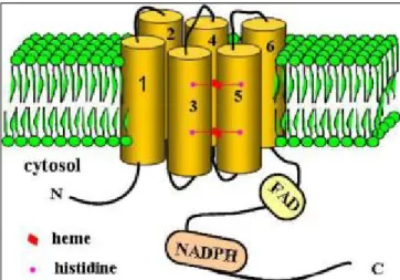
Nox2 (Doussiere et al., 1986; Doussiere et al., 1995). Directed mutagenesis showed that histidines in the third
(H101 and H115) and 5th transmembrane (H209 and H222) are responsible for anchoring the two hemes: one
is located near the cytosolic side coordinated by histidines H101 and H209 and the other is near the outer
extracellular coordinated by histidines H115 and H222 (Biberstine-Kinkade et al., 2001) (Fig. 7). Thus, the
electron path from NADPH to oxygen is mediated by several redox centers: one domain containing FAD
(cytosolic tail) and one containing two histidine residues that anchor two heme prosthetic groups. These
histidine residues are well conserved in all Nox family members and are essential for trans-membrane electron
transport.
al., 2000; Cheng et al., 2001) sequence identity respectively. Nox5 is more distantly with an overall homology
to Nox2 of 27% (Banfi et al., 2001). It consists of 737 amino acids and contains an additional N-terminal
extension comprising four EF-hand motifs.
Duox proteins have a seventh trans-membrane domain at the NH2 terminus with an ecto-facing peroxidase
like domain, in addition to a Nox1-4 homology domain and a EF-hand region. The peroxidase-like domains of
Duox proteins are unusual in that they lack conserved histidine residues found in all other peroxidases,
considered essential for heme binding (Daiyasu et al., 2000). Within the Nox backbone, Duox isoforms share
~50% identity with Nox2 (De Deken et al., 2000).
Therefore, all Nox/Duox share a common backbone that contains the entire transmembrane redox
machinery and a cytoplasmic dehydrogenase domain with FAD and NADPH binding sites. The Nox have not
been crystallized but hydropathy plots and secondary structure prediction programs point to a structure with 6
transmembrane domains that are connected by 5 loops. 2 non-identical hemes are coordinated on the 3rd and
the 5th alpha helices through 2 pairs of His residues separated by 13 or 14 AA. One of the hemes is positioned
in membrane close to the cytosolic space; the second one is on the opposite side. The alignment of primary
sequences revealed that the length of some regions connecting the transmembrane domains or FAD and
NADPH binding domains vary differently among Nox members. These variations may influence the structure
and regulation of NADPH oxidase activity of different Nox (Figure 6).
By analyzing the sequence of 107 Nox enzymes, conserved regions were identified to have important
functions in Nox structure or activation. One such region is the cytosolic B-loop, which in Nox1-4 contains a
conserved polybasic region. The Nox2 B-loop shares considerable homology, suggesting a general role for
this loop in Nox function. In earlier studies of Nox2, this loop was proposed as a binding site for p47phox.
Furthermore, both mutagenesis of arginine residues in the Nox2 B-loop and peptides corresponding to the
Nox2 B-loop inhibited Nox2 activity and prevented p47phox/p67phox translocation to the membrane (DeLeo
et al., 1995; Park et al., 1997; Biberstine-Kinkade et al., 1999). Recent study proposed that the B-loop
provides the interface between the transmembrane and dehydrogenase domains of Nox enzymes by binding to
Figure 6. Linear Representation of the Protein Sequence of NADPH Oxidases. Nox family members have six
putative transmembrane helices, binding sites for NADPH and FAD. Nox5 has 4 EF-hand motifs. Duox proteins have a sequence highly homologous to that of the peroxidase (P), another transmembrane domain and 2 EF-hand motifs at the NH2 terminus.
In terms of structure related to the Nox activity, whatever the mechanism is involved, structural changes
are linked to 3 regulation types: (a). An allosteric transition between two conformation states upon activation
which has been illustrated by AFM (Paclet et al., 2000; Paclet et al., 2007); (b). A continuous phosphorylation
for sustained activity has been shown for all Nox; (c). Calcium binding either directly as with Nox5 and the
Duox, or indirectly through S100A8/A9 proteins or calmodulins as shown with Nox2 and Nox5 (Berthier et
al., 2003; Benedyk et al., 2007; Paclet et al., 2007).
Light on Nox4: Structural Properties
The structure of Nox4 has the same overall organization as Nox2 with 6 transmembrane domains, four
highly conserved heme-binding histidines, two in the 3rd and two in the 5th transmembrane domains, a
FAD-binding region in proximity of the most COOH-terminal transmembrane domain and a NADPH-binding
region site at the very COOH terminus. Two extracellular regions of Nox4 are different with other Nox
members: loop C is shorter while loop E is longer. Given that most studies report that Nox4 generate H2O2
instead of O2
-, the differences in the extracellular loops might be responsible for the unique ability of Nox4.
and dehydrogenase domain. This interaction was weakened with Nox4R96E B-loop corresponding to a
mutation that also markedly decreased the activity of Nox4 (Jackson et al., 2010).
2.2 Topology of NADPH oxidases
Figure 7. Topology of Nox2. Nox2 has six transmembrane domains and its NH2 terminus and its COOH terminus are facing the cytoplasm.
The structure of Nox2 was not resolved yet, the data concerning the topology derived from experimental
and predictive data. Computational analysis of the primary sequence of Nox2, as well as data from epitope
mapping point to a structure with 6 transmembrane domains connected by 5 loops, termed A-E (Imajoh-Ohmi
et al., 1992; Burritt et al., 2001). Antibody mapping studies demonstrate a cytoplasmic localization of the
COOH terminus (Rotrosen et al., 1990; Burritt et al., 2003). Sequencing data and antibody mapping confirm a
cytoplasmic NH2 terminus. The N- as well as C-terminal part of Nox reside in the cytosol, giving rise to two
intracellular loops (B- and D-loop) and three loops oriented away from the cytosol and towards the
extracellular space or intracellular compartments (A-, C-, and E-loop). Phage display library screening also
provides experimental data defining the extracellular domains (Nakamura et al., 1987; Imajoh-Ohmi et al.,
1992; Burritt et al., 2001). Loop C and E were identified as extracelluar part by identifying the epitopes of
monoclonal antibodies specific to Nox2 (Burritt et al., 2001; Yamauchi et al., 2001; Campion et al., 2007).
Loop B is involved in the interaction with the cytosolic factor p47phox and therefore intracellular localization
(DeLeo et al., 1995; Biberstine-Kinkade et al., 1999). Taken together, the available data suggest that Nox2 has
al., 2004) (Figure 7).
Light on Nox4: Topology
The topology information of Nox4 most comes from the computational analysis of Nox4 sequence and
the comparison with Nox2. The current model to study the structure-function of Nox4 is based on it.
2.3 Post-translational Modification of NADPH Oxidases
Nox2 protein was synthesized in an immature form in the endoplasmic reticulum which has a molecular
weight of 65kDa. It then undergoes maturation by high glycosylation that appears as a broad smear on
SDS-PAGE reflecting the heterogeneity of glycosylation. The fully glycosylated form runs with an apparent
molecular mass of ~70-90kDa. Removal of the carbohydrates by endoglycosidase F leaves a protein that runs
at 55kDa, demonstrating the extent of glycosylation (Harper et al., 1985). Mutagenesis approach shows that
the extracellular loops II and III contain N-linked glycosylation sites (Wallach et al., 1997). Through amino
acid analysis of the new Noxes, we can find consensus sequences of potential N-glycosylation (Asn-X-Ser/Thr,
X is any amino acid) (Table 2). In the case of Nox1, most studies suggest a molecular mass of Nox1 in a range
of 55-60 kDa by Western blot (Ambasta et al., 2004; Janiszewski et al., 2005; Cui et al., 2006). If these values
are correct, Nox1 is most likely not N-glycosylated, despite the presence of two NXT/S consensus
glycosylation sites in the extracellular domains. However, this fact remains to be confirmed given the lack of
truly specific antibodies directed against Nox1. Nox3 and Duox1/2 are glycosylated due to a decrease in
molecular weight by deglycosylation (De Deken et al., 2002; Nakano et al., 2007). Both Duox1 and Duox2
have two N-glycosylation states: the high mannose glycosylated form found in the ER, and a fully
glycosylated form found at the plasma membrane (De Deken et al., 2002). Nox5 has no predicted
N-glycosylation site.
Light on Nox4: N-glycosylation
Few studies concerned the post-translational modification of Nox4 by N-glycosylation. The theoretical
size of Nox4 is 66.9kDa. Since now, it has been reported that Nox4 antibody recognize two kinds of bands:
one of 75-80KDa and a second of 55-65KDa from both endogenous Nox4 expressing cells and/or
von Lohneysen et al., 2008). The subcellular distribution of the two bands was distinct (Hilenski et al., 2004).
The fact that two molecular masses are detected and that Nox4 contains four putative N-glycosylation sites
might suggest that Nox4 is glycosylated, although treatment with N-glycosidase F failed to reduce the protein
to a single band (Shiose et al., 2001).
Proteins Potential N-glycosylation sites Glycosylation MW kDa
N terminal Loop c Loop d
Nox1 - Asn162 Asn236 - 55/60
Nox2 - Asn132/149 Asn240 + 65 91
Nox3 - Asn163 Asn238 + 50 55
Nox4 - Asn130/133 Asn230/236 +/-? 65 75-80
Nox5 - - - - 75
Duox1 Asn94/342/354/461/534 - - + 160 190
Duox2 Asn100/348/382/455/537 - - + 160 190
Table 2. NADPH oxidases and glycosylation. Asparagines corresponding to the potential sites of N-glycosylation.
2.4 Isoforms of NADPH oxidases
Nox1 has another 2 isoforms. This isoform Nox1v lacks entire exon 11 which contains a domain
predicted binding of NADPH (Geiszt et al., 2004). It appears that this splice variant encodes a protein
incapable of producing superoxide. Two other Nox1 mRNA (c-type and f-type) have been described in mouse
due to the use of alternative promoter. These two mRNAs encode the same protein Nox1 (c/f-type) lying on
the N terminal capable of having an activity equivalent to Nox1 NADPH oxidase (Arakawa et al., 2006).
Besides, a very short isoform of Nox1 had been suggested, but it proved to be an experimental artifact due to
the formation of a stable loop in the Nox1 mRNA (Banfi et al., 2000; Geiszt et al., 2004; Harper et al., 2005).
In regard to Nox2, a truncated isoform of Nox-2S has been described (Heidari et al., 2004). It includes a
previously unidentified exon (IIIa) and encodes an in-frame stop codon.Nox-2S displays a widespread pattern
of expression in mouse tissues and human cells, with high levels present in the myeloid cell line HL-60. The
function of Nox-2S awaits elucidation.
Nox5 (Nox5 ) has another four isoforms (Nox5 , - , - , - ). Nox5 isoforms (Nox5 - ) described by
Banfi et al. distinguish themselves from Nox1-4 enzymes by the presence of a long intracellular NH2 terminus
containing a Ca2+-binding EF-hand domain (Banfi et al., 2001; Banfi et al., 2004). Nox5 lacks the EF-hand
Light on Nox4: isoforms
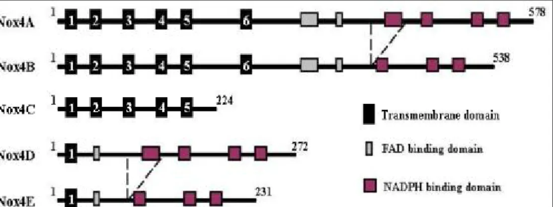
It has been suggested that Nox4 has another 4 isoforms except the prototype Nox4A (Goyal et al., 2005)
(Fig. 8). Nox4B differs from Nox4A by splicing of exon 14 that causes a deletion of the first binding domain
of NADPH, therefore results in the lack of the first NADPH binding site in the protein. Nox4C is a result of a
splicing of exon 9-11, which generates a frame shift in mRNA, and hence a new stop codon in the beginning
of exon 12. Consequently, Nox4C lacks the complete C-terminus including FAD and NADPH binding
domains in protein. Both variants B and C acted as dominant-negative molecules, since A549 cells transfected
with these variants had lower ROS concentration as compared to control plasmid transfected cells. Meanwhile,
the similar inhibitory effect on ROS generation between variant B and C indicated that the first NADPH
binding site seems to have a strong impact on NADPH oxidase activity and cannot be fully compensated by
the other NADPH and FADH binding sites. Nox4D and Nox4E are both a consequence of splicing of exons
3-11 whereas variant E has an additional splicing of exon 14. Therefore, Nox4D has only the first
transmembrane domain and contains all FADH and NADPH binding sites while Nox4E has only the first
transmembrane domain and additionally lacks one NADPH binding site similar to variant B. Interestingly,
Nox4D was shown to have NADPH oxidase activity comparable to that of Nox4A brings into question of the
necessity of conserved histidines distributed within the transmembrane containing N-terminus for enzymatic
activity. These histidines are postulated to bind iron in complex with the heme group that is required for
electron transfer to oxygen (Lambeth, 2000). Both Nox4D and Nox4E appear as glycosylated forms. As both
of them lack the hydrophobic putative transmembrane domains, they are hence considered as soluble variants.
3 Regulation of the NADPH Oxidase Activity
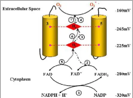
3.1 Electron Transfer Mechanism of NADPH Oxidase Nox2 (Cross et al., 2004)
The NADPH oxidase catalyzes the production of superoxide by electron transfer from NADPH to
molecular oxygen. The central component of the complex is the flavocytochrome b558, a membrane-bound
heterodimeric protein consisting of two subunits: a larger glycoprotein (gp91phox) and a smaller
nonglycosylated protein (p22phox). These two subunits are in a 1:1 stoichiometry.The mechanism of electron
transfer is well documented for the Nox2 protein. gp91phox possesses the entire electron transport machinery
(Geiszt et al., 2004; Lambeth, 2004; Bedard et al., 2007). It is the catalytic center of this enzyme. There are
seven separate steps of electron transfer during a single turnover of NADPH oxidase, as illustrated in Figure. 9.
The first step is the transfer of two electrons from NADPH to oxidized FAD. The second electron transfer step
is from reduced FAD (FADH2) to the inner haem, generating the FAD semiquinone (FAD°). In the third and
fourth steps, the electron is passed from the inner to the outer haem and then to oxygen forming superoxide.
The fifth, sixth and seventh steps recapitulate the third, fourth and fifth, the exception being in the fifth step,
where it is the flavin semiquinone that is the donor to the inner haem.
Figure 9. Electron transfer pathways within flavocytochrome b558 (Cross et al., 2004). The seven electron
transfer steps are numbered. The transfer of two electrons takes place from NADPH (step 1). The first electron transport is from FADH2 (step 2,3,4) and the second electron transport is fromFAD° (step 5,6,7).
can be distinguished from the NADPH oxidase activity by using INT (iodonitrotetrazolium) as a direct FAD
electron acceptor, in PLB985 cells expressing the whole protein (Pessach et al., 2001) and truncated protein
containing only cytosolic part (Pessach et al., 2006). Experiments with Nox2 truncated forms produced by
bacteria were able to determine the region responsible for this activity, which corresponds to amino acids 221
to 570. The diaphorase activity is intrinsic and does not require the presence of cytosolic factors. However,
these cytosolic factors (p67phox and Rac1) are capable of stimulating the activity (Han et al., 2001; Nisimoto
et al., 2004).
Light on Nox4: Diaphorase Activity
Recently, the cytosol-facing dehydrogenase (DH) domain (AA: 298-578) containing a binding site for
NADPH and one for FAD (Rotrosen et al., 1992; Segal et al., 1992; Sumimoto et al., 1992) from Nox4 was
expressed and purified to measure the electron transferase activity toward several artificial electron acceptors
(Nisimoto et al., 2010). In these experiments, the Nox4 DH domain showed significant rates of electron
transfer (65-470 min-1 depending on the electron acceptor), and these activities were inhibited by DPI.
3.2 Nox Subunits and Regulation Proteins
Despite their similar structure and enzymatic function, Nox family differs in their mechanism of
activation. The components involved in Nox activation include the membrane-bound p22phox, the cytosolic
proteins p47phox, p67phox, the small GTPase Rac, and the modulatory p40phox, which together lead to the
activation of the Nox enzyme.
3.2.1. p22phox: Indispensible Partner of NADPH Oxidases
p22phox is a membrane protein, which closely associates with Nox2 in a 1:1 complex (Huang et al.,
1995). This protein is encoded by the gene CYBA located on chromosome 16 (locus 16q24), it appears as a
band at 22kDa by western blot and seems not glycosylated (Parkos et al., 1987). At the moment, the topology
and functional domains within p22phox are not clearly characterized. The membrane topology of p22phox is
difficult to predict based on hydropathy plots, computational analysis of primary sequence of p22phox
suggests that p22phox contains 2-4 membrane-spanning segments (Taylor et al., 2004). However, the weight
p22phox and cytochrome b558 proteolytic digestion favors the presence of 2 transmembrane domains (Taylor
et al., 2004) and the cytosolic localization of the N- and C-terminal regions of the protein (Imajoh-Ohmi et al.,
1992; Burritt et al., 1998). p22phox has a proline-rich region (amino acids 151-160) that serves as an anchor
for cytosolic factors during oxidase activation. Moreover, the absence of antibodies and the protease sensitive
sites against an extracellular portion of p22phox indicates that a very small portion of the protein is exposed to
the extracellular surface.
The association of p22phox with Nox2 contributes to its maturation and stabilization. Nox2 or p22phox
alone is degraded by the proteasome (DeLeo et al., 2000; Block et al., 2007). The interaction with p22phox
and incorporation of heme are necessary for Nox2 maturation through the N-glycosylation (Yu et al., 1999).
Studies in the subcellular distribution of p22phox show that p22phox localization is a function of the Nox
isoform coexpressed with p22phox in a given cell type. The p22phox subunit has 2 major functions. It binds
and stabilizes Nox proteins, and serves as a membrane anchor for cytosolic regulatory factors by binding
organizer subunits. It was recently shown that p22phox also associates with Nox1 and Nox3 (Ambasta et al.,
2004; Ueno et al., 2005), suggesting a central role of p22phox in the cellular production of reactive oxygen
species. p22phox was also essential for the maturation of Nox3 (Nakano et al., 2007) and would be stabilized
by interaction with Nox1 (Ambasta et al., 2004; Martyn et al., 2006). The association of p22phox with
Nox1-3 suggests that Nox proteins and the p22phox protein are stable only as a heterodimer, while monomers
are degraded by the proteasome (DeLeo et al., 2000). Consistent with this concept, Nox2-deficient CGD
patients do not have detectable p22phox protein within phagocytes, and p22phox-deficient CGD patients do
not have detectable Nox2 protein (Parkos et al., 1989). Nox5 does not require p22phox for activity, as
demonstrated by siRNA suppression of p22phox leading to a decrease in the activity of Nox1-3, but not of
Nox5 (Kawahara et al., 2005; Martyn et al., 2006). Human Duox2 protein reportedly coimmunoprecipitates
with p22phox (Ameziane-El-Hassani et al., 2005), but there is little or no effect of p22phox on Duox
enzymatic activity.
Light on Nox4: Interaction between Nox4 and p22phox
Nox4 is also a p22phox dependent enzyme. siRNA-mediated p22phox down-regulation leads to a
decrease function of Nox4. Nox4 colocalizes and coimmunoprecipitates with p22phox. It also stabilizes the
domain leads to a loss of activation of Nox1, Nox2, and Nox3 (Leusen et al., 1994; Kawahara et al., 2005) but
not Nox4 in agreement with the concept that Nox4 activation does not need cytosolic organizer subunits.
Interestingly, a p22phox mutant (p22phox Y121H) is capable of distinguishing between Nox1-3 and Nox4 by
forming a functional complex only with Nox4, further suggesting the unique structural features in Nox4 (von
Lohneysen et al., 2008).
Recent structure-function analyses of complexes between Nox2 or Nox4 and the subunit p22phox
documented specific regions and amino acid residues in p22phox necessary for complex formation and
oxidase activity (Zhu et al., 2006; von Lohneysen et al., 2008; Helmcke et al., 2009). Extensive
structure-function analysis of the oxidase complex showed that B-loop was crucial for the activity (Jackson et
al., 2010; von Lohneysen et al., 2010) of both Nox enzymes and Nox4 D-loop is part of structural elements
required in bridging dimerization of Nox4 with p22phox Y121H mutant (Helmcke et al., 2009; von
Lohneysen et al., 2010).
3.2.2. p47phox, p67phox, p40phox and Rac: Cytosolic Regulatory Protein of Phagocyte NADPH
Oxidase
3.2.2.1 Organizer Subunits: p47phox, NoxO1
Organizer subunits p47phox and NoxO1 shares ~25% sequence identity. p47phox contains a phagocyte
oxidase (PX) domain, tandem SH3 domains, an autoinhibitory region (AIR) and a PRR (Figure. 10). The two
SH3 domains cooperatively interact with the PRR in the C-terminal cytoplasmic region of p22phox, which is
essential for both membrane translocation of p47phox and oxidase activation (Leto et al., 1994; Sumimoto et
al., 1994). In the resting state, the two SH3 domains of p47phox are masked via an intramolecular interaction
with AIR (Groemping et al., 2003; Yuzawa et al., 2004; Yuzawa et al., 2004). During phagocytosis of
invading microbes or with soluble stimuli such as N-formyl chemotactic peptide and PMA, p47phox
undergoes phosphorylation at multiple Ser residues, several of which are present in the AIR (el Benna et al.,
1994; Inanami et al., 1998), induces unmasking of the SH3 domains (Shiose et al., 2000), therefore interacts
with the PRR of p22phox. NoxO1 exhibits a domain architecture similar to that of p47phox, except that it
lacks an AIR (Figure. 10), suggesting that NoxO1 is constitutively active. NoxO1 plays an essential role in
Nox1 activation which functions via its PRR by interacting with NoxA1 and via its SH3 domains by binding
Figure 10. The two organizer homologs, p47phox and NoxO1, share a similar set of motifs. PX, phagocyte
oxidase domain; SH3, Src homology 3 domain; AIR, autoinhibitory region; PRR, proline rich region. See the detail of interaction of p47phox or NoxO1 with other protein in text (3.2.2.1).
3.2.2.2 Activator Subunits: p67phox and NoxA1
Activator subunits p67phox and NoxA1 share ~28% amino acid identity and their overall domain
structure is similar (Figure. 11). Both contain an NH2-terminal tetratricopeptide repeat (TPR, interact with Rac)
(Koga et al., 1999; Lapouge et al., 2000; Grizot et al., 2001), a highly conserved activation domain (AD,
interact with Nox proteins) (Han et al., 1998; Nisimoto et al., 1999; Geiszt et al., 2004), a less conserved
Phox and Bem 1 domain (PB1, interact with the PC domain of p40phox) and a COOH-terminal SH3
domain (binding to the C-terminal PRR of p47phox) (Finan et al., 1994; Leusen et al., 1995). However, there
are some differences between two activator subunits: the PB1 domain of NoxA1 has important differences
from that of p67phox, thus fails to interact with p40phox (Takeya et al., 2003). It also lacks the central SH3
domain found in p67phox (Banfi et al., 2003; Geiszt et al., 2003). Although the first SH3 domain is the most
conserved region in p67phox (Mizuki et al., 1998), its function is still unknown.