Université de Montréal
Exploring the Roles of Atypical MAP Kinases ERK3 and ERK4
During Inflammation
Par: Michelle Barbagallo
Département de Pharmacologie et Physiologie Faculté de Médecine
Mémoire présenté en vue de l’obtention de grade de M. Sc en Pharmacologie
Août, 2018
ii
Résumé
L’insuffisance rénale aiguë est un évènement complexe marqué par une lésion tissulaire causée par une ischémie ou une exposition aux néphrotoxines. Cette lésion est suivie par l’activation des cellules intrarénales qui déclenchent une réponse au stress provoquant l’infiltration de leucocytes. Ensemble, ils aident à éliminer les tissus endommagés pour ensuite jouer un rôle dans le processus de guérison du rein. Les mécanismes par lesquels le système immunitaire régule les évènements dans l’AKI ne sont pas entièrement compris. Des travaux récents ont révélé un rôle clé des MAP kinase atypiques ERK3 et ERK4 dans la promotion de l’inflammation, en régulant la synthèse des chimiokines CCL2 et CCL5 et en jouant un rôle dans la migration et l’invasion des cellules immunitaires aux tissue enflammés/endommagés.
On suppose que l’activité de ERK3 et ERK4 aide à la migration des cellules immunitaire par des mécanismes intrinsèques et favorise une cascade inflammatoire. Également, on pense que ERK3 et ERK4 jouent un rôle dans la maturation et l’activation des cellules immunitaire et que le blocage de leur activité peut inhiber ce processus. En utilisant des cellules rénales épithéliales murines, nous démontrons qu’une perte de ERK3 atténue la production de CCL2 et CCL5 in vitro dans les cas de stimulation avec TNFα et TWEAK. De plus, la perte de ERK3 n’affecte pas la maturation des cellules de la moelle osseuse en macrophages ou leur polarisation. Cependant, l’absence d’ERK3 dans les macrophages inhibe leur capacité à migrer.
En résumé, nous fournissons des preuves que les membres du TNFSF peuvent réguler l’expression de ERK3 et que ERK3 joue un rôle dans la production de chimioattractants des macrophages, qui aident à favoriser la cascade inflammatoire nécessaire a l’infiltration des macrophages. De plus, nous démontrons que ERK3 joue un rôle dans la capacité intrinsèque des macrophages à migrer dans les tissus. Ces résultats suggèrent que ERK3 a plusieurs fonctions biologiques durant l’inflammation.
Mots-clés: MAPK atypique; ERK3; ERK4; insuffisance rénale aiguë; inflammation; CCL2;
iii
Abstract
Acute kidney injury is a complex event marked by renal tissue injury after ischemia or exposure to nephrotoxins. It is followed by the activation of intrarenal cells that trigger a stress response causing the infiltration of leukocytes that together help clear damaged tissue and later play a role in the healing process of the kidney. The mechanisms in which the immune system regulates events in AKI are not fully understood. Recent work has unveiled the key role of the atypical MAP kinases ERK3 and ERK4 in promoting inflammation, in part through regulating the synthesis of chemokines like CCL2 and CCL5 and playing a role in migration/invasion capabilities of immune cells to inflamed/damaged tissues.
It is hypothesised that ERK3 and ERK4 activity promotes immune cell migration through intrinsic mechanisms and by fostering an inflammatory cascade. It is also believed that ERK3 and ERK4 promote immune cell maturation and activation and that blocking ERK3 and ERK4 activity can inhibit these processes. Using murine tubule epithelial kidney cells and bone marrow derived macrophages; we demonstrate that ERK3 deficiency attenuates the production of CCL2 and CCL5 in tubule cells stimulated with TNFα and TWEAK. Additionally, ERK3 loss does not impair bone marrow to mature into macrophages or polarize into pro-inflammatory or anti-inflammatory phenotypes. However, the absence of ERK3 in BMDMs inhibits their basal migration abilities in a scratch assay.
Taken together, we provide evidence that TNFSF members may regulate ERK3 expression and play a role in the production of macrophage chemoattractant, which help foster the inflammatory cascade needed for macrophage infiltration. Moreover, we demonstrate that ERK3 plays a role in inherent ability for macrophages to migrate. All suggesting that ERK3 has multiple biological functions during inflammation.
Keywords: Atypical MAPK; ERK3; ERK4; AKI; inflammation; CCL2; CCL5; macrophages;
iv
Table of Contents
Résumé ... ii Abstract ... iii List of Figures ... vi List of Tables ... vii List of Abbreviations ... viii Acknowledgements ... xi Introduction ... 13Mitogen-activated protein kinases ... 13
Extracellular signal-regulated kinase 3 and 4 ... 15
Identification and structure ... 15
Expression ... 16
Activators, Regulators & Substrates ... 16
Cellular functions ... 23
ERK3 and Immunity ... 29
Acute kidney injury ... 30
Pathophysiology of AKI ... 30
AKI and Inflammation ... 32
Rational and Objectives ... 46
Materials & Methods ... 49
Reagents and antibodies ... 49
Plasmid constructs ... 49
Mice ... 49
Cell Culture and lentiviral infections ... 50
Mouse Embryonic Fibroblasts (MEFs) ... 50
Inflammasome activation ... 50
Enzyme-linked immunosorbent assay (ELISA) ... 51
Bone marrow derived macrophages ... 51
Flow cytometry ... 51 TNFSF stimulation ... 51 BMDM polarization ... 52 Migration assays ... 52 Immunoblotting ... 52 Quantitative PCR analysis ... 53 Results ... 54
v
MEFs and the investigation of inflammasome signaling ... 54
ERK3 is expressed in renal and macrophage cell lines ... 55
ERK3 expression is upregulated by members of the TNFSF family in tubule cells ... 56
ERK3 deficiency reduces mRNA expression of CCL2 and CCL5 upon TNFSF stimulation in tubule cells ... 57
ERK3 loss does not effect monocyte to macrophage differentiation ... 60
ERK3 loss does not effect macrophage polarization ... 61
ERK3 loss reduces basal macrophage migration ... 64
Discussion ... 66
Conclusion and Perspectives ... 71
vi
List of Figures
Figure 1: MAPK signaling molecules 13
Figure 2: Structural representations of conventional and atypical MAPKs 14
Figure 3: Regulators of ERK3 and ERK4 18
Figure 4: Substrates of ERK3 and ERK4 20
Figure 5: ERK3 and ERK4 interaction and activation of MK5 22
Figure 6: ERK3 and ERK4 signaling in cancer 28
Figure 7: Idealized phases of AKI 31
Figure 8: MYD88-dependant and MYD88-indepenant TLR signaling 34 Figure 9: Two signal pathway and activation of the NLRP3 inflammasome 36
Figure 10: Potential role of ERK3 and ERK4 in AKI 47
Figure 11: NLRP3 inflammasome regulation of pro-IL-1β mRNA in Mapk6LacZ and
ERK4-/- MEFs 55
Figure 12: ERK3 and ERK4 expression in murine kidney and murine and human monocyte/macrophage cell lines
56
Figure 13: ERK3 expression upon TNFSF stimulation in MCT cells 57 Figure 14: Lentiviral CRISPR knockout of ERK3 in MCT cells 58 Figure 15: ERK3 regulation of CCL2 and CCL5 upon TNFα stimulation 59 Figure 16: ERK3 regulation of CCL2 and CCL5 upon TWEAK stimulation 60 Figure 17: ERK3 deficiency does not impact BMDM maturation 61 Figure 18: Impact of catalytically inactive ERK3 BMDMs on macrophage polarization 63
vii
List of Tables
Table 1: Forward and reverse RNA sequence guides 49
viii
List of Abbreviations
ACTB β-actin
AKI Acute kidney injury APC Antigen presenting cell
Arg1 Arginase-1
ASC Apoptosis-associated speck-like protein bFGF Basic fibroblast growth factor
BMDM Bone marrow-derived macrophages BORGS Binders of Rho GTPases
CARD8 Caspase recruitment domain family member 9 CCAC Canadian Council on Animal Research
CCL2 C-C motif chemokine protein 2 (also, MCP-1) CCL5 C-C motif chemokine protein 5 (also, RANTES) CCR2 C-C motif chemokine receptor type 2
CD Common docking
CSF-1 Colony stimulating factor-1
CX3CL1 C-X3-C motif ligand 1 (also, fractalkine) CX3CR1 C-X3-C motif receptor 1
CXCL1 C-X-C motif ligand 1
DAMPs Damage-associated molecular patters DCs Dendritic cells
DMEM Dulbecco's modified Eagle's medium
DN Double negative
DT Dipheria toxin
DUSP2 Dual-specificity phosphatase 2 Elk-1 ETS domain-containing protein ERK Extracellular signal-regulated kinases Fn14 Fibronectin 14
GFR Glomerular filtration rate HIF-1α Hypoxia-inducible factor 1-α HMBG-1 High mobility-group B1 HSP1 Heat shock protein 1 HSP27 Heat shock protein 27 HSPs Heat shock proteins
HUVEC(s) Human umbilical cord vein endothelial cell(s) ICAM-1 Intracellular adhesion molecule 1
IFN Interferon
IGF2BP1 Insulin-like growth factor 2 binding protein 1 IL-10 Interleukin-10
ix IL-12 Interleukin-12 IL-17 Interleukin-17 IL-17 Interleukin-17 IL-18 Interleukin-18 IL-1α Interleukin-1α IL-1β Interleukin-1β IL-23 Interleukin-23 IL-6 Interleukin-6 IL-8 Interleukin-8
IRI Renal ischemic injury
JNK1/2/3 c-Jun amino (N)-terminal kinase 1/2/3 KIM-1 Kidney injury molecule-1
M-CSF Macrophage colony stimulating factor mAb Monoclonal antibody
MAP-1 Microtubule associated protein 1 MAPK(s) Mitogen activated protein kinase(s) MAPKK(s) Mitogen activated protein kinase kinase(s)
MAPKKK(s) Mitogen activated protein kinase kinase kinase(s) MCP-1 Monocyte chemoattractant protein-1 (also, CCL2) MCT Mouse cortical tubule
MEF Mouse embryonic fibroblast
MEK(s) Mitogen activated protein kinase kinase(s) MHCII Major histocompatibility complex class II MIP-2 Macrophage inflammatory protein-2
miRNA MicroRNA
MK Mitogen activated protein kinase activated protein kinase MKP Mitogen activated protein kinase phosphatase
NF-κB Nuclear factor κ light chain enhancer of activated B cells NGAL Neutrophil gelatinase-associated lipocalin
NK Natural Killer
NKG2D Natural killer group 2 member D NLK Nemo-like kinase
NLRPs NOD-like receptor family protein
NODs Nucleotide-oligomerization domain like receptors NOS2 Nitric oxide synthase 2
PAK1/2/3 p21-activated kinase 1/2/3
PAMPs Pathogen-associated molecular patters PRRs Pattern recognition receptors
Rae-1 Retinoic acid early inducible 1 RAG Recombination-activating genes
x
RANTES Regulated upon activation normal T cell expressed and secreted (also, CCL5) RNAi RNA interference
RPTECs Renal proximal tubule epithelial cells RSK1/2/3 Ribosomal s6 kinase
shRNA Short hairpin RNA siRNA Small interfering RNA Sept7 Septin 7
SRC-3 Steroid coactivator 3 T regs Regulatory T cells TCR T cell receptor TCRα T cell receptor α
TDP2 Tyrosyl DNA phosphodiesterase 2 TECs Tubular epithelial cells
TGFβ Transforming growth factor-β TLR Toll-like receptor
TNFR Tumour necrosis factor receptor TNFSF Tumour necrosis factor super family TNFα Tumour necrosis factor α
Top2 Topoisomerase 2
TWEAK Tumour necrosis factor like weak inducer of apoptosis USP20 Ubiquitin specific protease 20
VEGF Vascular endothelial growth factor
VEGFR Vascular endothelial growth factor receptor α-MSH α-melanocyte stimulating hormone
xi
Acknowledgements
There are many people I would like to thank for their help, support and encouragement throughout my master’s degree. First, I would like to thank my supervisor, Dr. Sylvain Meloche, for his guidance throughout this research project. Sylvain, thank you for teaching me to keep pushing through despite negative results and helping me become more resilient as a scientist. I am happy for the opportunity you have given me to do research in my two interests, pharmacology and immunology, and I am proud of everything I’ve learned and accomplished in the lab under your supervision.
I would also like to thank everyone in the Meloche Lab, both past and present. Thank you to everyone working on ERK3, especially Dr. Mathilde Soulez and Dr. Simon Mathien, you both were there for every question I had about ERK3 and were always ready to discuss recent results and hypothesizes I had about its function. Thank you, Charlotte Girondel, for all your guidance during my internship and throughout my master’s; your help with all things from immunology to helping me prepare for presentations is greatly appreciated. To Dr.Marc Saba-El-Leil, Dr. Mathilde Soulez and Charlotte for all your help with experiments that needed mice. Lastly to all my fellow lab members not mentioned above: Laure, Marjorie, Chloé, Kim, Eric, Patrick, Édith, and Joaquim, I will always be grateful for your help.
Outside of our lab, I would like to thank the Départment de Pharmacologie and Physiologie for taking me on as a graduate student and IRIC for being the institute that allowed me to grow as a scientist through the use of their amazing facilities. More specifically I would like to thank those at the Genomics, Flow Cytometry and Imaging, and In Vivo Biology platforms for all their help with this project.
More personally, I would like to thank Gabriella Bernal Astrain. Meeting you during the IRIC recruitment event two summers ago, to now being lunch buddies and great friends. I wanted to thank you so much for everything you have done for me in the past year. Meeting you was such a blessing and I believe we have created a lifelong friendship full of our combined love of science, food and great company!
xii
To my parents, thank you for everything you have done throughout the years, especially supporting me through my education. I know it must have been difficult to understand my scientific ramblings at the dinner table, but your understanding and emotional support though my scientific highs and lows means so much. Also, I would like to thank you for your strength helping me through my thesis writing this summer – we all went through a difficult time, but I know that your strength and love will guide us day-by-day in finding our new normal.
Finally, to Ryan, you are my rock through the ups and downs that accompany scientific research and the roller coaster we call life. Your support not only throughout the past two years, but since we met mean the world. I am so lucky to have you as a partner in life’s adventure - cheers to the next chapter!
13
Introduction
Mitogen-activated protein kinases
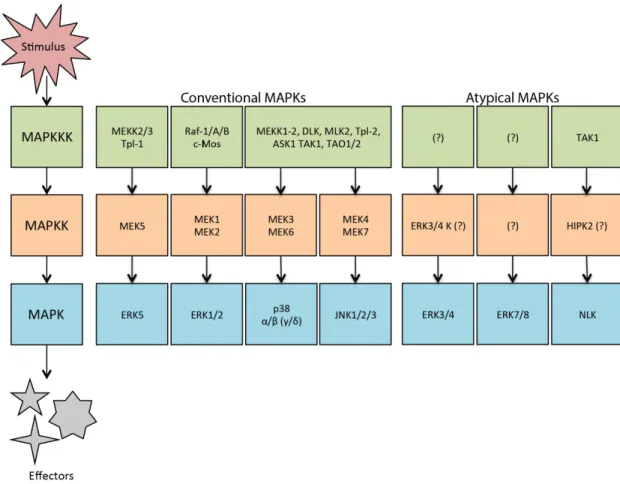
Mitogen activated protein kinases (MAPKs) are serine-threonine kinases involved MAPK signaling pathways conserved in eukaryotic cells.1 They convert extracellular signals into intracellular responses through MAPK activation modules. These modules consist of three sequentially phosphorylated, and therefore activated, kinases; the activation of MAP kinase kinase kinase (MAPKKK) followed by MAP kinase kinase (MAPKK or MEK) and then the activation of the final effector MAPK. Once activated, MAPKs then activate a wide range of substrates that coordinate and regulate gene expression, mitosis, metabolism, motility, survival, apoptosis, differentiation and immune responses. In mammals there are seven MAPK modules, four of which are considered conventional and three considered atypical (Figure 1).2,3
14
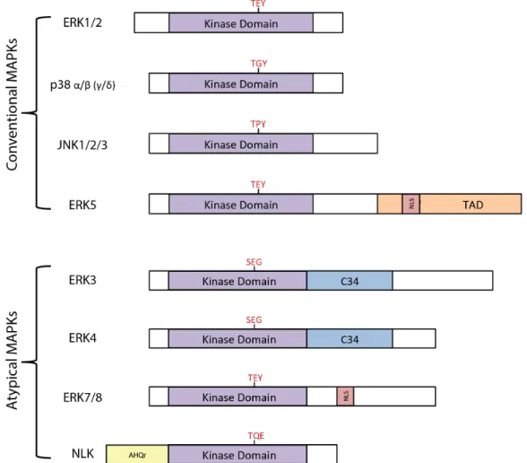
Conventional MAPKs, including extracellular signal-regulated kinases 1/2 (ERK1/2), c-Jun amino (N)-terminal kinases 1/2/3/(JNK1/2/3), p38 isoforms (α, β, γ and δ) and ERK5, adhere to the descried three-tiered MAPK module. They are activated by MAPKKs through the dual phosphorylation on the threonine and tyrosine residues part of a conserved Thr-Xaa-Tyr motif in the activate loop of their kinase domain. Atypical MAPKs, ERK3/4, ERK7/8 and Nemo-like kinase (NLK), above all do not contain the Thr-Xaa-Tyr motif (with exception to ERK7/8) and are not activated by conventional MAPKKs; thus they do not conform to the three-tiered model (Figure 2).2,3
FIGURE 2. Structural representations of conventional and atypical MAPKs. Activation loop motif marked in red. Adapted from reference 2.
15
While conventional MAPKs have been well studied, their activators, substrates and regulators have been characterized; the activators, substrates and regulators of atypical MAPKs remain unclear.
This master’s work focuses on the role atypical MAPKs, ERK3 and ERK4, during the inflammatory response following tissue damage, such as acute kidney injury (AKI). The next chapter aims at describing what is known about ERK3 and ERK4, in addition to events surrounding inflammatory responses during AKI.
Extracellular signal-regulated kinase 3 and 4
Identification and structure
The genes Mapk6 and MapK4 (coding for ERK3 and ERK4, respectively) were cloned by homology thanks to their similarity to MAPK ERK1 in 1991 and 1992, respectively.4,5 At first, rat Mapk6 was predicted to code a protein of 543-amino acids in length.4 Later the sequences for human and mouse Mapk6 were reported.6–8 Sequence comparison of the three orthologs revealed 90% sequence homology over their shared length; human and mouse Mapk6 being longer containing a c-terminal extension of 178 amino acids, which results in a larger predicted amino acid length of 721 and 720, respectively. Interestingly, closer inspection of rat Mapk6 revealed a missing nucleotide between codons 502 and 503 that had resulted in shift of the reading frame and the introduction of a premature stop codon.8,9 Reading frame corrected, rat Mapk6 gene translates to a 720-amino acid protein with 94% homology to the human protein. Additionally, in vitro translation of rat, human and mouse Mapk6 cDNA confirmed all three yield a protein which have a molecular mass of ~100kDa.10
Similar to Mapk6, the initial sequence for Mapk4 had been mistakenly sequenced, missing five nucleotides that resulted in a predicted length of 557-amino acids.5 The rectified Mapk 4 sequence corrects four amino acids and adjusts the reading frame resulting in a predicted length of 587-amino acids and a molecular mass of ~70kDa.5,11 Comparative genomic analysis of Mapk6 and Mapk 4 have revealed that the organization of exon/intron boundaries are similar, however differ from the genes that code for conventional MAPKs. Additionally, encoded proteins, ERK3 and ERK4, share 73% amino acid homology.10 Interestingly, while conventional
16
MAPK orthologs have been found in invertebrates and plants, orthologs of ERK3 and ERK4 have not, suggesting that ERK3 and ERK4 MAPK genes are restricted to vertebrates.12
Similarities between ERK3 and ERK4 are not only restricted to their identification. Both contain a kinase domain the N-terminus and a C-terminal extension (Figure 2). Looking at the C-terminal, ERK3 and ERK4 share ~50% identity in the first 150 residues which then diverge at the C-terminus extreme. Nonetheless, the C-terminus of both MAPK are conserved throughout vertebrate evolution signifying probable important cellular functions.13,14
In relation to kinase domain of conventional MAPK ERK1, ERK3 and ERK4 display 45% and 42% amino acid identity respectively and yet still diverge from their conventional counterpart due to two structural differences. Conventional MAPKs have two phospho-acceptors in the activation loop, while ERK3 and ERK4 contain one. More specifically, ERK3 and ERK4 contain a Ser-Glu-Gly motif differing from the Thr-Xaa-Tyr motif typically found in conventional MAPKs; because of this, ERK3 is a poor substrate for dual-specificity-MAPKKs (Figure 2).13,14 Secondly, all ERK3 and ERK4 orthologs have a Ser-Pro-Arg amino acid sequence instead of a Ala-Pro-Glu in subdomain VIII of their kinase domains – the only two MAPKs that have an arginine residue at this position.15
Expression
ERK3 messenger RNA (mRNA) is expressed ubiquitously in adult mammalian tissues.3 The highest expression is found in the brain, skeletal muscle and the gastrointestinal tract.8 ERK4 mRNA has a more restricted expression profile than ERK3. ERK4 is expressed in the brain, colon, eye, heart, kidney, lung, ovary, pancreas, placenta, prostate and skin. Similar to ERK3, ERK4 has the highest expression in brain tissue.5,15,16
Activators, Regulators & Substrates
The activation of MAPK is controlled by the phosphorylation by kinases upstream. However, they can also be regulated through dephosphorylation by phosphatases and other means like degradation. As mentioned, conventional MAPK, their activators, regulators and substrates, are well studied; those of ERK3 and ERK4 are more elusive.
17
Activators
The study of ERK3 and ERK4 have revealed their phosphorylation sites within their respective activation loops; serine 189 on ERK3 and its homolog serine 186 on ERK4, leading to the discovery of their respective activators.17–19 To date the only activator, hence the only kinases known to phosphorylate ERK3 and ERK4, are p21-activated kinases 1/2/3 (PAK1/2/3).20,21 Interestingly, and contrary to conventional MAPK, ERK3 and ERK4 phosphorylation is thought to be constitutive.19 Because the biological activity of ERK3 and ERK4 does not appear to be a dynamic function of its phosphorylation (due to the observation of it being constitutively active), looking at their regulation is important.
Regulators
While little work has been done on the regulator(s) of ERK4, it is believed that the regulation of ERK3 relies on the protein’s stability.
In mice, ERK3 and ERK4 gene expression markedly increases between embryonic days nine and eleven of development coinciding with early organogenesis.8,16,22,23 Numerous in vitro studies have shown that various stimuli are able to upregulate the mRNA expression of ERK3. Many of these show ERK3 upregulation during differentiation, namely the differentiation of P19 embryonic carcinoma cells into neuronal cells, C2C12 myoblast cells into muscle cells and the differentiation of Burkett’s lymphoma Raji cells.24–26 Of note, is the in vivo work that demonstrates that ERK3-deficient mice display impaired thymocyte development, suggesting ERK3 involvement. In a study, Marquis et. al. demonstrate the increased expression of ERK3 during thymocyte development from double-negative (DN) 1 to the DN4 stage which then decreases further on in development.27 Interestingly, ERK3 is also upregulated when T lymphocytes are stimulated through their T cell receptor (TCR) dependent on the RAF-MEK-ERK conventional MAPK signaling cascade.28 This observation is in agreement with the study conducted by Hoeflich, et. al. which demonstrates that an activating mutation in B-Raf coincides with an increased expression of ERK3.29 It has also been shown that mRNA ERK3 expression is upregulated upon stimulation with the cytokines TNFα and IL-1β and the growth factor basic fibroblast growth factor (bFGF) in human umbilical vein endothelial cells (HUVECs).30 The same study also suggests that the c-Jun transcription factor binds to the Mapk6 promoter and regulates Mapk6 transcription in response to TNFα stimulation.30 Lastly, two microRNAs
18
(miRNA) have been shown to regulate ERK3 expression. The first, miR499a, negatively regulates ERK3 expression in an in vitro model of hepatitis B induced hepatocellular carcinogenesis (Figure 3).31 While the second, let-7i, is involved in a BMI1-let-7i-ERK3 pathway whereby BMI1 upregulates ERK3 expression by suppressing let-7i which directly targets ERK3 mRNA (Figure 3).32
In contrast to ERK3, the context in which ERK4 expression is controlled is less known. However, it has been shown that IGF2BP1 (insulin-like growth factor 2 binding protein 1), a RNA binding protein, inhibits the translation of ERK4 mRNA (Figure 3).33
FIGURE 3. Regulators of ERK3 and ERK4.
ERK3 is a highly unstable protein with a half-life of 30-45mins in exponentially proliferating cells.25,34 It is the first MAPK family member whose activity has been found to be linked to protein turnover.25 In a study done by Zimmermann, et. al., transcriptional profiling identified Mapk6 as a gene significantly upregulated in response to treatment with a proteasome inhibitor.35 Further investigation identified the proteolysis of ERK3 is executed by the proteasome and is dependent on the polyubiquination of the protein; proteasome inhibitor treatment or inactivation of the ubiquitin-activating enzyme (E1) was shown by Coulombe et. al. to result in the accumulation of endogenous or ectopically-expressed ERK3 protein.25 In the same study, ERK3 turnover was found to be independent of its activation loop phosphorylation, enzymatic activity or its C-terminal extension. However, the analysis of a series of chimeras
19
made between stable ERK1 and ERK3 delimited two degrons in the N-terminal lobe of ERK3’s kinase domain that are both necessary and sufficient to target ERK3 for proteasomal degradation.25 Further investigation of the proteasomal degradation of ERK3 by our lab revealed that the deubiquinating enzyme, ubiquitin specific protease 20 (USP20) can regulate ERK3’s stability and thus its biological activity (Figure 3).36
In contrast to ERK3, ERK4 is a highly stable protein.16 In a recent study, the activity of ERK3 and ERK4 was shown to be regulated by the dual-specificity MAP kinase phosphatase (MKP) dual-specificity phosphatase 2 (DUSP2) (Figure 3). The interaction of DUSP2 with either ERK3 or ERK4 causes the dephosphorylation of serine 189 and 186, respectively, which also leads to the decrease in the phosphorylation of threonine 182 on MAP kinase-activated protein kinase 5 (MK5), a MAP kinase activated protein kinase (MK) and substrate of ERK3 and ERK4 (more on MK5 below).37
Away from specific regulators, cellular localization also appears to be important for ERK3 and ERK4 activity. While ERK3 is found in both the cytoplasm and in the nucleus, ERK4 is principally found in the cytoplasm in a large variety of cells types.11,38,39 Likewise, the cytoplasmic localization of ERK3 is facilitated by active transport dependent on CRM1, for which the C-terminus of ERK3 is necessary.38 However, in contrary to conventional MAPK, ERK3 and ERK4’s localization within the cell does not change in response to stimuli investigated, and a few studies have suggested ERK3’s localization changes during the cell cycle, yet they are not in agreement. Bind et. al. were the first group to suggest that ERK3 changes during mitosis by localizing to the Golgi apparatus in HeLa cells.9 This is in conflict with Aredia et. al.’s study that demonstrates ERK3’s homogenous distribution during mitosis.40 In addition to the above, ERK3 and MK5 interaction is known to cause them to localize to the cytoplasm.41,42
Substrates
While ERK3 and ERK4’s conventional counterparts, ERK1 and ERK2 are multifunctional and phosphorylate many different substrates, including microtubule associated protein 1 (MAP-1), c-Jun and ETS domain-containing protein (Elk-1), ERK3 and ERK4 appear to have a more restricted substrate repertoire.17,43 Still, preliminary studies have shown ERK3
20
involvement in activating tyrosyl DNA phosphodiesterase 2 (TDP2), steroid coactivator 3 (SRC-3) and MK5 (Figure 4).11,19,39,41,42,44–46
In cancerous cells, TDP2 repairs topoisomerase 2 (Top2)-linked DNA damage protecting cancer cells against Top2 inhibitor-induced growth inhibition and apoptosis thereby conferring chemoresistance. While the regulation of TDP2 activity is largely unknown, a recent study by Bian, et. al. suggests that ERK3 phosphorylates TDP2 at serine 60. This promotes TDP2’s phosphodiesterase activity, which upregulates TDP2-mediated DNA damage response and increases chemotherapeutic drug resistance.44
FIGURE 4. Substrates of ERK3 and ERK4.
SRC-3 acts as a coactivator of nuclear receptors and other transcription factors. It promotes cell proliferation, transformation and also cancer cell migration and invasion. Interestingly, when cells are depleted for ERK3, the ability of lung cancer cells to invade and form tumours is inhibited in a xenograft mouse model.45 In the same study, it was shown that ERK3 promotes invasiveness by phosphorylating SRC-3 at serine 857 – phosphorylation site determined through immunoprecipitation of the kinase in 293T cells.45 Conversely, unpublished data from our laboratory has shown that in 293T cells, the immunoprecipitation of ERK3 systematically yields the co-purification of MK5 (Déléris, P. and Meloche, S. unpublished data).
21
Interestingly, serine 857 on SRC-3 is situated in an Arg-Xaa-Xaa-pSer motif, the same motif recognized by MK5 and needed for phosphorylation by MK5, suggesting that SRC-3 activation by ERK3 likely passes through MK5. Additionally, it has been shown that ERK3 phosphorylates threonine 182 on MK5’s activation loop, which is followed by a proline, leading to the belief that, like other MAP kinases, ERK3 is a proline directed kinase; serine 857 of SRC-3 is followed by a leucine.19,45
MK5 is the only bona fide substrate of ERK3 and ERK4 characterized by multiple groups. As mentioned, ERK3 and ERK4 phosphorylate MK5 on threonine 182 of its activation loop, this causes a nuclear-cytoplasmic translocation of a ERK3/4-MK5 complex and the activation of MK5 that also stabilizes ERK3.11,39,41,42 Because of the stabilization effect, ERK3-MK5 involvement is more complex than the activation of ERK3-MK5 by ERK4 phosphorylation. It has been proposed that, while ERK3 directly phosphorylates MK5, it’s catalytic activity may be dispensable for MK5 activation as it can exert a scaffolding function needed for the auto-phosphorylation of MK5.41,42 This said, in vitro kinases assays did not support the scaffold protein hypothesis; nevertheless, ERK3/4-MK5 interaction is complex.19
MK5 has low affinity for and interacts transiently with unphosphorylated ERK3/4. The physical interaction of ERK3/4 with MK5 promotes the recruitment and/or activation of ERK3/4 activation loop kinase, PAK1/2/3, catalyzing the phosphorylation of serine 189/186 of ERK3 and ERK4 respectively. The phosphorylation of ERK3/4 strengthens the interaction of the ERK3/4-MK5 complex subsequently phosphorylating MK5 on threonine 182 and activating it. Activated MK5 then phosphorylates ERK3/4 outside of their respective activation loops. It is believed that these further activated forms of ERK3 and ERK4 are competent to then phosphorylate additional physiological substrates to relay the signaling cascade further downstream (Figure 5). However, the identity and regulatory impact of MK5 phosphorylation of extra-activation loop sites on ERK3 and ERK4 remain elusive.19,46
22
FIGURE 5. ERK3 and ERK4 interaction and activation of MK5. A) Unphosphorylated ERK3 interacts transiently with MK5 recruiting PAK1/2/3 that then phosphorylates ERK3. B) The phosphorylation of ERK3 strengthens the interaction with MK5 leading the phosphorylation of MK5 on threonine 182. C) Activated MK5 then phosphorylates ERK3 outside its activation loop. D) The additional phosphorylation of ERK3 allows activation of downstream cellular substrates. E) ERK3-MK5 complex disassembles. Note, ERK3 is used for simplicity, and can be replaced for ERK4. Adapted from reference 19.
Furthermore, the fact that ERK3 and ERK4 phosphorylation strengthens the interaction with MK5 differs from how conventional MAPK interact with their substrates. The classical MAPK
23
ERK1/2 and p38 interact with the downstream MK’s, ribosomal s6 kinases 1/2/3 (RSK1/2/3) and MK2/3, through conserved clusters of acidic amino acids which form a common docking (CD) domain. This CD domain is dispensable for the interaction of ERK3 and ERK4 with MK5, in its place ERK3 and ERK4 interact with MK5 through a FRIEDE motif within ERK3 and ERK4 which is essential for ERK3 and ERK4 binding to the C-terminal region of MK5.47 Cellular functions
Development and Differentiation
Observations made through the creation of ERK3-deficient mice have led researchers to the idea that ERK3 is involved in development and differentiation.
Initially, the targeted disruption of the Mapk6 in mice was accomplished by the insertion of the LacZ gene in-frame with the ATG initiation codon found in exon 2, creating Mapk6LacZ mice. The loss of ERK3 in Mapk6LacZ homozygous mice leads to intrauterine growth restriction, delayed lung maturation associated with defective type II pneumocyte differentiation, and neuromuscular abnormalities. Furthermore, 40% of Mapk6LacZ mice died after delivery from respiratory distress syndrome while the other 60% survived the initial neonatal interval but died with 24 hours from an unknown cause.22 Hence the perinatal lethality of these Mapk6LacZ homozygous mice has stopped the analysis of ERK3 functions in post-natal development and growth.
To address the importance of ERK3 kinase activity and to study the role of ERK3 in post-natal development, two novel genetically engineered mouse models were generated. Mice that express a catalytically inactive (kinase dead) allele of ERK3 (ERK3KI) are born at normal Mendelian ratios and do not exhibit signs of respiratory distress or neuromuscular abnormalities. Unexpectedly, mice with a conditional disruption of the ERK3 gene were crossed to obtain an ERK3 knockout (ERK3Δ/Δ) also survived into adulthood, demonstrating that ERK3 expression and activity is dispensable for post-natal survival; however, kinase activity is necessary for optimal post-natal growth in mice (Soulez, M., Meloche, S., et. al. manuscript in preparation). These recent results dispute the original phenotypes of the original Mapk6LacZ mutant mice, and it is believed that the observed phenotypes of these mice were not directly due to the loss of
24
ERK3 but due to the LacZ construct used to target ERK3 (Soulez, M., Meloche, S., et. al. manuscript in preparation).
Unlike ERK3, the targeted disruption of the Mapk4 gene in mice did not present with the same phenotypes. ERK4-deficient (ERK4-/-) mice are viable, fertile and develop normally exhibiting no gross morphological or physiological irregularities. Additionally, ERK4 loss is not compensated by ERK3; there is no change in ERK3 catalytic activity or expression. Additionally, the loss of ERK4 does not exacerbate the phenotypes seen in the Mapk6LacZ mice nor does it compromise the viability of Mapk6LacZ neonates. Survival into adulthood of ERK4-deficient mice allowed for behavioral phenotyping revealing that they display depression-like behavior in a forced-swim test.16
Apart from development studies done through depletion of ERK3 and ERK4 in mice, studies have looked at its substrate MK5 as well. MK5-deficient mice display impaired spine formation of hippocampal neurons in vivo. It was found that ERK3 interacts with septin 7 (Sept7) to form an ERK3-MK5-Sept7 ternary complex that can then phosphorylate binders of Rho GTPases (BORGS). The same study showed that in transfected primary neurons, the ERK3-MK5 module stimulates Sept-7 dendrite development and spine formation, suggesting that the regulation of neuronal morphogenesis is a physiological function of ERK3-MK5 signaling.48
Classical MAPK are known to play a part in the signaling pathways activated during various steps of T cell development; for instance ERK1/2’s essential role in β-selection and positive selection during thymocyte differentiation.49 Studies have shown that ERK3 also plays a role in the development of thymocytes. A study conducted by Marquis, et. al. in collaboration with our lab has shown that ERK3-deficiency leads to a 50% reduction in CD4+CD8+ double positive (DP) thymocyte number. As a result, ERK3-deficient DP thymocytes have a decreased half-life associated with a higher level of apoptosis and their ability to make successful T cell receptor alpha (TCRα) gene rearrangement is compromised. These observations are linked to ERK3 catalytic activity as non-functioning ERK3 fails to rescue the reduction in DP thymocytes and establishes that ERK3 is essential to sustain DP survival during recombination-activating genes (RAG)-mediated rearrangements.27 Further investigation has found that using a knock-in
25
mouse model in which the coding sequence for ERK3 is replaced by the gene encoding β-galactosidase reporter, that ERK3 is expressed by DP thymocytes undergoing positive selection. ERK3-deficient mice that have a polyclonal TCR repertoire have reduced positive selection that is also observed when ERK3-deficent mice are backcrossed to class I and class II restricted TCR transgenic mice; and TCR stimulation of DP thymocytes was strongly reduced in ERK3-deficient mice. These results show that ERK3 expression following TCR signaling is critical for accurate thymic positive selection.28
Cell Cycle and Proliferation
While indications in the literature have suggested that ERK3 plays a role in proliferation, it has not been well characterized.
Functional studies have shown that ERK3 expression is increased during the differentiation of P9 and PC12 cells into neurons and during the differentiation of C2C12 myoblasts into myotubes; differentiation of these cells are linked to a stop in proliferation.4,25 Moreover, the overexpression of stable ERK3 inhibits fibroblast entry into the S phase.25,38 Plus, in cellular models of hepatocellular carcinoma, it appears that miR499a promotes proliferation by inhibiting the expression of ERK3.31 On the contrary to ERK3’s suggested role as an inhibitor of proliferation, ERK3 appears to be a positive regulator of proliferation in endothelial cells and T cells in response to TCR simulation.27,30,45 However, it has also been shown that the loss of ERK3 expression does not effect the proliferation of various cell lines, including mouse embryonic fibroblasts (MEFs) isolated from Mapk6LacZ mice (Tanguay, P.L. and Meloche, S. unpublished data).
In an attempt to dissect a mechanism for ERK3’s role in proliferation, molecular studies have suggested that ERK3 interacts with proteins cyclin D3 and Cdc14A/B which are cell cycle regulators.50,51 In fact, ERK3 is phosphorylated on its C-terminal extension during mitotic entry and is dephosphorylated during the M/G1 transition.50 However a defined role still remains elusive.
Very little is known about ERK4’s involvement in cell cycle and proliferation; nevertheless, it has been shown that shRNA knock-down of ERK4 in HUVECs causes a
26
reduction in their proliferation.52 Intriguingly, another study in the same cells has shown that an siRNA knock-down of ERK3 also causes a decrease in proliferation.30 Sadly, a study was never conducted looking at the depletion of ERK3 and ERK4 on proliferation, to evaluate the redundancy between the two kinases.
ERK3, ERK4 and Cancer
The above roles of ERK3 and ERK4 in differentiation and proliferation have led many researchers to believe that both ERK3 and ERK4 may be involved in cancer.
Using patients cohorts, it has been found that ERK3 expression is increased in chewing tobacco associated oral squamous cell carcinoma.53 The same study showed that those with elevated ERK3 in cancerous tissues also have a high expression of ERK3 in peripheral blood cells, which was absent in healthy individuals.53 Moreover, Rai, et. al. suggested that an overexpression of ERK3 in normal healthy individuals may be indicative of increased risk of developing oral cancers.53 Along the same line, 50% of individuals with colorectal or gastric cancers have increased ERK3 protein expression in cancerous tissue versus adjacent healthy tissue.54,55 Plus, large-scale transcriptome studies have revealed that ERK3 expression is also increased in melanomas and cells derived from breast cancers.56,57 Likewise, the study by Long et. al. that identified SRC-3 as a substrate of ERK3 also demonstrates that ERK3 is elevated in human pulmonary carcinomas.45
In models of oncogenesis, studies have suggested ERK3 involvement. In the Smgb-Tag transgenic mouse model, a tool that can identify genes that are deregulated in salivary gland carcinomas, ERK3 is elevated in dysplastic tissues and adenocarcinomas compared to healthy tissues.58 Additionally, the oncogenic activating mutation of B-Raf, V600E, upregulates the mRNA and protein expression of ERK3 in cultured mouse fibroblasts.29 As for ERK4, the profiling of pulmonary carcinomas induced by oncogene K-Ras in a transgenic K-Ras mouse model has shown elevated ERK4 mRNA expression.59
Apart from variances in expression, studies have found ERK3 and ERK4 mutations in cancer; however, none have shown the consequence of their mutation. Mutations found for ERK3 through the portal of the TCGA project show that a R399Q mutation is present in uterine,
27
breast and colorectal cancer tissues as well as melanomas.60 The same portal also identifies the R114C/H mutation found in the kinase domain of ERK4, which has been found in colorectal carcinomas, gliomas, non-small cell pulmonary carcinomas and melanomas.60 Other described mutations are H333D/N and E331D/D336N that appear in ERK3 and ERK4 respectively; and are important as they are required for the interaction with MK5.47 More recently, Alsaran, et. al. described that L290P/V mutations in the kinase domain of ERK3 exist in several cancers.61 Their work suggests that these mutations offer increased activity in promoting cancer cell migration and invasion but have little/no impact on ERK3’s role in cell proliferation compared to wild type controls. Furthermore, while there is no clear effect on kinase activity, L290P/V mutations enhance ERK3’s localization to the cytoplasm by increasing its integration with nuclear export factor CRM1.61 Together, their findings suggest that ERK3 plays a role in cancer invasiveness.
Apart from looking at databases, experimental models and the mutations found, there is also work done in cancerous cell lines that further suggests that ERK3 (and therefore potentially ERK4) has a role in cancer, more precisely tumour progression via cellular migration and invasion.
Long, et. al.’s study, whereby SRC-3 is the downstream substrate of ERK3, suggests that the loss of ERK3 inhibits the migration of a pulmonary carcinoma cell line; additionally, the loss of ERK3 and therefore SRC-3 signaling, has been shown to inhibit the migration of human endothelial cells as discovered by Wang, et. al (Figure 6).30,45 Also, pro-invasive studies through SRC-3 have been shown through in vitro and in vivo experiments.45 To further these findings, another study demonstrated that the overexpression of ERK3 modifies the organization of the actin cytoskeleton and promotes the migration speed and adhesion of assorted breast cancer cell lines.62 In a recent study, oncogenic polycomb group protein, BMI1, has been revealed to be a positive regulator of ERK3 levels in head and neck cancer cells.32 Mechanistically, BMI1 upregulates ERK3 expression by suppressing the tumour suppressive miRNA let-7i that directly targets ERK3 mRNA. ERK3 can then act as an important downstream mediator of BMI1 in promoting cell cancer migration.32 In terms of ERK4, it has been shown that ERK4 signals through a ERK4-MK5-heat shock protein-(HSP)-27 pathway
28
that controls the motility of U2OS cells, a human osteosarcoma cell line.33 Furthermore, it is believed that IGF2BP1 inhibits ERK4-MK5-HSP27 pathway by inhibiting the translation of ERK4 mRNA thereby preventing MK5 activation and the phosphorylation of HSP27, which sequesters actin monomers available for F-actin polymerization (Figure 6). Likewise, HSP27-β-actin (ACTB) association is reduced, mobilizing cellular G-actin for polymerization in order to promote the velocity of cell migration.33
29
In other studies, in addition to ERK3’s role in migration, ERK3 depletion inhibits the proliferation of HUVECs – further implicating ERK3 in tumour progression.30 Additionally, HUVECs can form tube structures in vitro when stimulated with cytokines like TNFα, therefore they can be used as a model of angiogenesis. When HUVECs are depleted of ERK3 through siRNA, tube formation is inhibited and thus suggests that ERK3 plays a role in the progression of solid tumours through angiogenesis.30 While the exact mechanism is not understood, the same study defines ERK3 involvement for the vascular endothelial growth factor (VEGF) receptor, VEGFR2, expression through SRC-3 signaling (Figure 6).30 Although this study does not investigate ERK4, HUVECs appear to require ERK4 for normal proliferation in vitro.52 Also, as previously mentioned, our lab believes that the activation of SRC-3 by ERK3 is not direct but through MK5 activation (Figure 6).
In other studies, apart for a direct role in tumorigenesis itself, ERK3 has been described as a regulator of TDP2, and its action confers chemoresistance to cancerous cells; ERK3’s inhibition can therefore sensitize cancerous cells to chemotherapies involved in DNA damage (Figure 6).44 There has been no work done on ERK4’s involvement in DNA damage response and chemoresistance.
ERK3 and Immunity
While there has been work looking that ERK3’s involvement during T cell development and that ERK3 expression is crucial for TCR signaling and thymic positive selection, little to no work has investigated the role of ERK3 and/or ERK4 in other areas of immunity; be it during the responses of the innate immune system and inflammation, or other dynamics of adaptive immunity.27,28
As described, MK5 is the only bona fide substrate of ERK3 and ERK4, and even its biological role remains largely unknown. However, there is some research that suggests that MK5 may be implicated in the inflammatory response, through the phosphorylation of cPLA2 at serine 727 and hnRNAP at serine 84 in in vitro assays. However, their phosphorylation in vivo remains unclear and the biological significance in relation to immunity and inflammation and ERK3 and ERK4 are unknown.63,64 Additionally, while we know there is ERK4-MK5-HSP27 (and potentially ERK3-MK5-ERK4-MK5-HSP27) involvement in cell migration, MK5-ERK4-MK5-HSP27 is
30
also activated in response to the activation of the cAMP-PKA (protein kinase A) pathway and there has not been a study looking at MK5-HSP27 or even ERK3/4-MK5-HSP27 involvement during cellular stress like that of inflammation.65
Finally, since ERK3 and ERK4 are understudied kinases there is a lot still to be learned about their biological functions and therefore it is advantageous to look at both in various contexts, including its potential function(s) in various immune responses to pathogens and tissue injury.
Acute kidney injury
Acute kidney injury (AKI) is defined as a rapid decrease in glomerular filtration rate (GFR) caused by vascular and tubular factors that include ischemia, hypoxia and nephrotoxicity.66–68 Up to 7% of hospitalized patients develop AKI, this increases to 25% in intensive care unit patients, of which 5% will need renal replacement therapy.69,70 It is usually diagnosed by increases in serum creatinine or blood urea nitrogen; however, with the increased study of AKI, biomarkers such as IL-18, kidney injury molecule-1 (KIM-1) and neutrophil gelatinase-associated lipocalin (NGAL) have been used for diagnosis.71 While AKI incidence has decreased and the use of biomarkers contributes to earlier detection, AKI is still associated with a high mortality rate.72 Additionally, it is now believed that inflammation plays a major role in the pathophysiology of AKI.73,74 Endothelial injury increases macrovascular permeability leading to the recruitment of immune cells to the injured kidney during AKI; likewise damage to tubular cells produces pro-inflammatory factors that also result in immune cell recruitment.75,76 This insight has led to the use of various anti-inflammatory therapies, which include the use of mycophenolate, α-melanocyte stimulating hormone (α-MSH), targeting pro-inflammatory pathways implicated in AKI, as well as lymphocyte or macrophage depletion.77 To this end, further understanding of the inflammatory response in AKI can advance our understanding and ameliorate the treatment of AKI.
Pathophysiology of AKI
While the pathophysiology of AKI remains unclear, recent progress in identifying and elucidating the mechanisms and mediators of AKI can help identify a global definition of the disease progress and help reveal novel potential therapeutic targets for AKI prevention or early
31
treatment.78,79 AKI can be broken down into three idealized phases: cell damage and cell death caused by kidney insult, acute inflammation and repair and regeneration of the kidney (Figure 7).80
FIGURE 7. Idealized phases of AKI. After acute kidney injury, cell damage and death trigger activation of kidney resident cells causing an acute inflammatory response through the production of cytokines and chemokines and the recruitment of immune cells. If properly regulated, the acute inflammatory response evolves into repair and regeneration through the plasticity of immune cells, resolving AKI. Adapted from reference 80.
The initial phase of AKI is governed by cell damage and cell death that varies in severity depending on the specific nature of the insult, be it nephrotoxicity, ischemia or sepsis. This phase can last minutes to hours and involves various cells types: epithelial, endothelial and other parenchymal cells in the affected zone or the entire injured kidney generating a broad range of factors that induce an acute inflammatory response.78,79,81,82
Acute inflammation the next phase, which overlaps with the previous, can last hours to days and is mediated primarily though immune cells. Important immediate inflammatory cell
32
responders are resident macrophages and dendritic cells.83,84 Additionally, there are also rapid responses that occur in the bloodstream involving endothelial adherence and activation of monocytes and neutrophils which lead to the infiltration of neutrophils, macrophages, T cells and other myeloid and lymphoid effectors.83,85–89 The activation of these resident and recruited cells are mediated by the production of classic pro-inflammatory mediators such as cytokines, chemokines, free radical species, enzymes and lipid mediators that have high cytotoxic potential, amplifying and extending cell damage and death while also signaling tissue injury.81,83 Because of this, the inflammatory phase of AKI can be seen as both consolidating and worsening kidney dysfunction emphasizing the need for an improved understanding of cellular and molecular components of AKI in an attempt to limit organ damage in the early phases of AKI. Interestingly, recent studies have highlighted that the acute inflammatory response also incorporates counter-regulatory components, such as the production of anti-inflammatory cytokines (e.g. IL-10).90–92
These counter-regulatory mediators, while involved in during inflammation are key players in the third and final phase of AKI, repair and regeneration.79,93,94 Experimental models have shown that programmed transition in the phenotypes of immune effect cells, especially macrophages, involves specific alternative intracellular signaling pathways and mediators.83,84,93–95 Understanding phenotypic plasticity of these effector cells and their restorative mechanisms can help harness them to actively promote repair after AKI as well as acute organ injury in general.72,79,96
AKI and Inflammation
Renal vascular endothelium and tubular epithelium
The initial phase of AKI causes changes to the integrity to the endothelial cell layer leading to endothelial dysfunction causing the infiltration of immune cells into the injured kidney.75,77,97 Brodsky et. al. show in a rat model of renal ischemic injury (IRI) that there is a loss of endothelial cells from afferent arterioles and an interruption of endothelial contacts, which can be reversed though endothelial cell transfer.98 Interestingly, increased microvascular permeability after IRI was attenuated in mice deficient in CD3+ T cells, suggesting that molecules like sphingosine-1-phospate (S1P, a regulator of both immune systems and vascular
33
function) and immune system effector, like T cells, can also be mediators of increased vascular permeability after renal injury.99 In fact, treatment with activation of S1P with the pro-drug FTY-720 does attenuate renal injury in the mouse model of IRI.100
Additionally, IRI regulates the expression of adhesion molecules that facilitate endothelial-leukocyte cell interactions. In particular, the expression of intracellular adhesion molecule 1 (ICAM-1) increases after kidney IRI and mice lacking ICAM-1 are protected from renal injury attenuating leukocyte adhesion to endothelial cells reducing inflammation and the extension of cellular injury.101 Moreover, renal endothelial cells upregulate the expression of C-X3-C motif ligand 1 (CX3CL1, also fractalkine), a substrate for the C-C-X3-C motif receptor 1 (CX3CR1) which is highly expressed on macrophages and acts as a mediator of macrophage recruitment during inflammation; pretreatment with a neutralizing CX3CR1 monoclonal antibody reduced the severity of AKI.102
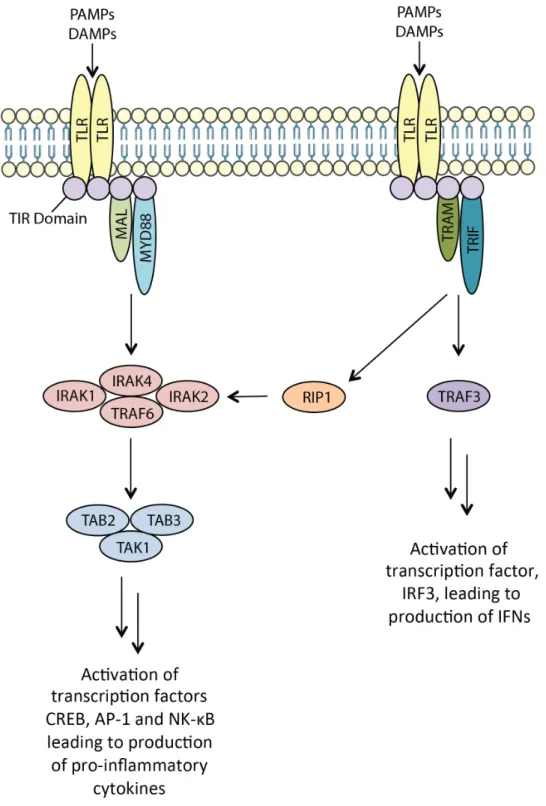
In addition to regulating vascular permeability, renal endothelial cells along with renal tubular epithelial cells (TECs) express toll-like receptors (TLRs) that recognize and interact with microbial and non-microbial endogenous substances released by damaged cells to evoke immune responses.103 For instance the release of high mobility-group B1 (HMBG-1) proteins that can activate inflammation in wild type mice, but not TLR4-deficient mice.104,105 Similarly, mice that lack either TLR2 or TLR4 display blunted cytokine and chemokine production and reduced macrophage and neutrophil infiltration in response to models of IRI or cisplatin-induced AKI.106–109 It is also known that both TLR2 and TLR4 are upregulated on TECs in response to kidney injury.108,110,111 Additionally, it is believed that both MyD88-dependant and independent pathways of TLR activation are involved during injury (Figure 8).112
34
35
Taken together, these studies suggest that the renal endothelium and epithelium play roles in the inflammatory response to kidney injury by promoting the production of cytokines and chemokines thereby promoting the accumulation of immune cells.
Cytokines and Chemokines
Cellular damage/death and its associated molecular products are the key triggers for inflammation after kidney insult.113 Once insult occurs, resident kidney cells release damage-associated molecular patters (DAMPs) such as HMBG-1, histones, heat shock proteins (HSPs), fibronectin, and biglycan into extracellular spaces. These activate pattern recognition receptors (PRRs) including TLRs, and nucleotide-oligomerization domain like receptors (NODs) that are found on epithelial, endothelial and resident leukocytes and thereby initiating a transcriptional response through transcription factors like nuclear factor κ light chain enhancer of activated B cells (NF-κB), HSP1 and hypoxia inducible factor 1-α (HIF-1α).79,83,114–118 Their activation stimulates the synthesis of time-dependent pro-inflammatory and anti-inflammatory cytokines and chemokines throughout all stages of AKI such as IL-1β, Il-6, Il-18, IL-4, IL-10, TNFα, monocyte chemoattractant protein-1 (MCP-1, also known as CCL2), regulated upon activation normal T cell expressed and secreted (RANTES, also known as CCL5), CX3CL1, macrophage inflammatory protein-2 (MIP-2), ICAM-1, and transforming growth factor-β (TGF-β).79,83,119– 123 While there are a vast number of studies and reviews that investigate specific mediators of inflammation, be it a cytokine or chemokine, a few of interest will be further discussed here.
Interleukin-1β (IL-1β)
IL-1β cytokine levels are increased in mice following ischemic insult and recruits leukocytes to the area of injury.101 TLRs expressed on intrarenal cells activated by DAMPs initiate a NF-κB transcriptional response producing immature forms of IL-1β and IL-18, termed pro-IL-1β as well as pro-IL-18. To mature these two cytokines, a second signal leads to the formation and activation of an intracellular multi-protein complex called the inflammasome that is comprised of a NOD-like receptor (NLR) family protein (NLRPs, the best characterized being NLRP3), apoptosis-associated speck-like (ASC) protein, caspase recruitment domain family member 9 (CARD9) and pro-caspase 1 enzyme. This process matures pro-caspase 1 into caspase 1 which then can proteolytically cleave pro-IL-1β and pro-Il-18 into their mature forms for secretion (Figure 9).124–127
36
FIGURE 9. Two signal pathway and activation of the NLRP3 inflammasome.
Studies investigating the blockade of IL-1β signaling during AKI have revealed that using receptor antagonists or IL-1β-deficient mice reduces neutrophil infiltration into the kidney
37
following ischemic injury but had no effect on the resultant loss of renal function.128 However, more investigation in required.
Tumour Necrosis Factor-α (TNFα)
TNFα is a potent pro-inflammatory cytokine and mediator of inflammatory tissue damage and is increased in both ischemic and cisplatin-induced AKI.129–131 In rats, splenectomy attenuated renal injury by decreasing the production of TNFα and other pro-inflammatory cytokines.129 Similarly, genetic or pharmacological inhibition of TNFα reduced the expression of other inflammatory cytokines, namely IL-1β, CCL2 and CCL5.130 Moreover, TNFα knockout or inhibited mice were resistant to cisplatin nephrotoxicity.130 A study by Zhang, et. al. using chimeric mice in which the bone marrow was ablated and replaced with donor bone marrow cells from wild type or TNFα knock-out mice determined that TNFα production was largely produced by kidney resident cells rather than infiltrating leukocytes.132 Conversely, there is also support that infiltrating cells do contribute to TNFα production as the deletion of T cells protected against AKI and reduced levels of TNFα.131 TNFα mediates biological activities through two different receptors, tumour necrosis factor receptor (TNFR) 1 and TNFR2. Interestingly, TNFR2-deficient mice had lower TNFα serum levels and developed significantly less severe renal dysfunction compared to TNFR1-deficient mice in a cisplatin-induced model of AKI; suggesting that TNFR2 is more important during AKI.133
Tumour Necrosis Factor-like Weak Inducer of Apoptosis (TWEAK)
Tumour necrosis factor-like weak inducer of apoptosis (TWEAK, Apo3L, TNFSF12) is a member of the TNF superfamily (TNFSF).134 TNFSFs are widely expressed on many different cell types and play important roles in immune responses, inflammation, cell homeostasis and tissue repair.135 TWEAK and its receptor, fibronectin 14 (Fn14), have multiple functions that depend on the microenvironment, the cell type and the state of activation; and therefore the cytokine is poorly understood. So far, TWEAK has been found to regulate cell proliferation, cell death, cell migration, cell differentiation, tissue regeneration, neo-angiogenesis and inflammation.136–138 During tissue injury, repair and remodeling (events that occur in AKI), Fn14 expression is strongly unregulated and TWEAK expression, while not as strong as Fn14, is also increased.137,139,140 TWEAK-induced inflammation is considered to be mediated by the canonical NF-κB pathway; numerous studies have confirmed NF-κB activation during AKI.141–
38
143 In addition, Sanz, et. al. have also described the activation of non-canonical NF-κB pathway through TWEAK involving NFκB2 and RelB during AKI.144 Targeting TWEAK in a folic acid-induced model of AKI decreased TEC expression of NF-κB inducible CCL2 and CCL5, as well as NFκB2/RelB inducible CCL21 and interstitial inflammation by recruited macrophages and T cells.141,144 Moreover, TWEAK-deficient mice showed a decrease in peak TEC apoptosis and a decreased peak of proliferation; likewise, Fn14 blockade reduced apoptosis in ischemic-induced AKI.145,146 Interestingly, while it has been shown that TWEAK modulates IL-6 expression in cultured TECs, it is not translated to in vivo models, and therefore it is believed that there is another, more potent, modulator of IL-6 during AKI – thus adding to the complexity of AKI and identifying challenges between in vitro and in vivo investigation.141 Because of this, and TWEAK’s multifactorial biological functions, it is a promising cytokine for further investigation during AKI.
CCL2; Monocyte chemoattractant protein-1 (MCP-1)
MCP-1 is also known as C-C motif chemokine 2 (CCL2). CCL2 mRNA is upregulated during ischemic and nephrotoxic-induced AKI and enhances the infiltration and activation of macrophages thought its receptor, C-C motif chemokine receptor type 2 (CCR2).147 Recently CCL2 has been used as a biomarker of the mononuclear inflammatory processes that occur in AKI.148 In a setting of human kidney transplant (in which AKI is a common risk factor), donor kidneys with a TLR4 loss-of-function allele produce less TNFα and less CCL2, exhibiting a higher rate of immediate graft function.105 Moreover, the delivery of truncated CCL2 protein
into mice with ischemia-induced AKI also blocks the activation of CCR2 and thereby inhibits macrophage infiltration.149,150 Interestingly, CCL2 deficiency alone does not have an effect on renal macrophage infiltration; suggesting ligand redundancy for CCR2 and further demonstrates the complexity of inflammation during tissue injury and AKI.151 Furthermore, CCL2/CCR2 have been shown to guide regulatory T cells (T regs) in other pathologies and therefore may also drive T reg accumulation in AKI.147,152,153 Taken together, CCL2/CCR2 axis plays a role in macrophage infiltration into the kidney as well as other immune cell types.
CCL5; Regulated on activation normal T cell expressed and secreted (RANTES)
RANTES (also known as CCL5), is a member of the C-C motif chemokine family secreted by various tissues and is well known for its chemoattractant effect on immune cells
39
including neutrophils, monocytes and lymphocytes.154–156 While the role of CCL5 during AKI is not well known, high levels of CCL5 have been found on interstitial cell surfaces and has been shown to act as a powerful activator of leukocytes during ischemic AKI.157 Mice deficient in CCL5 show impaired recruitment of inflammatory cells to the injured kidney when compared to wild type mice as well as a reduction in IL-1β, TNFα and CCL2.118 Furthermore, in other forms of ischemia (atherosclerosis, stroke, and myocardial infarction) CCL5 has been shown to play a role during inflammatory cell infiltration and the release of inflammatory cytokines during post-ischemic inflammation.157–161 Interestingly, CCL5 antagonists reduced tissue damage in these ischemic settings by significantly reducing the infiltration of leukocytes and lymphocytes.157,160 Furthermore, CCL5-deficiency or treatment with an anti-RANTES monoclonal antibody (mAb) leads to a significant reduction in inflammatory cytokines including IL-6, IL-10, TNFα, and CCL2.118,157,159,160 Thus it can be hypothesized that CCL5 plays similar roles in ischemic-AKI as well as in tissue injury in general.
Inflammatory Cells Neutrophils
Neutrophils are critical mediators of innate immunity and respond rapidly to invading pathogens and tissue injury by phagocytizing pathogens and particles, generating reactive oxygen species, and releasing anti-microbial peptides. Mouse models of IRI and cisplatin-induced AKI have confirmed neutrophil accumulation during the early phase of AKI.87,101,162– 164 Moreover the depletion or the prevention of neutrophil infiltration to the kidney reduces injury.101,165 Some studies in other species (apart from mice) reported a lack of significant neutrophil infiltration into the injured kidney therefore conferring no benefit after neutrophil depletion; these results however, may be related to the differences in experimental models and limitations of various neutrophil depletion strategies.123 Furthermore, neutrophils have been found on the biopsies of AKI patients.74,166
It is also known that many factors that affect neutrophil infiltration/activation, such as neutrophil elastase, tissue type plasminogen activator, hepatocyte growth factor and CD44 expression also contribute to injury during AKI.167–170 Apart from releasing granules, neutrophils also can produce pro-inflammatory cytokines such as interferon (IFN)-γ, IL-17 and
40
the chemokine C-X-C motif ligand 1 (CXCL1), in the injured kidney.87,165 All suggesting that neutrophils play a role in the pathophysiology of AKI.
Macrophages
Macrophages are phagocytic cells that arise from monocytes in the blood and are also present as resident cells in some tissues. They can act as both an effector cell and antigen-presenting cell (APC) thereby connecting the innate and adaptive immune systems. During AKI, macrophage numbers increase rapidly after insult and their infiltration is mediated by the CX3CL1/CX3CR1 and CCL2/CCR2 signaling pathways.102,147,151,171 These macrophages have a distinct ‘inflamed’ F4/80lowLy6ChighGR-1+CX3CR1low phenotype.151 In both ischemia and cisplatin-induced AKI, CX3CL1 expression increases on endothelial cells facilitating the infiltration of monocytes and macrophages that express its receptor, CX3CR1. Blocking this pathway via a CX3CR1 antibody effectively reduced the severity of AKI while the adoptive transfer of activated RAW264.7 macrophages (a murine macrophage cell line) exacerbates injury.102,151 Comparably, macrophages lacking CCR2 do not infiltrate the injured kidney and the resultant injury is less severe.151 Similarly, depletion of macrophages by liposomal clodronate prior to kidney ischemia reduces renal injury and adoptive transfer of macrophages reverses the effect.172,173 Conversely, while macrophage infiltration is seen in cisplatin-induced experimental AKI, blocking macrophage infiltration to the injured kidney did not reduce renal injury.174
Once in the kidney, macrophages are found to be key sources of many pro-inflammatory cytokines, including 6 and TNFα, as demonstrated by flow cytometry; as well as 1α, IL-8, and IL-12p40/70.96,151,175,176 While the release of these cytokines promotes inflammation, an increased awareness of macrophage plasticity has arisen and has lead researchers to investigate the idea that macrophages can either promote inflammation by displaying a pro-inflammatory phenotype, termed M1 macrophages, or inhibit inflammation by exhibiting an anti-inflammatory phenotype, termed M2 macrophages throughout AKI.177
In a well-designed study by Lee, et. al. macrophages in the early stages of AKI display a M1 phenotype, however, several days later the same M1 macrophages adopt an M2 phenotype that is involved in subsiding inflammation and is vital to the repair process.95 Therefore