HAL Id: tel-03208760
https://tel.archives-ouvertes.fr/tel-03208760
Submitted on 26 Apr 2021
HAL is a multi-disciplinary open access
archive for the deposit and dissemination of sci-entific research documents, whether they are pub-lished or not. The documents may come from teaching and research institutions in France or abroad, or from public or private research centers.
L’archive ouverte pluridisciplinaire HAL, est destinée au dépôt et à la diffusion de documents scientifiques de niveau recherche, publiés ou non, émanant des établissements d’enseignement et de recherche français ou étrangers, des laboratoires publics ou privés.
Fuctional characterization of different candidate effectors
from the root rot oomycete Aphanomyces euteiches
Laurent Camborde
To cite this version:
Laurent Camborde. Fuctional characterization of different candidate effectors from the root rot oomycete Aphanomyces euteiches. Vegetal Biology. Université Paul Sabatier - Toulouse III, 2020. English. �NNT : 2020TOU30227�. �tel-03208760�
Abstract
Oomycetes are eukaryote pathogens able to infect plants and animals. During host interaction, oomycetes secrete various molecules, named effectors, to counteract plant defence and modulate plant immunity. Two different classes of cytoplasmic effectors have been described to date, Crinklers (CRNs) and RxLR proteins. The translocation process allowing the entrance into the host cells is still unclear, and while extended research gave insight into some molecular targets and role during infection, most of effectors have not been characterized.
In the root rot pathogen of legumes Aphanomyces euteiches, only the CRNs are present. Based on a previous study reported by our research group, we published an opinion paper focused on the emergence of DNA damaging effectors and their role during infection.
Previous experiments indicate that one of these Crinklers, AeCRN5, harbours a functional translocation domain and once the protein reaches host nuclei, dramatically disturbs root development. Here we reveal that AeCRN5 binds to RNA and interferes with biogenesis of various small RNAs, implicated in defence mechanisms or plant development.
Furthermore, comparative genetic analyses revealed a new class of putative effectors specific to Aphanomyces euteiches, composed by a large repertoire of small-secreted protein coding genes (SSP), potentially involved during root infection. Preliminary results on these SSPs point out that AeSSP1256, which contains a functional nuclear localisation signal, enhances host susceptibility.
Functional characterisation of AeSSP1256 evidenced that this effector binds to RNA, relocalizes a plant RNA helicase and interferes with its activity, causing stress on plant ribosome biogenesis.
This work highlights that various effectors target nucleic acids and reveals that two effectors from distinct family are able to interact with plant RNA in order to interfere with RNA related defence mechanisms and plant development to promote pathogen infection.
Remerciements
Alors par qui commencer… les membres de mon jury bien sûr, à commencer par Claire Veneault-Fourrey et Bruno Favery qui ont accepté d’évaluer mon travail. En ces temps de Covid et à l’heure où j’écris ces lignes, je ne sais pas encore si on se verra masqués, ou par écrans interposés, mais je vous remercie sincèrement pour le temps que vous m’accordez. Merci aussi à Christophe Roux pour avoir gentiment accepté de faire parti de mon jury. Un grand merci à Bernard Dumas, ancien chef de l’équipe lors de mon arrivée au laboratoire. Merci pour la confiance que tu m’as accordé et pour m’avoir un peu poussé à faire une thèse. Bon pour m’avoir beaucoup poussé à faire une thèse. Beaucoup.
Mention spéciale à la plateforme d’imagerie, pour leur compétence, leur disponibilité et leur gentillesse en commençant par Alain Jauneau, avec qui j’ai passé des heures autour d’un laser, d’un microscope et d’un tableau Velleda, pour apprendre le FRET-FLIM. Il existe des gens qui rendent tout intéressant. Je n’oublie pas bien sûr Cécile Pouzet, avec qui j’ai beaucoup de plaisir à travailler, et qui en plus veut bien me prêter ses jouets à 500 000 euros. Merci aussi à Yves Martinez et Aurélie Le Ru, d’une patience rare, même si Aurélie me coûte plus cher en bières. Heureusement que vous êtes là.
Merci à Jean Philippe Combier pour les discussions et les suggestions apportées. 5 min de discussion avec JP, c’est 5 mois de manips derrière. Faut pas y aller trop souvent non plus… Merci aux personnes avec qui j’ai travaillé sur ce sujet de thèse, notamment Annelyse, Amandine et Marie Alexane que j’ai eu en stage, Chiel Pel et Sarah Courbier qui ont initié le projet sur les SSP et avec qui j’ai passé de très bons moments. Diana Ramirez qui a effectué sa thèse sur les CRNs, ce qui nous a permis de découvrir le monde merveilleux des amphibiens, l’odeur de l’animal, les inséminations de grenouilles femelles, les injections d’ARN dans des centaines d’embryons. En fait on oublie, mais vraiment, travailler sur les plantes, c’est bien. Là c’est le paragraphe dédié aux gens qui m’ont rendu la vie plus facile. Par exemple David, qui réceptionne mes bons de commandes, donc mes erreurs hebdomadaires, dans une ambiance de zénitude et d’encens qui rappelle que rien n’est jamais grave avec David, c’est reposant. Catherine, notre gestionnaire, qui a su aussi être très patiente, mais sans la musique zen et l’encens. Son “CammmmmBBBOOORDEEEE” résonne encore dans ma tête.
Merci aux membres de mon équipe, ancienne et actuelle, à ceux du LabCom, à Thomas et Olivier pour les discussions et conseils, c’est toujours agréable de parler avec vous. Merci à Charlène qui m’a soutenu, surveillé mes réinscriptions et ma phobie administrative, à Malo, à Andreï. Merci EMILIE, oui en majuscule, parce que tu m’as beaucoup aidé dans la gestion des affaires courantes comme on dit, en se partageant les tâches à merveille. En gros tu t’occupais de tout ce qui ne me plaisait pas!
Enfin, un grand merci à ma directrice de thèse, Elodie Gaulin, qui a su me guider et me soutenir durant ce travail. Ta patience et la facilité avec laquelle tu évacues la pression m’ont beaucoup aidé. C’est un vrai plaisir de travailler avec toi!
A ma famille, dont mes parents, éternelle source d’inspiration, à mes enfants, qui vont retrouver leur pôpa, et à ma femme, pour tout ce qu’elle est.
List of abbreviations
Ae Aphanomyces euteichesAEPs Apoplastic Effector Proteins AM Arbuscular Mycorrhiza
Avr Avirulence
Bd Batrachochytrium dendrobatidis
BIC Biotrophic Interfacial Complex CSEPs Candidate Secreted Effector Proteins CBEL Cellulose Binding ELicitor
CRN Crinkling and Necrosis
CSEPs Candidate Secreted Effector Proteins CWDEs Cell Wall Degrading Enzymes ECM Ectomycorrhizal Fungi
ER Endoplasmic Reticulum
ESTs Expressed Sequence Tags ETI Effector-Triggered Immunity EVs Extracellular Vesicles
FLIM Fluorescence Lifetime Imaging Microscopy FRET Fluorescence Resonance Energy Transfer GFP Green Fluorescent Protein
GWAS Genome Wide Association Study HGT Horizontal Gene Transfer HIGS Host-Induced Gene Silencing HR Hypersensitive Response HRP HorseRadish Peroxidase
HSP Heat Shock Protein
JA Jasmonic Acid
MAMP Microbe-Associated Molecular Pattern
MAPKKK Mitogen Activated Protein (MAP) Kinase Kinase Kinase MAX Magnaporthe Avrs and ToX B-like effectors
NES Nuclear Export Signal
NLRs Leucine-Rich Repeat proteins NLS Nuclear Localization Signal
PAMP Pattern-Associated Molecular Pattern PTGS Post-Transcriptional Gene Silencing PTI PAMPs-Triggered Immunity PBS Phosphate-Buffered Saline PCW Plant Cell Wall
PR Pathogenesis Related PTI PAMP-Triggered Immunity QTL Quantitative Trait Loci
R Resistance
RALPHs RnAse‐Like Proteins Associated with Haustoria REase Restriction Endonuclease (REase) superfamily RIP Repeat-Induced Point mutations
RBPs RNA Binding Protein
rDNA Ribosomal DNA
ROS Reactive Oxygen Species
SA Salicylic Acid
SAR Systemic Acquired Resistance
SAR Stramenopiles, Alveolates, and Rhizaria SDS Sodium Dodecyl Sulfate
siRNA Small Interfering RNAs SSPs Small-Secreted Proteins
SNPs Single Nucleotide Polymorphism TALE Transcription Activator-Like Effector TBS Tris-Buffered Saline
TE Transposable Element
TFs Transcription Factors
Ubi Ubiquitin
WGA Wheat Germ Agglutinin
Y2H Yeast-2-Hybrid
Table des matières
Abstract ... List of abbreviations ...
I – CHAPTER I: General Introduction ... 1
I-1. Oomycetes and fungi, The World Is Not Enough ... 1
I-1.1. The Phantom menace ... 1
I-1.2. Defence and Resistance against pathogens ... 2
I-2. Oomycetes, so close and yet so far from Fungi ... 6
I-2.1. The false brothers ... 6
I-2.2. Lifestyle: oomycete and fungi in front of the mirror ... 6
I-2.3. Oomycete phylogeny, still a growing tree ... 8
I-2.4. Oomycetes, origin(s) and evolution ... 9
I-3. Effectors, the infectious Swiss knife ... 10
I-3.1. Effector genes evolution ... 11
I-3.2. Apoplastic and intracellular effectors ... 13
I-3.2 a. Apoplastic effectors: in front of the Wall ... 13
I-3.2 b. Intracellular effectors: Destroy from within ... 15
I-3.3. Intracellular effectors targets: hit the defence key players ... 21
I-4. Aphanomyces: an oomycete genus to study effectors and host adaptation ... 24
I-4.1. Aphanomyces euteiches, the Legume threat ... 25
I-4.2. Aphanomyces euteiches – Medicago truncatula pathosystem ... 27
I-4.3. Insight into Aphanomyces euteiches intracellular effectors ... 29
I-5. Scope of the thesis ... 31
II – CHAPTER II: DNA-Damaging Effectors: New Players in the Effector Arena (Camborde et al. TIPS, 2019) ... 33
III – CHAPTER III: AeCRN5 effector from A. euteiches targets plant RNA and perturbs RNA silencing ... 42
IV – CHAPTER IV: Genomics analysis of Aphanomyces spp. identifies a new class of oomycete effector associated with host adaptation (Gaulin et al. BMC Biol, 2018) ... 74
V – CHAPTER V: A DEAD-Box RNA helicase from Medicago truncatula is hijacked by an RNA-binding effector from the root pathogen Aphanomyces euteiches to facilitate host infection ... 97
Complementary results: Aphanomyces euteiches effectors from two different families interact and modulate their activity. ... 135
General discussion and perspectives ... 142
Chapter I
1
I – CHAPTER I: General Introduction
I-1. Oomycetes and fungi, The World Is Not Enough
I-1.1. The Phantom menace
Plants and animals have to face constantly with abiotic stresses, like environmental modifications due to climate change, including higher temperature, pH variation or long drought for instance, but also various biotic stresses due to multiple interactions with other organisms, from bacteria to nematodes, via fungi, oomycetes, viruses or insects. Unlike animals, who can move to find a better environment, plants are rooted in place and must adapt very quickly to changes or attacks. One of the major biotic threats are eukaryotic filamentous microorganisms, represented by oomycetes and fungi, which comprise several of the most devastating plant and animal pathogens, considered as a major threat for agriculture, but also for natural terrestrial or oceanic ecosystems (Beakes et al., 2012).
Even if humans and most mammals are remarkably resistant to invasive fungal diseases, in the same time entire ecosystems are currently devastated by fungal pathogens (Fisher et al., 2012; Casadevall, 2017). Bats or reptiles are threatened with extinction due to pathogenic fungi (Fisher et al., 2012; Casadevall, 2017). Another example of feared fungus is
Batrachochytrium dendrobatidis, considered as the major threat for amphibians causing a
catastrophic loss of biodiversity (Fisher et al., 2012; Scheele et al., 2019). Fungal diseases also impact plant crops, destroying a third of all food crops annually and impacting the most important crops (rice, wheat, maize, potatoes, and soybean) (Fisher et al., 2012; Almeida et al., 2019). For instance, the wheat stem rust caused by the fungus P. gramini sf. tritici, which has being threating wheat cultures since 1998, had disastrous impact in the Middle East and West Asia, with reduction in yields up to 40% (Pennisi, 2010). Very recently, researchers warned and reported the re-emergence of this fungus in Western Europe (Saunders et al., 2019; Bhattacharya, 2017). Another example is the rice blast disease agent Magnaporthe
2
species including wheat. Only on rice, annual yield losses can reach 20% in many production zones but the entire harvest can be lost when significant outbreak occur (Prabhu et al., 2009).
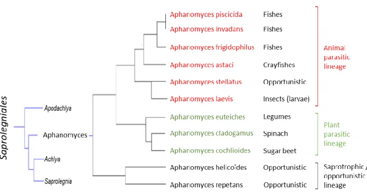
Oomycetes also comprise devastating pathogens and represent the most problematic group of disease-causing organisms in both agriculture and aquaculture (Derevnina et al., 2016b). However, oomycetes stand as notorious plant pathogens with remarkable examples, like Phytophthora infestans causing late blight triggering the Irish potato famine in 1840 (Haas et al., 2009). Phytophthora species are responsible of serious diseases affecting crop yields. The annual economic loss on tomato and potato due to P. infestans was estimated at $ 6.7 billion (Haas et al., 2009). On soybean, for North America, the average annual yield loss caused by P. sojae was estimated at 1.1 million tons, from 2007 to 2014 (Allen et al., 2017). Others notable species are P. palmivora and P. capsici, causing agents of cocoa black pod causing yield losses of 20–30% annually (Adeniyi, 2019). On legumes, Aphanomyces euteiches, the causing agent of root rot, represent one of the major limitations to pea production worldwide (Wu et al., 2018). All those examples highlight the important impact of plant pathogen oomycetes, but some species are also responsible for devastating diseases in natural ecosystems or in aquaculture. For instance, members of the Saprolegnia genus, such as S. parasitica infecting freshwater fish, are involved in the decline of wild salmon populations around the world (Phillips et al., 2008; van West, 2006). Another example of killing agent is Aphanomyces astaci, parasite of fresh-water decapods and causing crayfish plague. Originate from North America, it is now present in Europe and has been nominated among the “100 of the World’s Worst Invasive Alien Species” in the Global Invasive Species Database (GISD).
I-1.2. Defence and Resistance against pathogens
Despite the impact of these diseases and the increase of dedicated research, it is still challenging to control fungal or oomycete attacks. To reach high-quality crops with optimal yields, modern agriculture had resort to intensive use of fungicides that frequently became ineffective due to high adaptation frequency, caused by gene mutations, leading to the emergence of new fungal races (Zhou et al., 2007; Lucas et al., 2015). Same problem occurs
3
with oomycete diseases control, where complex fungicidal mixtures were used for many years, often inefficient due to wide range of intrinsic sensitivities (Judelson and Senthil, 2005) or because resistance evolved against most single-site inhibitors in many oomycete pathogen species (Gisi and Sierotzki, 2015). Some fungicides are also inefficient because the metabolic pathways or key molecules they target in fungi are absent in some oomycete species. For instance, the class of triazole pesticides, representing the largest class of fungicides which target CYP51 enzymes involved in sterol biosynthesis, should not be used against
Phytophthora or Pythium species since these oomycetes do not possess CYP51 enzymes (Tyler
et al., 2006; Sello et al., 2015) and are sterol auxotrophs (Kazan and Gardiner, 2017), leading these fungicides to be inefficient against diseases caused by these pathogens (Gaulin et al., 2010).
Fortunately, chemicals are not the only way to counteract pathogen attacks. Hosts have evolved innate immunity due to their long coevolution with microorganisms. The first layer of plant defence is based on the recognition of essential molecules derived from microorganisms. When the host perceives those molecules that are specific to microorganisms and indispensable for its life cycle and called pathogen-associated molecular patterns (PAMPs), it triggers and activates numerous defence responses. The PAMPs-Triggered Immunity (PTI), comprises a set of responses including callose deposition, oxidative bursts or activation of defence gene (Jones and Dangl, 2006; Nicaise et al., 2009). One of the most famous identified PAMPs is the bacterial flg22, a conserved peptide from the protein flagellin, a major component of the motility organ flagellum, which is recognized by most plants thanks to an LRR Receptor–like Kinase (Gómez-Gómez and Boller, 2000). Numerous eukaryotic PAMPs correspond to cell wall components, like Pep-13, a highly conserved amino acid fragment within the cell wall glycoprotein GP42 from the oomycete Phytophthora sojae (Brunner et al., 2002), or NPP1,a cell-wall protein identified in several Phytophthora species as eliciting immune responses in plants (Fellbrich et al., 2002), or CBM1 from the cell wall protein CBEL from P. parasitica (Gaulin et al., 2006; Larroque et al., 2012). PAMPs are not only proteins as β-Glucans also represent a common fungal and oomycete PAMPs derived from cell wall fractions. Most plants recognize chitin, the main component of fungal cell wall, but also the branched β-Glucans from oomycete cell wall. As example branched
glucan-4
chitosaccharides from the oomycete Aphanomyces euteiches induce defence and calcium signals in Medicago truncatula root cells (Nars et al., 2013).
Faced to PTI, microorganisms evolved and secreted hundreds of pathogenesis-related molecules, named effectors, to modulate immunity and facilitate host colonization. In turn, some hosts evolve to detect specifically those molecules, leading to the Effector-Triggered-Immunity (ETI). Perception is mediated by receptors know as resistance proteins (R) that directly or indirectly recognize some secreted effectors, then called avirulence proteins (AVR). This process was previously named gene-for-gene resistance (R/AVR) (Van Der Biezen and Jones, 1998). This recognition is frequently associated to a hypersensitive response (HR), a localized host cell death to confine the pathogen at the infection site (Jones and Dangl, 2006). Then, major R genes have been used by breeding companies to protect crops against fungal plant diseases (Stuthman et al., 2007). However, the strategy using a single resistance gene often turns out to be inefficient due to adaptation of pathogen populations, which have a high evolutionary potential and rapidly evolve by AVR genes mutations to become virulent. For instance, the fungus Leptosphaeria maculans, the causing agent of the phoma stem canker disease on oilseed rape (Brassica napus), produce new strains by mutations of genesrendering the corresponding major host resistance genes ineffective in only three years (Sprague et al., 2006). Similarly, appearance of new and more virulent pathotypes of the downy mildew (Plasmopara halstedii) in sunflower leads researchers to identify new R genes in order to combine them in varieties carrying a wide range of resistance genes (Pecrix et al., 2019, 2018). Nowadays, major R genes are deployed in cultivars in combination with sustainable disease management practices like precise chemical treatments in order to prolong the use of those resistance genes (Mitrousia et al., 2018).
In addition, another aspect of genetic resistance is related to a quantitative resistance with a partial reduction of symptoms and disease severity (Kamoun et al., 1999). This partial resistance is due to quantitative resistance genes localized in genome area named Quantitative Trait Loci (QTLs). Even if this resistance is frequently less efficient than gene-for-gene resistance like R-AVR gene-for-gene interaction (Hu et al., 2008; Pilet-Nayel et al., 2017), it appeared to be more durable, with a lower selection pressure for pathogens, which limit mutations, and resistance acquired by the expression of different QTLs is more difficult to
5
circumvent (Poland et al., 2009). For instance, no resistant pea, lentil cultivars are available against the oomycete Aphanomyces euteiches that causes the devastating root rot diseases of legumes. However, genome-wide association studies based on the model legume Medicago
truncatula identified one major and several minors QTLs contributing to the tolerance (Badis
et al., 2015; Bonhomme et al., 2014, 2019). Then some Aphanomyces resistance QTLs were identified in pea but fine mapping to identify underlying genes is still challenging (Hamon et al., 2013; Desgroux et al., 2016). In lentil, numerous QTLs were recently detected and some genes are under validation (Ma et al., 2020; Marzougui et al., 2019). Similarly, the oilseed rape (Brassica napus), threatened by stem rot caused by the fungus Sclerotinia sclerotiorum, represents another crop with absence of resistant lines. Currently, breeding for Sclerotinia resistance in B. napus is only based on germplasms with quantitative resistance genes (Wu et al., 2013) and the identification of new QTLs is still an active research (Qasim et al., 2020). In rice, where many R genes were characterized, QTLs were also identified. Then, the resistance in cultivars to the blast fungus Magnaporthe oryzae is controlled by a combination of both major genes and QTLs (Kang et al., 2016).
The use of chemicals to threat animal pathogens invasion triggered also the development of chemical-resistance coupled with negative side-effects on the ecosystem. Then, alternative strategies have to be developed. In aquatic culture for instance, biological control strategies are under development to control zoosporic diseases due to chytrid fungus and oomycetes (Frenken et al., 2019). This include for example a project of immunization against the oomycete Saprolegnia parasitica using a serine protease, the identification of stimuli able to increase the production of natural antifungal peptides produced by the skin of amphibians, or the modification of the pathogen fitness using secondary parasites (Frenken et al., 2019). While those projects are promising, much work still needs to be done to
implement biological-control applications in aquaculture (Frenken et al., 2019). Biocontrol strategies are also currently develop to protect plant against pathogens (Köhl et al., 2019).
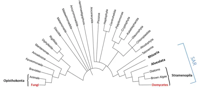
Figure 1: Simplified tree of eukaryotes showing the distant relationship between oomycetes
and fungi.
SAR is an acronym of its constituents: Stramenopiles, Alveolates, and Rhizaria. Oomycetes and Fungi are highlight in red. Adapted from (McGowan and Fitzpatrick, 2020).
6
I-2. Oomycetes, so close and yet so far from Fungi
I-2.1. The false brothers
Oomycetes were originally considered as members of the kingdom of Mycota, in the Opisthokonta clade, with the same classification level as the ascomycetes and basidiomycetes within Fungi (Lévesque, 2011). Even if oomycetes and fungi share common characteristics, as filamentous growth in the form of tip-growing branching hyphae, or similar ecological role and feeding behaviour (Beakes et al., 2012), oomycetes form a phylogenetic lineage distinct from fungi, closely related to brown algae and diatoms among Stramenopiles (Straminipila) (Cavalier-Smith and Chao, 2006). Stramenopiles constitute one of the major eukaryotic clades, branching with Rhizaria and Alveolata within the ‘supergroup’ SAR (Derelle et al., 2016) (Figure
1). Major differences at morphological and molecular levels are now evidenced, as oomycetes
are diploid organisms while fungi are haploid during the majority of their life cycle, disseminate mainly asexually with biflagellated zoospores and are mostly auxotrophic for sterols (with few exception like Aphanomyces euteiches (Gaulin et al., 2010)). Oomycetes develop mostly non-septate hyphae and unlike true fungi, the main structural polysaccharide of the oomycete cell wall is cellulose and not chitin (Judelson, 2017), with few exception like
A. euteiches which contains chitin derivate in the cell wall (Badreddine et al., 2008). Then,
molecular analysis based on combined protein data and rDNA sequences, and more recently, large-scale genome phylogenetic studies confirmed the distant relation of oomycetes from true fungi (Baldauf et al., 2000; Burki, 2014; Derelle et al., 2016).
I-2.2. Lifestyle: oomycete and fungi in front of the mirror
Although oomycetes and fungi are evolutionarily very distantly related, both taxa evolved similar lifestyles. The saprophytic species, which represent a large group of fungi but also numerous oomycetes related to Pythium and some Saprolegnian species (Lamour and Kamoun, 2009), are able to develop on dead host tissue and perform the initial steps in the decomposition macromolecules, like cellulose or lignin on plant cells (Berg et al., 2014). On the other hand, many fungal and oomycete species are obligate biotrophs, meaning that they
7
are unculturable on artificial media, and grow only on living cells. Those species require metabolic active tissues to achieve their life cycle and then are highly adapted to their host, such as downy mildewPlasmopara viticola, which infects grapevine (Vitis vinifera), Albugo candida, the causing agent of white rust on crucifers (Kamoun et al., 2015) or the pathogenic
fungus Blumeria graminis causing powdery mildew on barley (Thomas et al., 2001) and the smut fungus Ustilago maydis on corn (Banuett and Herskowitz, 1996). By contrast, many plant pathogenic oomycetes or fungi, especially species of the genus Phytophthora, or fungi/Ascomycota like Colletotrichum or Magnaporthe, display an intermediate lifestyle called hemibiotrophy, starting infections like biotrophs by establishing a transient biotrophic relationship with the host, then switch to necrotrophic phase later in the disease cycle (Latijnhouwers et al., 2003; Lamour and Kamoun, 2009; Thines, 2018). Finally, necrotrophic pathogens kill host tissues to feed during the colonisation like the fungusBotrytis cinerea, the
causal agent of gray mold, an economically devastating disease, which serves as a model species for plant-necrotroph interactions (Petrasch et al., 2019). Pythium represent the largest genus of necrotrophic oomycetes, but some aquatic pathogenic oomycetes likeLagenisma coscinodisci are also efficient necrotrophic organisms, able to kill marine diatoms in few days
by hijacking the host’s alkaloid metabolism (Vallet et al., 2019). However, the classification in hemibiotrophy or necrotrophy is not always clear, as for the oomycete Aphanomyces
euteiches that causes root rot of legumes (Judelson and Ah-Fong, 2019).
Both oomycetes and fungi share similar traits for host interaction. Dispersal of oomycetes is mediated by water or wind through asexual sporangia or directly by the release of asexual motile zoopores from sporangia (Tyler, 2002). Once oomycete zoospores have reached host surface, they encyst by shedding their flagella and secrete adhesion molecules (Hardham and Shan, 2009; Carzaniga et al., 2001). Asexual spores of fungi as conidies are transported by wind and water, before an adhesion step to the host due to the secretion of adhesion molecules. The germinated cyst produce hyphae able to penetrate inside cell layers, mainly by using a pathogenic structure called appressorium, then vegetative hyphae grow in intercellular space and develop haustoria which penetrated inside host cells (Fawke et al., 2015). Oomycetes and fungi hyphae can also penetrate by natural opening such as stomata
A
8
Figure 2: Phylogeny of the Oomycetes.
(A) Consensus phylogeny of the oomycete class within the greater SAR grouping, including information pertaining to various taxa. Adapted from (McCarthy and Fitzpatrick, 2017). (B) Maximum-likelihood phylogeny of the 65 oomycete species based on the concatenation of 102 conserved BUSCO sequences. The stramenopile
Hyphochytrium catenoides is included as an outgroup. All nodes have 100% bootstrap support except where
indicated. Species are colored according to their order. Phytophthora clades are indicated as designated by Blair, Coffey, Park, Geiser, and Kang (2008) and Pythium clades are as designated by de Cock et al. (2015). From (McGowan and Fitzpatrick, 2020).
(Lucas, 2020). Numerous enzymes to break the host barriers (i.e. cell wall, cutin) are also produced by oomycetes and fungi during the penetration and colonization steps. However, some oomycetes do not form haustoria, like Pythium ultimum or neither appressorium, such as Aphanomyces euteiches (Gaulin et al., 2008). Finally, they complete their life cycle by producing new asexual spores and/or by making their sexual life cycle/stage.
I-2.3. Oomycete phylogeny, still a growing tree
Oomycete phylogeny is still subject to revision due to new genome availability. To date, 65 oomycete species have publicly available genome sequences deposited in databases (McGowan and Fitzpatrick, 2020) and although many species are yet unsampled, the current consensus phylogeny of the oomycetes split them into a basal order and four major “crown” orders: the Peronosporales, Pythiales, Albuginales, and Saprolegniales (Beakes et al., 2014; McCarthy and Fitzpatrick, 2017; McGowan and Fitzpatrick, 2020) (Figure 2a). The basal order of oomycetes includes exclusively marine organisms which are predominantly parasites of seaweeds, nematodes or arthropods (Beakes et al., 2012). The Saprolegniales order is the most basal of the four major crown orders and includes saprophytes and animal parasites, such as the fish pathogen Saprolegnia (Hulvey et al., 2007), and also the plant and animal pathogenic Aphanomyces genus (Gaulin et al., 2007) (Figure 2a and b). The Peronosporales order includes the largest group of terrestrial organisms and represent the best studied order, comprising the well-known oomycete Phytophthora genus. It is also composed by the phytopathogenic Phytopythium genus as well as downy mildew such as Hyaloperonospora,
Plasmopara or Sclerospora genera (Fletcher et al., 2019; McCarthy and Fitzpatrick, 2017;
9
plant pathogens, but also comprises some species able to parasitize fungi and other oomycetes, such as Pythium oligandrum. These mycoparasites are used as new types of biocontrol agents (Benhamou et al., 2012; Faure et al., 2020). The last member of the four crowns are the Albuginales, which include the plant pathogenic Albugo genus (Figure 2a and
b) which causes “white blister rust” on many valuable crop species. Additionally, few species
are members of the Lagenidiales genus, a complex taxon still unclearly defines (Spies et al., 2016). The phylogeny of the 65 available sequenced oomycete species exposed in the recent paper of Mc Gowan and Fitzpatrick is presented in Figure 2b.
I-2.4. Oomycetes, origin(s) and evolution
The history and the evolution of oomycetes are still an ongoing research, partially under debate and regularly update due to the increasing number of available genomes. To date, the consensus hypothesis is that Stramenopiles originate from the enslavement of algal ancestors by a biflagellate photosynthetic organism. Then oomycetes evolved by multiple losses of plastids and genes for phototropism (Cavalier-Smith and Chao, 2006), even if some lineages like some Phytophthora species still conserve photosynthesis-related genes (Tyler et al., 2006).
Molecular clock studies, based on complete genome analyses, estimated the origin of oomycetes around the mid-Palaeozoic Era, up to 430 million years ago (Matari and Blair, 2014). This is supported by the discovery of preserved oomycete structures in the fossil records from the Carboniferous period (approximately 360 to 300 million years ago during the late Paleozoic Era) (Krings et al., 2011). In addition, fossils from the same period evidenced the parasitic lifestyle of oomycetes towards plants (Strullu-Derrien et al., 2011). By the way, parasitism is widespread in oomycetes lineage, reflecting the radical reconfiguration of lifestyle and trophic mechanism from the oomycetes ancestor, changing from carbon fixation by photosynthesis and/or digests microbes inside the cell, to a cellular form that processes complex substrates in the extracellular environment for transportation into the cell
Figure 3: Gene acquisitions from horizontal gene transfer (HGT) in oomycetes.
The three major flows of genes are annotated in ovals. Only three broad group of eukaryotes are drawn schematically on the tree. Arrows indicate the direction of gene transfer. Multiple acquisitions have occurred from different fungal species and bacterial species. Adapted from (Jiang and Tyler, 2012).
Aphanomyces s
10
(Savory et al., 2015; Beakes et al., 2012). It is thought that horizontal gene transfer (HGT), especially from bacteria and fungi, supported this evolution for pathogenicity and virulence genes (Jiang and Tyler, 2012; Savory et al., 2015; McCarthy and Fitzpatrick, 2016) (Figure 3). Notably, HGT had a major impact upon the evolution of the secretomes of oomycetes, which represent all the molecules released out of the cell into the external environment such as hydrolytic enzymes, toxins and effectors (Jiang and Tyler, 2012; Savory et al., 2015).
I-3. Effectors, the infectious Swiss knife
This diversity of lifestyle, coupled with the wide host range and various environment displayed by oomycetes and fungi raised questions about genetic and molecular mechanisms involved in their evolution and rapid adaptation to their hosts and environmental changes (Raffaele and Kamoun, 2012; Judelson, 2012). One answer is that for both oomycetes and fungi, success of infection mainly relies on large repertoires of secreted proteins defining the secretome. The secretome represents all the molecules secreted by the microbe to adapt to new environmental resources or changes in his close environment (McCotter et al., 2016). The estimated size of fungi / oomycete secretome range from 4–15% of the total gene number (Girard et al., 2013; Pellegrin et al., 2015), with a highly variable composition closely related to the niche the microbes reside in (Soanes et al., 2008). This comprises a wide range of proteases, lipases, enzymes and small-secreted proteins (SSPs) to achieve functions such as nutrient acquisition, detoxification or cell wall manufacture (Feldman et al., 2020; Pellegrin et al., 2015). Among secreted proteins, some affect host physiology to neutralize plant defences and promote microorganism colonisation, the so-called effectors. Effectors include mainly proteins, secondary metabolites but also nucleic acids (e.g. small RNAs) (Wang et al., 2019). Therefore, secreted effectors evolved quickly, have different function, localization and may affect various host processes to enhance infection.
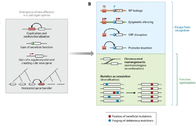
Figure 4: The evolutionary birth and death of effectors.
(A) New effectors can emerge through gene duplication or the gain of a secretion function. Effector genes may also evolve de novo from noncoding sequences through the gain of a regulatory element or be acquired horizontally from a different pathogen species. (B) Effector genes can undergo rapid sequence evolution upon recognition of the encoded effector by the host. The major mechanism leading to the loss of an effector gene is the presence and activity of nearby transposable elements (TEs). The effects of the transposable elements can include repeat-induced point (RIP) mutations, epigenetic silencing or the disruption of the gene sequence. Escape from recognition can also be mediated by chromosomal rearrangements or the fixation of beneficial mutations. Rearrangements and selection for beneficial mutations are also major routes for effectors to optimize their function. Abbreviations: ORF, open reading frame; P, promoter regions. From (Sánchez-Vallet et al., 2018).
A
11
I-3.1. Effector genes evolution
Effectors show rapid evolution within a given genome as a result of co-evolution with their hosts and are often associated with transfers to unrelated host (Dong et al., 2015; Raffaele et al., 2010). For instance, protease inhibitors produced by two sisters Phytophthora species evolved to target plant proteases of their respective unrelated hosts, linking effector specialization and host diversification (Dong et al., 2014). This close link between effectors and host adaptation was also revealed by comparative fungal genomic studies showing evidences of rapid evolution of effectors in related pathogens with different host ranges (Meerupati et al., 2013; Condon et al., 2013; O’Connell et al., 2012; Richards et al., 2019). Evasion of host recognition and effector functional optimization is achieved by sequence modification, gene deletion, modulation of effector genes expression and the gain of new effectors by horizontal gene transfer (Figure 4a) (Lo Presti et al., 2015b).
Some HGT have been evidenced, like for the transfer of ToxA between three unrelated wheat pathogens, leading to isolates that are more virulent (Friesen et al., 2006). Another example was reported in the cotton fungal pathogenVerticillium dahlia where lineage-specific
region that might have originated from Fusarium oxysporum increased virulence on cotton but not on other hosts (Chen et al., 2018). Even if the main mechanisms leading to HGT are poorly understood, it seems that necrotrophic pathogens are far more susceptible to the acquisition of effector genes, particularly with host-specific toxin coding genes (Sánchez-Vallet et al., 2018). In oomycetes, gene acquisition by HGT was also evidenced for a cutinase gene from bacteria to Phytophthora species (Belbahri et al., 2008), and more extensively reported between fungi and oomycetes, at least in Peronosporales (Richards et al., 2011). In addition, changes in secretome of Saprolegniales oomycetes due to HGT from bacterial and fungal donor lineages were evidenced (Misner et al., 2014).
In addition to HGT, other genetic events occurred to evolve effector genes. For instance, gene duplications combined with mutations were shown to generate new effector genes in the smut fungus Ustilago maydis (Dutheil et al., 2016) (Figure 4a).
Transposable elements (TEs) were evidenced to play a major role in gene duplication and are significantly associated in the formation of virulence gene clusters through non-homologous recombination (Dutheil et al., 2016). The last generation of sequencing strategies
12
greatly increased the quality of genome assemblies and gave new insight into effector evolution and genome organization. Firstly, it revealed that TEs content was often underestimated. For example, the last version of Colletotrichum higginsianum genome contains 7% TEs whereas it was estimated to only 1.2% in the first assembly (Dallery et al., 2017). Then, it is now clear that many effector genes are not randomly distributed across the genomes and are associated with TEs and repetitive sequences in specific genome compartments. These results have led to the “two-speed genome” model in which some pathogen genomes have a bipartite architecture with essential genes in the core genome, protected from deleterious mutations, and the accessory genome where effector genes take place in a rapid evolutionary compartments (Raffaele and Kamoun, 2012; Croll and McDonald, 2012).
Rapid host adaptation can lead to effector recognition that triggers host defence. Hence, adaptive pressure on effector gene sequence can force mutations in order to modify, modulate or delete a given effector to escape host recognition. The most efficient mechanism leading to the loss of an effector gene is related to the activity of TEs. TEs can drive multiple effects on gene sequence, from gene disruption to repeat-induced point (RIP) mutations (Figure 4b). Adaptive loss of function was reported in the fungal pathogen of wheat
Zymoseptoria tritici, where gene losses affected more than 10% of all genes in the genome,
including both effectors and genes with conserved functions such as secondary metabolite gene clusters (Hartmann and Croll, 2017).
In addition to TEs activity, two types of mutations are known to modulate effector genes evolution (Figure 4b) (reviewed in (Sánchez-Vallet et al., 2018)). The first type of mutation consists in substitutions, insertions or deletions that change the protein properties of a given effector. The second type of mutation concerns neutral mutations with weak but cumulative effects. Fixation of beneficial mutations leads to optimization of the effector function and can infer the past selective history at the effector locus (Sánchez-Vallet et al., 2018).
Transcriptional silencing of an effector gene is another mechanism involved to escape host recognition, which preserve the effector sequence (Gijzen et al., 2014; Whisson et al., 2012). This was observed for the Phytophthora sojae effector gene Avr3a that is recognized in soybean plants carrying the resistance gene Rps3a. Silenced Avr3a alleles were transmitted
13
and persisted over multiple generations suggesting that transgenerational gene silencing at this locus mediated the gain of virulence phenotype (Qutob et al., 2013).
I-3.2. Apoplastic and intracellular effectors
During eukaryotic filamentous pathogens-plant interactions, two types of effectors can be distinguished depending on their localization. Apoplastic effectors proteins (AEPs) stay in the plant extracellular space (i.e. apoplast) while intracellular effectors proteins traffic into the host cell in various compartments.
I-3.2 a. Apoplastic effectors: in front of the Wall
The apoplast is a hostile environment notably due to secreted basal defence compounds like proteases, secondary metabolites or hydrolytic enzymes like chitinases (Doehlemann and Hemetsberger, 2013; Jashni et al., 2015). The release of PAMPs in the apoplast due to activity of plant chitinases or β-glucanases that disrupt microbial cell wall integrity leads to their perception through cell surface-localized immune receptors, such as Lysin motif (LysM)-containing proteins, which activates plant immune system (Cook et al., 2015; Liu et al., 2012). Thus, to counteract this first recognition process, numerous fungal and some oomycete AEPs have been characterized to evade glycan-triggered immunity or to protect cell wall microorganism from degradation (reviewed in (Rocafort et al., 2020)).
Phytophthora spp. for instance secrete glucanase inhibitor proteins (GIPs) to inhibit the
degradation of pathogen β-1,3/1,6-glucans and the release of defence-eliciting oligosaccharides by host endoglucanases (Rose, 2002; Damasceno et al., 2008). The tomato fungal pathogen Cladosporium fulvum secretes two characterized AEPs, the chitin-binding effector protein Avr4, which protects fungal hyphae against hydrolysis by plant chitinases (van den Burg et al., 2006), and Ecp6, an effector which uses LysM domains that competitively sequesters chitin oligomers from host immune receptors leading to the perturbation of chitin-triggered host immunity (Sánchez-Vallet et al., 2013). Other LysM effectors have been shown to contribute to virulence through chitin binding in other plant pathogenic fungi like
Magnaporthe oryzae, Colletotrichum higginsianum and Verticillium dahlia (Kombrink et al.,
2017; Mentlak et al., 2012; Takahara et al., 2016). Interestingly, AEPs with similar roles to both Avr4 and Ecp6 have been described in the fungal wheat pathogen Zymoseptoria tritici
14
(Marshall et al., 2011; Sánchez-Vallet et al., 2020) but also in the mutualistic fungus
Trichoderma atroviride and in the arbuscular mycorrhizal fungus Rhizophagus irregularis
(Zeng et al., 2020; Romero-Contreras et al., 2019). This indicates that both pathogenic and mutualistic microbes use AEPs to evade glycan-triggered immunity.
Thus, many characterized AEPs act to supress this glycan-triggered immunity (Rocafort et al., 2020) but other families of AEPs have been described. One large group of apoplastic effectors commonly found in fungi and oomycetes are cell wall degrading enzymes (CWDEs), which play a major role in pathogenicity, contributing to plant cell wall degradation. Thus, this family includes hundreds genes coding for enzymes such as cellulases, hemicellulases, pectinases, β-1,3-glucanases, glyceraldehyde hydrolases, carbohydrate binding molecules and other proteases able to degrade glycoproteins. The aim is to reduce the complexity of the cell wall structure to facilitate entry and colonization of the host. In animal pathogen interaction, those enzymes are absent and replaced by other specific enzymes. For instance, the plant pathogen oomycete A. euteiches possesses a large repertoire of CWDEs coding genes that target plant cell wall polysaccharides, absent in Aphanomyces astaci, the causing agent of crayfish plague. In turn, A. astaci shows an expansion of protease genes predicted to target chitin, the main component of the crayfish shell ((Gaulin et al., 2018) and see CHAPTER IV). Recently, it has been shown that a CWDE effector was protected by another AEP, acting as a decoy. Indeed, Phytophthora sojae displays an apoplastic effector, called PsXLP1, able to promote infection by protecting PsXEG1, another effector with xyloglucanase activity essential for full virulence but targeted for inhibition byGmGIP1, a soybean protein. Then, PsXLP1 binds to GmGIP1 and functions as a decoy to protect PsXEG1 from the inhibitory action of GmGIP1 (Ma et al., 2017).
Some AEPs are considered as toxins, called necrosis-inducing proteins (NLPs), able to cause cell death. NLPs were first identified from culture filtrate of Fusarium oxysporum but have been isolated in oomycetes, fungi and bacteria, and have the ability to induce cell death and ethylene accumulation in plants (Gijzen and Nürnberger, 2006; Cobos et al., 2019). The structure of NLPs is remarkably conserved among long phylogenetic distance, from bacteria to oomycetes (Feng et al., 2014; Ottmann et al., 2009). However, the role of NLPs during infection is unclear. When studies reported evidences that NLPs function as virulence factors that increase pathogen growth in host plants or extend the host range (Veit et al., 2001;
15
Mattinen et al., 2004; Pemberton et al., 2005), others revealed that mutations in some NLP genes from various fungi like Fusarium oxysporum or Botrytis cinerea do not reduce their virulence (Cuesta Arenas et al., 2010; Bailey et al., 2002). In addition, most identified NLPs are perceived by the host as PAMPs leading to the stimulation of PTI, such as NLPs from
Phytophthora species in Arabidopsis (Qutob et al., 2006, 2002), or from Pythium in various
dicotyledonous plants (Veit et al., 2001).
In oomycetes, particularly in Phytophthora and Pythium species, elicitins represent another family of small AEPs and display similar characteristics with NLPs. Elicitins are structurally conserved and induce a sustained oxidative burst that leads to hypersensitive response (HR) cell death in most case (Derevnina et al., 2016). Plants from different botanical families perceived elicitins as MAMPs, which induce activation of defence through MAMP-triggered immunity (MTI) (Derevnina et al., 2016). Then, like NLPs, the role of elicitins is still unclear. Since elicitins bind sterol and other lipids (Osman et al., 2001) and given the fact that most oomycetes including Phytophthora are sterol auxotrophs, elicitins are proposed to act as sterol-carrier proteins (Mikes et al., 1998). As sterols and fatty acids stimulate sexual reproduction and oospore production in Phytophthora, elicitins could contribute to the appearance of more virulent strains (Chepsergon et al., 2020).
Finally it is anticipated that some apoplastic effectors, especially cyclic peptides, could play a role in self-defence against competitor antimicrobial compounds, or in manipulating the apoplastic microbiome to promote host colonization (Snelders et al., 2018; Rocafort et al., 2020).
I-3.2 b. Intracellular effectors: Destroy from within
The second class of effectors are secreted proteins translocated to the host cytoplasm or intracellular compartments. In oomycetes, the first (and the largest) family of cytoplasmic effectors, named RxLR effectors, were identified by comparative sequence analysis of predicted secreted avirulence proteins from several oomycete species, leading to the identification of a conserved amino acid motif, namely the RxLR-EER motif (Rehmany et al., 2005). Thus more than 350 RxLRs effectors characterized by their R (arginine) – X (any amino
16
acid) – L (Lysine) – R (arginine) motif after signal peptide sequence, were predicted in
Phytophthora species (Tyler et al., 2006). Then presence of RxLR genes was evidenced in
numerous Phytophthora species, where several hundred putative RxLRs were predicted (Haas et al., 2009; Jiang and Tyler, 2012), but only one in Saprolegniales species (Trusch et al., 2018). Finally, RxLR and RxLR-like effectors may also be present in fungi (Kale and Tyler, 2011) as in the endophytic fungus Piriformospora indica (named later Serendipita indica) in which 5
proteins with a degenerated RxLR motif were predicted to be secreted but none of them were found to be up-regulated during colonization of barley roots (Zuccaro et al., 2011).
RxLR proteins contain a conserved N-terminal motif in addition to a predicted signal peptide and a highly variable C-terminal part that allows biological function (Birch et al., 2006). It was proposed that the RxLR motif acts as a signal for host delivery (Whisson et al., 2007; Dou et al., 2008). In addition, RxLR effectors have been reported to translocate into host cells in the absence of the pathogen, after binding of the RxLR motif to lipids via phospholipid-mediated endocytosis (Kale and Tyler, 2011; Kale et al., 2010). However, studies made on other RxLR effectors could not observed this entry mechanism and finally exclude the phospholipid binding as a general host entry mechanism (Gan et al., 2010; Yaeno and Shirasu, 2013; Wawra et al., 2012). Then, pathogen-independent translocation of effectors into plant cells is controversially discussed and the entry mechanism of effectors is still unclear (Wawra et al., 2013). A recent study demonstrated that the RxLR motif of the Phytophthora infestans effector AVR3a was cleaved before secretion (Wawra et al., 2017). Even more recently, in the oomycete Saprolegnia parasitica, it was reported that the uptake process of the RxLR protein SpHtp3 is guided by a gp96-like receptor via its C-terminal region, but not by the N-terminal RxLR motif (Trusch et al., 2018). After translocation into host cell, a major part of RxLR effectors target nucleus, but some have a nucleo-cytoplasmic localization when others accumulate in membranes (Sperschneider et al., 2017), as described for the oomycete
Hyaloperonospora arabidopsidis (Caillaud et al., 2012).
The identification of RxLR effectors with conserved motif and the availability of
Phytophthora infestans genome lead to the discovery of another family of intracellular
effectors named CRNs, for CRinkling and Necrosis effectors. CRNs were first identified in the plant pathogenic oomycete Phytophthora infestans. To identify pathogen-secreted proteins potentially involved in the manipulation of host processes, a large screen of cDNA coding for
17
secreted proteins were expressed in N. benthamiana and tomato leaves. Two of which, named CRN1 and CRN2, presenting similarities at the sequence level were found to cause a CRinkling and Necrosis (CRN) phenotype when expressed in plant tissue (Torto et al., 2003). Like RxLR proteins, CRNs present a modular architecture with a conserved N-ter signal characterized by LxLFLAK-derived amino acid sequence (with possible variation) followed by a highly variable C-ter domain (Schornack et al., 2010). With the increasing number of available genomes, many studies performed on other oomycetes revealed that, in contrast to the RxLR protein family, CRN coding genes are widespread in oomycete lineage, and were found in all plant pathogenic oomycetes sequenced to date including Peronosporales (Haas et al., 2009; Tyler et al., 2006; Baxter et al., 2010), Albuginales (Kemen et al., 2011), Pythiales (Adhikari et al., 2013; Lévesque et al., 2010) and Saprolegniales (Gaulin et al., 2008). Some CRN-like coding genes were also predicted in the animal pathogen Aphanomyces astaci, the causing agent of the Crayfish plague ((Gaulin et al., 2018) and see CHAPTER IV). The identification of hundreds CRNs genes in Aphanomyces species, which are early divergent species among the “crown” oomycetes, suggests that CRNs are an ancient class of conserved oomycete effector proteins (Schornack et al., 2010).
CRNs have a modular architecture with two distinct protein regions. The N-terminus domains, composed around 130 amino acids (aa), contains a conserved LxLFLAK or LxLFLAK-derivate motifs (within the first 60 aa) and more diversified DWL domains. Another highly conserved HVLVxxP motif marks the end of the N-terminal region (Figure 5a). This N-terminal part is presumed to specify the secretion and the translocation of the protein into the host. The functionality of CRNs N-termini domain was initially tested via an infection-translocation assay ((Schornack et al., 2010) and see P29-30 of this manuscript for more details). In this study, three CRN N-termini of P. infestans (CRN2, CRN8 and CRN16) and one A. euteiches (AeCRN5) were fused to C-terminal domain of the P. infestans Avr3a RxLR protein, and introduced in Phytophthora capsici. Those strains were used to infect transgenic N.
Figure 5: CRNs structure analysis (adapted from (Amaro et al., 2017)) .
(A) Initially CRN N-termini contain a conserved structure featuring: a signal peptide for secretion; an LXLFLAK domain containing the respective LXLFLAK motif connected with translocation; and a DWL domain that ends in a conserved HVLVVVP motif that marks the end of CRN N-terminus. This site is predicted as a hot spot for recombination events. In contrast, CRN C-termini were shown to exhibit a large variety of domain structures (not depicted here). (B) Zhang et al. (2016) redefined CRN structure. CRN N-termini (renamed header domains) from the two Phytophthora species analyzed (P. infestans and P. sojae) all feature an Ubiquitin like (Ubl) domain that is thought to be responsible for secretion and translocation into the host cell. CRN C-termini (also named CR-toxin domains) feature distinct domain architectures, having enzymatic origins. The majority of Phytophthora CRN C-termini contained the depicted domain structure (NTPase + HTH + REase). (C) Summary of domain architectures predicted to occur in Phytophthora (from Zhang et al., 2016). The number of CRN proteins with each given domain architecture/composition are indicated between brackets. Figure from (Amaro et al., 2017). A
B
18
As chimera proteins induced avirulence by the recognition of the R3a protein, it was concluded that those N-termini domains allow secretion and translocation of C-termini CRN proteins into host cells. Even more, mutations in the LxLFLAK conserved motif indicate that these motifs are necessary for translocation function. Importantly, N-termini domains of AeCRN5 and CRN16 were demonstrated to be functional, even if no signal peptide were predicted in the first 30 amino acids. These results demonstrate that despite the absence of signal peptide, which was reported for numerous CRNs (Stam et al., 2013b; Voß et al., 2018; Amaro et al., 2017; Adhikari et al., 2013; Gaulin et al., 2018), an unpredictable secretion signal is present in this region and ensure secretion of CRNs in oomycetes (Schornack et al., 2010).
A recent study proposed to reconsider the requirement of LxLFLAK motifs in CRN translocation and challenged the classification of CRNs proteins as members of a larger order of Eukaryotic effectors (Zhang et al., 2016). In this paper, authors performed multiple in silico analyses using a combination of sequence alignments and structure prediction programs, coupled to comparative genomics to assess CRN occurrence across the Eukaryote taxon. Results of those analyses ruled out the presence of signal peptides and indicate that the proteins containing the LxLFLAK motif but also numerous proteins lacking this motif were predicted to have an ubiquitin-like structure, similar to those found in the N-terminal region of SSK1 (mitogen activated protein kinase) / Mcs4 (mitotic catastrophe suppressor 4) signalling proteins in fungi. The LxLFLAK motif was located in strand 2 and 3 of this ubiquitin-like domain (Ubl), suggesting that structural features rather than sequence conservation underpin CRN translocation (Zhang et al., 2016). Authors then renamed the N-terminal region as a Header Domain (Figure 5b). SSK1 orthologs play important roles in stress responses in various true fungi, such as oxidative and osmotic shock, and in some cases in a phosphorylation-dependent manner, employing an interaction between their N-terminal domains and a MAPKKK heteromer (Morigasaki and Shiozaki, 2013; Yu et al., 2016). From this, the authors suggest that CRN Ubl N-terminal domains could facilitate translocation inside the host by analogous mechanisms (Zhang et al., 2016) but functional studies are require to support this new concept. Similarly, the classification of CRN C-termini was challenged by the study of Zhang and colleagues (Zhang et al., 2016). Initially 36 different conserved subdomains that can assemble in different combinations defining C-terminal subfamilies were identified in P. infestans (Haas et al., 2009). Then it has been proposed that the highly variable
19
organisation of C-termini was the result of recombination events between subdomains. Following studies indicated that among oomycetes, most of these subfamilies are present and that specific subfamilies can also be found. For example, in P. capsici, 30 of these subfamilies are present while 7 new subfamilies appear specific to this species (Stam et al., 2013b). Similarly, in A. euteiches, 160 CRN gene models have been described, among which 12 C-termini domains are novel subdomains (Gaulin et al., 2008). In the study of Zhang and colleagues, even if high levels of diversity are still present, the re-classification of CRN C-terminal domains led to a limited set of domain configurations that were found to be prevalent. Numerous CRN C-termini are related to enzymatic activities (Figure 5c). For instance, one-fourth of all CRN C-termini analysed contains a P-loop NTPase domain, coupled with a nuclease domain of the restriction endonuclease (REase) superfamily. In the same way, approximately one-sixth of the C-termini domains harbour REase superfamily domain combined with protein kinase domain. Then, those NTPase or Kinase domains, but also other domains like HNH domain, could regulate REase activity or affinity toward nucleic acids (Zhang et al., 2016), suggesting that targeting nucleic acids could be a shared feature among CRNs. This hypothesis is supported by two studies that report the DNA binding activity of CRN proteins in addition to the nuclear localization of numerous CRNs (Song et al., 2015; Ramirez-Garcés et al., 2016; Amaro et al., 2017).
CRN-like proteins were identified outside oomycete lineage, in the fungal pathogen
Batrachochytrium dendrobatidis (Bd) and in the fungal symbiont Rhizophagus irregularis (Sun
et al., 2011; Lin et al., 2014).The presence of CRN-like proteins in such different organisms suggests a HGT event or the presence CRN genes in early eukaryote progenitors (Sun et al., 2011; Lin et al., 2014). Bd causes chitridiomycosis and is responsible for the declines of amphibian population worldwide (Fisher et al., 2012; Scheele et al., 2019). Genome analyses of Bd strains revealed 84 CRN-like sequences presenting up to 46.5 % of similarity to CRNs of
P. infestans, with a conserved modular architecture, comprising both LxLFLAK-derived signal
and C-ter domains organization (Sun et al., 2011; Joneson et al., 2011). Comparative analyses of Bd with its closest relative, the non-pathogenic chytrid fungus Homolaphlyctis polyrhiza, highlight the absence of CRN-like sequences, suggesting a link between pathogenic processes and CRN effectors (Joneson et al., 2011). However, 42 genes were predicted with high sequence similarity and canonical amino acid motifs of CRNs in the arbuscular
20
endomycorrhizal (AM) fungus Rhizophagus irregularis (Lin et al., 2014). The functional characterization of the RiCRN1 protein evidenced the biological role of this Crinkler in the establishment of the symbiosis, especially on the initiation of arbuscule development (Voß et al., 2018). Furthermore, the study of Zhang et al. revealed that CRN effectors are also present in free-living eukaryotes and land plants that are not known to have a pathogenic lifestyle, indicating that CRNs are widespread in Eukaryotes (Zhang et al., 2016). Thus it was proposed that CRN proteins could be firstly involved in inter-organismal conflicts, after which in some host-pathogen interactions, these proteins were co-opted as effectors (Zhang et al., 2016; Amaro et al., 2017).
In contrast to oomycetes, intracellular fungal effector lacks a conserved sequence that facilitate their prediction; therefore, their identification relies on small-secreted proteins (SSPs). Typical fungal pathogens possess several hundreds and sometimes more than a thousand of these SSP effectors. SSPs are defined as proteins of less than 300 amino acids with a signal peptide and devoid of any functional domains. Many SSPs are coded by orphan genes, lacking known domains or similarities to known sequences, and usually are cysteine-rich proteins. Large repertoires of SSPs have been evidenced upon genome annotation of fungi interacting with plants (Duplessis et al., 2011; O’Connell et al., 2012), nematodes (Meerupati et al., 2013), insects (Hu et al., 2014) and human (Vivek-Ananth et al., 2018). Thus, SSPs were initially described as virulence effectors produced by pathogens, but finally large repertoire of SSPs were also predicted in mycorrhizal fungi (Martin and Selosse, 2008; Kamel et al., 2017) and their role in the establishment of symbiosis evidenced, like MiSSP7 from the ectomycorrhizal fungus Laccaria bicolor (Plett et al., 2011, 2014). SSPs were also reported in bacteria in the plant pathogen Pseudomonas syringae (Shindo et al., 2016), and finally within the scope of this PhD study, SSPs were described in oomycete genomes ((Gaulin et al., 2018) and see
CHAPTER IV).
Within the fungal kingdom, the proportion of SSPs ranges from 40 to 60% of the secretome across all lifestyles and phylogenetic groups (Feldman et al., 2020; Pellegrin et al., 2015; Kim et al., 2016). However, it seems that this proportion may vary depending on the lifestyle. For instance obligate biotrophs likely encode more and diverse effector-like SSPs to suppress host defence compared to necrotrophs, which generally use cell wall degrading enzymes and phytotoxins to kill hosts (Kim et al., 2016). Comparative analyses of secretomes
21
also identified shared or lifestyle-specific SSPs between saprotrophic and Ectomycorrhizal (ECM) fungi, indicating that presence of SSPs is not limited to fungi interacting with living plants (Pellegrin et al., 2015).
Some sequence similarity leads to the classification of SSPs in superfamily as in
Blumeria graminis. Sequence analyses of candidate secreted effector proteins (CSEPs) of the
powdery mildew revealed that 25% of those CSEPs, highly expressed in haustoria, contain features resembling catalytically inactive RNases. Thus, they are part of the superfamily of RnAse‐Like Proteins associated with Haustoria, the so‐called ‘RALPH’ effectors (Pedersen et al., 2012). Recently, a new family of small fungal effectors, that has particularly expanded in the fungus Magnaporthe oryzae, was described (de Guillen et al., 2015) and was called the MAX family for Magnaporthe Avrs and ToxB-like effectors. Despite sharing little protein sequence similarity, MAX effectors are characterized by a conserved structure. Those effectors have different shapes and surface properties suggesting that they target different host processes.
How SSPs are addressed within the host cytoplasm is still an opening question, but when transiently express in planta, numerous SSPs localized in nucleus or nucleolus, but some can be found in mitochondria or chloroplasts (Petre et al., 2015). Recently, plant Golgi, peroxisomes and microtubules were also reported as targets for fungal SSPs (Robin et al., 2018).
I-3.3. Intracellular effectors targets: hit the defence key players
To promote microbial colonization, effectors could favour microbial growth by manipulating plant defences and/or by enhancing invader nutrition. Thus, functions and targets of intracellular effectors are diverse and range from altering plant cellular metabolic pathways, signalling cascades, RNA silencing, transcription, trafficking and interfering with DNA machinery.
One of the primary mechanism targeted by intracellular effectors is to supress the host response by targeting crucial compounds. For instance, the two essential defence phytohormones salicylic acid (SA) and jasmonic acid (JA) that act antagonistically in response to pathogen infection (Niki et al., 1998) can be modulated by effectors. Cmu1, from the maize pathogenic fungus Ustilago maydis, is secreted into the host cell and acts as a chorismate
22
mutase to reduce SA levels during infection (Djamei et al., 2011). Similarly, effectors Pslsc1 from Phytophthora sojae and Vdlsc1 from Verticillium dahliae reduce the amount of SA by hydrolizing isochorismate, a precursor to SA, when expressed within plant cells (Liu et al., 2014). HaRxLR44 from the oomycete Hyaloperonospora arabidopsidis degrades Mediator subunit 19a (MED19A) to alter the balance of JA and SA, which affects defence-related transcriptional changes (Caillaud et al., 2013). Other plant metabolites can also be modulated by effectors. The SSP Tin2, from Ustilago maydis, prevents the degradation of the maize protein kinase ZmTTK1, which is responsible for the activation of genes involved in anthocyanin biosynthesis. This overproduction of anthocyanins turns to plant defence detriment, sinceanthocyanin biosynthesis competes with tissue lignification, promoting the pathogen to reach vascular tissue due to a lower content of lignin (Tanaka et al., 2014). Additionally, the RxLR PSE1 from Phytophthora parasitica interferes with auxin physiology through the redistribution of auxin efflux carrier proteins, modulating auxin content which enhances pathogen infection (Evangelisti et al., 2013).
Another key point of plant defence response is the reactive oxygen species (ROS) production, which plays a role in MTI, phytoalexin production, callose deposition and systemic acquired resistance (SAR) (O’Brien et al., 2012). Crinkler PsCRN63 from Phytophthora sojae interacts and destabilizes plant catalases to promote plant cell death, whilst PsCRN115 inhibits the catalases degradation to maintain the proper H202 levels and block plant cell death (Zhang et al., 2015).
Plant defence responses are also dependent on signalling pathways like MAPK cascades, which are essential for both MTI and ETI. Then it is not surprising to find effectors that evolved to block these pathways. For instance, the RxLR PexRD2 from P. infestans interacts with the kinase domain of MAPKKKε to interrupt plant immunity-related signalling (King et al., 2014).
Some effectors play a role in the disruption of various trafficking pathways that lead to the secretion of defence proteins. In Blumeria graminis, BEC4 Interacts with ARF-GAP protein, a key player of membrane vesicle trafficking in eukaryotic cells (Schmidt et al., 2014). Pi03192, an RxLR from P. infestans is able to prevent the re-localisation of two plant NAC transcription factors from the endoplasmic reticulum to the nucleus (McLellan et al., 2013). To ended, still in P. infestans, PexRD54 stimulate autophagosome formation through binding to the autophagy protein ATG8CL (Dagdas et al., 2016). This activation of autophagy suggests that