REPORTS
Serum ct-Tocopherol
Concentration in Relation to
Subsequent Colorectal Cancer:
Pooled Data From Five Cohorts
Matthew P. Longnecker*
Jose-Maria Martin-Moreno, Paul
Knekt, Abraham M. Y. Nomura,
Susan E. Schober, Hannes B.
Stdhelin, Nicholas J. Wald, K.
Fred Gey, Walter C. Willett
Background: Vitamin E is an
antioxi-dant that inhibits mutagenesis and cell transformation. Previous findings in five prospective epidemiologic studies suggested that the level of serum oc-tocopherol, the predominant form of vitamin E in the blood, was lower in subjects who subsequently developed colorectal cancer than in control sub-jects. However, the difference was neither obvious nor statistically sig-nificant in any one of these five studies.
Purpose: To evaluate in greater detail
the association between serum a-to-copherol concentration and risk of colorectal cancer, we pooled and analyzed the original data from the five studies. Our analyses were designed to (a) test the hypothesis with greater statistical power, (b) examine the as-sociation after adjustment for serum cholesterol levels, and (c) evaluate the association after uniform exclusion of cases diagnosed shortly after blood specimens were drawn. Methods: Data for individual subjects were analyzed. To make the design of the component investigations uniformly nested case-control studies with individual match-ing, we matched controls to cases in
two of the cohorts. Subjects were categorized according to study-specific quartile of serum a-tocopherol level within the study. The pooled analysis included 289 cases of colorectal cancer and 1267 matched controls. Results: For cancers of the colon and rectum combined, the matched odds ratio (OR) for the highest quartile of serum a-tocopherol concentration compared with the lowest was 0.6 (95% con-fidence interval [CI] = 0.4-1.0). Adjust-ment for serum cholesterol level attenuated the OR to 0.7 (95% CI = 0.4-1.1). Conclusion: The results sug-gest that serum a-tocopherol concen-tration may be inversely related to risk of colorectal cancer. It is unclear whether an association exists, however, because the association between serum a-tocopherol level and decreased risk of colorectal cancer was modest, the CIs were wide, and, overall, the tests for trend in effect were not significant.
Implications: Larger observational studies
with concurrent dietary data are needed to determine whether vitamin E has a modest but potentially important protective effect against colorectal can-cer. [J Natl Cancer Inst 84:430-435, 1992]
Vitamin E inhibits mutagenesis and cell transformation, probably by blocking peroxidation of lipids in membranes (/). In some animal models, vitamin E is anti-carcinogenic, but the study results are not entirely consistent (1*2).
Although few epidemiologic studies have examined vitamin E intake in rela-tion to risk of cancer (3), serum a-to-copherol concentration, which reflects in-take of vitamin E, has been examined in relation to risk of cancer in several prospective studies (4-10). In reviewing these studies, we noted results suggesting that the level of serum a-tocopherol was lower in subjects who subsequently
de-veloped colorectal cancer, compared with that in control subjects. However, the dif-ference in a-tocopherol level was neither obvious nor statistically significant in any one of the studies. While the prospective design in these observational studies was methodologically strong, the statistical power in any given study was somewhat limited. Further, the interpretation of the findings in some investigations was un-clear because the inverse association might have been due to confounding by serum cholesterol level, which is corre-lated with serum a-tocopherol, or to an effect of preclinical disease on the con-centration of a-tocopherol in the blood.
To evaluate the association between serum a-tocopherol concentration and
Received July 17, 1991; revised November 4, 1991; accepted December 13, 1991.
Supported in part by the American Cancer Society and the Medical Foundation, Inc. (M. P. Longneck-er).
M. P. Longnecker, Department of Epidemiology, University of California, Los Angeles, School of Public Health.
J.-M. Martin-Moreno, Escuela Nacional de Sanidad, Madrid, Spain.
P. Knekt, Research Institute for Social Security, The Social Insurance Institution, Helsinki, Finland.
A. M. Y. Nomura, Japan-Hawaii Cancer Study, Kuakini Medical Center, Honolulu, Hawaii.
S. E. Schober, Division of Epidemiology and Prevention Research, National Institute on Drug Abuse, Rockville, Md.
H. B. Stahelin, Medizinisch-geriatrische Klinik, Kantonsspital Basel, Basel, Switzerland.
N. J. Wald, Department of Environmental and Preventive Medicine, Medical College of St. Bar-tholomew. London, England.
K. F. Gey, Vitamin Unit, Institute of Biochemistry and Molecular Biology, University of Berne, Switzerland.
W. C. Willett, Departments of Epidemiology and Nutrition, Harvard School of Public Health, and the Channing Laboratory, Department of Medicine, Harvard Medical School and Brigham and Women's Hospital. Boston. Mass.
We thank Fran Fullerton, Roger Newson, Chris Frost, and Jean de Traversay. We especially thank the investigators who collected the data that were analyzed.
Correspondence to: Matthew P. Longnecker.
M.D., Sc.D.. Department of Epidemiology, UCLA School of Public Health. 10833 Le Come Ave., Los Angeles, CA 90024-1772.
risk of colorectal cancer in greater detail, we pooled and analyzed the original data from the five prospective, nested case-control studies of this relationship. Analysis of the combined data allowed us to (a) test the hypothesis with greater statistical power, (b) examine the associa-tion after adjustment for serum choles-terol levels, and (c) evaluate the associa-tion after uniform exclusion of cases diagnosed shortly after blood specimens were drawn.
Subjects and Methods
By 1988, authors of five prospective studies had presented data on serum oc-tocopherol concentration in relation to risk of colorectal cancer (5-9); no new studies of this type have been published since.
In the two other prospective studies of serum a-tocopherol level in relation to subsequent cancer, the colorectal cancer cases were grouped either with all other cancers (4) or with other gastrointestinal cancers (10). We estimated that there were fewer than 10 cases of colorectal cancer in each of these studies and ex-cluded them because little additional in-formation would have been obtained. In these two studies, serum oc-tocopherol levels for cases were slightly lower than those for controls.
Table 1 describes the five original studies included in the analysis. The fol-low-up of subjects was virtually complete in four studies, but the completeness of follow-up was not determined in one study (7). Incident cases of colorectal cancer were used as the end point in all but one of the studies (8), in which death from colorectal cancer was the end point. In addition, the duration and temperature for storage of blood specimens varied considerably from study to study.
For the present analysis, the authors of the five studies provided data on serum oc-tocopherol and cholesterol concentra-tion, cigarette smoking, the date the blood was drawn, and the date of diagnosis for each subject in the original reports.
The method of selecting controls varied among studies. In three of the studies (5,7,9), controls had been individ-ually matched to cases and the matched data were supplied for the current analysis. In the study by Nomura et al.
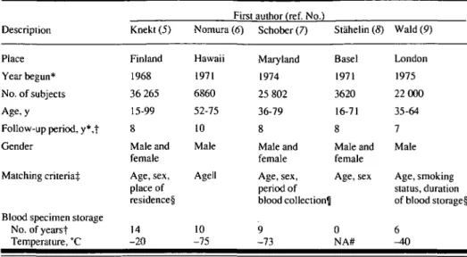
Table 1. Description of the five prospective studies of serum a-tocopherol concentration in relation to risk of colorectal cancer that were included in the pooled analysis
Description Place Year begun* No. of subjects Age,y
Follow-up period, y*,t Gender
Matching criteria^
Blood specimen storage No. of yearst Temperature, °C Knekt (5) Finland 1968 36 265 15-99 8 Male and female Age, sex, place of residence§ 14 -20
First author (ref. No.) Nomura (6) Hawaii 1971 6860 52-75 10 Male Agell 10 -75 Schober (7) Maryland 1974 25 802 36-79 8 Male and female Age, sex, period of blood collection^ 9 -73 Stahelin (S) Basel 1971 3620 16-71 8 Male and female Age, sex 0 NA# Wald (9) London 1975 22 000 35-64 7 Male Age, smoking status, duration of blood storage§ 6 ^ 0 *Approximate. tAverage.
^Refers to original authors' analyses; all were nested case-control studies. §Effectively matched on period of blood collection.
Illn the original analysis done by Nomura et al. (6), subjects were frequency matched by age; all other studies were individually matched by age. See "Subjects and Methods" section for description of matching procedure used in pooled analysis.
^Subjects were also matched according to whether they had been included in a private census of county residents.
#Not applicable.
(6), controls had originally been frequen-cy matched to cases. Stahelin et al. (8) supplied data on their entire cohort before selection of controls. To facilitate anal-ysis of the combined data, the data from Nomura et al. (6) and Stahelin et al. (8) were reorganized so that each control in these two studies was individually matched to a case. The resulting matched sets each had one case and one or more controls. The matching factors were 5-year-age category, gender, and 3-month period since blood was drawn. Because no match was available, five cases and 41 controls were excluded from the study by Nomura et al. (6), and 2896 controls were excluded from the study by Stahelin et al.
(8). Thus, data for 289 cases and 1267
controls matched by age, gender, and period of blood collection were pooled for the present analysis (Table 2).
The SAS statistical software package was used to perform tabular analyses
(11). To evaluate the average difference
between the serum a-tocopherol concen-trations for cases and their matched con-trols, a general linear model was fitted to the data. The dependent variable was serum a-tocopherol concentration, and the following parameters were estimated
for the independent variables: grand (overall) mean, a fixed-effect term for case-control status, and a random-effect term representing set membership (12). A likelihood ratio test was used to evaluate the difference in fit between models, with and without the term for case-control status. Dose-response and multivariate analyses were conducted with conditional logistic regression models fitted with the EGRET statistical software package (75).
The relationship of serum a-tocopherol to risk of colorectal cancer was modeled in two ways. 1) The quartiles of the a-to-copherol distribution in controls were determined for each study, and summary odds ratios (ORs) (across all studies) ac-cording to quartile were calculated. This approach assumed that the OR for a given quartile was constant across studies. 2) The OR per 20 (imol/L increase in serum a-tocopherol was calculated. Dietary sup-plementation with 800 IU/d of a-to-copherol results in an increase in serum a-tocopherol of approximately 20 |J.mol/L
(14). This approach assumed that the
rela-tive risk per unit of a-tocopherol was linear on the log scale. The study-specific distributions of serum a-tocopherol levels were skewed, long tail to the right.
How-Table 2. Serum a-tocopherol level for cases of colon cancer or rectal cancer and controls, by study* Source of
data, first author (ref. No.) Knekt (5) Nomura (6) Schober (7) Stahelin (5) Wald (9) Total* No. of subjects Colon cancer Cases 21 78 72 18 17 206 Controls 37 188 143 536 34 938 Rectal cancer Cases 37 30 0 5 11 83 Controls 71 72 0 165 21 329 a-Tocopherol, Colon cancer Cases 21.318.2 32.0+ 14.0 27.1±8.5 36.2 ± 10.2 25.4 ±11.0 29.0111.8 Controls 22.9 18.3 32.0+ 12.7 29.51 12.4 36.4110.4 25.319.0 30.01 11.7 umol/Lt Rectal cancer Cases 22.7 + 6.2 27.817.1 — 31.4 + 7.9 21.817.8 24.9 1 7.4 Controls 23.1 1 10.2 30.1110.8 — 36.8+ 11.6 24.0+14.4 25.9112.7 *Results based on data from five prospective case-control studies of colorectal cancer. Nos. of subjects in original papers of Stahelin et al. (S) and Nomura et al. (6) differ from those included in present analysis. See "Subjects and Methods" section for explanation. The current study includes females from the Stahelin et al. cohort, whereas the original report did not.
fValues = means 1 SD.
t-The overall mean of the values for serum a-tocopherol levels in controls was calculated as the arithmetic mean of values for cases plus the mean case-control dif-ference estimated from the generalized linear model described in the "Subjects and Methods" section. The crude analysis indicated that for colon cancer the case mean was significantly different from the control mean (/><.03). However, the difference after adjustment for serum cholesterol was not significant (P = .13). All
other case-control differences in the table were not statistically significant at the P = .05 level.
ever, analyses performed with log-trans-formed serum a-tocopherol levels led to the same conclusions as those conducted with the untransformed values. Results based on untransformed values were presented to ease interpretation.
Results
The results of the individual studies were relatively homogeneous. Within each cohort, the concentration of a-tocopherol tended to be similar among cases and controls, although in a few in-stances mean levels for cases were 1 |imol/L or more less than those for con-trols (Table 2). The standard deviations for the serum a-tocopherol concentra-tions were large in each study; i.e., the distributions overlapped substantially among the cohorts. For colon cancer, the overall average difference between case
and control serum a-tocopherol levels was of borderline significance (P = .03); for rectal cancer, the difference was not significant (P = .17). When the average difference was examined after adjustment for serum cholesterol level, there were no statistically significant differences be-tween levels for cases and controls (colon, P - . 13; rectum, P - AS).
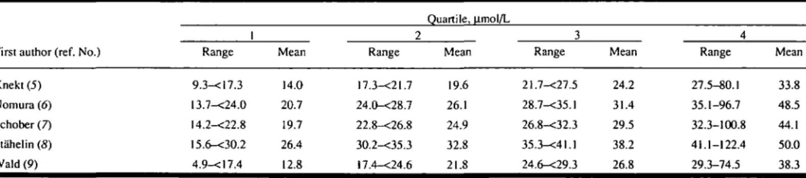
The mean concentration for the highest quartile of the serum a-tocopherol dis-tribution in each study was approximately 25 |imol/L greater than that for the lowest quartile (Table 3). The values for crude OR according to quartile of serum a-to-copherol concentration were generally similar for cancer of the colon and for cancer of the rectum (Table 4). For can-cers of the colon and rectum combined, the crude OR for the highest quartile compared with the lowest was 0,6 (95% confidence interval [CI] = 0.4-1.0).
How-ever, serum a-tocopherol and cholesterol levels were correlated (Pearson r = .27, P
= .0001; calculated with log-transformed
a-tocopherol), and serum cholesterol level was inversely associated with risk of colorectal cancer. Thus, when the ORs were adjusted for serum cholesterol level, the association between serum a-tocoph-erol concentration and colorectal cancer risk was reduced (OR = 0.7; 95% CI = 0.4-1.1). Adjustment for cigarette smok-ing status (current, past, or never) did not affect the results.
To check for evidence of a threshold effect for cancers of the colon and rectum combined, a quadratic term was added to the model, with a-tocopherol as a con-tinuous variable. The additional term did not improve the fit (P>.80). Using a similar model, a test for heterogeneity in effect across studies was performed, and the results were consistent with
homo-Table 3. Ranges and means forquartiles of serum a-tocopherol concentration (in umol/L), by study*
First author (ref. No.) Knekt (5) Nomura (6) Schober (7) Stahelin (8) Wald (9) 1 Range 9.3-<17.3 13.7-<24.0 14.2-<22.8 !5.6-<30.2 4.9-<17.4 Mean 14.0 20.7 19.7 26.4 12.8 2 Range 17.3-<21.7 24.0-<28.7 22.8-<26.8 30.2-<35.3 !7.4-<24.6 Quartile, u.mol/L Mean 19.6 26.1 24.9 32.8 21.8 3 Range 21.7-<27.5 28.7-<35.1 26.8-<32.3 35.3-<4l.l 24.6-<29.3 Mean 24.2 31.4 29.5 38.2 26.8 4 Range 27.5-80.1 35.1-96.7 32.3-100.8 41.1-122.4 29.3-74.5 Mean 33.8 48.5 44.1 50.0 38.3 •Results based on data from five prospective case-control studies of colorectal cancer.
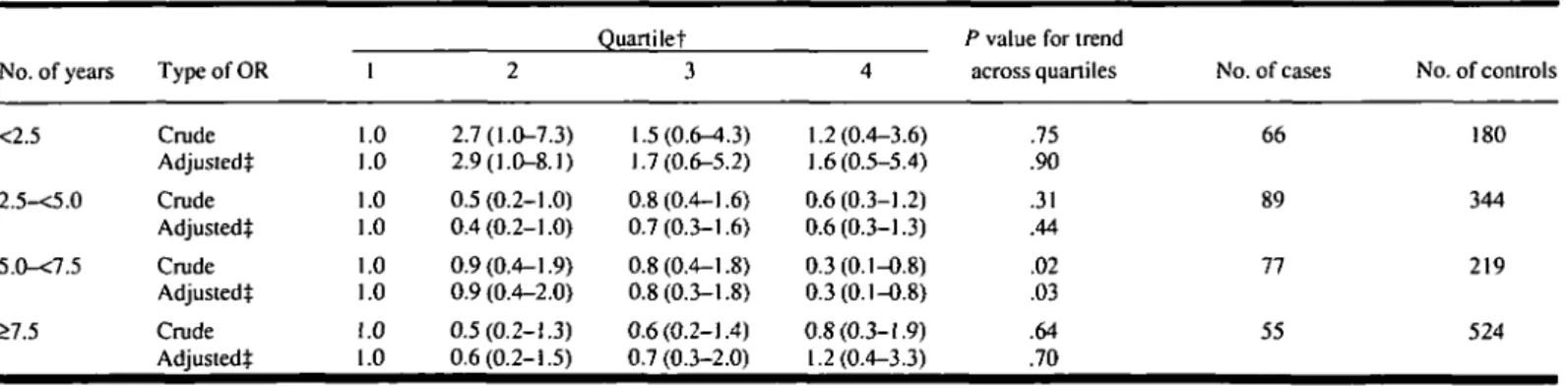
geneity (P = .90). The association be-tween serum a-tocopherol level and risk of colorectal cancer was not appreciably modified by cigarette smoking, age, or gender. In the first 2.5 years of follow-up, the relative risks of colorectal cancer in subjects with serum a-tocopherol levels in quartiles 2, 3, arid 4 were greater than 1 (Table 5). In later periods, the association was almost uniformly inverse.
As noted above, serum cholesterol con-centration was inversely associated with risk of colorectal cancer. The OR per mmol/L increase was 0.9 (95% CI = 0.8-1.0). The OR for colorectal cancer among those in the highest quartile of serum cholesterol level relative to those in the lowest was 0.6 (95% CI = 0.4-0.9). The association did not vary with the number of years between blood collection and diagnosis. The ORs per mmol/L choles-terol according to number of years were the following: 0.9 for 0-2.4 years, 0.9 for 2.5-5.0 years, 0.9 for 5.0-7.5 years, and 0.8 for 7.5 years or more.
Discussion
The results suggest that serum a-to-copherol concentration may be inversely related to risk of colorectal cancer. The laboratory procedures and the matching procedures employed in the five studies we used, together with the procedures used in our current analysis, ensured that differences between case and control values, where present, were not due to unequal length of specimen storage or to variation over time in laboratory meas-urement of serum a-tocopherol. How-ever, it is unclear whether an association exists between serum a-tocopherol level and decreased risk of colorectal cancer because, in our study, the association was modest, the CIs were wide, and, overall, the tests for trend in effect were not sig-nificant.
Because colorectal cancer is common, even a modest protective effect could have important public health implica-tions. Dietary intake of vitamin E is a
determinant of serum a-tocopherol level
(15). If an increase in daily vitamin E
in-take of 800 IU truly reduces the relative risk of colorectal cancer by 20% or 30% (within the CI of our estimate), this infor-mation could be useful for preventive health care.
One strength of the pooled analysis lies in overcoming the problem of lack of power in any individual study, especially when subgroups are analyzed (16). Another advantage is that uniform adjust-ment for confounding factors is facili-tated. If we had combined data from well-conducted, randomized clinical trials, we would have no serious concern about bias in the summary findings (16). However, because the results of the present analysis reflect findings in observational studies, biases, if any, in the component investi-gations will be reflected in the summary.
The apparent association between a-tocopherol level and decreased risk of colorectal cancer may be due to uncon-trolled confounding in our data from the
Table 4. ORs for risk of colorectal cancer according to quartile of serum a-tocopherol level and per 20-nmol/L increase in level, by cancer site*
Cancer site Colon Rectum Colon and rectum
Type of OR Crude Adjusted! Crude Adjusted! Crude Adjusted! 1 1.0 1.0 1.0 1.0 1.0 1.0 2 0.9(0.5-1.4) 0.9(0.6-1.5) 0.8(0.4-1.8) 0.8(0.4-1.8) 0.9(0.6-1.3) 0.9(0.6-1.3) Ouartile 3 0.7(0.4-1.2) 0.8(0.5-1.3) 1.0(0.4-2.2) 1.0(0.4-2.2) 0.8(0.5-1.2) 0.8(0.5-1.3) 4 0.6(0.4-1.1) 0.7(0.4-1.3) 0.6(0.2-1.6) 0.6(0.2-1.5) 0.6(0.4-1.0) 0.7(0.4-1.1)
P value for trend
across quartiles .06 .26 .32 .41 .03 .15
Per 20 |imol/L increase 0.82(0.61-1.11) 0.90(0.63-1.27) 0.59(0.30-1.16) 0.59(0.28-1.20) 0.77(0.58-1.02) 0.82(0.60-1.11) P value for increase .21 .55 .13 .15 .07 .20 •Results based on pooled data from five prospective case-control studies of colorectal cancer. Estimates were from conditional logistic regression models. Values in parentheses = 95% CIs. Subjects with the lowest serum a-tocopherol concentrations are in quartile 1.
tAdjusted for serum cholesterol level.
Table 5. ORs for risk of colorectal cancer according to quartile for serum a-tocopherol level, by No. of years between blood collection and diagnosis*
No. of years <2.5 2.5-<5.0 5.0-<7.5 >7.5 Type of OR Crude Adjustedt Crude Adjustedt Crude Adjustedt Crude Adjustedt 1 .0 .0 .0 .0 .0 .0 .0 .0 2 2.7(1.0-7.3) 2.9(1.0-8.1) 0.5(0.2-1.0) 0.4(0.2-1.0) 0.9(0.4-1.9) 0.9 (0.4-2.0) 0.5(0.2-1.3) 0.6(0.2-1.5) Quartilet 3 1.5(0.6-4.3) 1.7(0.6-5.2) 0.8(0.4-1.6) 0.7(0.3-1.6) 0.8(0.4-1.8) 0.8(0.3-1.8) 0.6(0.2-1.4) 0.7 (0.3-2.0) 4 1.2(0.4-3.6) 1.6(0.5-5.4) 0.6(0.3-1.2) 0.6(0.3-1.3) 0.3(0.1-0.8) 0.3(0.1-0.8) 0.8(0.3-1.9) 1.2(0.4-3.3)
P value for trend
across quartiles .75 .90 .31 .44 .02 .03 .64 .70 No. of cases 66 89 77 55 No. of controls 180 344 219 524
•Estimates were from conditional logistic regression models.
jSubjects with the lowest serum a-tocopherol concentrations are in quartile 1. Study-specific quartiles are specified in Table 3. tAdjusted for serum cholesterol level.
five studies we used. In the United States, fats, oils, and vegetables are the major sources of dietary vitamin E (77); an ob-served protective association could be due to constituents of these foods other than.vitamin E or to behaviors associated with eating such foods.
Serum a-tocopherol level is deter-mined, in part, by serum lipoprotein con-centration (18). Serum cholesterol level can be reduced in persons with cancer that is not yet clinically detectable, in-cluding colorectal cancer (19-22). The ef-fect is usually greatest within 2 years before diagnosis, although Tomberg et al.
(21) found that, for all cancers combined,
persons with a low baseline cholesterol level still had a slightly greater risk of cancer even 10 years later. In the current data, the length of time after blood collec-tion did not modify the relacollec-tionship be-tween cholesterol level and cancer risk, even though it did in one of the con-stituent studies (9). Although the precise reasons for the inverse relationship be-tween serum cholesterol level and risk of colorectal cancer are not entirely clear, the association of serum a-tocopherol concentration with risk of colorectal can-cer was adjusted for serum cholesterol concentration to avoid confounding.
The serum specimens collected by Stahelin et al. (8) were analyzed immedi-ately after the blood was drawn; vitamin E levels among those subjects were the highest in the five cohorts. The specimens collected by Knekt et al. (5) were stored for a longer period than in any of the other studies, and vitamin E levels among the subjects were the lowest in the five cohorts. The rate of decay of serum a-to-copherol concentration in sera frozen at -40 °C was observed by Wald et al. (9) to be 1.2 |omol L"'y"'. If one assumes that the decay of a-tocopherol in our speci-mens was proportional to storage temper-ature and length of storage, it is possible to estimate, based on Wald's decay rate at -40 °C, the mean serum a-tocopherol level in each cohort at the time the blood was drawn. Such estimates suggest that the mean a-tocopherol levels in the specimens from the cohorts were more similar at the time the blood was drawn than in the stored specimens. However, Gey et al. (23) found that the
a-tocoph-erol concentration in freshly analyzed sera from subjects from Finland was re-markably similar to the levels Knekt et al. (5) observed in sera after storage. Thus, the differences in serum a-tocopherol levels observed among cohorts may have been largely due to true population dif-ferences rather than to storage conditions.
If the rates of degradation of a-tocoph-erol were equivalent for the corres-ponding cases and controls within case-control sets, degradation would have had no effect on the observed ORs. Any departure from equality of case-control degradation rates would have resulted in misclassification of relative levels and, thus, less precise and possibly biased re-sults. For example, serum cholesterol and, by inference, serum lipoprotein levels were associated with serum a-to-copherol levels. If lipoprotein concentra-tion in the stored specimens was asso-ciated with rate of loss of a-tocopherol, the results might be biased toward the null. Although the observed association might have been more precise, and pos-sibly stronger, in the absence of degrada-tion, the fact that the findings did not vary markedly across studies suggests that our findings were not seriously distorted by differences in specimen preservation.
Although vitamin E inhibits mutagen-esis and cell transformation (/), its anti-carcinogenic effects, if any, may be through other mechanisms. For example, because vitamin E is excreted in bile (24), it might influence large-bowel carcino-genesis in the lumen of the gut. Dion et al. (25) showed that dietary supplementa-tion with vitamins E and C reduces the mutagenicity of feces. Nonetheless, ani-mal data regarding large-bowel cancer and vitamin E are contradictory (7). Results of experimental studies of polyp recurrence in humans are compatible with a modest inhibitory effect of vitamin E
(26,27), but the effects have not been
statistically significant and could be due to concurrently administered vitamin C. In the present analysis, the evidence sup-porting a causal relationship between vi-tamin E level and colorectal cancer is equivocal. If vitamin E truly has a subtle protective effect against colorectal can-cer, one might expect results like that ob-served—a monotonic modest decrease in
risk with increasing quartile of serum a-tocopherol concentration and little statis-tical precision.
The evidence for a protective effect of vitamin E is weak—certainly not strong enough to justify an intervention study powerful enough to detect an effect, if in-deed one exists. In the meantime, larger observational studies with control for confounding by other dietary factors will be needed to determine whether vitamin E has a modest but potentially important protective effect against colorectal can-cer.
References
(/) CHEN LH, BOISSONNEAULT GA, GLAUERT HP:
Vitamin C, vitamin E, and cancer. Anticancer Res 8:739-748, 1988
(2) WANG YM, PUREWAL M, NIXON B, ET AL:
Vitamin E and cancer prevention in an animal model. Ann N Y Acad Sci 570:383-390, 1989
(3) ROGERS AE, LONGNECKER MP: Dietary and
nutritional influences on cancer: A review of epidemiologic and experimental data. Lab in-vest 59:729-759, 1988
(4) SALONEN JT, SALONEN R, LAPPETELAINEN R,
ET AL: Risk of cancer in relation to serum con-centrations of selenium and vitamins A and E: Matched case-control analysis of prospective data. Br Med J 290:417-420, 1985
(5) KNEKT P, AROMAA A, MAATELA J, ET AL:
Serum vitamin E, serum selenium and the risk of gastrointestinal cancer. Int J Cancer 42:846-850, 1988
(6) NOMURA AM, STEMMERMANN GN, HEILBRUN
LK, ET AL: Serum vitamin levels and the risk of cancer of specific sites in men of Japanese ancestry in Hawaii. Cancer Res 45:2369-2372, 1985
(7) SCHOBER SE, COMSTOCK GW, HELSING KJ, ET
AL: Serologic precursors of cancer. I. Prediag-nostic serum nutrients and colon cancer risk. Am J Epidemiol 126:1033-1041, 1987
(8) STAHELIN HB, ROSEL F, BUESS E, ET AL:
Can-cer, vitamins, and plasma lipids: Prospective Basel study. JNCI 73:1463-1468, 1984
(9) WALD NJ, THOMPSON SG, DENSEM JW, ET AL:
Serum vitamin E and subsequent risk of can-cer. Br J Cancer 56:69-72, 1987
(10) WILLETT WC, POLK BF, UNDERWOOD BA, ET
AL: Relation of serum vitamins A and E and carotenoids to the risk of cancer. N Engl J Med 310:430-434, 1984
(//) SAS Institute, Inc. Statistical Analysis System. Cary.NC, 1987
(12) BMDP Statistical Software, Inc. Los Angeles,
Calif, 1990
(13) Statistics and Epidemiology Research
Cor-poration. Epidemiological Graphics, Estima-tion, and Testing Package. Seattle, Wash, 1987
(14) WILLETT WC, STAMPFER MJ. UNDERWOOD BA,
ET AL: Vitamins A, E, and carotene: Effects of supplementation on their plasma levels. Am J ClinNutr 38:559-566, 1983
(15) KNEKT P, SEPPANEN R, AARAN RK:
Deter-minants of serum a-tocopherol in Finnish adults. Prev Med 17:725-735. 1988
(16) SPECTOR TD, THOMPSON SG: The potential
and limitations of meta-analysis. J Epidemiol Community Health 45:89-92, 1991
(17) MURPHY SP, SUBAR AF, BLOCK G: Vitamin E
intakes and sources in the United States. Am J ClinNutr 52:361-367, 1990
(18) BURTON GW, TRABER MG: Vitamin E:
An-tioxidant activity, biokinetics, and bioavaila-bility. Annu Rev Nutr 10:357-382, 1990
(19) ROSE G, BLACKBURN H, KEYS A, ET AL: Colon
cancer and blood-cholesterol. Lancet 1:181-183,1974
(20) WALD NJ, THOMPSON SG, Low MR, ET AL:
Serum cholesterol and subsequent risk of can-cer: Results from the BUPA study. Br J Can-cer 59:936-938, 1989
(2/) TORNBERO SA, HOLM LE. CARSTENSEN JM, ET
AL: Cancer incidence and cancer mortality in relation to serum cholesterol. J Natl Cancer Inst 81:1917-1921,1989
(22) KNEKT P, REUNANEN A. AROMAA A, ET AL:
Serum cholesterol and risk of cancer in a cohort of 39,000 men and women. J Clin Epidemiol41:519-530, 1988
(23) GEY KF, BRUBACHER GB, STAHELIN HB:
Plas-ma levels of antioxidant vitamins in relation to ischemic heart disease and cancer. Am J Clin Nutr 45:1368-1377, 1987
(24) TRABER MG, KAYDEN HJ: Preferential
incor-poration of a-tocopherol vs y-tocopherol in human lipoproteins. Am J Clin Nutr 49:517-526,1989
(25) DION PW, BRIGHT-SEE EB, SMITH CC, ET AL:
The effect of dietary ascorbic acid and a-to-copherol on fecal mutagenicity. Mutat Res
102:27-37,1982
(26) MCKEOWN-EYSSEN G, HOLLOWAY C, JAZMAJI
V, ETAL: A randomized trial of vitamins C and E in the prevention of recurrence of colorectal polyps. Cancer Res 48:4701^*705, 1988
(27) DECOSSE JJ, MILLER HH, LESSER ML: Effect of
wheat fiber and vitamins C and E on rectal polyps in patients with familial adenomatous polyposis. J Nad Cancer Inst 81:1290-1297, 1989
Phase II Study: Treatment of
Non-Hodgkin's Lymphoma
With an Oral Antitumor
Derivative of
Bis(2,6-dioxopiperazine)
Ryuzo Ohno* Kazumasa
Yamada, Masami Hirano, Shigeru
Shirakawa, Masao Tanaka,
Takashi Oguri, Yoshihisa Kodera,
Yasuharu Mitomo, Yasushi Ikeda,
Shozo Yokomaku, Osamu Kamiya,
Masahide Kobayashi, Hidehiko
Saito, Kiyoji Kimura, The Tokai
Blood Cancer Study Group
Background: Although razoxane
(ICRF-159), a derivative of
bis(2,6-dioxopiper-azine), has shown significant antitumor activity in several murine tumors, in-adequate bioavailability has limited its clinical efficacy. Sobuzoxane (MST-16), another derivative of bis(2,6-dioxopi-perazine), has shown activity against a broad spectrum of murine tumors and human tumor xenografts in nude mice and a lack of cross-resistance to vin-cristine, doxorubicin, cyclophospha-mide, fluorouracil, etoposide, and mitomycin C. These findings suggest that MST-16 has a mode of cytocidal activity different from that of other antitumor agents. Purpose: The present late phase II study was conducted to evaluate the clinical efficacy and toxicity of MST-16 in non-Hodgkin's lymphoma (NHL). Methods: As part of a multi-institutional cooperative study, we conducted a study of MST-16 in 27 patients with NHL who were assessable for drug efficacy and toxicity. MST-16, a bis(2,6-dioxopiperazine) analogue and an inhibitor of topoisomerase II, was administered orally at a dose of 1600 mg/m2 a day for 5-7 days at
inter-vals of 2-3 weeks. Results: Response consisted of one complete remission and seven partial remissions in 27 as-sessable patients. Response was achieved at a median of 13 days (range, 9-62 days) after initiation of therapy and lasted a median of 46 days (range, 29-155 days). Major toxic effects were leukopenia in 70% of the patients, thrombocytopenia in 44%, and gastro-intestinal disorders in 37%.
Conclu-sions: MST-16 was shown to be
ef-fective in NHL and deserves further clinical trial, since the drug shows little cross-resistance to available antitumor drugs. Implications: Phase II clinical studies of MST-16 in treatment of breast cancer, gastric cancer, and adult T-cell leukemia and lymphoma are also being conducted in Japan. Future trials of combination chemotherapy using MST-16 with other antitumor drugs are warranted in view of the additive effects observed in studies of MOLT-3 cells and studies of L1210 leukemia in mice. [J Natl Cancer Inst 84:435-438, 1992]
4,4'-(l,2-Ethanediyl)bis(l-isobutoxy-carbonyloxymethyl-2,6-piperazinedione)
(MST-16, sobuzoxane) (Fig. 1) is an anti-tumor agent that can be administered orally. It is a derivative of bis(2,6-dioxo-piperazine) (1). Although ICRF-159 (razoxane), another derivative of bis-(2,6-dioxopiperazine), showed signif-icant antitumor activity in several murine tumors, its inadequate bio-availability has limited its clinical ef-ficacy (2-5). MST-16 has been selected from a number of synthetic derivatives of bis(2,6-dioxopiperazine) as the most promising compound for clinical use
(6,7). It has shown a broad spectrum of
activity against murine tumors includ-ing L1210 leukemia, P388 leukemia, B16 melanoma, Colon26 carcinoma, and Lewis lung carcinoma, as well as against human tumor xenografts such as MX-1 breast carcinoma, CO-4 colon carcinoma, and LX-1 lung carcinoma in nude mice. Results of these studies demonstrate a higher therapeutic ratio than that for ICRF-159 (7). MST-16 demonstrated no cross-resistance to vin-cristine, doxorubicin, cyclophosphamide, fluorouracil, etoposide, or mitomycin-C,
Received August 5, 1991; revised December 16, 1991; accepted December 23, 1991.
R. Ohno, K. Yamada, Department of Medicine, Nagoya University School of Medicine, The Branch Hospital, Nagoya, Japan.
M. Hirano, Department of Medicine, Fujita Health University Hospital, Toyoake, Japan.
S. Shirakawa, Department of Medicine, Mie University Hospital, Tsu, Japan.
M. Tanaka, K. Kimura, Department of Medicine, Nagoya National Hospital, Nagoya.
T. Oguri, Department of Medicine, Aichi Medical University Hospital, Nagakutecho, Japan.
Y. Kodera, Department of Medicine, Nagoya First Red Cross Hospital, Nagoya.
Y. Mitomo, Department of Medicine, Nagoya City University Hospital, Nagoya.
Y. Ikeda, Department of Medicine, Hamamatsu University Hospital, Hamamatsu, Japan.
S. Yokomaku, Department of Medicine, Aichi Shokuin Hospital, Nagoya.
O. Kamiya, Department of Medicine, Anjo Kosei Hospital, Anjo, Japan.
M. Kobayashi, Department of Hematology, Hamamatsu Iryo Center, Hamamatsu.
H. Saito, Department of Medicine, Nagoya University Hospital, Nagoya.
The Tokai Blood Cancer Study Group, Nagoya University School of Medicine, The Branch Hospi-tal.
We thank participating physicians from 12 institu-tions for their cooperation, T. Narita and M. Takeyama for the analysis of data, and T. Ushiroku for the preparation of the manuscript.
Correspondence to: Ryuzo Ohno, M.D.,
Depart-ment of Medicine, Nagoya University School of Medicine, The Branch Hospital, 1-1-20 Daiko-minami, Higashiku, Nagoya 461, Japan.