Neuropsychological long-term sequelae after
posterior fossa tumour resection during childhood
Maja Steinlin,
1Sara Imfeld,
1,4Prisca Zulauf,
1,4Eugen Boltshauser,
2Karl-Olaf LoÈvblad,
3Annette Ridol® LuÈthy,
1Walter Perrig
4and Franz Kaufmann
1Departments of1Paediatrics,3Neuroradiology and
4Psychology, University of Bern, Bern, and 2Department of
Paediatrics, University of ZuÈrich, ZuÈrich, Switzerland
Correspondence to: Maja Steinlin MD, Division of Paediatric Neurology, University Children's Hospital, Inselspital, 3010 Bern, Switzerland
E-mail: maja.steinlin@insel.ch
Summary
The importance of the cerebellum for non-motor func-tions is becoming more and more evident. The in¯uence on cognitive functions from acquired cerebellar lesions during childhood, however, is not well known. We present follow-up data from 24 patients, who were operated upon during childhood for benign cerebellar tumours. The benign histology of these tumours required neither radiotherapy nor chemotherapy. Post-operatively, these children were of normal intelligence with a mean IQ of 99.1, performance intelligence quotient (PIQ) of 101.3 and verbal intelligence quotient (VIQ) of 96.8. However, 57% of patients showed abnormalities in subtesting. In addition, more extensive neuropsychological testing revealed signi®cant problems for attention, memory, processing speed and inter-ference. Visuo-constructive problems were marked for copying the Rey ®gure, but less pronounced for recall of the ®gure. Verbal ¯uency was more affected than
design ¯uency. Behavioural de®cits could be detected in 33% of patients. Attention de®cit problems were marked in 12.5%, whereas others demonstrated psy-chiatric symptoms such as mutism, addiction problems, anorexia, uncontrolled temper tantrums and phobia. Age at tumour operation and size of tumour had no in¯uence on outcome. Vermis involvement was related to an increase in neuropsychological and psychiatric problems. The observation that patients with left-sided cerebellar tumours were more affected than patients with right-sided tumours is probably also in¯uenced by a more pronounced vermian involvement in the former group. In summary, this study con®rms the importance of the cerebellum for cognitive development and points to the necessity of careful follow-up for these children to provide them with the necessary help to achieve full integration into professional life.
Keywords: cerebellum; posterior fossa tumour; cognition; intellect; behaviour
Abbreviations: FS IQ = full scale intelligence quotient; HAWIK-R and HAWIE-R = Hamburg±Wechsler intelligence scale for children (revision 1983) and adults (revision 1991); PIQ = performance intelligence quotient; RVDLT = Rey visual design learning test; VIQ = verbal intelligence quotient; VLMT = Verbal learning memory test
Introduction
The importance of the cerebellum for non-motor functions such as language, thought modulation, emotions and the ability to plan is becoming more and more evident (Schmahmann, 1991; Schmahmann and Sherman, 1998; Fiez et al., 1992; Leiner et al., 1993). In their study of adult patients, Schmahmann and colleagues introduced the term `cerebellar cognitive affective syndrome', characterized by the four major problems of executive function, impaired spatial cognition, linguistic dif®culties and personality changes. An increasing number of clinical studies have con®rmed cognitive de®cits in children with congenital or acquired cerebellar problems (Steinlin et al., 1999; Riva and
Giorgi, 2000; Levisohn et al., 2000; Karatekin et al., 2000). In our previous study (Steinlin et al., 1999), we could show that children with congenital non-progressive cerebellar ataxia have decreased full-scale IQ varying between 30 and 90. Patients with IQ above 60 revealed in further neuropsycho-logical testing a better verbal than non-verbal performance and major problems in visuospatial tasks. Posterior fossa tumours are relatively common and account for half of all brain tumours in children. Many studies show outcome after cerebellar tumours, but results are in¯uenced by further treatments such as radiotheraphy or chemotherapy (Konrad et al., 1998). Riva and Giorgi (2000) have shown neuropsy-Brain 126 ã Guarantors of neuropsy-Brain 2003; all rights reserved
chological problems a few weeks after cerebellar tumour resection, prior to further treatment such as radiotherapy or chemotheraphy. Their results reveal a localization related pattern, with problems of auditory sequential memory and language processing after right-sided cerebellar tumour and de®cits in spatial and visual memory after left-sided tumour. Lesions to the vermis led to post-surgical mutism, which evolved into speech and language disorders as well as behavioural disturbances ranging from irritability to those reminiscent of mutism. Levisohn and colleagues presented a retrospective study of neuropsychological problems of chil-dren during the ®rst 2 years after resection of a cerebellar tumour (Levisohn et al., 2000). These children had problems comparable with the cognitive affective syndrome in adults with dysfunction of visual±spatial tasks, language sequen-cing, memory and regulation of affect. There was no localization related pattern as in the patients of Riva and Giorgi (2000). Karatekin et al. (2000) studied the effect of isolated cerebellar hemispheric tumours post-operatively and compared them with the effect of temporal tumours. After cerebellar lesions, children had a neuropsychological pattern characterized by executive function problems, which was different to those who had suffered temporal tumours. The aim of our study is to analyse long-term consequences of patients who have undergone isolated cerebellar tumour resection, without adjuvant chemotheraphy or radiation therapy, during childhood. The main aims were to determine patterns of neuropsychological problems, the role of age at diagnosis/operation and the role of tumour localization.
Patients and methods
Patients
Between 1981 and 1998, 46 children underwent surgical resection of a cerebellar tumour without radiotheraphy or chemotherapy at the Children's University Hospitals of Bern and Zurich. Thirty-®ve could still be contacted and 27 were willing to undergo more extensive neuropsychological test-ing. From these 27 children, two were excluded due to severe psychomotor retardation, which rendered neuropsychological testing impossible. In both children, cerebellar tumours were incidentally diagnosed during evaluation of longstanding developmental problems. In one, a pilocytic astrocytoma was resected 1 year after diagnosis. There had been no obvious tumour growth during that year. In the second case, the tumour was never removed and showed no changes over a 10-year follow up (probably haemangioblastoma). One patient was only 4.5 years at the time of the study. He had a full-scale IQ of 112 in Kaufman assessment battery for children testing, but was too young to undergo more extensive neuropsycho-logical testing.
Hence 24 patients (seven girls/women and 17 boys/men) entered the study. They were aged between 7.6 and 26.7 years with an average of 15.04 years. Age at operation/diagnosis (tumour biopsy was performed in one child only) was
between 3.6 and 15.5 years with an average of 8.25 years. Follow-up after tumour operation was between 2.1 and 18.25 years with an average of 7.5 years. The social class of the patients was de®ned as in the study by De Spiegelare and colleagues (De Spiegelare et al., 1998): group 1 parents were members of upper management and professional groups; group 2 were white collar workers; group 3 were self-employed individuals and technicians; group 4 were manual workers; and group 5 were unemployed. Of the 24 patients, ®ve belonged to group 1, three to group 2, four to group 3 and 12 to group 4.
Detailed clinical neurological data are summarized in Table 1, together with the results of a standardized neuromotor assessment, as suggested by Largo to quantify the motor handicap of the limbs (Largo et al., 2000). Fine motor testing with a pegboard was performed twice with each hand. Despite only minimal ®ne motor problems at clinical examination (as mild tremor) and almost no subjective problems, this test yielded an overall mild motor handicap. Fifteen of the 22 tested patients showed at least two out of the four tests to be >1 SD from the norm. The two results of the dominant hand are listed in Table 1.
Informed consent was given by patients and/or parents and the study was approved by the ethics committee of the Canton of Bern, Switzerland.
Brain tumours
Histology of the brain tumours revealed pilocytic astro-cytomas in all but ®ve patients. There were two children with choroid plexus papilloma originating from the fourth ven-tricle and one child each with astrocytoma grade II, ganglio-cytoma and haemangioblastoma. These data are summarized in Table 2, together with pre- and post-operative imaging ®ndings. Subgroups of localization were de®ned as follows: (i) right-sided tumour resection without damage of the vermis; (ii) right-sided tumour resection with damage of the vermis; (iii) left-sided tumour resection with damage of the vermis (Fig. 1); and (iv) vermis resection showing atrophy without signi®cant hemispheric damage (Fig. 2).
Neuropsychological assessment
Neuropsychological assessment was performed in all patients by two neuropsychologists (S.I. and P.Z.) during one or two sessions of 2±4 h each at the hospital. Sessions lasting longer than 2 h were interrupted by a break of ~30 min. The different tests were always performed in the same order.
Due to the diversity of age and gender in our group, evaluation of results was correlated with published reference values for age and transferred to z-values for comparison. Subtests of intelligence quotient are given in points (mean 10; SD 6 3).
Neuropsychological tests
All tests administered are summarized in Table 3, together with normal values and references. Chosen tests were, on the whole, identical to tests we administered in a previous study for evaluation of cognitive impairments in patients with non-progressive congenital ataxia (Steinlin et al., 1999).
Verbal and performance IQ was tested by HAWIK-R and HAWIE-R [Hamburg±Wechsler intelligence scales for children (revision 1983) and adults (revision 1991)], respect-ively. These are the German versions of the Wechsler intelligence scales (Tewes, 1983, 1994, respectively). Attention was assessed by the TAP test (Testbatterie zuÈr AufmerksamkeitspruÈfung)Ðthree tests of a computer-supported test series by Zimmermann and Fimm (1973): (i) alertness by the reaction to visual stimuli with and without acoustic warning; (ii) selective attention by selective reaction to predetermined stimuli mixed in a series of irrelevant stimuli; and (iii) sustained visual attention (10 min ), which was tested at the end of the half day by marking irregular optic stimuli in a series of regular optic stimuli.
Learning, memory and executive functions were tested as summarized in Table 3. Normal values were given only for
ages >18 years for the block board test (a test of visual and spatial memory). For younger patients <5 years, correct tappings were considered abnormal (Schellig, 1997). For verbal ¯uency, generating words beginning with the letters F/S or FBL, or animal naming was utilized depending upon the age of patients. For the Five Point test, normal values for divergent thinking were only available for ages <14 years. For older patients, we administered the highest given normal values (Regard et al., 1982). For perseveration (repeated ®gures compared with number of total ®gures), a score <10 is considered to be normal for all ages. Testing habitual response in favour of an unusual one by the Stroop test (Osterrieth, 1944) was only possible in patients who were able to read.
Statistical analyses
We calculated for mean values of the group and SDs and used the t-test and ANOVA (analysis of variance) for analyses of signi®cance in parametric groups. The Mann± Whitney U test and Kruskal-Wallis test were used to analyse data that did not ful®l the requirements of Table 1 Summary of clinical ®ndings
Patient Age at
diagnosis Age attest Pre-operative ®ndings Follow-up FM FU
1 4 3/4 7 1/2 HA, VO, R-sided ataxia R-predom dysdiadocho 0/1
2 3 1/2 8 HA, VO, HT, PO, mild truncal ataxia minimal truncal ataxia ±
3 5 3/4 8 1/3 VO, PO mild truncal ataxia 3/3
4 8 8 3/4 HA VO PO mild truncal ataxia, dysmetria normal 3/3
5 5 8 3/4 HA, VO, appetite ¯, vigilance ¯ mild truncal ataxia 1/1
6 5 3/4 10 1/2 HA, HT hemi R, aggressive po: extended
somnolence, VI palsy mild hemi R ±
7 6 1/2 12 HA, VO, PO, VII + VI L, ataxia L-predom increased processing time 1/1
8 7 3/4 12 HA, VO, PO po: extended somnolence,
VII bil, VI L, affect lability, ataxia depressive, mutism mild truncalataxia, tremor bil 0/0
9 12 1/4 12 3/4 Diplopia, limited gaze to R nystagmus to R 0/0
10 5 3/4 13 1/4 HA, VO, diplopia, mild truncal ataxia R dysmetria 2/2
11 9 1/2 14 tired, aggressive, V, VI, VII L, mild truncal
and R-sided ataxia ADHD mild V+ VII L mild ataxia L 2/1
12 12 14 HA, PO?, ataxia R-predom phobias, fears, tremor R 2/1
13 7 3/4 15 1/3 VO, mild truncal + L sided ataxia slow, stiff movements 1/1
14 4 1/4 15 1/2 HA, VO, HT, PO, mild truncal ataxia minimal dysmetria 0/0
15 11 3/4 15 3/4 HA VO, PO, mild truncal ataxia, vertical
nystagmus, disturbed saccades, anorexia, mutism mild hemi R, dysmetria R 1/0
16 14 16 HA, VO, PO, dysmetria R dysdiadocho R 3/1
17 10 1/3 17 2/3 HA, VO, PO, truncal ataxia normal 1/1
18 9 1/2 18 1/4 HA, VO, PO, diplopia, temper tantrums temper tantrums, ataxia L 2/1
19 8 1/3 18 1/4 HA, VO, PO, diplopia increased processing time ±
20 7 20 1/4 HA, VO, PO, mild truncal ataxia and bilateral
tremor rotatory nystagmus, minimal R-ataxia 3/3
21 5 3/4 22 activity ¯, mild ataxia and possible Babinskipo:
confusions + seizure no exact information 0/0
22 4 1/4 22 1/2 HA, VO, HT, PO, minimal ataxia no exact information 0/0
23 10 3/4 25 HA, dysdiadocho dysdiadocho 1/0
24 15 1/2 26 1/2 HA, VO, weight loss normal 0/0
ADHD = attention de®cit hyperactivity disorder; bil = bilateral; dysdiadocho = dysdiadochokinesis; FM FU = Finemotor follow-up, standard deviations from norm for dominant hand (two tests); HA = headache; hemi = hemisyndrome with pyramidal signs; HT = head tilt; L = left-sided; PO = papilloedema; po = post-operative; predom = predominant; R = right-sided; VO = vomiting.
parametric testing. Only parametric results are reported if there was no difference between the parametric and nonparametric analyses. The Kolmogorov±Smirnov test was used to control for normal distribution. The c2-test
(Bortz, 1993) was used to compare variance of the patient group with the normal population. Finally, we correlated subtest results to see whether any correlation existed between any of the neuropsychological functions.
Results
Intelligence
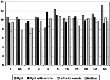
Mean full scale IQ together with verbal intelligence quotient (VIQ) and performance intelligence quotient (PIQ) are summarized in Table 4. Only three patients had full-scale IQ values below 85 (two patients of 68 and one of 72). Mean values for all subtests of HAWIK are shown in Fig. 3, revealing lower than normal values for subtests of verbal performance, signi®cantly abnormal for digit span (t = ±1.91 and P < 0.05) and vocabulary (t = ±2.38 and P < 0.025). Out of the 21 patients with full-scale IQ values >85, only seven had no subtests with <7 points and seven patients had 3±5 subtests with <7 points.
Table 2 Summary of histology and MRI/CT ®ndings
Patient Histology Tumour localization/size SL*
Hydropre-operative Vermis post-operative Hemispheres post-operative
1 PA R/5 3 4 cm 1 + normal small resection R median
2 PA R 2 0 atrophy resection R 1/3
3 PA ML/7.5 cm 2 0 median resection resection R 2/3
4 PA ML®L/4 cm 3 + atrophy resection L 1/4
5 PA ML®L/5 3 5 cm 4 +++ median resection atrophy
6 PA ML/4 3 5 cm 3 +++ superior resection resection L 1/4
7 PA L/5 cm 3 ++ atrophy resection L 1/6
8 PA No information 3 median resection resection L
9 GC ML®R/6 3 4 cm 4 0 resection lower parts resection ML
10 PA ML®R/5 cm 2 +++ resection superior parts resection ML-R 1/8
11 PP ML/4 cm 4 +++ superior atrophy normal
12 PA R/6 cm 1 + normal resection R >3/4 13 PA ML®L/4 3 5 cm 3** 0 no information no information 14 PA ML®L/5 cm 3** 0 no information no information 15 PA R®ML/5 3 3.5 cm 2 +++ atrophy resection R 16 HB R/6 3 4 3 5 cm 2 0 atrophy resection R 1/3 17 PA R/3 3 7.5 1** + no information no information 18 PA L®ML/5 3 4 cm 3 +++ atrophy resection L 1/8 19 PA R/7 3 7 cm 3 ++ atrophy resection L 2/3
20 PA ML/7 3 7 cm 2 0 median resection resection R 3/4
21 PP R/4 3 3 cm 4 0 atrophy superior parts atrophy
22 PA R/4 cm 1 +++ normal resection R minimal
23 PA R®ML/7.5 3 5 cm 2 + resection lower parts resection R 3/4
24 Astro II R/big 1** + no information no information
**Localization by pre-operative ®ndings. SL* = subgroups of localization: 1 = right sided lesion; 2 = right sided and vermal lesion; 3 = left sided and vermal lesion; 4 = vermis atrophy or resection. + = mild; ++ = signi®cant; +++ = with periventricular CSF leak; ++++ = severe with herniation. Astro = astrocytoma; GC = gangliocytoma; HB = haemangioblastoma; L = left-sided; ML = midline; PA = pilocytic astrocytoma; PP = plexus papilloma; R = right-sided.
Fig. 1 Axial T2weighted image at the level of the posterior fossa [4 mm thick slice; TR (transition time) = 4220 ms; TE (echo time) = 80 ms; FOV (®eld of view) = 200 mm], demonstrating a partial resection of the right sided cerebellar hemisphere including a signi®cant lesion of the midline (Patient 23).
Attention and processing speed
In the test for phasic alertness, the patients showed a mean z-value of ±0.27 (SD 0.84). In selective attention (Go/No Go test) and in divided attention, mean z-values were ±0.561 (SD 0.96) and ±0.591 (SD 0.97) respectively. Processing speed in the whole group revealed a z-value of 0.57 (SD 1.03). There was a signi®cant deviation from the norm for selective
attention and processing speed (P < 0.01) and for divided attention (P < 0.005), with a trend of signi®cance (P < 0.1) for phasic alertness.
Memory and learning
Immediate recall (block board test and digit span) showed mean z-values of ±0.19 (SD 1.17) and mean subtest values of 8.8 (SD 2.28). There was signi®cant deviation from the norm for digit span, with P < 0.05. The block board test was dif®cult to evaluate. Patients >18 years all gave normal z-values. Six out of 13 of the younger patients (<16 years) had abnormal results. Four of the ®ve patients <10 years showed abnormal values, although the validity of reference values for younger patients might not be adequate.
Subtest results of semantic memory (information, vocabu-lary and picture completion) revealed mean values of 9.0 (SD 3.08), 8.5 (SD 2.73) and 10.2 (SD 3.48), respectively, with signi®cance of abnormality only for vocabulary (P < 0.05).
Verbal free recall, recognition and learning [verbal learn-ing memory test (VLMT)] in all except three patients were
Table 3 Tests performed, their normal values and literature references
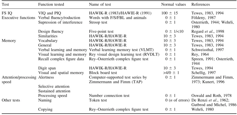
Test Function tested Name of test Normal values References
FS IQ VIQ and PIQ HAWIK-R (1983)/HAWIE-R (1991) 100 6 15 Tewes, 1983, 1994
Executive functions Verbal ¯uency/roduction Words with F/S/FBL and animals 0 6 1 FoÈldeny, 1987
Supression of interference Stroop test 0 6 1 Osterrieth, 1944; Wehrli,
1980
Design ¯uency Five-point test 0 6 1/<10 Regard et al., 1998
Similarities HAWIK-R/HAWIE-R 10 6 3 Tewes, 1983, 1994
Memory Vocabulary HAWIK-R/HAWIE-R 10 6 3 Tewes, 1983, 1994
General HAWIK-R/HAWIE-R 10 6 3 Tewes, 1983, 1994
Verbal learning and memory Verbal learning memory test (VLMT) 0 6 1 Schweissthal, 1997 Visual learning and memory Rey visual design learning test (RVDLT) 0 6 1 Rey, 1964
Recall complex ®gure data Rey±Osterrieth complex ®gure test 0 6 1 Spreen, 1991; Osterrieth, 1944
Digit span HAWIK-R/HAWIE-R 10 6 3 Tewes, 1994
Visual and spatial memory Block board test >4/0 6 1 Schellig, 1997
Attention/processing
speed Alertness Computer-supported test series byZimmermann and Fimm (TAP) 0 6 1 Zimmermann and Fimm,1973; Kunert, 1996 Selective attention
Sustained attention
Processing speed Number connection test 0 6 1 Oswald and Roth, 1978
Other tests Naming Token test 0 (n of errors) De Renzi et al., 1962;
Gutbrod and Michel, 1986
Copying Rey±Osterrieth complex ®gure test 0 6 1 Wehrli, 1980
Fig. 2 Sagittal T1weighted image (5 mm thick slice; TR = 440 ms; TE = 19 ms; FOV = 220 mm) demonstrating partial resection of the vermis (Patient 20).
Table 4 Summary of full-scale, verbal and performance IQ of all patients
Mean value (SD) t-test Abnormal n (percentage)
Full scale IQ 99.1 (15.38) ±0.28 3 (12.5)
Verbal IQ 96.8 (16.14) ±1.05 6 (25)
within normal limits with mean z-values of 0.8 (SD 0.71), 0.4 (SD 0.63) and 3.3 (SD 0.71), respectively. However, all three patients with z-values <0 were tested in a language other than their mother tongue, which might have in¯uenced their results. Visual free recall and recognition [Rey visual design learning test (RVDLT)] was normal with mean z-values of 1.16 (SD 0.68) and 1.43 (SD 0.87).
Recall of the Rey ®gure was abnormal in six patients; the mean z-value was ±0.66 (SD 1.19). See below for comparison of copying and recall of the Rey ®gure.
Executive functions
Verbal ¯uency assessed by the F/S and FBL-test showed a mean z-value of ±0.81 with a large SD of 1.46. There was a signi®cant deviation from the normal population with P < 0.001. Design ¯uency was normal for all but two patients with z-values of ±1.3 and ±2.0. The mean z-value was 0.67 (P > 0.05); however, for this test there was a signi®cant SD of
1.26 (P < 0.05). Perseveration for this test was abnormal in six out of 24 patients (25%). Test for interference (Stroop test) was abnormal for all three subtests: reading of coloured words showed a mean z-value of ±1.25 (SD 1.1), reading of coloured lines ±1.37 (SD 1.08) and interference ±1.19 (SD 1.4). All three subtests were abnormal with P < 0.001. Large standard deviations for all three subtests reveal the increased scattering of the group. Results for similarities are shown in Fig. 3.
Further testing
Naming by the Token test showed these patients to have no problems; z-values were all within the norm.
Copying of the Rey ®gure was the most affected function for these patients. The mean z-value for copying was ±1.92 (SD 1.21), this being signi®cantly worse than mean z-value for later recall of the ®gure (P < 0.001 versus P < 0.01).
Summary of test results
A summary of all results with statistically signi®cant deviations from the norm in different subtests is given in Table 5. This reveals that memory, attention, visual±spatial abilities and interference are especially affected. Although most patients had normal values for full-scale IQ and needed no special schooling, these partial neuropsychological de®cits were not without importance in the daily life of these patients. Many parents report additional school problems for these children compared with their siblings and adult patients complain about these mild de®cits.
Age
Figure 4 shows the comparison of subtest values of HAWIK-R or HAWIE-HAWIK-R for the following three age groups: age at diagnosis/operation for group 1, the preschool group, was 3.5±6.5 years (n = 10); for group 2, the early school age
Table 5 Overview on statistically signi®cant abnormal results of different cognitive functions of all patients
Cognitive function Test t±test Level of
signi®cance
Immediate memory Digit span (DS) ±1.91 P < 0.05
Semantic memory Vocabulary (V) ±2.38 P < 0.05
Free recall (visual) RVDLT* 3.67* P < 0.01
Recognition (visual) RVDLT* 5.72* P < 0.001
Visual memory Rey±Osterrieth ®gure (recall) ±3.29 P < 0.005
Selective attention TAP (Go/Nogo) ±2.69 P < 0.01
Divided attention TAP (divided attention) ±3.34 P < 0.005
Processing speed Number connection test ±2.79 P < 0.01
Visuo-constructive Rey±Osterrieth ®gure (copy) ±9.39 P < 0.001
Verbal F-test, FBL-test ±3.88 P < 0.001
Interference Stroop test ±4.91 P < 0.001
*Performance compared with normal population superior; all other tests show inferior performance. Fig. 3 Mean values of subtest in HAWIK-R or HAWIE-R in all
patients. Bold horizontal line represents the normal values. A = arithmetic; BD = block design; DS = digit span; I = information; PA = picture arrangement; PC = pictures
completion; S = similarities; OA = object assembling; SS = symbol search; V = vocabulary.
group, it was 7±9.5 years (n = 7); and for group 3, it was 10± 15.5 years (n = 7). Figure 4 reveals that group 2 was most affected for almost all subtest functions, but most pronounced for subtests of verbal performance. In addition, group 2 was also slightly more disturbed for memory and learning as seen in verbal free recall and recognition. However, a signi®cant difference between the three groups was only reached for information, where group 2 with a mean value of 7.57 was signi®cantly more disturbed than group 3 with a mean value of 10.29 (P < 0.05). These data were in contrast to the data for alertness, with group 3 being most disturbed (mean z-values of 0.2 for group 1, 0.43 for group 2 and ±0.99 for group 3), with P < 0.05 for the comparison of group 1 or 2 with group 3.
Localization
The mean full-scale intelligence quotient (FS IQ) of the whole group was 99.1; patients with right-sided tumours
showed a mean FS IQ of 105.8 and those with left-sided tumours a mean FS IQ of 91.9. The mean PIQ was 101.3; in children with right-sided tumours, the mean was 104.2 and, in those with left-sided tumours, it was 97.7. The difference between children with right-sided and left-sided tumours was more pronounced with respect to VIQ (103.9 versus 88.2; overall mean 96.8). Table 6 gives an overview of statistically signi®cant differences of functions with respect to tumour lateralization. FS IQ in respect to subgroups of localization of residual cerebellar lesions showed an effect of lateralization of tumour, but also an important effect of vermis involvement (as shown in Fig. 5 and especially Fig. 6). However, these results only reached statistical signi®cance for in¯uence of localization on subgroups with selective attention (P = 0.0093) and a trend of signi®cance for digit span (P = 0.027).
A correlation matrix between subtests revealed high correlations (r > 0.5) between (i) arithmetic, digit span, block tapping, and (ii) copy and recall of the complex ®gure
Table 6 Overview of statistically signi®cant differences in tests comparing results in patient with right- and left-sided lesions
Right-sided lesion Left-sided lesion t-test U-test
Mean (6SD) Mean (6SD) Full scale IQ1 105.8 (10.26) 91.9 (16.7) ±2.35* Verbal IQ1 103.9 (8.93) 88.2 (19.34) 25.50+ Information2 10.0 (2.83) 7.7 (2.55) ±1.92+ Vocabulary2 9.9 (2.02) 7.0 (3.80) ±2.19* Digit span2 10.2 (2.18) 7.7 (1.58) ±2.89** Figures2 11.5 (1.86) 9.11 (2.42) ±2.54* Stroop B3 ±0.89 (0.74) ±1.93 ( 1.176) ±2.01+ Stroop C3 ±0.39 (0.65) ±1.86 (15.10) 13.00+
1Value in IQ points, norm 100 6 15;2Values in subtest scaled scores, norm 10 6 3;3z-values, norm 0 6 1; *P < 0.05; **P < 0.001; + = trend to signi®cance.
Fig. 4 Comparison of subtest values of HAWIK-R or HAWIE-R in different age groups. A = arithmetic; BD = block design; C = comprehension; CO = codes; DS = digit span; I = information; PA = picture arrangement; PC = pictures completion;
S = similarities; OA = object assembling; V = vocabulary; VIQ = verbal intelligence quotient; PIQ = performance intelligence quotient.
Fig. 5 Mean values for subtests related to localization of tumour. A = arithmetic; BD = block design; C = comprehension; DS = digit span; I = information; PA = picture arrangement; PC = pictures completion; S = similarities; OA = object assembling; SS = symbol search; V = vocabulary.
of Rey. For both subgroups of the tests, the scores were summarized to a combined scoreÐthe ®rst group represent-ing executive function tests and the second subgroup visual constructive function tests. Further statistical analyses by parametric ANOVA and non-parametric Kruskal±Wallis testing revealed the side location of the lesion in different test subgroups to be signi®cant for executive function tests (P = 0.011 for ANOVA testing and P = 0.021 for Kruskal± Wallis testing), while lesion of the vermis showed a trend to signi®cance for the visual±constructive function tests (P = 0.056 for ANOVA testing and P = 0.075 for Kruskal± Wallis test).
Psychopathology
Although no formal testing of behaviour was performed, eight out of 24 patients were diagnosed with special behaviour problems. These behaviour problems were evident during testing and/or from the patient's history. They were con®rmed by previous psychological assessments or the patients received psychiatric treatment for these problems. Three children suffered from severe problems of attention de®cit, which interfered signi®cantly with their school performances. The other ®ve patients had one of the following: selective mutism, phobia, anorexia, addiction to gambling or uncontrolled temper tantrums. It is possible, that with more extensive psychiatric exploration, even more patients would have been detected. It seems worth mention-ing that all but one patient with behavioural or psychiatric problems had vermal lesions.
Discussion
Although FS IQ as well as VIQ and PIQ for the whole group show mean values within normal limits (100/97/101) and only ®ve and three out of 24 children showed VIQ and PIQ values, respectively, below the normal, analyses of subtests and further tests reveal signi®cant neuropsychological sequelae in children after resection of posterior fossa tumours. Analyses of subtests show two-thirds of our patients have de®cits in selected domains of cognitive processing.
Thirty-six partial neuropsychological de®cits were detected in 21 patients with normal full-scale IQ. This is signi®cantly above the frequency of the general healthy population, where 4±7% of all children have partial neuropsychological de®cits (Remschmidt, 1997). The fact that these children have a typical pattern of neuropsychological de®cit rather than a diffuse dysfunction supports the idea that these problems are related to cerebellar lesion rather than a potential impact on intellectual and emotional development by psychological trauma induced by a major medical or neurosurgical disease. This assumption is further supported by the study of Karatekin et al. (2000), which showed that neuropsycholo-gical de®cits of children after a cerebellar tumour differ from those seen in children after temporal tumours. The similarity of the pattern of abnormalities to those described by Schmahmann and Sherman (1998), as the cerebellar cogni-tive affeccogni-tive syndrome in adults, and to those demonstrated in similar studies in children after cerebellar tumours (Levisohn et al., 2000; Riva and Giorgi, 2000) are additional points in favour of the existence of a cerebellar cognitive syndrome.
The most prominent problems within general intelligence tests were an impairment in the performance of digit span and vocabulary, which are subtests for short-term memory and semantic memory, respectively. Short-term memory was more frequently disturbed with an auditive verbal test (digit span) and less in a visual-spatial test (Black board test). These results con®rm the ®ndings of Riva and Giorgi (2000) and those of Levisohn et al. (2000). Short-term memory problems in patients with cerebellar abnormalities might be explained by the theory of Keele and Ivry (1990) that the cerebellum has an important role for timing. Due to aberrations in timing, the capability of encoding and organising the incoming informa-tion might be disturbed. The fact that the patients achieved a normal or even high normal performance in learning tests such as VLMT and RVDLT shows that they have normal abilities to learn numbers and ®gures when correct succession or timing was not mandatory. The difference between auditory±verbal and visual±spatial short-term memory might be explained by the additional in¯uence of (mute) articulation speed for the capacity of remembering the numbers (Baddeley, 1990).
Alertness was normal in our patients during testing for phasic alertness, but there were dif®culties noted in selective and, even more notably, for sustained attention. These ®ndings are consistent with the results from Akshoomoff et al. (1992). These dysfunctions can also be seen in patients with frontal lesions. Sturm and Zimmermann (2000) showed that alertness in these patients is disturbed by increased interference, but also ®xation on an expected result (perseveration). Additional tests in our patients (¯uency test and Stroop interference test) led us to assume that alertness in patients with cerebellar disorders is disturbed by the same basic dysfunctions as in patients with frontal lesions. Sustained and selective attention are functions of the working memory, supporting the results of Mandolesi et al. (2001),
Fig. 6 FS IQ, verbal IQ and performance IQ related to residual vermis ®ndings, showing normal vermis, atrophic vermis and resectioned vermis.
revealing that working memory is not only disturbed by frontal, but also by cerebellar lesions.
Further hints at frontal dysfunction are problems in organization and planning, as shown by verbal and design ¯uency. Verbal ¯uency is more affected than design ¯uency, possibly due either to the important function of semantic organization strategies in verbal ¯uency or to the effect of the cerebellum in word generation. Performance in design ¯uency was relatively low with an additional tendency to perseverate, revealing a de®cit of inhibitory alertness and organization strategies. Executive functions of interference were disturbed for all three levels of interference in the Stroop testÐa cerebellar dysfunction that was also shown by Fiez et al. (1992). Interference is a function of processing time and memory span. Patients with increased interference also show dif®culties with attention (especially sustained attention) and vice versa. This illustrates that the dif®culties of patients with cerebellar problems for attention, interference and processing time might aggravate each other reciprocally. Our patients con®rm the growing evidence that cerebellar dysfunction includes a mild frontal dysfunction, explained by the cerebello-frontal connections (Grafman et al., 1992; Leiner et al., 1995; Schmahmann and Sherman, 1998).
There were signi®cant dif®culties in copying and recall of the Rey ®gure, con®rming the results of Levisohn et al. (2000). The major dif®culties in copying the Rey ®gure reveal signi®cant planning and organization problems, but less pronounced visual±spatial memory problems. The fact that all but two patients improved signi®cantly for recall compared with copying has to be interpreted as planning of copying the ®gure being signi®cantly disturbed, which results in dif®culty for memorising being due to primary insuf®-ciency of copying. Dif®culties in planning and organization tasks for cerebellar patients are largely explained by fronto-cerebellar pathways (Schmahmann and Pandya, 1995, 1997). However, the signi®cantly pronounced dif®culties in the planning and organization task of copying the Rey ®gure compared with other planning and organization tasks (for example, ¯uency) might be related to an additional dysfunc-tion in the spatial domain. The most likely explanadysfunc-tion is a disconnection of pathways from the cerebellum to the posterior parietal regions and vice versa (Brodal, 1978; Schmahmann and Pandya, 1989; Middleton and Strick, 1994).
The number connecting test is known to correlate well with full-scale IQ (Oswald and Roth, 1978). However, our patients demonstrated signi®cant slowing for this function. Mild motor problems might in¯uence this result, but articulation dif®culties might also be involved in this dysfunction, as a mute naming of the numbers increases the speed for this test. Results for semantic memory are a further clue for the importance of the cerebellum in word ®nding. Semantic memory was normal for pictures, but slightly disturbed for vocabulary and also information. There was a gap for vocabulary in children who suffered the acute episode of tumour treatment between the ages of 5 and 10 years, pointing
to the possibility that the cerebellum has an important impact, during this time, upon vocabulary development, as well as information.
More than 30% of our patients had psychopathological symptoms. This is in concordance with the study of Levisohn et al. (2000), where 32% of the children showed de®cits in affect regulation. Over the last few years, several studies have pointed to the importance of the vermis in patients with psychopathological problems such as attention de®cit hyper-activity disorder (Berquin et al., 1998; Mostofsky et al., 1998) and schizophrenia (Ackermann and Daum, 1995). The majority of our patients with psychopathological problems had an affected vermisÐa ®nding that supports the postula-tion of Schmahmann and Sherman (1998) of a cerebellar limbic system within the vermis.
Left-sided tumour to right-sided tumour comparison reveals a difference for almost all different functionsÐthe children with left-sided tumours being slightly more affected than those with right-sided tumours (Table 6). Most import-antly, there was a difference for FS IQ (91 versus 106), vocabulary, digit span and ®gures. These data are contrary to the ®ndings of Riva and Georgi (2000), where left-sided tumour led to more pronounced visual±constructive problems and right-sided tumours to more verbal problems. Comparison of our data with those of Riva and Giorgi (2000) showed that the lesions of their children seemed to be more localized to one hemisphere, whereas the majority of our patients had signi®cant involvement of the vermis. Figure 6 shows FS IQ, VIQ and PIQ in relation to extent of vermal lesion. Although statistically not signi®cant, there is a tendency for vermal lesion to affect cognitive function. On reanalysing our data, it became obvious that our children with left-sided tumours had, in general, more signi®cant vermal involvement than those with right-sided tumours. Therefore, it might be that, in our study, lateralization of dysfunction was overshadowed by vermal dysfunction
Statistical analyses of the effect of localizations to speci®c neuropsychological functions are limited in our study and the results have to be discussed carefully. Adequate extension of residual lesion by MRI and, therefore, allocation to different subgroups was sometimes dif®cult. In addition, the numbers in the different groups were mostly small. However, there are still some interesting points, which are worth mentioning. The results of our study show that there is a tendency for left-sided lesions and vermal lesions to produce further neuro-psychological de®cits (Figs 5 and 6). Comparing the subgroup of executive functions to the subgroup of visual± constructive functions reveals left cerebellar hemispheres to be important for the executive functions (P = 0.01) and vermis to be more important for visual±constructive functions (P = 0.06).
Comparison with our previous data from patients with congenital non-progressive cerebellar ataxia (Steinlin et al., 1999) showed a similar pattern of dysfunction for attention and executive functions. However, this recent study showed more dysfunction for verbal than performance IQ, which is
contrary to the previous study. We suggested in this previous publication that better plasticity of the left cerebral hemi-sphere compared with the right hemihemi-sphere (Woods and Teuber, 1973; Glos and Pavlovkin, 1985) might explain these ®ndings. The recent results suggest that this postulated difference of plasticity disappears later in life. Another explanation for the pronounced verbal dif®culties of the patients in this study might be a more pronounced effect of vermal lesion for the patients after cerebellar tumours compared with patients with congenital non-progressive cerebellar ataxia and more generalized affection of cere-bellum. The idea from our previous study (Steinlin et al., 1999) that the earlier the damage in life, the more important cognitive problems will be, was not con®rmed by this present study. The results of this study are more suggestive for a vulnerable age between 5 to 10 years. An explanation for these ®ndings might be that generalized cerebellar dysfunc-tion leads to the most pronounced cognitive problems. However, more localized lesions of the cerebellum showed an age-dependent effect on later cognitive function with a vulnerable age around early school age.
In conclusion, our study supports previous ®ndings that the cerebellum is important in cognitive functioning. Besides the similarity of frontal impairment of executive functions, there are also additional challenges especially in visual±spatial domains, attention and short-term memory. The pattern of these dysfunctions, involving many different cognitive func-tions, supports the idea that cerebellar cognitive and affective symptoms can not be explained by one uniform cerebellar dysfunction, but are rather a consequence of the many pathways connecting the cerebellum and the cerebrum in both directions (Schmahmann and Sherman, 1998). It is important to point to the psychopathological problems of these children. Both early and long-term neuropsychological follow-up and speci®c intervention until integration into professional life seems mandatory for children after posterior fossa tumours, irrespective of further administered therapies such as radio-therapy or chemoradio-therapy.
Acknowledgements
We wish to thank Susan Blaser for her help in preparing this manuscript.
References
Ackermann H, Daum I. Kleinhirn und Kognition ± psychopathologische, neuropsychologische und neuroradiologische Befunde. Fortschr Neurol Psychiatr 1995; 63: 30±7.
Akshoomoff NA, Courchesne E, Press GA, Iragui V. Contribution of the cerebellum to neuropsychological functioning: evidence from a case of cerebellar degenerative disorder. Neuropsychologia 1992; 30: 315±28.
Baddeley AD. Human memory. Theory and practice. London: Lawrence Erlbaum; 1990.
Berquin PC, Giedd JN, Jacobsen LK, Hamburger SD, Krain AL Rapoport JL, et al. Cerebellum in attention-de®cit hyperactivity disorder: a morphometric MRI study. Neurology 1998; 50: 1087±93.
Bortz J. Statistik fuÈr Sozialwissenschaftler (4. vollst. uÈberarb. Au¯.). Berlin: Springer; 1993.
Brodal P. The corticopontine projection in the rhesus monkey. Origin and principles of organization. Brain 1978; 101: 251±83. De Renzi E, Vignolo LA. The Token test: a sensitive test to detect receptive disturbances in aphasics. Brain 1962; 85: 665±678. De Spiegelaere M, Dramaix M, Hennart P. Socioeconomic status and changes in body mass from 3 to 5 years. Arch Dis Child 1998; 78: 477±8.
Fiez JA, Petersen SE, Cheney MK, Raichle ME. Impaired non-motor learning and error detection associated with cerebellar damage. Brain 1992; 115: 155±78.
FoÈldenyi M. Neuropsychologische Untersuchungen bei Kindern nach SHT. Eine katamnestische Studie [thesis]. ZuÈrich: UniversitaÈt ZuÈrich, 1987.
Glos J, Pavlovkin M. Pro®le of intellectual achievements in the WISC of children with hemiplegic form of cerebral palsy (their dependence on the hemispheric localization of the brain damage). Stud Psychol 1985; 23: 285±9.
Grafman J, Litvan I, Massaquoi S, Stewart M, Sirigu A, Hallett M. Cognitive planning de®cit in patients with cerebellar atrophy. Neurology 1992; 42: 1493±6.
Gutbrod K, Michel M. Zur klinischen ValiditaÈt des token tests bei hirngeschaÈdigten Kindern mit und ohne Aphasie. Diagnostica 1986; 32: 118±128.
Karatekin C, Lazareff JA, Asarnow RF. Relevance of the cerebellar hemispheres for executive functions. Pediatr Neurol 2000; 22: 106±12.
Keele SW, Ivry R. Does the cerebellum provide a common computation for diverse tasks? A timing hypothesis. Ann NY Acad Sci 1990; 608: 179±211.
Konrad K, Gauggel S, Jansen HT. Hirntumourerkrankungen im Kindesalter: Kognitive, affektive und psychosoziale Langzeitfolgen. Kindheit Entwicklung 1998; 7: 154±62.
Kunert HJ, Derichs G, Irle E. Entwicklung von
Aufmerksamkeitsfunktionen im Kindesalter: Ergebnisse einer vorlaÈu®gen Normierung der computergestuÈtzten Testbatterie zur AufmerksamkeitspruÈfung (TAP) an 9±12 jaÈhrigen Kindern. Z Neuropsychol 1996; 2: 92±113.
Largo RH, Ca¯isch JA, Hug F, Muggli K, Molnar AA, Molinari L, et al. Neuromotor development from 5 to 18 years. Part 1: timed performance. Dev Med Child Neurol 2001; 43: 436±43.
Leiner HC, Leiner AL, Dow RS. Cognitive and language functions of the human cerebellum. Trends Neurosci 1993; 16: 444±7. Leiner HC, Leiner AL, Dow RS. The underestimated cerebellum. Hum Brain Mapp 1995; 2: 244±54.
Levisohn L, Cronin-Golomb A, Schmahmann JD.
in children. Cerebellar cognitive affective syndrome in a paediatric population. Brain 2000; 123: 1041±50.
Mandolesi L, Leggio MG, Graziano A, Neri P, Petrosini L. Cerebellar contribution to spatial event processing: involvement in procedural and working memory components. Eur J Neurosci 2001; 14: 2011±22.
Middleton FA, Strick PL. Anatomical evidence for cerebellar and basal ganglia involvement in higher cognitive function. Science 1994; 266: 458±61.
Mostofsky SH, Reiss AL, Lockhart P, Denckla MB. Evaluation of cerebellar size in attention- de®cit hyperactivity disorder. J Child Neurol 1998; 13: 434±9.
Osterrieth PA. Le test de copie d'une ®gure complexe: contribution a l'eÂtude de la perception et de la meÂmoire. Arch Psychol 1944; 30: 286±356.
Oswald W, Roth E. Der Zahlenverbindungstest (ZVT) Handanweisung (2. uÈberarbeitete und erweiterte Au¯age). GoÈttingen: Hogrefe; 1978.
Regard M, Strauss E, Knapp P. Children's production on verbal and non-verbal ¯uency tasks. Percept Mot Skills 1982; 55: 839±44. Remschmidt H. Kinder- und Jugendpsychiatrie. Eine praktische EinfuÈhrung. Stuttgart: Thieme; 1997.
Riva D, Giorgi C. The cerebellum contributes to higher functions during development. Evidence from a series of children surgically treated for posterior fossa tumours. Brain 2000; 123: 1051±61. Schellig D. B-T-T. Block-tapping-test. Frankfurt am Main: Swets Test Services; 1997.
Schmahmann JD. An emerging concept: the cerebellar contribution to higher function. Arch Neurol 1991; 48: 1178±87.
Schmahmann JD. The cerebellum and cognition. International review of neurobiology, Vol. 41. San Diego: Academic Press; 1997. Schmahmann JD, Pandy DN. Anatomical investigation of projections to the basis pontis from posterior parietal association cortices in rheusus monkey. J Comp Neurol 1989; 289: 53±73. Schmahmann JD, Pandya DN. Preferential cortex projections to the
basilar pons in rhesus monkey: implications for the cerebellar contribution to higher function. Neurosci Lett 1995; 199: 175±8. Schmahmann JD, Pandya DN. Anatomic organization of the basilar pontine projections from prefrontal cortices in rhesus monkey. J Neurosci 1997; 17: 438±58.
Schmahmann JD, Sherman JC. The cerebellar cognitive affective syndrome. Brain 1998; 121: 561±79.
Schweissthal B. Die Leistungen von 5±15 jaÈhrigen Kindern im verbalen Lern- und MerkfaÈhigkeitstest (VLMT). Z Neuropsychol 1997; 2: 129±36.
Spreen O, Strauss E. A compendium of neuropsychological tests. Administration, norms and commentary. 2nd ed. New York: Oxford University Press; 1991.
Steinlin M, Styger M, Boltshauser E. Cognitive impairments in patients with congenital nonprogressive cerebellar ataxia. Neurology 1999; 53: 966±73.
Sturm W, Zimmermann P. AufmerksamkeitsstoÈrungen. In: Sturm W, Herrmann C, Wallesch CH, editors. Lehrbuch der klinischen Neuropsychologie. Grundlagen, Methoden, Diagnostik, Therapie. Lisse: Swets und Zeitlinger; 2000. p. 345±65.
Tewes U. Hamburg-Wechsler-Intelligenztest fuÈr Kinder, Revision. Bern: Verlag Hans Huber; 1983.
Tewes U. Hamburg-Wechsler Intelligenztest fuÈr Erwachsene. Revision 1991. HAWIE-R 2. korrigierte Au¯age. Bern: Verlag Hans Huber; 1994.
Wehrli A. Neuropsychologische Untersuchungen im Kindesalter [thesis]. ZuÈrich: UniversitaÈt ZuÈrich; 1980.
Woods BT, Teuber HL. Early onset of complementary specialization of cerebral hemispheres in man. Trans Am Neurol Assoc 1973; 98: 113±7.
Zimmermann P, Fimm B. Testbatterie zur AufmerksamkeitspruÈfung (TAP). Herzogenrath: Psytest; 1973.
Received June 16, 2002. Revised December 2, 2002. Second revision April 12, 2003. Accepted April 16, 2003
![Fig. 1 Axial T 2 weighted image at the level of the posterior fossa [4 mm thick slice; TR (transition time) = 4220 ms; TE (echo time) = 80 ms; FOV (®eld of view) = 200 mm], demonstrating a partial resection of the right sided cerebellar hemisphere includin](https://thumb-eu.123doks.com/thumbv2/123doknet/14893965.650541/4.916.478.826.682.1039/weighted-posterior-transition-demonstrating-resection-cerebellar-hemisphere-includin.webp)