Eur Radiol (2003) 13:1608–1611 DOI 10.1007/s00330-002-1717-3
Received: 19 March 2002 Revised: 5 August 2002 Accepted: 5 September 2002 Published online: 1 November 2002 © Springer-Verlag 2002
Abstract Our objective was to eval-uate tumor hemodynamics of para-gangliomas of the head and neck. A 2D magnetic resonance projection angiography (MRPA) protocol was developed. Six patients with histo-logically proven paragangliomas were included in the study. The se-quence protocol and data post-pro-cessing procedure were optimized in view of a high temporal resolution and maximum contrast-to-noise ra-tio. Image-time series of two freely orientated slabs could be obtained. Correlation analysis was performed to generate selective arterial and ve-nous weighted angiographic images. Glomus tumors showed a rapid and intense homogeneous enhancement following the intravenous adminis-tration of contrast material. The smallest tumors investigated mea-sured 10 mm and could be visualized clearly. Time-resolved analysis of the tumor enhancement revealed an early bolus arrival within the vascu-lar bed of the tumor and prolonged
incomplete washout indicating blood pooling. The dynamic contrast en-hancement of glomus tumors can be evaluated non-invasively with MRPA providing a high temporal resolution and high image quality. The characteristic contrast enhance-ment of glomus tumors can be help-ful in the diagnostic workup of le-sions that may mimic glomus tu-mors.
Keywords Paraganglioma · Glomus tumor · MR imaging · MR projection angiography H E A D A N D N E C K Sebastian M. Arnold Ralph Strecker Klaus Scheffler Joachim Spreer Jörg Schipper Hartmut P. H. Neumann Joachim Klisch
Dynamic contrast enhancement
of paragangliomas of the head and neck:
evaluation with time-resolved 2D MR
projection angiography
Introduction
Paragangliomas are rare highly vascularized tumors in adults. The most common locations of head and neck are the carotid space and the jugular fossa. Familial para-gangliomas appear in 7% of cases [1]. Recently, a gene focus in families with inherited paragangliomas or glo-mus tumors has been identified [2, 3]. Despite paragang-liomas are classified as benign tumors WHO grade I,
dis-tant metastases have been published [4, 5]. On the other hand, 10% of glomus tumors may be multicentric in ori-gin. Neuroimaging shows a well-circumscribed, strongly enhancing mass associated with paraganglionic struc-tures indicating abundant blood supply. Both CT and MR may show erosions of adjacent bony structures. Paragangliomas are of variable size, encapsulated, with non-invasive but expansive growth. A “salt-and-pepper” appearance of the tumor due to flow voids and small S.M. Arnold (
✉
) · J. Spreer · J. KlischDepartment of Neuroradiology, University of Freiburg, 79106 Freiburg, Germany e-mail: [email protected] Tel.: +41-61-2652525 Fax: +41-61-2655471 R. Strecker · K. Scheffler Department of MR Physics, University of Freiburg, 79106 Freiburg, Germany S.M. Arnold Department of Radiology,
University Hospital, Petersgraben 4, 4031 Basel, Switzerland J. Schipper Department of Otolaryngology, University of Freiburg, 79106 Freiburg, Germany H.P.H. Neumann
Department of Nephrology and Hypertension, University of Freiburg,
areas of subacute hemorrhage is characteristic [6, 7]. Time-resolved contrast-enhanced magnetic resonance angiography is increasingly gaining interest since it of-fers anatomical and functional information about the vascular system [8, 9, 10].
In this study we evaluated magnetic resonance projec-tion angiography (MRPA) for the visualizaprojec-tion of glo-mus tumors of the jugular foramen and the carotid bifur-cation. We propose that MRPA is a valuable method for detection of glomus tumors.
Materials and methods
Six patients suffering from histologically proven paragangliomas – 4 glomus caroticum tumors (3 unilateral, 1 bilateral) and 2 glomus jugulare tumors – were investigated in the study. The tumor size varied between 1 and 5 cm. All patients gave informed consent to the study protocol. The examinations were performed on a 1.5-T scanner (Magnetom Vision, Siemens, Erlangen, Germany) using the standard whole-body gradient set with 25 mT/m amplitude and 600 µs rise time. Images were acquired using the standard cp head coil or cp neck array. A radio-frequency-spoiled snapshot fast low-angle shot (FLASH) sequence with a slice selective Shinnar-Le Roux excitation pulse and a minimum TE of 1.5 ms and TR of 4.2 ms was used. The read out acquisition time was 1.56 ms for 160 sampling points, starting 32 points before the echo center. A minimum field of view of 220 mm was used. Slice thickness and orientation was selected freely including the whole volume of in-terest, i.e., both hemispheres. The typical slice thickness was 80 mm. A significant improvement of the temporal resolution could be achieved by the use of view sharing and temporal inter-polation [11]. Using a matrix size of 256¥256 a temporal resolu-tion of four images per second was possible. In a double-slab pro-tocol two images per second could be acquired, respectively. A high-resolution protocol permitted a matrix size of 512¥512. The images were inline mask subtracted to a temporal averaged image acquired prior to contrast bolus arrival. This technique was used to provide a better delineation of small vessels and to suppress signal cancellations caused by magnitude subtraction at boundary areas of different susceptibilities. Correlation analysis was used to selec-tively derive functional angiograms of the arterial and venous phase with a significantly improved signal-to-noise ratio (SNR) [12]. Contrast agent administration of 10–15 ml gadoteridol (Pro-hance, Byk Gulden, Germany) was started with a 10-s delay using a power injector with a flow of 4–5 ml/s flushed by 20 ml of sa-line. The total acquisition time was below 1 min.
Results
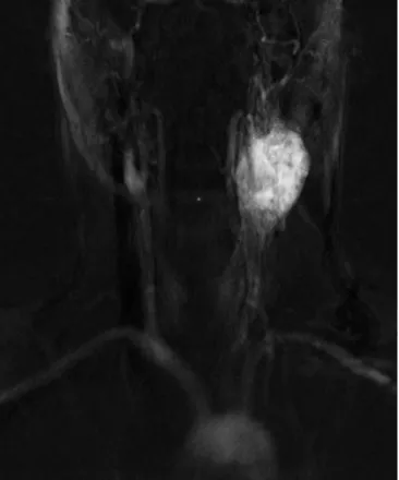
After contrast application, in all cases an intense and ho-mogeneous tumor blush was demonstrated (Fig. 1). In the case of larger tumors the carotid bifurcation was splayed. Tumor borders were well defined; however, in extended lesions the carotid bifurcation could be ob-scured by the tumor. In these cases conventional T1- and T2-weighted images were superior in demonstrating the course of the adjacent vessels. In small tumors biplane illustrations of the carotid bifurcation in coronal and sag-ittal orientation were advantageous. Concerning contrast dynamics, all glomus tumors showed an early and rapid
1609
Fig. 1 Coronal magnetic resonance projection angiography image of a large left-sided glomus caroticum tumor in early arterial phase (matrix size 256¥256, data acquisition of four images per second)
Fig. 2 Normalized signal intensity/time curves by setting the maxi-mum signal intensity to unity are plotted. Signal/time curves are shown for three different regions of interest placed in the common carotid artery, the left-sided paraganglioma and the jugular vein, re-spectively. For comparison, time-dependent enhancement of an in-tracranial meningioma of another patient is outlined (asterisks)
enhancement after contrast administration very similar to the arterial vascular enhancement (Fig. 2); however, due to summation effects, the maximum of intensity could differ significantly. In the venous phase the signal inten-sity of glomus tumors persisted on a higher level indicat-ing blood poolindicat-ing. Postprocessindicat-ing enables a selective vi-sualization of the tumor with complete suppression of venous signal. In one patient we performed a follow-up examination after combined preoperative tumor emboli-zation and surgery, which found the tumor had complete-ly vanished. Contrast enhancement of the glomus jugu-lare tumors was quite similar to the early enhancement in glomus caroticum tumors; however, as the growth pat-tern of glomus tumors in the region of the jugular fora-men is known to be highly variable with possibly extra-and intracranial compartments, the whole extent of the tumor is more ill defined. Furthermore, both tumors were investigated after tumor surgery with clinical evidence of tumor recurrence. For this reason additional scar forma-tion could not be excluded.
Discussion
The diagnostic value of functional imaging in the assess-ment of masses of the head and neck suggestive of para-gangliomas is still under debate. In our study we could show a typical time-dependent enhancement pattern of glomus caroticum tumors and glomus jugulare tumors using MRPA. The selective imaging of vascular and tu-mor enhancement easily points out also small lesions, which might open a new diagnostic field in screening in-vestigations. The MRPA is a non-invasive technique with minimal time demand and could be simply included within a routine clinical MRI scanning protocol.
Dynamic MR digital subtraction angiography by us-ing fast acquisition, contrast enhancement, and complex subtraction was first proposed by Wang et al. [10]. The sequence protocol we used in our study was published by Hennig et al. [9] and has already reached a temporal resolution in the subsecond domain. An improved SNR of the projection images up to a factor three could be achieved by using correlation analysis similar to the al-gorithms used in functional MRI [12]. Some applications of the method in fast MR angiography with subsecond resolution have been published recently [13, 14, 15, 16].
The combination of MR projection angiography with view sharing and inline background subtraction offers several advantages [11]. Firstly, the SNR in the single images of the time series is improved due to temporal in-terpolation without reducing spatial resolution. Secondly, steady-state magnetization is increased and leads to a further gain in the SNR. Thirdly, the matrix size can be increased to 512¥512 while retaining the temporal reso-lution in the subsecond domain. The increase of tempo-ral resolution due to view sharing allows multiple slab
measurement with subsecond temporal resolution within one examination.
In a study by Vogl et al. [17] contrast enhancement of glomus tumors was assessed dynamically by the use of a single-slice gradient-recalled sequence with a temporal resolution of 6 s. They found a dropout effect in form of an initial dip during early enhancement, which they con-cluded might be a specific sign for glomus tumors. They postulated that the initial signal decline might be the re-sult of susceptibility effects of the highly concentrated intravascular contrast agent. Our data could not confirm such a strong susceptibility effect after intravenous con-trast agent administration, although it is a well-known effect after intra-arterial contrast administration in MR angiography [18]. Naegele et al. [19] used a fast snap-shot-FLASH single-slice imaging technique allowing dynamic contrast enhancement imaging with measure-ment times of 1 s per image. They could demonstrate a very early contrast enhancement of glomus tumors as compared to neurinomas and meningiomas. Our study confirms that glomus caroticum tumors and glomus jug-ulare tumors show a typical temporal contrast enhance-ment (Fig. 2). The high vascularization of the tumors with high blood flow and sometimes arterial-venous shunting leads to nearly simultaneous contrast approach in tumor tissue and supplying arteries. Due to the sum-mation effect within the slice, the absolute signal of tu-mor and adjacent arteries could vary considerably. Con-cerning the whole extent of the tumor blush, however, MRPA was postulated to be superior to conventional an-giography [15]. Particularly tumors fed by several arteri-al systems may only become partiarteri-ally opacified in con-ventional angiography. Non-enhanced blood from other feeding vessels may lead to a selective washout effect of the injected contrast agent. Due to non-selective bolus application, MRPA visualizes all vessels within the se-lected volume.
In comparison with the arterial concentration time curve, the venous signal of glomus tumors was pro-longed on a higher level. One explanation is the delayed washout of contrast agent from the tumor into the vascu-lar bed. On the other hand, the recirculation of contrast agent may explain an increase in baseline signal indicat-ed by the signal approach of the mean arterial and ve-nous signal. To some unknown extent, disruption of blood-brain barrier should be taken into account.
In the case of small tumor size, there could be a con-siderable overlap with the bulb of the internal carotid ar-tery, which can obscure the tumor in the carotid bifurca-tion. The use of an additional second plane can over-come this limitation by taking into account the anatomi-cal configuration of the carotid bifurcation. Although MRPA is not able to offer spatial depth information, bi-plane imaging may help to evaluate, for example, the carotid bifurcation in detail, and to distinguish it from other carotid-space pathologies.
1611
Scar formation after surgical approach is often diffi-cult to differentiate from recurrent tumor or residual tu-mor [20, 21]. Grees et al. [22] used a T1 fast-field-echo sequence to study dynamic contrast enhancement after surgery for glomus jugulare tumors. They found clearly different dynamic contrast appearance of scars and
re-current or rest tumor. Whereas rere-current or rest tumor showed a strong enhancement and a washout phenome-non, scars showed much less contrast enhancement per-sisting on a low level. Although we could not demon-strate scar formation in our patients, MRPA gave addi-tional information in these difficult cases.
References
1. Lantos PL, Vandenberg SR, Keihues P (1997) Tumours of the nervous system. In: Graham DI, Lantos PL (eds) Green-field’s neuropathology, 6th edn. Arnold, London, pp 583–795
2. Baysal BE, Ferrell RE, Willett-Brozick JE et al. (2000) Mutations in SDHD, a mitochondrial complex II gene, in hereditary paraganglioma. Science 287:848–851
3. Niemann S, Müller U (2000) Mutations in SDHC cause autosomal dominant paraganglioma, type 3. Nat Genet 26:268–270
4. Brewis C, Bottrill ID, Wharton SB et al. (2000) Glomus jugulare tumour with metastases to cervical lymph nodes. J Laryngol Otol 114:67–69 5. Mossner R, Keidel M (2000)
Malig-nant pheochromocytoma with progres-sive paraparesis in von Hippel-Lindau disease. Eur J Neurol 7:439–442 6. Osborn AG, Tong KA (1996)
Hand-book of neuroradiology: brain and skull, 2nd edn. Mosby, St. Louis 7. Rao AB, Koeller KK, Adair CF (1999)
Paragangliomas of the head and neck: radiologic–pathologic correlation. Radiographics 19:1605–1632 8. Korosec FR, Frayne R, Grist TM et al.
(1996) Time-resolved contrast-enhanc-ed 3D MR angiography. Magn Reson Med 36:345–351
9. Hennig J, Scheffler K, Laubenberger J et al. (1997) Time-resolved projection angiography after bolus injection of contrast agent. Magn Reson Med 37:341–345
10. Wang Y, Johnston DL, Breen JF et al. (1996) Dynamic MR digital subtrac-tion angiography using contrast en-hancement, fast data acquisition, and complex subtraction. Magn Reson Med 36:551–556
11. Strecker R, Arnold S, Scheffler K et al. (2001) In: Proc Joint Annual Meeting, ISMRM and ESMRMB, Glasgow, p 301
12. Strecker R, Scheffler K, Klisch J et al. (2000) Fast functional MRA using time-resolved projection MR angiogra-phy with correlation analysis. Magn Reson Med 43:303–309
13. Klisch J, Strecker R, Hennig J et al. (2000) Time-resolved projection MRA: clinical application in intracranial vas-cular malformations. Neuroradiology 42:104–107
14. Wetzel SG, Bilecen D, Lyrer P et al. (2000) Cerebral dural arteriovenous fistulas: detection by dynamic MRPA. Am J Roentgenol 174:1293–1295 15. Yoshikawa T, Aoki S, Hori M et al.
(2000) Time-resolved two-dimensional thick-slice magnetic resonance digital subtraction angiography in assessing brain tumors. Eur Radiol 10:736–744 16. Griffiths PD, Hoggard N, Warren DJ et al. (2000) Brain arteriovenous mal-formations: assessment with dynamic MR digital subtraction angiography. Am J Neuroradiol 21:1892–1899
17. Vogl TJ, Mack MG, Juergens M et al. (1993) Skull base tumors: gadodiamide injection-enhanced MR imaging: drop-out effect in the early enhancement pat-tern of paragangliomas versus different tumors. Radiology 188:339–348 18. Serfaty JM, Atalar E, Declerck J et al.
(2000) Real-time projection MR angi-ography: feasibility study. Radiology 217:290–295
19. Naegele T, Petersen D, Klose U et al. (1993) Dynamic contrast enhancement of intracranial tumors with snapshot-FLASH MRI. Am J Neuroradiol 14:89–98
20. Schumacher A, Jonas M, Rummeny E et al. (1996) Diagnosis of recurrence of a glomus carotid artery tumor by somatostatin receptor scintigraphy. Nuklearmedizin 35:38–41
21. Konig H, Kurtz B, Strohm M (1984) High-resolution and dynamic comput-ed tomography in the diagnosis of glo-mus tympanicum and gloglo-mus jugulare tumors. Fortschr Geb Roentgenstr Nuklearmed 141:642–646
22. Grees H, Bentzien S, Tomandl B et al. (2000) Rezidivdiagnostik von Glomus-jugulare-Tumoren mittels dynamischer Kontrastmittel-MR-Anflutung. Fort-schr Geb Roentgenstr Nuklearmed 172:91