Introduction
Intravenous thrombolysis (IVT) with recombinant
tissue plasminogen activator (rt-PA) and local
intra-arterial thrombolysis (IAT) using recombinant
pro-urokinase or pro-urokinase have been found effective for
treatment of acute ischemic stroke in randomized
trials [
8
,
18
,
22
]. In routine clinical practice both
treatment modalities are safe and efficacious when
guidelines are followed [
7
,
15
,
21
]. Most guidelines
recommend IVT within 3 hours of symptom onset,
though a more recent metaanalysis indicates a benefit
up to 4.5 hours [
12
]. Nevertheless, this narrow
treat-ment window limits the number of patients eligible for
IVT. In multicenter surveys 1.6% to 6.8% of patients
have received IVT [
13
,
14
,
20
,
24
]. Individual hospitals
where special referring systems for stroke patients had
been created to accelerate admission, thrombolysed
from 15% to 18% of their patients [
9
,
10
].
The IAT trials have shown that IAT can be applied
up to 6 hours after stroke onset in patients with M1 or
M2 segment occlusion of the middle cerebral artery
(MCA). As time is one of the main limiting factors for
J. Isenegger
K. Nedeltchev
M. Arnold
U. Fischer
G. Schroth
L. Remonda
H.P. Mattle
Reasons to withhold intra-arterial
thrombolysis in clinical practice
Received: 11 February 2005
Received in revised form: 23 July 2005 Accepted: 12 August 2005
j
Abstract Background
In
se-lected stroke centers intra-arterial
thrombolysis (IAT) is used for the
treatment of acute stroke patients
presenting within 6 hours of
symptom onset. However, data
about eligibility of acute stroke
patients for IAT in clinical practice
are very scarce. Methods We
col-lected prospectively data on
indi-cations advising for or against IAT
of 230 consecutive stroke patients
in a tertiary stroke center. Results
76 patients (33.0%) presented
within 3 hours, 69 (30%) between 3
and 6 hours of symptom onset and
85 (37%) later than 6 hours.
Arte-riography was performed in 71
patients (31%) and IAT in 46
(20%). In 11 patients no or only
peripheral branch occlusions were
seen on arteriography and
there-fore IAT was not performed. In 9
patients the ICA was occluded and
barred IAT and in five anatomical
or technical difficulties made IAT
impossible. 72 patients presenting
within 6 hours did not undergo
arteriography and thrombolysis,
mostly because of mild (n = 44) or
rapidly improving neurological
deficits (n = 13). Other reasons to
withhold IAT were CT and/or
clin-ical findings suggesting lacunar
stroke due to small vessel occlusion
(n = 7), limiting comorbidty
(n = 7) and baseline international
normalized ratio > 1.7 (n = 1).
Conclusions
A third of the patients
underwent diagnostic
arteriogra-phy and one fifth received IAT. The
most important reasons to
with-hold thrombolysis were
presenta-tion beyond the 6 hours time
window and mild or rapidly
improving symptoms.
j
Key words
stroke Æ
acute Æ stroke management Æ
thrombolytic therapy
JON
2220
J. Isenegger, MD Æ U. Fischer Dept. of Internal Medicine University of Bern
Bern, 3010 Bern, Switzerland K. Nedeltchev, MD Æ M. Arnold, MD H. P. Mattle (&)
Dept. of Neurology University of Bern
Inselspital, 3010 Bern, Switzerland Tel.: +41-31/632-3332 Fax: +41-31/632-0321 E-Mail: heinrich.mattle@insel.ch G. Schroth, MD Æ L. Remonda, MD Dept. of Neuroradiology University of Bern
IVT, the extended time window for IAT should result
in more patients amenable to thrombolysis. However,
whether the extended time window for IAT,
consid-ering the technical and logistical complexity of IAT,
will result in more thrombolysed patients is unknown.
The purpose of this study was to analyze the number
of patients undergoing IAT and the reasons to
with-hold it in a single tertiary care center.
Methods
j Stroke center and catchment area
The Inselspital is a tertiary care center. IAT, started in 1992, is now used as the preferred stroke treatment and offered 24 hours a day. Some results and the referral practice in our hospital have been published previously [1, 3, 4, 16, 19, 25]. Patients are referred mainly from the city area of approximately 300000 inhabitants and some selected patients from a larger region of up to 1500000 people. At the time of the study, thrombolysis for stroke was not performed in any of the community hospitals.
j Patient selection
All ischemic stroke patients aged 16 to 80 years admitted from 1 July 2001, until 30 June 2002, were included in this study. Data were collected prospectively and recorded in a stroke registry. Stroke was diagnosed if symptoms or signs of a focal neurological deficit evolved rapidly and lasted longer than 24 hours. Hemorrhage was ruled out by CT or MRI.
The neurological status was assessed after admission by a neurologist using the NIHSS score [6]. Time of stroke onset was considered as the moment when the patient or a witness noted a neurological abnormality. If the symptoms were noted on awak-ening, stroke onset was defined as the moment the patient was last seen well. Patients were categorized into five groups depending on the time from symptom onset to admission: < 3 hours, 3–6 hours, 6–12 hours, 12–24 hours and > 24 hours. Patients of the two first groups underwent angiography when considered eligible for IAT [4]. All reasons for exclusion from IAT were analyzed. For patients who presented after 6 hours, the reasons for delayed admission
were recorded. Outcome was assessed 3 months after the ictus by clinical examination using the modified Rankin scale (mRS) in patients who underwent arteriography or thrombolysis [23].
j Statistical analysis
Statistical analysis was performed using SPSS 10 statistical software (SPSS Inc). Subgroup analyses were performed for patients admitted directly to the Inselspital and for those referred by sur-rounding community hospitals. The v2and the Mann-Whitney tests were used to assess different ways of referral.
Results
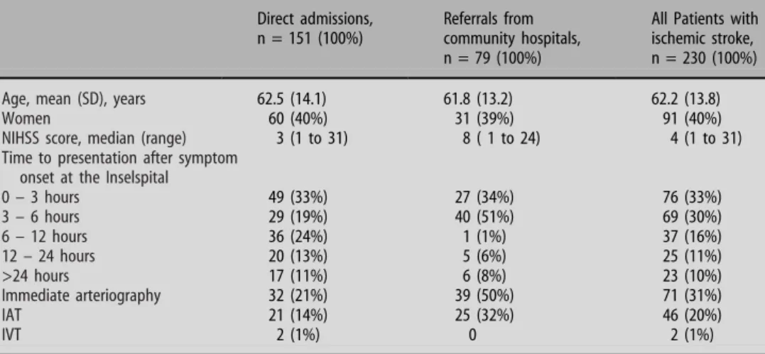
During the study period 230 patients aged 16 to
80 years with acute ischemic stroke were admitted
(Table
1
). The mean age was 62.2 ± 13.8 years. 39.6%
were women. 76 patients (33%) presented within
3 hours, 69 (30%) from 3 to 6 hours, 37 (16%) from 6
to 12 hours, 25 (11%) from 12 to 24 hours and 23
(10%) after 24 hours. 151 patients (66%) were
admitted directly and 79 (34%) via community
hos-pitals. 71 patients (31%) underwent diagnostic
arte-riography and 46 (20% of all patients; 65% of those
with arteriograms) received IAT. Patients referred
from community hospitals were more likely to qualify
for arteriography (50% of 79) and IAT (32% of 79)
than patients who had been admitted directly (21%
and 14% of 151; p < 0.0001). The median NIHSS
score of all patients was 4 (range 1 to 31), of those
treated with IAT 14 (range 3 to 24) and of those not
receiving thrombolysis 3 (range 1 to 31). 115 patients
had a NIHSS score ‡ 4 at admission.
Two patients (1%) were treated within 3 hours of
symptom onset with intravenous recombinant tissue
plasmogen activator (rt-PA), one because of a
dis-sected and occluded internal carotid artery that
pre-vented intraarterial access to the occluded middle
Table 1 Demographic data, time to presentation and rates of diagnostic arteriography, IAT or IVT
Direct admissions, n = 151 (100%)
Referrals from community hospitals, n = 79 (100%)
All Patients with ischemic stroke, n = 230 (100%)
Age, mean (SD), years 62.5 (14.1) 61.8 (13.2) 62.2 (13.8)
Women 60 (40%) 31 (39%) 91 (40%)
NIHSS score, median (range) 3 (1 to 31) 8 ( 1 to 24) 4 (1 to 31)
Time to presentation after symptom onset at the Inselspital
0 – 3 hours 49 (33%) 27 (34%) 76 (33%) 3 – 6 hours 29 (19%) 40 (51%) 69 (30%) 6 – 12 hours 36 (24%) 1 (1%) 37 (16%) 12 – 24 hours 20 (13%) 5 (6%) 25 (11%) >24 hours 17 (11%) 6 (8%) 23 (10%) Immediate arteriography 32 (21%) 39 (50%) 71 (31%) IAT 21 (14%) 25 (32%) 46 (20%) IVT 2 (1%) 0 2 (1%)
NIHSS score indicates National Institutes of Health Stroke Scale Score; IAT, intraarterial thrombolysis; IVT indicates intravenous thrombolysis
cerebral artery, and the other because both
neurora-diologists were busy with other interventions.
No thrombolysis followed the diagnostic
arteriog-raphy in 24 patients (10.5% of 230), in 11 of them no
or only peripheral branch occlusions (M3 or M4) were
seen on arteriography. In one patient an
atheroma-tous pseudoocclusion of the ICA was responsible for
the patient’s symptoms. He was successfully treated
with PTA and stenting. In 9 patients the ICA was
occluded and rendered local IAT of the intracranially
occluded vessels technically impossible. One
men-tioned above received IVT after the arteriography
within three hours of symptom onset. An additional
patient with a hypoplastic vertebral artery on one side
and an occluded vertebral artery on the other side
could not be treated with IAT either. In two patients
the time window for IAT elapsed while trying to reach
the occluded intracranial vessel and in one patient
arterial hypertension was not controllable.
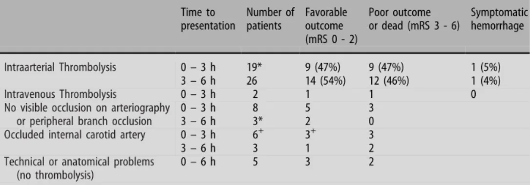
Up to 3 hours 35 of 76 patients underwent
arteri-ography and 19 received IAT. In the 3 to 6 hours
window the numbers are 35 arteriograms in 69
pa-tients and 26 IATs. A patient with a basilar artery
occlusion was treated at 6.5 hours. One patient each
treated with IAT in the groups 0–3 hours and 3–
6 hours had a symptomatic cerebral hemorrhage
(5%).
The reasons for excluding patients presenting
within 6 hours from diagnostic arteriography and
thrombolysis are given in Table
2
. Most frequently
mild symptoms were the reasons. 44 patients (19% of
230) had a NIHSS score £ 4. 13 patients (6%) were
excluded despite NIHSS score ‡ 4 because of rapid
improvement.
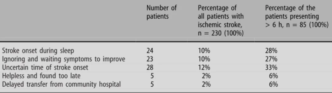
Reasons for delayed presentation after 6 hours are
summarized in Table
3
. The most frequent reasons
were stroke onset during sleep (n = 24), uncertain
time of onset (n = 28), and waiting and expecting
symptoms to improve (n = 23).
Clinical outcome of patients who underwent
thrombolysis and of those patients who were not
treated with thrombolysis after arteriography are
presented in Table
4
.
Discussion
Our data demonstrate that the indications for
arteri-ography with the intention to use IAT for stroke
treatment are given in approximately one of three
patients. Of three patients who undergo arteriography
two would probably receive IAT ultimately, i.e.,
approximately one of five stroke patients or 20% of all
admitted.
Table 2 Reasons to withhold arteriography and thrombolysis in patients presenting within 6 hours of symptom onset Number of patients Percentage of all patients presenting < 6 h, n = 145 Percentage of all patients with ischemic stroke, n = 230
Mild symptoms (NIHSS score < 4, no isolated aphasia or hemianopsia)
44 30% 19%
Rapidly improving neurological deficit 13 9% 6%
CT and clinical findings suggesting lacunar stroke due to small vessel occlusion
7 5% 3%
Limiting comorbidity * 7 5% 2%
Baseline international normalized ratio > 1.7
1 0.7% 0.4%
* Limiting comorbidity: Metastatic cancer n = 2, severe chronic heart failure n = 2, severe chronic obstructive lung disease n = 1, history of stroke with severe disability (n = 2)
Table 3 Reasons for delayed presentation past 6 hours after symptom onset at the Inselspital
Number of patients
Percentage of all patients with ischemic stroke, n = 230 (100%)
Percentage of the patients presenting > 6 h, n = 85 (100%)
Stroke onset during sleep 24 10% 28%
Ignoring and waiting symptoms to improve 23 10% 27%
Uncertain time of stroke onset 28 12% 33%
Helpless and found too late 5 2% 6%
In the first 3 hours 35 of 76 patients qualified for
arteriography. If we had been practicing IVT, they all
would have undergone IVT. However, after
arteriog-raphy 19 patients only received IAT. Six patients had an
occluded ICA that made intracranial IAT technically
impossible. One of them received subsequently IVT;
five did not receive active treatment. They might have
possibly derived a benefit from IVT. Therefore, we have
changed our policy since. When CT or MR angiography
shows carotid occlusion in MCA stroke, we perform
IVT or attempt mechanical recanalization [
17
].
Eight patients presenting within 3 hours did not
show any vessel occlusion on arteriography that could
have been recanalized. Five of them had a favorable
outcome (mRS £ 2), similar to 75% of our experience
in a larger series [
2
]. How much their benefit from
IVT and how big their risk of hemorrhage would have
been remains speculative, because trials with direct
comparisons of IVT, IAT and combined IVT/IAT
have not been published to date. It is likely that
ad-vances in MR and CT imaging will help to select ideal
patients for intravenous or intraarterial treatment
[
11
].
Patients in the 3 to 6 hours time span would not have
been treated with IVT. Therefore they represent the
cohort that has drawn a benefit from the prolongation
of the treatment window because of the intraarterial
approach. Overall, the patients who received IAT in the
3 to 6 hours time span represent 11% of all admissions
and 54% had a favorable outcome.
Patients with a low NIHSS score are likely to
re-cover without any intervention. In randomized trials
patients with a NIHSS score of 4 or more were
in-cluded, a score indicating that some degree of
dis-ability is likely to remain. The NIHSS score at
admission was £ 4 in 115 patients. Of the other 115
patients with a NIHSS score ‡ 4 42% were treated.
The biggest obstacle to administering i.v. rt-PA to
stroke patients is the narrow 3 hours treatment
win-dow. Despite substantial efforts many patients still
arrive between 3 to 6 hours and thus too late for IVT
[
9
]. For these patients IAT or IVT based on clinical
findings and imaging represent worthwhile treatment
that can be implemented in larger institutions.
How-ever, more than a third of all patients presented
be-yond 6 hours, when IAT is generally no longer an
option. In a study by Barber et al. 29% of those
pre-senting after 3 hour had recognized their symptoms
but had chosen not to seek medical attention
imme-diately [
5
]. In our cohort 27% of those presenting
after 6 hours indicated the same reason for late
pre-sentation. Other reasons for delayed presentation or
withholding thrombolysis were stroke during sleep or
uncertain time of symptom onset. Future trials will
show if such patients could benefit from thrombolysis
or mechanical recanalization if they are selected by
combined clinical and imaging criteria and not solely
on the basis of time to treatment.
In conclusion, our data demonstrate that tertiary
care centers with continuously available IAT can offer
thrombolysis to a substantial proportion of patients.
However, many patients still present too late.
There-fore, further widespread efforts are needed to increase
public awareness of stroke and to optimize
organi-zation of emergency services and stroke centers.
j Acknowledgments and Funding We thank Pietro Ballinari, PhD,
for statistical advice. This study was supported in part by a grant of ‘‘Stiftung zur Fo¨rderung der wissenschaftlichen Forschung an der Universita¨t Bern’’
Table 4 Clinical outcome three months after stroke in patients who underwent arteriography Time to presentation Number of patients Favorable outcome (mRS 0 - 2) Poor outcome or dead (mRS 3 - 6) Symptomatic hemorrhage Intraarterial Thrombolysis 0 – 3 h 19* 9 (47%) 9 (47%) 1 (5%) 3 – 6 h 26 14 (54%) 12 (46%) 1 (4%) Intravenous Thrombolysis 0 – 3 h 2 1 1 0
No visible occlusion on arteriography or peripheral branch occlusion
0 – 3 h 8 5 3
3 – 6 h 3* 2 0
Occluded internal carotid artery 0 – 3 h 6+ 3+ 3
3 – 6 h 3 1 2
Technical or anatomical problems (no thrombolysis)
0 – 6 h 5 3 2
* Outcome data missing for one patient,+One patient treated with Intravenous Thrombolysis mRS indicates modified Rankin Scale score
References
1. Arnold M, Nedeltchev K, Mattle HP, Loher TJ, Stepper F, Schroth G, Bre-kenfeld C, Sturzenegger M, Remonda L (2003) Intra-arterial thrombolysis in 24 consecutive patients with internal car-otid artery T occlusions. J Neurol Neurosurg Psychiatry 74:739–742 2. Arnold M, Nedeltchev K, Brekenfeld C,
Fischer U, Remonda L, Schroth G, Mattle H (2004) Outcome of acute stroke patients without visible occlu-sion on early arteriography. Stroke 35:1135–1140
3. Arnold M, Nedeltchev K, Schroth G, Baumgartner RW, Remonda L, Loher TJ, Stepper F, Sturzenegger M, Schuknecht B, Mattle HP (2004) Clini-cal and radiologiClini-cal predictors of re-canalisation and outcome of 40 patients with acute basilar artery occlusion treated with intra-arterial thromboly-sis. J Neurol Neurosurg Psychiatry 75:857–862
4. Arnold M, Schroth G, Nedeltchev K, Loher T, Remonda L, Stepper F, Stur-zenegger M, Mattle HP (2002) Intra-arterial thrombolysis in 100 patients with acute stroke due to middle cere-bral artery occlusion. Stroke 33:1828– 1833
5. Barber PA, Zhang J, Demchuk AM, Hill MD, Buchan AM (2001) Why are stroke patients excluded from TPA therapy? An analysis of patient eligibility. Neurology 56:1015–1020
6. Brott T, Adams HP Jr., Olinger CP, Marler JR, Barsan WG, Biller J, Spilker J, Holleran R, Eberle R, Hertzberg V (1989) Measurements of acute cerebral infarction: a clinical examination scale. Stroke 20:864–870
7. Buchan AM, Barber PA, Newcommon N, Karbalai HG, Demchuk AM, Hoyte KM, Klein GM, Feasby TE (2000) Effectiveness of t-PA in acute ischemic stroke: outcome relates to appropri-ateness. Neurology 54:679–684 8. Furlan A, Higashida R, Wechsler L,
Gent M, Rowley H, Kase C, Pessin M, Ahuja A, Callahan F, Clark WM, Silver F, Rivera F (1999) Intra-arterial pro-urokinase for acute ischemic stroke. The PROACT II study: a randomized controlled trial. Prolyse in Acute Cere-bral Thromboembolism. JAMA 282:2003–2011
9. Grond M, Stenzel C, Schmulling S, Rudolf J, Neveling M, Lechleuthner A, Schneweis S, Heiss WD (1998) Early intravenous thrombolysis for acute ischemic stroke in a community-based approach. Stroke 29:1544–1549 10. Grotta JC, Burgin WS, El-Mitwalli A,
Long M, Campbell M, Morgenstern LB, Malkoff M, Alexandrov AV (2001) Intravenous tissue-type plasminogen activator therapy for ischemic stroke: Houston experience 1996 to 2000. Arch Neurol 58:2009–2013
11. Hacke W, Albers G, Al-Rawi Y, Bo-gousslavsky J, Davalos A, Eliasziw M, Fischer M, Furlan A, Kaste M, Lees KR, Soehngen M, Warach S; DIAS Study Group (2005) The Desmoteplase in Acute Ischemic Stroke Trial (DIAS): a phase II MRI-based 9-hour window acute stroke thrombolysis trial with intravenous desmoteplase. Stroke 36:66–73
12. Hacke W, Donnan G, Fieschi C, Kaste M, von Kummer R, Broderick JP, Brott T, Frankel M, Grotta JC, Haley EC Jr, Kwiatkowski T, Levine SR, Lewandow-ski C, Lu M, Lyden P, Marler JR, Patel S, Tilley BC, Albers G, Bluhmki E, Wilhelm M, Hamilton S, ATLANTIS Trials Investigators; ECASS Trials Investigators, NINDS rt-PA Study Group Investigators (2004) Association of outcome with early stroke treatment: pooled analysis of ATLANTIS, ECASS, and NINDS rt-PA stroke trials. Lancet 363:768–774
13. Heuschmann PU, Berger K, Misselwitz B, Hermanek P, Leffmann C, Adelmann M, Buecker-Nott HJ, Rother J, Neun-doerfer B, Kolominsky-Rabas PL (2003) Frequency of thrombolytic therapy in patients with acute ischemic stroke and the risk of in-hospital mortality: the German Stroke Registers Study Group. Stroke 34:1106–1113 14. Katzan IL, Graber TM, Furlan AJ,
Sundararajan S, Sila CA, Houser G, Landis DM. Cuyahoga (2003) County Operation Stroke Speed of Emergency Department Evaluation and Compli-ance With National Institutes of Neu-rological Disorders and Stroke Time Targets. Stroke 36:6
15. Lindsberg PJ, Soinne L, Roine RO, Salonen O, Tatlisumak T, Kallela M, Happola O, Tiainen M, Haapaniemi E, Kuisma M, Kaste M (2003) Commu-nity-based thrombolytic therapy of acute ischemic stroke in Helsinki. Stroke 34:1443–1449
16. Nedeltchev K, Arnold M, Brekenfeld C, Isenegger J, Remonda L, Schroth G, Mattle HP (2003) Pre- and in-hospital delays from stroke onset to intra-arte-rial thrombolysis. Stroke 34:1230–1234 17. Nedeltchev K, Brekenfeld C, Remonda L, Ozdoba CH, Do D, Arnold M, Mattle HP, Schroth G. Stenting Of The Inter-nal Carotid Artery in Acute Stroke: Preliminary Results Of 25 Patients. Radiology, in press
18. Macleod MR, Davis SM, Mitchell PJ, Gerraty RP, Fitt G, Hankey GJ, Stewart-Wynne EG, Rosen D, McNeil JJ, Bladin CF, Chambers BR, Herkes GK, Young D, Donnan GA (2005) Results of a Multicentre, Randomised Controlled Trial of Intra-Arterial Urokinase in the Treatment of Acute Posterior Circula-tion Ischaemic Stroke. Cerebrovasc Dis. 20(1):12–17
19. Mattle HP, Kappeler L, Arnold M, Fi-scher U, Nedeltchev K, Remonda L, Jakob SM, Schroth G (2005) Blood pressure and vessel recanalization in the first hours after ischemic stroke. Stroke 36:264–268
20. Reed SD, Cramer SC, Blough DK, Meyer K, Jarvik JG (2001) Treatment with tissue plasminogen activator and inpatient mortality rates for patients with ischemic stroke treated in com-munity hospitals. Stroke 32:1832–1840 21. Schmulling S, Grond M, Rudolf J, Heiss
WD (2000) One-year follow-Up in acute stroke patients treated with rtPA in clinical routine. Stroke 31:1552–1554 22. The National Institute of Neurological Disorders, Stroke rt-PA Stroke Study Group (1995) Tissue plasminogen activator for acute ischemic stroke. N Engl J Med 333:1581–1587
23. van Swieten JC, Koudstaal PJ, Visser MC, Schouten JH, von Gijn J (1988) Interobserver agreement for the assessment of handicap in stroke pa-tients. Stroke 19:604–607
24. Wang DZ, Rose JA, Honings DS, Gar-wacki DJ, Milbrandt JC (2000) Treating acute stroke patients with intravenous tPA. The OSF stroke network experi-ence. Stroke 31:77–81
25. Weber J, Remonda L, Mattle HP, Ko-erner U, Baumgartner RW, Sturzeneg-ger M, Ozdoba C, Koerner F, Schroth G (1998) Selective intra-arterial fibrino-lysis of acute central retinal artery occlusion. Stroke 29:2076–2079