• Aucun résultat trouvé

Posttraumatic lateral ankle pain

N/A
N/A
Protected

Academic year: 2021

Partager "Posttraumatic lateral ankle pain"

Copied!
2
0
0

Texte intégral

(1)

TEST YOURSELF: QUESTION

Posttraumatic lateral ankle pain

Michele Pansini

&

Stefan Schlosser

&

Aline R. Buck

&

Florian M. Buck

Received: 13 October 2010 / Revised: 6 January 2011 / Accepted: 24 January 2011 / Published online: 22 February 2011 # ISS 2011

History

A 25-year-old woman presented with pain at the lateral

contour of her left ankle and foot after having suffered a

motorcycle accident with contusion of the left foot 3 weeks

earlier. After an interval of decreasing pain for some days,

pain recurred and the patient presented to the emergency

department of our hospital. During the clinical examination

the patient complained about a circumscribed painful area

at the lateral contour of the left foot with evidence of local

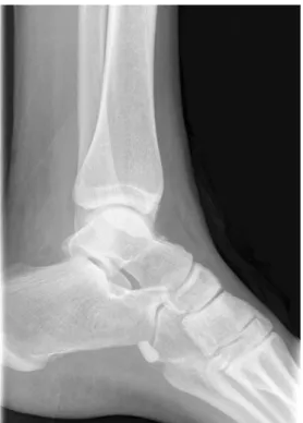
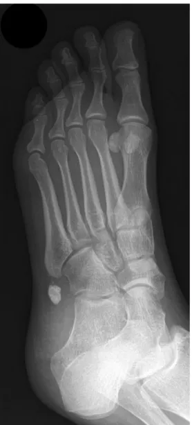
inflammation. Standard lateral (Fig.

1

), dorsoplantar

(Fig.

2

), and oblique radiographs (Fig.

3

) of the foot were

performed.

Fig. 1 Lateral radiograph of the foot The diagnosis can be found at doi:10.1007/s00256-011-1116-4.

The authors declare that there is no conflict of interest. M. Pansini

:

F. M. Buck (*)

Department of Radiology and Nuclear Medicine, University Hospital of Basel,

Petersgraben 4,

CH-4031 Basel, Switzerland e-mail: florian.buck@gmail.com S. Schlosser

Department of Surgery, University Hospital of Basel, Petersgraben 4,

CH-4031 Basel, Switzerland A. R. Buck

Department of Internal Medicine, Triemli City Hospital, Birmensdorferstrasse 497,

CH-8063 Zurich, Switzerland Skeletal Radiol (2011) 40:771–772 DOI 10.1007/s00256-011-1117-3

(2)

Fig. 3 Oblique radiograph of the foot Fig. 2 Dorsoplantar of the foot

Figure

Fig. 1 Lateral radiograph of the footThe diagnosis can be found at doi:10.1007/s00256-011-1116-4.
Fig. 3 Oblique radiograph of the foot Fig. 2 Dorsoplantar of the foot

Références

Documents relatifs

In this section, and throughout the text, the basic method for solving an equation in one variable involves writing a sequence of equivalent equations until we finally reach

Si 45 lanternes identiques coˆ utent 2569,50 $, combien coˆute chaque

Plantar flexion activation patterns initiate the foot movement by first contracting muscles that allow the plantar flexion of the ankle (Tab 1, bottom lines): 1) The first

81 However, mechanochromic materials that displayed a macroscopically discernible change of the absorption were only obtained when mechanical stresses led to a very

Higher Grade sprains that include CFL injury result in significant decreases in rotation stiffness, peak torque, substantial alteration of contact mechanics at the ankle joint,

Interestingly, the Foot and Ankle Ability Measure (FAAM), which is one of few outcome scores validated for ankle instability, was not used in any of the selected articles

This sudden increase in inversion and internal rotation forces, combined with either dorsi- or plantarflexion, produces sufficient strains to rupture the ankle lateral

The sole of the foot was driven by an imposed harmonic displacement y in (t). Masses m b and m c were computed according to the study of Matsumoto and Griffin, 2003, considering