ORIGINAL ARTICLE
Ultrafast nuclear myocardial perfusion imaging
on a new gamma camera with semiconductor detector
technique: first clinical validation
Ronny R. Buechel&Bernhard A. Herzog&Lars Husmann&Irene A. Burger&
Aju P. Pazhenkottil&Valerie Treyer&Ines Valenta&Patrick von Schulthess&
René Nkoulou&Christophe A. Wyss&Philipp A. Kaufmann
Received: 18 August 2009 / Accepted: 3 November 2009 / Published online: 27 January 2010 # Springer-Verlag 2010
Abstract
Purpose To assess the diagnostic performance of a novel ultrafast cardiac gamma camera with cadmium-zinc-telluride (CZT) solid-state semiconductor detectors for nuclear myocardial perfusion imaging (MPI).
Methods The study group comprised 75 consecutive patients (55 men, BMI range 19–45 kg/m2
) who underwent a 1-day 99mTc-tetrofosmin adenosine-stress/rest imaging protocol. Scanning was performed first on a conventional dual-detector SPECT gamma camera (Ventri, GE Health-care) with a 15-min acquisition time each for stress and rest. All scans were immediately repeated on an ultrafast CZT camera (Discovery 530 NMc, GE Healthcare) with a 3-min scan time for stress and a 2-min scan time for rest. Clinical agreement (normal, ischaemia, scar) between CZT and SPECT was assessed for each patient and for each coronary territory using SPECT MPI as the reference standard. Segmental myocardial tracer uptake values (percent of maximum) using a 20-segment model and left ventricular ejection fraction (EF) values obtained using CZT were compared with those obtained using
convention-al SPECT by intraclass correlation and by cconvention-alculating Bland-Altman limits of agreement.
Results There was excellent clinical agreement between CZT and conventional SPECT on a per-patient basis (96.0%) and on a per-vessel territory basis (96.4%) as shown by a highly significant correlation between segmen-tal tracer uptake values (r=0.901, p<0.001). Similarly, EF values for both scanners were highly correlated (r=0.976, p<0.001) with narrow Bland-Altman limits of agreement (−5.5–10.6%).
Conclusion The novel CZT camera allows a more than fivefold reduction in scan time and provides clinical information equivalent to conventional standard SPECT MPI.
Keywords Ultrafast cardiac gamma camera . Myocardial perfusion imaging . Scan time reduction . Cadmium-zinc-telluride
Introduction
Myocardial perfusion imaging (MPI) with single photon emission CT (SPECT) is the most widely used nuclear cardiac imaging technique for the noninvasive assessment of cardiac disease including prognosis and choice of the most appropriate treatment strategies for patients with known or suspected ischaemic coronary artery disease (CAD) [1]. Nevertheless, several drawbacks such as, for example, time-consuming acquisition prone to motion artefacts, cumbersome MPI protocols, and radiation expo-sure still affect SPECT MPI. Despite numerous refinements regarding iterative reconstruction algorithms [2], early-imaging protocols [3], and tracer developments [4], fundamental advances in the hardware equipment utilized
Ronny R. Buechel and Bernhard A. Herzog contributed equally to this work.
R. R. Buechel
:
B. A. Herzog:
L. Husmann:
I. A. Burger:
A. P. Pazhenkottil:
V. Treyer:
I. Valenta:
P. von Schulthess:
R. Nkoulou:
C. A. Wyss:
P. A. Kaufmann (*)Cardiac Imaging, University Hospital Zurich, Ramistrasse 100,
CH-8091 Zurich, Switzerland e-mail: [email protected]
P. A. Kaufmann
Zurich Center for Integrative Human Physiology (ZIHP), University of Zurich,
have lagged behind. This is particularly evident when compared to the impressive advances in the field of multislice CT coronary angiography [5, 6]. It is in this setting that the latest generation of gamma cameras [7] with a novel semiconductor cadmium-zinc-telluride (CZT) de-tector technology instead of the conventional sodium iodide crystals is perceived as the anticipated technical milestone in nuclear cardiac MPI. In fact, preliminary results indicate that incorporation of CZT detectors into cardiac gamma cameras allows a more than fivefold shortening of scan time [8] enabled by the increased system sensitivity due to the use of semiconductors and the stationary detector array. The latter is made possible by the miniaturized CZT detectors and a multipinhole design that allow a serial alignment of the detectors around the patient, thus covering the entire heart at all times. This renders camera rotation around the patient unnecessary.
However, no data are available on the clinical and diagnostic performance of rapid scanning with such a device. Therefore, the aim of the present study was to assess the agreement between MPI obtained using ultrafast scanning with a novel CZT camera and MPI acquired on a conventional SPECT camera with standard scan duration.
Subjects and methods Study protocol
The study group comprised 75 consecutive patients referred to SPECT MPI for evaluation of CAD who underwent MPI on a conventional standard dual-detector SPECT camera (Ventri, GE Healthcare) and immediately thereafter on a novel ultrafast CZT camera (Discovery NM 530c, GE Healthcare). All patients underwent a 1-day
99m
Tc-tetrofosmin adenosine-stress/rest imaging protocol in accordance with the guidelines of the European Association of Nuclear Medicine [9]. Pharmacological stress was induced by adenosine (continuous infusion at 140 μg/kg per minute) or dobutamine (incrementally infused, starting at 5μg/kg per minute and increasing at 1-min intervals to a maximum of 60 μg/kg per minute until 85% of the age-predicted heart rate had been reached). 99mTc-tetrofosmin was injected after 3 min of induced stress. The injection was followed by a waiting time of 90 min before image acquisition using the SPECT camera, which was immedi-ately followed by acquisition using the CZT camera. Rest MPI was then performed with the identical acquisition protocol after injection of a three times higher dose of
99m
Tc-tetrofosmin.
The study protocol was approved by the local ethics committee and written informed consent was obtained from every patient.
Image acquisition
With the SPECT camera scans were acquired using a low-energy, high-resolution collimator, a 20% symmetrical window at 140 keV, a 64×64 matrix, and an elliptical orbit with step-and-shoot acquisition at 3° intervals over a 180° arc (45° anterior oblique to 45° left posterior oblique) with 30 steps (60 views). Electrocardiogram-gated scans were acquired using 16 bins. Scan time was set to 25 s per frame for stress and rest, resulting in a total scan time (including inter-step rotation time) of 14 min 52 s according to the guidelines.
With the CZT camera scans were acquired using a multi-pinhole collimator (effective diameter aperture of 5.1 mm) and 19 stationary detectors simultaneously imaging 19 views of the heart. Each detector contained 32×32 pixelated (2.46×2.46 mm) CZT elements. The system design allowed acquisition without detector or collimator motion. A 10% symmetrical energy window at 140 keV was used. Scans with the CZT camera were acquired over 3 min for stress and 2 min for rest, which has recently been identified as the optimized protocol for obtaining adequate image quality with the shortest scan time [8]. The system design with CZT detectors aligned around the patient, covering the entire heart, allowed acquisition of all cardiac views simultaneously and therefore avoided the need for detector rotation, as opposed to the SPECT acquisition.
MPI reconstruction
Conventional SPECT images were reconstructed on a dedicated workstation using a standard iterative recon-struction algorithm with ordered subset expectation max-imization (OSEM). CZT images were reconstructed on the same workstation using a new dedicated iterative algo-rithm with maximum likelihood expectation maximization (MLEM). The software package Myovation for Alcyone (GE Healthcare) was used for analysis and a Butterworth postfilter was applied to the reconstructed slices. All images were reconstructed in the standard axes (short axis, vertical long axis, horizontal long axis) and polar maps of the left ventricle were created.
Quantitative analysis
Quantitative analysis was performed on MPI polar maps using a 20-segment model for the left ventricle [10]. Uptake was normalized to 100% peak activity and relative myocardial uptake (percent of maximum myocardial up-take) was assessed for each segment from the SPECT and CZT data. Automated analysis on gated SPECT was performed to determine the left ventricular ejection fraction (EF).
Visual analysis
Each single scan from both conventional SPECT and CZT was analysed by two experienced nuclear cardiologists in a randomized and mixed sequence with regard to patients and scans (i.e. stress and rest scans from one patient were not analysed consecutively) and blinded for any information as to patient identification, stress or rest scan, or the camera utilized. The presence or absence of perfusion defects was evaluated on a per-patient basis as well as on a per-vessel territory basis (i.e. left anterior descending artery, segments 1, 2, 7, 8, 13, 14, 15, 19 and 20; circumflex artery, segments 5, 6, 11, 12, 17 and 18; right coronary artery, segments 3, 4, 9, 10 and 16). Image quality was assessed subjectively on a four-point scale (1 poor, 2 fair, 3 good, 4 excellent). Differences between the readers were resolved by consensus.
Statistical analysis
Quantitative variables are expressed as means±SD and categorical variables as percentages or means. The concor-dance between percent tracer uptake and EF from SPECT and CZT images was evaluated using intraclass correlation analysis. In addition, Bland-Altman limits of agreement were calculated. Clinical interobserver agreement was assessed by kappa statistics. A two-tailed probability value of 0.05 or less was considered significant and 95% confidence intervals are presented. SPSS 15.0 (SPSS, Chicago, IL) was used for statistical testing.
Results
All 75 patients successfully underwent MPI with both cameras. The patient characteristics are given in Table 1. Patients were referred to SPECT MPI for the following reasons: suspected CAD (n=36, 48%), follow-up of a known CAD (n=29, 39%), preoperative risk assessment before major noncardiac surgery (n=10, 13%). In 70 patients (93%) pharmacological stress was induced with adenosine and in 5 patients (7%) with dobutamine. For stress and rest the mean doses of 99mTc-tetrofosmin administered were 337±29 MBq (range 303–455 MBq) and 948±79 MBq (range 894–1275 MBq), respectively. Quantitative analysis
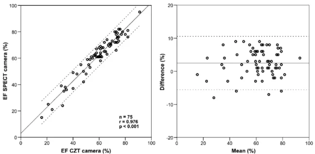
The intraclass correlation coefficient for percent segmental tracer uptake was 0.901 (CI 0.893–0.907; p<0.001) and Bland-Altman limits of agreement were−16.3–15.4%. The quantitative comparison of the EF resulted in an intraclass correlation coefficient of 0.976 (CI 0.936–0.989; p<0.001)
with Bland-Altman limits of agreement of −5.6–10.6% (Fig. 1).
Visual analysis
For the conventional SPECT scans, image quality was good in 12 patients (16%) and excellent in 63 patients (84%), compared to good image quality in 11 (15%) and excellent quality in 64 (85%) for the CZT scans. The interobserver agreement regarding clinical findings was excellent (kappa 0.926).
SPECT MPI revealed myocardial scar tissue in 48 patients (64%) and ischaemia in 3 patients (4%). Compa-rably, CZT revealed scar tissue in 45 patients (60%) and
Table 1 Patient baseline characteristics (n=75)
Characteristic Value Male, n (%) 55 (74) Age (years) Mean±SD 65±10 Range 43–87 BMI (kg/m2) Mean±SD 27±4 Range 19–45
Cardiovascular risk factors, n (%)
Obesity (BMI >30 kg/m2) 17 (23)
Smoking 17 (23)
Diabetes mellitus 13 (17)
Hypertension 49 (65)
Dyslipidaemia 40 (53)
Positive family history 14 (19)
Clinical symptoms, n (%)
Typical angina pectoris 13 (17)
Atypical chest pain 28 (37)
Dyspnoea 8 (11)
No cardiac symptoms 26 (35)
Clinical findings, n (%)
Abnormal resting ECG 7 (9)
Abnormal exercise ECG 10 (13)
Abnormal echocardiography 1 (1) Previous cardiac events, n (%)
Myocardial infarction 20 (27)
Invasive coronary angiography 27 (36) Percutaneous coronary intervention 20 (27) Coronary artery bypass grafting 10 (13) Current cardiac medication, n (%)
Aspirin 32 (43)
Beta-blocker 30 (40)
ACE/angiotensin II inhibitor 29 (39)
ischaemia in 3 patients (4%), yielding an agreement of 96.0% between the CZT camera and SPECT MPI (Table 2).
Of the 225 analysed coronary territories, 69 (31%) revealed a perfusion defect in both the conventional SPECT scans and the CZT scans. The CZT camera reached an overall agreement of 96.4% per vessel territory. The results for each vessel territory (left anterior descending, left circumflex, and right coronary arteries) are illustrated in Fig.2.
Discussion
The present study is the first to clinically validate MPI on a novel ultrafast camera with static CZT semiconductor detector technology against MPI obtained on a clinically established standard SPECT gamma camera using a widely
used routine protocol. The CZT camera allowed rapid scanning (2 and 3 min compared to standard 15 min), with an image quality at least as good as scanning on a standard SPECT camera (Fig. 3). Furthermore, and even more importantly, our data showed an excellent diagnostic performance with the rapid scanning compared to conven-tional SPECT. This substantial reduction in scan time not only offers greater patient comfort which may translate into reduced probability of motion artefacts, but also potentially allows an increase in patient throughput and therefore improved scanner efficiency. Alternatively, the higher sensitivity of the ultrafast camera system may allow a reduction in tracer activity and, instead of scan duration, consequently, a decrease in effective radiation dose.
Interestingly, the excellent comparability of the CZT and SPECT results also held true for functional data such as EF, although gated acquisition is associated with a further
Fig. 1 Linear regression analysis (left) and Bland-Altman plot (right) comparing left ventricular EF from CZT and that from conventional SPECT MPI. Bland-Altman limits of agreement were−5.6–10.6%
Table 2 Diagnostic performance of CZT compared to conventional SPECT n True positive False positive True negative False negative Sensitivity (%) Specificity (%) Positive predictive value (%) Negative predictive value (%) Accuracy (%) Per patient 75 48 0 24 3 94 100 89 100 96 Per territory All vessels 225 65 4 152 4 94 97 97 94 96 Left anterior descending artery 75 12 2 58 3 80 97 95 86 93
Left circumflex artery 75 7 1 66 1 88 99 99 88 97
decrease in count statistics and increased noise. In nuclear imaging the trade-off between image acquisition time and noise levels has determined the standard protocols and scan time. Recently, new iterative reconstruction algorithms incorporating noise regularization and resolution recovery that allow shorter acquisition times have been reported [11,
12]. In the present study, such algorithms were implemented for reconstruction of images acquired on the CZT camera.
The CZT detectors directly convert radiation into electrical signals [13], yielding an improved energy discrimination and spatial resolution (radial resolution 4.3 mm). The miniaturization of the semiconductors has enabled a more compact detector design, facilitating the engineering of a circular and therefore stationary detector
array which simultaneously acquires all views necessary for tomographic reconstruction, avoiding the time-consuming rotation around the patient. The integration of these develop-ments into a clinical device allows substantial shortening of scan duration from 15 min to 2–3 min [8]. Our results establish the clinical validity of rapid scanning on a novel system, which incorporates all the mentioned develop-ments, including latest semiconductor detectors, novel design geometry, and innovative reconstruction algorithms. A limitation of the present study could be that we did not use coronary angiography as the standard of reference. However, we felt that perfusion from CZT would be most appropriately compared to a standard for perfusion. By contrast, coronary angiography is a flawed gold standard for myocardial perfusion, as the pathophysiological relevance of a lesion may depend on many factors that cannot be assessed from anatomy alone. In addition, we did not use any correction for tissue attenuation although we included patients representing a broad range of BMI. However, the impact of attenuation correction on image quality and agreement of MPI with CZT was beyond the scope of this study. Furthermore, improvements in software components may have contributed, at least in part, to the increased CZT system sensitivity. However, although some of these components may have the potential to improve conventional SPECT performance, their implementation would require extensive scanner-specific validation of each component
Fig. 2 Agreement between CZT and conventional SPECT MPI in revealing perfusion defects (LAD left anterior descending artery, LCX left circumflex artery, RCA right coronary artery)
Fig. 3 MPI in a 78-year old man with a history of anteroapical myocardial infarction reveals a large anteroapical fixed defect indicating a scar without ischaemia. The images show a clear clinical
agreement between conventional SPECT (with 15-min acquisition times) and CZT scans (with 3- and 2-min acquisition times)
[11,12]. As we intended to validate a new system against the established standard, we adhered to the standard of reference according to the EANM procedural guidelines [9] using a conventional SPECT camera with standard scan durations and established reconstruction algorithms.
Conclusion
Our results demonstrate that the novel ultrafast MPI camera using CZT semiconductor detector technology provides an excellent clinical agreement compared to a conventional SPECT camera with significantly reduced scan times.
Acknowledgments This study was supported by a grant from the Swiss National Science Foundation and by the ZIHP (Zurich Center for Integrative Human Physiology, University of Zurich). We would like to thank Edlira Loga, Ennio Mueller, Josephine Trinckauf, Verena Wechselbaumer and Mirjam De Bloeme for their technical support. Conflicts of interest The University Hospital Zurich holds a research contract with GE Healthcare.
References
1. Thomas GS, Miyamoto MI, Morello AP 3rd, Majmundar H, Thomas JJ, Sampson CH, et al. Technetium 99m sestamibi myocardial perfusion imaging predicts clinical outcome in the community outpatient setting. The Nuclear Utility in the Com-munity (NUC) Study. J Am Coll Cardiol 2004;43:213–23. 2. Seo Y, Mari C, Hasegawa BH. Technological development and
advances in single-photon emission computed tomography/ computed tomography. Semin Nucl Med 2008;38:177–98. 3. Giorgetti A, Rossi M, Stanislao M, Valle G, Bertolaccini P,
Maneschi A, et al. Feasibility and diagnostic accuracy of a gated
SPECT early-imaging protocol: a multicenter study of the Myo-view Imaging Optimization Group. J Nucl Med 2007;48:1670–5. 4. Kapur A, Latus KA, Davies G, Dhawan RT, Eastick S, Jarritt PH, et al. A comparison of three radionuclide myocardial perfusion tracers in clinical practice: the ROBUST study. Eur J Nucl Med Mol Imaging 2002;29:1608–16.
5. Husmann L, Valenta I, Gaemperli O, Adda O, Treyer V, Wyss CA, et al. Feasibility of low-dose coronary CT angiography: first experience with prospective ECG-gating. Eur Heart J 2008;29:191–7.
6. Rybicki FJ, Otero HJ, Steigner ML, Vorobiof G, Nallamshetty L, Mitsouras D, et al. Initial evaluation of coronary images from 320-detector row computed tomography. Int J Cardiovasc Imaging 2008;24:535–46.
7. Berman DS, Kang X, Tamarappoo B, Wolak A, Hayes SW, Nakazato R, et al. Stress thallium-201/rest technetium-99m sequential dual isotope high-speed myocardial perfusion imaging. JACC Cardiovasc Imaging 2009;2:273–82.
8. Herzog BA, Buechel RR, Katz R, Brueckner M, Husmann L, Burger IA, et al. Nuclear myocardial perfusion imaging with a novel cadmium-zinc-telluride detector technique: optimized protocol for scan time reduction. J Nucl Med 2010;51:46–51. 9. Hesse B, Tagil K, Cuocolo A, Anagnostopoulos C, Bardies M,
Bax J, et al. EANM/ESC procedural guidelines for myocardial perfusion imaging in nuclear cardiology. Eur J Nucl Med Mol Imaging 2005;32:855–97.
10. Hansen CL, Goldstein RA, Akinboboye OO, Berman DS, Botvinick EH, Churchwell KB, et al. Myocardial perfusion and function: single photon emission computed tomography. J Nucl Cardiol 2007;14:e39–60.
11. Ali I, Ruddy TD, Almgrahi A, Anstett FG, Wells RG. Half-time SPECT myocardial perfusion imaging with attenuation correction. J Nucl Med 2009;50:554–62.
12. Valenta I, Treyer V, Husmann L, Gaemperli O, Schindler MJ, Herzog BA, et al. New reconstruction algorithm allows shortened acquisition time for myocardial perfusion SPECT. Eur J Nucl Med Mol Imaging 2009;in press. doi:10.1007/s00259-009-1300-0
13. Madsen MT. Recent advances in SPECT imaging. J Nucl Med 2007;48:661–73.