HAL Id: dumas-03087034
https://dumas.ccsd.cnrs.fr/dumas-03087034
Submitted on 23 Dec 2020HAL is a multi-disciplinary open access archive for the deposit and dissemination of sci-entific research documents, whether they are
pub-L’archive ouverte pluridisciplinaire HAL, est destinée au dépôt et à la diffusion de documents scientifiques de niveau recherche, publiés ou non,
Development of a new right ventricle evaluation
technique using a gamma camera cadmium-zinc-telluride
Antoine Apert
To cite this version:
Antoine Apert. Development of a new right ventricle evaluation technique using a gamma camera cadmium-zinc-telluride. Human health and pathology. 2020. �dumas-03087034�
AVERTISSEMENT
Ce document est le fruit d'un long travail approuvé par le
jury de soutenance et mis à disposition de l'ensemble de la
communauté universitaire élargie.
Il n’a pas été réévalué depuis la date de soutenance.
Il est soumis à la propriété intellectuelle de l'auteur. Ceci
implique une obligation de citation et de référencement
lors de l’utilisation de ce document.
D’autre part, toute contrefaçon, plagiat, reproduction illicite
encourt une poursuite pénale.
Contact au SID de Grenoble :
[email protected]
LIENS
LIENS
Code de la Propriété Intellectuelle. articles L 122. 4
Code de la Propriété Intellectuelle. articles L 335.2- L 335.10
http://www.cfcopies.com/juridique/droit-auteurUNIVERSITÉ GRENOBLE ALPES
UFR DE MÉDECINE DE GRENOBLE
Année : 2020
DEVELOPMENT OF A NEW RIGHT VENTRICLE EVALUATION TECHNIQUE
USING A GAMMA CAMERA CADMIUM-ZINC-TELLURIDE
THÈSE
PRÉSENTÉE POUR L’OBTENTION DU TITRE DE DOCTEUR EN MÉDECINE DIPLÔME D’ÉTAT
Antoine APERT
THÈSE SOUTENUE PUBLIQUEMENT À LA FACULTÉ DE MÉDECINE DE GRENOBLE Le : 21/04/2020
DEVANT LE JURY COMPOSÉ DE Président du jury :
Monsieur le Professeur Gérald VANZETTO Membres :
Monsieur le Professeur Gilles BARONE-ROCHETTE (directeur de thèse) Monsieur le Professeur Daniel FAGRET
Madame le Docteur Aude BOIGNARD Monsieur le Docteur Loïc DJAILEB Monsieur le Docteur Adrien JANKOWSKI
REMERCIEMENTS
À Monsieur le Professeur Gérald VANZETTO, vous me faites l’honneur de présider ce jury. Vos connaissances, votre pédagogie et votre calme à toute épreuve ne cessent de m’impressionner. Veuillez trouver ici le témoignage de toute ma reconnaissance.
À Monsieur le Professeur Gilles BARONE-ROCHETTE, pour tes idées à l’origine de cette thèse.Je te remercie de tes conseils avisés, de ton accompagnement précieux tout au long de mon internat, de ta disponibilité et ton dynamisme à tout moment.
À Monsieur le Professeur Daniel FAGRET, pour avoir accepté de siéger à ce jury et d’évaluer ce travail ; veuillez trouver ici le témoignage de toute ma gratitude.
À Madame le Docteur Aude BOIGNARD, pour ta gentillesse, ta discrétion associée à un grand professionnalisme. Je garde un excellent souvenir de mon stage en chirurgie cardiaque à tes cotés.
À Monsieur le Docteur Loïc DJAILEB, pour ta participation soutenue dans la réalisation de ce travail ainsi que pour tes précieux conseils. Merci pour ta bonne humeur, ton enthousiasme et ta disponibilité, en toutes circonstances. Ce fut un plaisir de travailler avec toi.
À Monsieur le Docteur Adrien JANKOWSKI, pour votre présence dans ce jury et l’attention que vous porterez à ce travail ; veuillez trouver ici l’expression de ma sincère reconnaissance.
7
À mes parents, pour votre amour et votre patience. J’ai bien conscience que vous avez toujours été là pour moi durant toutes ces années, merci de m’avoir toujours soutenu dans mes projets. Je n’y serais pas arrivé sans vous.
À ma sœur Juliette, pour tous ces bons moments passés ensemble et surtout ceux à venir. Sache que je suis très fier d’avoir une petite sœur comme toi-même si je ne te le dis pas tout le temps. Prépare toi, c’est bientôt ton tour !
À ma grand-mère Jacqueline, pour ta bienveillance, ta droiture et ton esprit de famille qui me laissent admiratif. Et bien sûr pour ta patience quand nous faisions « la Java » entre cousins. À ma grand-mère Françoise, pour ta douceur, ta gentillesse pour veiller à ce que nous ne manquions de rien, pour m’avoir toujours encouragé. Merci mamie.
À mon grand-père Pierre, j’ai sans doute suivi ta voie en prenant le chemin de la médecine et bien évidemment tu n’y es certainement pas pour rien. Je suis très fier d’avoir un papi comme toi. Très cher collègue !
À mon grand-père René, tu m’as laissé le gout de l’espagnol assurément, j’imagine que tu aurais été heureux de nous voir grandir ainsi avec Juju. Aussi longtemps que je me souvienne, tu auras été un papi formidable.
À toute ma famille avec mes oncles et tantes, Jean - Yves pour ces parties de ping-pong et sorties en vélo dans la plus belle région du monde. Renée tout aussi discrète que sympathique. Anne un peu moins discrète, tu respires le bonheur et la joie de vivre. Jean Luc, solitaire, passionné de montagne et concepteur de planeur. A mes cousins préférés Claire et Vincent et ces formidables moment d’insouciance dans notre jeunesse, les vacances ensemble à faire enrager les grands parents, mais surtout les parties de Uno où je vous mettais de belles pâtées !
Aux médecins et paramédicaux que j’ai côtoyés pendant mon internat
À Carole Saunier, pour ton humour et tes précieux conseils en échographie. Deux domaines où tu excelles. J’ai vraiment adoré ce dernier semestre en ta compagnie. Encore merci pour le comté !
À Charlotte Casset, pour ta gentillesse, ta bienveillance et ta patience lors de mes premiers pas d’interne.
À Muriel Salvat, pour ton dynamisme, ta sensibilité et toujours à l’écoute ! Merci encore de m’avoir aidé à recruter quelques patients pour ce travail.
À Caroline Augier, si dévouée, patiente et toujours souriante.
À Marion Maurin, pour ta gentillesse et tes compétences.
À Estelle Vautrin, pour ton sens de l’humour et tes supers visites au 8C. À Stéphanie Marlière, pour ton dynamisme et ton efficacité.
À Pierre-Vladimir Ennezat, jamais avare de bons conseils, je vous remercie pour votre éternelle bonne humeur dans toutes situations !
À Peggy Jacon, pour ta disponibilité et ta bienveillance. À Hélène Bouvaist, pour votre abnégation sans faille. À Sandrine Venier, pour ta bonne humeur.
À Pascal Defaye, pour vos compétences.
À Olivier Ormezzano, pour m’avoir soutenu lors de mon premier semestre. À Bernard Bertrand, pour votre phrasé inégalable.
9
À Adrien Carabelli, pour ton calme, ta sympathie, ton accessibilité, ne change rien ! À Marjorie Canu, pour ta douceur, ta générosité, tes compétences et ta modestie. Tu fais une formidable assistante.
À l’équipe médicale de chirurgie cardiaque, merci de votre accueil pendant mon semestre. Une pensée particulière à Cécile et Maria pour votre bienveillance.
À l’équipe médicale de la réanimation CVT, pour ce semestre plein de découvertes et d’apprentissages au sein de votre belle équipe.
À l’équipe médicale et paramédicale du service de cardiologie du GHM.
Aux équipes soignantes côtoyées pendant mon internat et en général. Vous êtes formidables, merci pour tout le travail que vous accomplissez pour les patients et les familles. J’ai toujours pris beaucoup de plaisir à vous côtoyer. Vous êtes constamment en première ligne et toujours les premiers à nous réconforter dans nos moments de doutes. Mais ce que je retiens le plus, ce sont les nombreux moments de rigolade !
À l’équipe médicale et paramédicale du service de cardiologie du CH d’Annecy. J’ai hâte de continuer ma route auprès de vous.
À mes co internes en or.
À Thomas, pour notre semestre à l’USIC et nos soccers. Ce fut un réel plaisir de travailler avec toi et de te côtoyer en général. Ton calme, ta sensibilité et ton sens de l’humour font de toi une personne en or. J’espère que l’on gardera contact encore longtemps Toto.
À Anne, pour notre 1er semestre laborieux mais heureux. Tu es aussi discrète que rigolote,
toujours positive. Flo et Raph ont bien de la chance de t’avoir. Je suis ravi de te retrouver dans quelques mois.
À Léa, pour ta gentillesse, ta douceur. Tu es toujours prête à rendre service. Je ne garde que des excellents souvenirs lors de nos semestres ensemble. Je te souhaite plein de bonheur pour ton futur.
À Lauriane, pour ton énergie débordante, ta bonne humeur permanente. Nous n’avons jamais réalisé de semestre ensemble mais nous avons tous les deux passé la coronathése par contre… Plein de bonheur à toute ta petite famille.
À mes ainés qui m’ont toujours soutenu, Aure-Elise, Mathieu, Thomas, Lisa, Océane, Wassima, Johanne.
Aux plus jeunes, Victor, Lucie, Sarah, Elodie, Antoine, Estelle, Guillaume, Benjamin, Rémi, Hala, Julien, Benoit.
À mes co internes de Réa CVT, je crois que je ne pouvais pas rêver plus belle équipe. Il faudra que nous continuions à manger de bonnes raclettes ensemble. Marjoo, pour ta naïveté et ta curiosité. J’ai bien compris que tu ne ferais pas ton droit au remord en cardio ;). La CEC, aussi rigolote que rigoureuse. Théo et Jordy, toujours indissociables, le mot finesse me vient à l’esprit quand je pense à vous…A nos belles séances de rigolade.
Aux amis dijonnais, parce que Dijon c’est Capitale.
À Thomas ou Turk, je me dis que finalement nous avons bien avancé depuis notre discussion philosophique entre midi et deux au Lycée... Je te souhaite que du bonheur avec ta formidable Lulu.
À Hadrien, pour ta modestie légendaire !
11
À Hugo, ou ma victime. J’ai hâte de rencontrer ton petit bout. Encore félicitations à Caro et toi.
À JB et Polo, même si l’on s’est un peu perdu de vue, vous restez de fidèles amis. À Ninnin, Marco, Luis, Ninni.
Aux copains de Fac, SamSam, Mayo, les 2 Justine, Lila, Jeanne, Claire, Guigui, les Lech, Jean-Léo, Adrien Nava, Théo, Tiffen, Alice. Une belle bande de copains.
Aux grenoblois, Lulu et Rémi (vous me faites bien rire les deux), Sinus, Gégé, Aurélie. À la touconnie, j’ai nommé Blublu, Aude et Théophile, Greg, PP, Vivien, Estelle, James, Romain...sans oublier les Laura bien sûr.
À Michel et Michel qui se reconnaitront, nos réunions intellectuelles à 3 me manquent, j’espère que l’on pourra y remédier rapidement. Me voilà soulagé de ne pas être le dernier à passer ma thèse.
Enfin, à Laura, ma plus belle rencontre. Je mesure la chance de t’avoir à mes côtés. Merci pour ta patience, tes sourires, ton soutien au quotidien. Chaque moment passé en ta compagnie n’est que du bonheur. Tu es ma force. J’ai hâte de continuer avec toi mon cœur. Le meilleur est à venir.
TABLE DES MATIÈRES
REMERCIEMENTS ... 6
LISTE DES TABLEAUX ... 13
LISTE DES FIGURES ... 14
ABRÉVIATIONS ... 15
INTRODUCTION ... 17
PATIENTS AND METHODS ... 19
RESULTS ... 28
DISCUSSION ... 31
FIGURES ... 38
TABLES ... 45
13
LISTE DES TABLEAUX
Table 1:Baseline characteristics of population
Table 2: Baseline imaging characteristics of population
Table 3: Reliability between CMR, radionuclide ventriculography and 3D echocardiography for right ventricle function
LISTE DES FIGURES
Figure 1: Alignment stage in 3D echography Figure 2: Landmarks
Figure 3: Review (end diastole + end systole) Figure 4: Dynamic results
Figure 5: First step of the analysis with centering of cardiac cavities in ventriculography Figure 6: Dynamic images generated by QBS software of right and left ventricles Figure 7: Manual contouring of cardiac cavities with QBS software
Figure 8: Final reconstruction of right (blue) and left (red) ventricles with fractions and volumes.
Figure 9: Flow chart
Figure 10: RV ejection fraction: The relationship between radionuclide ventriculography and cardiac magnetic resonance
Figure 11: RV ejection fraction: The relationship between 3D echocardiography and cardiac magnetic resonance
Figure 12: RV end diastolic volume: The relationship between radionuclide ventriculography and cardiac magnetic resonance
Figure 13: RV end diastolic volume: The relationship between 3D echocardiography and Cardiac magnetic resonance
Figure 14: RV end systolic volume: The relationship between radionuclide ventriculography and cardiac magnetic resonance
Figure 15: RV end systolic volume: The relationship between 3D echocardiography and cardiac magnetic resonance
15
ABRÉVIATIONS
CMR: cardiac magnetic resonance
COPT: chronic obstructive pulmonary disease
CZT: cadmium-zinc-telluride
EDV: end diastolic volume
EF: ejection fraction
ESV: end systolic volume
ICC: intraclass correlation coefficient
ICD: implantable cardioverter defibrillator
ICM: ischemic cardiomyopathy
LV: left ventricle
LVAD: left ventricle assitance device
LVEF: left ventricle ejection fraction
NYHA: New York heart association
RIMP: right ventricular index of myocardial performance
RNV: radionuclide ventriculography
RNV CZT: tomoventriculography with a gamma camera cadmium-zinc-telluride
RV: right ventricle
RVEDV: right ventricle end diastolic volume
RVEF: right ventricle ejection fraction
RVESV: right ventricle end systolic volume
RVFS: right ventricle fractional shortening
SD: standard deviation
TAPSE: tricuspid annular plane systolic elevation
TV: tricuspid valve
TTE: trans thoracic echocardiography
2D TTE: two dimensional trans thoracic echocardiography
17
INTRODUCTION
Heart failure is a major public health problem since its prevalence is very high in the world. According to the European Society of Cardiology, it is between 1 and 2% in developed countries (1). French cardiology federation estimates that one million people would be hit in France (2). Terminal heart failure accounts for 2 to 5% of heart failure overall. Mortality at 1 year is then more than 50% and sometimes more than 80% in case of degradation towards cardiogenic shock (3–5). At this stage of the disease, the reference treatment remains heart transplantation. Access is unfortunately limited by the organ shortage. In France in 2017, only 53.4% of patients on the waiting list received a transplant (6). The REMATCH study marked a turning point in the management of these patients by proving the value of a left ventricular assistance device (LVAD) in destination therapy in patients with refractory heart failure who were rejected for heart transplantation, with a clearly demonstrated benefit on survival and quality of life, compared to continuing simple medical treatment (7).
Right ventricular function is recognized as an important prognostic marker in multiple cardiopulmonary diseases including heart failure (8), pulmonary arterial hypertension (PAH) (9) and chronic obstructive pulmonary disease (COPD) (10). Epidemiologically, incidence of right ventricle (RV) failure after left ventricular assistance in studies is highly variable, and ranges from 15 to 57% (11–13). Because RV is not assisted, its function is a critical determinant of the hemodynamic in patients with LVADs and contributes significantly to postoperative morbidity and mortality. Mortality rate in patients with post-operative right ventricular dysfunction is variable, depending on the series, but high, since evaluated from 19% to 43% (13–15). RV failure remains a challenge in the area of continuous-flow LVADs. Consequently, the assessment and follow-up of the RV volumes and function are some of the major challenges in clinical practice (16).
Cardiac magnetic resonance (CMR) remains the benchmark for assessing the RV function (17-20). But it is very difficult to perform in hemodynamically precarious patients and patients with implantable cardioverter defibrillator (ICD). Quantification of the RV function in 2D transthoracic echocardiograms (2D TTE) remains a challenge. RV has the shape of a truncated pyramid, with a triangular base, wrapped in a crescent around left ventricle and consists of three separate chambers with a base (admission chamber), the apex and the infundibulum (ejection chamber). The thin, compacted and trabeculated wall alters the echogenicity, generating causing difficulties in the delimitation of myocardial contours. Thus, no transthoracic echocardiogram (TTE) section fully displays the right ventricle and no mathematical model can perfectly estimate its volume (20-21). Right ventricle three-dimensional echography has been developing for the last ten years with interesting results but more or less correlated with CMR according to the studies (21-27).
Radionuclide ventriculography (RNV) is a non-invasive technique that allows both a qualitative and quantitative study of the heart function. Advances in RNV include single photo emission computed tomography (SPECT), which allows for calculation of additional variables on end systolic (ESV) and end diastolic volumes (EDV) for both ventricles (28-31). Since 2005, new dedicated cardiac gamma camera types equipped with cadmium-zinc-telluride (CZT) detectors have been commercially available, with improved spatial resolution, faster acquisition times and potentially lower radiation doses as well as improved reproducibility (32-34).
Objective of this study was to develop a new technique for analyzing RV using a gamma camera CZT in tomoventriculography (RNV-CZT). Then we assessed correlation between this technique compare to CMR in the evaluation of right ventricular function.
19
PATIENTS AND METHODS
Patients included in this cohort were extracted from the 3 C REGISTRY (NCT03479580). This registry analyses the long-term prognostic value of macrovascular and microvascular coronary artery stenosis in each type of cardiomyopathy (ischemic, dilated, hypertrophic and restrictive) and use multimodal imaging to assess cardiomyopathies. Included patients were admitted to cardiology unit for diagnostic and prognostic evaluation of cardiomyopathies between November 2019 and February 2020. Then, all patient underwent TTE (2D and 3D analysis), CMR and RNV-CZT. 3 reviews had to be carried out within 15 days. Consecutive patients who presented cardiomyopathies with possible evolution toward LVAD were included (ischemic and dilated cardiomyopathies).
Clinical parameters:
Patient’s medical history, blood pressure, heart rate, weight, height and treatments and New York Heart Association (NYHA) stage were prospectively recorded, and other information was retrieved from medical files and from review of hospital records.
Two and three dimensional echocardiography:
Transthoracic examination was carried out with the patient lying in left lateral recumbent position. Views and measurements were recorded according to the guidelines of the American Society of Echocardiography. Images were obtained using a GE medical healthcare Vivid E95. Standard 2D acquisitions have been carried out in the parasternal, apical and subcostal views. RV function was assessed by tricuspid annular plane systolic elevation (TAPSE), RV fractional shortening (RVFS),
RV annular peak systolic velocity by pulsed wave tissue Doppler and right ventricular index of myocardial performance (RIMP) by pulsed wawe tissue Doppler.
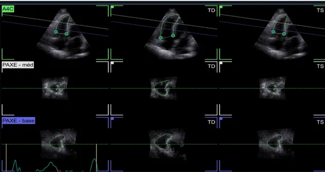
3D analysis was performed using new dedicated software (4D Auto RVQ) to measure RV volumes and ejection fraction (EF) using a semi-automated algorithm based on a software platform for data management (EchoPAC, GE healthcare). Full-volume acquisition was performed using electrocardiogram gating over 3 consecutive cardiac cycles in the apical 4 chamber view. Then at the alignment stage (see figure 1) one can quickly adjust the vertical axis so that it crosses through the tricuspid valve (TV) center point and the RV apex (on the vertical plane). The lower horizontal plane is parallel to the tricuspid valve at the base, while the upper horizontal plane is at the center of the RV. The alignment is done in 4 chamber and orthogonal 4 chamber views. Six Landmark points (see figure 2) are then placed (two tricuspid annulus points and the RV apex point in the 4 chamber view, and the RV/LV posterior and anterior points plus the RV free wall point in the short axis mid view). Then for review stage (see figure 3), the contours can easily be edited through a flexible interface, either in an ED/ES 3 by 3 layout, or in a dynamic layout where the dynamic RV model is also shown. From end diastolic and end systolic volumes, right ventricular ejection fraction (RVEF) was then automatically calculated (see figure 4).
21
Figure 1. Alignment stage in 3D echography
23
Figures 4. Dynamic results
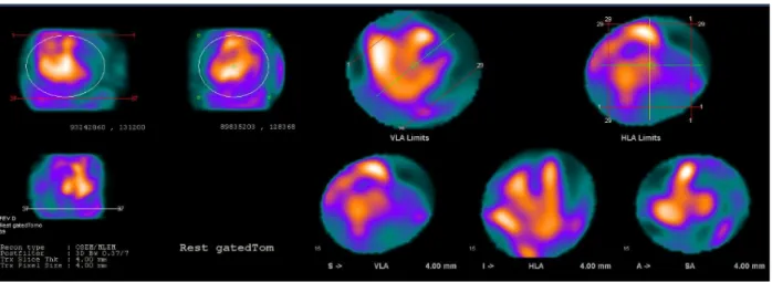
Radionuclide ventriculography:
All acquisitions were performed at the department of Nuclear Medicine at CHUGA hospital. Gated blood-pool SPECT is a volumetric technique based on visualization of the contour of the 99mTc-labeled blood pool in the right ventricular cavity in diastole and systole. Patients were injected with 840 MBq (22.7 mCi) of in vitro labelled erythrocyte solution. Acquisitions were performed
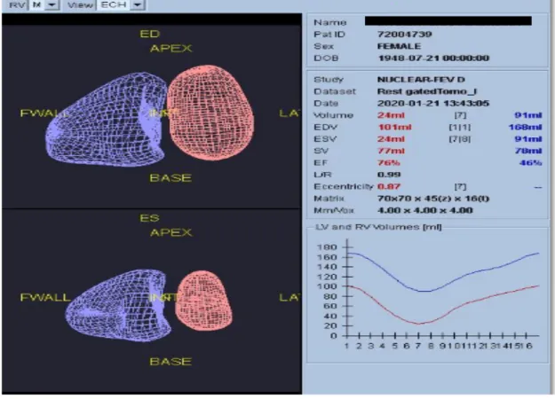
30 min after injection using a CZT Discovery NM530C camera (GE Healthcare, Haifa, Israel). A solid-state semiconductor CZT gamma camera system (Discovery NM 530c, GE Healthcare Ltd.) compound of a multiple pinhole collimator and 19 stationary detectors imaging simultaneously 19 views of the heart without motion of the camera was used. Each detector contained 32x32 pixelated (2.46 x 2.46mm) CZT elements. Acquisition time was 5 minutes. Assessment of left and right ventricular ejection fraction as well as estimations of end diastolic and end systolic volumes (EDV and ESV) were carried out with the Cedars-Sinai QBS processing software (Cedars-Sinai, Ca, USA—revision 2009) after ensuring that images were properly synchronized to electrocardiogram. Contours of RV and left ventricle (LV) were determined and checked automatically (see figures 2 and 4). In the event of incorrect contours, they were manually modified by repositioning edge of the ventricular cavity (see figure 3).
25
Figure 6. Dynamic images generated by QBS software of right (yellow border) and left ventricles (white border)
Figure 7. Manual contouring of cardiac cavities with QBS software. It is possible to reposition tricuspid and pulmonary planes as well as inter-ventricular septum
Figure 8. Final reconstruction of right (blue) and left (red) ventricles with fractions and volumes
Cardiac Magnetic resonance:
Using a 1.5-T and 3-T CMR system (Altea and Skyra, Siemens, Erlangen, Germany), 10 to 12 consecutive short-axis images covering the entire left ventricle (LV) and Right ventricle (RV) were obtained. Respectively, single 2-, 3-, and 4-chamber long-axis images were acquired using a cine steady-state free-precession sequence to allow for the assessment of myocardial function and mass. About 10 to 15 min after the injection of 0.2 mmol/kg gadolinium-based contrast agent, identical prescriptions of short- and long-axis slices were acquired using a 2 or 3 dimensional inversion recovery sequence allowing for the assessment of LGE.
CMR images were analyzed using the freely available software Segment Version 1.8 (Medviso AB, Lund, Sweden). LV volumes, RV volume, LV ejection fraction (EF), RVEF and regional wall
27
Statistical analysis:
Analysis was performed using SPSS 21 software (SPSS Inc., Chicago, IL). Continuous variables are expressed as mean ± SD or median (25th, 75th percentile) and discrete variables as percentage. The mean values of continuous variables was compared using the Mann-Whitney rank sum test. The correlation between variables was assessed using Spearman correlation analysis and expressed by r. Agreement between the two methods was assessed by Bland-Altman analysis. Furthermore, to assess the variability of the measures using RVEF by RNV-CZT, all patients were evaluated twice by the same observer for intra-observer variability, and by two different observers for interobserver variability. Variability was quantified computing the intraclass correlation coefficient (ICC). Interobserver reliability for measurement of the RVEF was assessed by using two-way random single-measure ICC analysis. Intraobserver and intrasubject reliability was assessed by using one way random two-measure ICC analysis. A p-value < 0.05 was considered as statistically significant for all the analyses.
RESULTS
28 patients have benefited from a multimodal RV assessment. 3 were excluded due to an arrhythmia with more than 50% of cycles rejected. Of the 25 patients, 4 other patients could not be evaluated in ventriculography due to a centering defect during acquisition making it impossible to quantify RV activity. Figure 9 shows flow chart of study. The average irradiation in ventriculography was 7 ± 1 mSv. The mean time from CMR to RNV was 3 days with a median of 2 days. Characteristics of study population are shown in Table 1 Mean age of the patients was 54 ± 19 years and 62% were male. Etiology of heart failure was non-ischemic dilated in 86% and ischemic cardiomyopathy (ICM) in 14 %. 81% of patients were New York Heart Association (NYHA) Functional Class I or II. Table 2 presents imaging data of study patients. The mean left ventricle ejection fraction LVEF was 41.8 ± 14.3%.
Ejection Fraction:
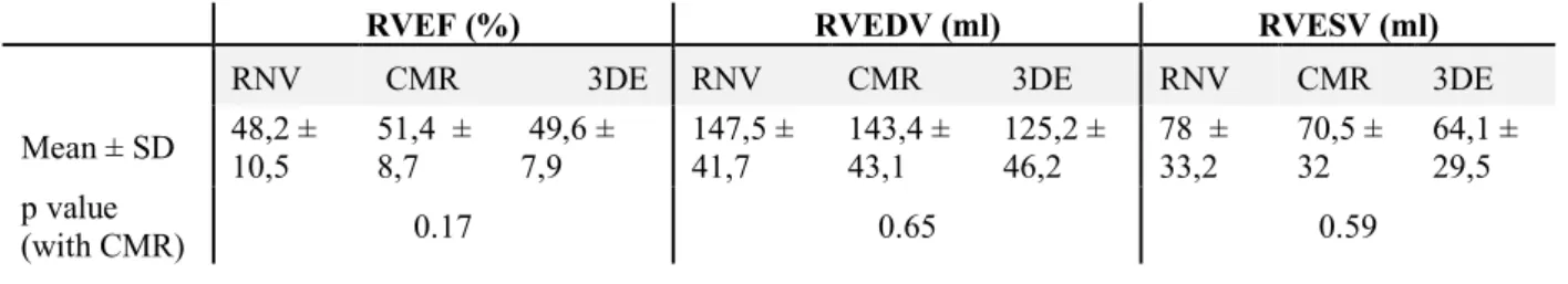
Correlations and Bland Altman analysis between RVEF obtained by CMR and RNV, and CMR with 3D TTE are shown in figures 10 and 11. Mean values between the three modalities were similar with no statistical differences (51.4 ± 8.7% for CMR, 48.2 ± 10.5% for RNV; p = 0.17 and 49.6 ± 7.9% for 3D TTE; p = 0.17) (see table 3). Correlations were quite good, slightly higher for RNV (for RNV: r = 0.72, p < 0.0001, for 3D TTE: r = 0.64, p < 0.001). Bland-Altman analysis showed small mean difference in both cases (-3.6 ± 5.9 % for RNV and -2.7 ± 5.3 % for 3D TTE). In both studies, there was only one value lying outside 2 standard deviations (2 SD).
29
End diastolic volume:
There were no statistical differences between end diastolic volume obtained by CMR (143.4 ± 51 ml) and RNV (147.5 ± 41.7 ml; p = 0.65) (see table 3). Correlation between CMR and RNV was good (r = 0.80, p < 0.0001). Bland-Altman analysis showed small mean differences but rather wide limits of agreement (-3.4 ± 27.6 ml) (see figure 12). No value was lying outside 2 SD. The slope and intercept of the regression plot and dispersion of data on the Bland–Altman scattergram revealed a tendency for RNV to underestimate large RV volumes compared to CMR.
Right ventricle end diastolic volume (RVEDV) obtained by 3D TTE was 125.2 ± 46.2 ml with no statistical differences compare to CMR (p = 0.65). There was close correlation between CMR and 3D TTE (r = 0.94, p < 0.0001). Bland-Altman analysis showed a systematic difference with a quite large mean difference (-18.9 ± 17.3 ml) (see figure 13). This difference reflects an overall underestimation of right end diastolic volumes by 3D TTE compared to CMR. As ventriculography, this underestimation appears more important in large RV volumes compared to CMR.
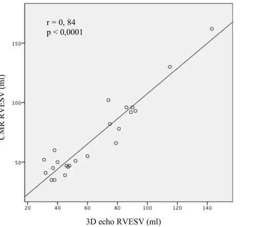
End systolic volume:
Correlations and Bland Altman between RV end systolic volume obtained by CMR and isotopic ventriculography, and CMR with 3D TTE are shown in figures 14 and 15 respectively. There was no statistical differences between end systolic volume obtained by CMR (70.5 ± 32 ml) and RNV (78.0 ± 33.2 ml; p = 0.59) and between 3D TTE (64.1 ± 29.5 ml; p = 0.59) and CMR, with close correlation (for RNV: r = 0.94, p < 0.0001, for 3D TTE: r = 0.84, p < 0.0001). Bland-Altman analysis showed small mean difference in both cases (-4.3 ± 11.9 ml for RNV and -5.5 ± 10.2 ml for 3D TTE). For 3D TTE, there was only one value lying outside 2 SD and two for RNV (one on the outside stage).
Reproducibility:
Intraobserver, and interobserver reproducibility were good for RVEF (ICC.0.85 [95% CI: 0.68 to 0.93], 0.91 [95% CI: 0.79 to 0.96], respectively.
31
DISCUSSION
RNV CZT showed good correlation with CMR for assessment of RV in our study. Correlations were close for RVEF, RVEDV and RVESV without statistical differences. However, radionuclide ventriculography tended to underestimate the highest CMR volumes for EDV, particularly for volumes more than 200mL. These underestimates are probably due to the dilution of radiation with largest volume Adjustments may be needed for the highest volumes to obtain optimal accuracy. These results confirm data in the literature. Nichols et al. showed an average underestimation of 28 ml of EDV by ventriculography compared to CMR (31). However, majority of studies do not use the CZT gamma camera. Those cameras with solid state CZT detectors have better count sensitivity and spatial resolution than conventional sodium iodine detectors, allowing for significant radiotracer dose and/or acquisition time reductions. Previous studies (36-41) have shown that the dose of radiotracers injected to patients can be reduced and image acquisition time can be shortened using new imaging technologies with highly efficient gamma detection and dedicated small field of view image reconstruction (42, 43).These studies focus only on ischemia, but they provide a good understanding of the benefits of CZT camera. More specifically, extraordinarily sensitive SPECT cameras with solid state cadmium-zinc-telluride detectors have better count sensitivity and spatial resolution than those with conventional sodium iodine detectors.
Ventriculography has the potential advantage over CMR and TTE of deriving its volumetric information from detected gamma rays rather than from geometrical considerations so that RV trabeculations should not interfere with received counts.Moreover, irradiation rate is low with only 7 ± 1 mSv. Others advantages of this technique are linked to advantages of the gamma camera mentioned above. In particular, image acquisition time was reduced to about 5 minutes compared to 15 minutes with a conventional camera. RNV with gamma camera is a new promising method
for evaluating right ventricular function, especially since there are few data in the literature about RNV with gamma camera using QBS software (32-33). Jensen et al. studied inter- and intra-observer variability with excellent results for LV but less good for RV. Whereas, our study found very interesting results on the variability of RV. Finally, ventriculography can be used in in hemodynamically precarious patients or patients with ICDs.
Arrhythmia is generally an important limiting factor in cardiac imaging. In our study, there were 3 exclusions of patients in atrial fibrillation with more than 50% of cardiac cycles rejected due to lack of synchronization with electrocardiogram.It would therefore appear that atrial fibrillation is a real limiting factor in the interpretation of RNV since the 3 patients with atrial fibrillation in the study were excluded. However 2 patients with ventricular hyperexcitability could be included with cardiac cycle rejection rates close to 20%. This limitation remains problematic for patients on amines.4 others RNV could not be interpreted due to a centering defect. Contrary to arrhythmia, this limit can be lifted by the use of an experienced manipulator. Finally, although edge detection is less important than in volumetric methods, inaccuracies may result from chamber overlap and background activity; image projections must separate the RV from right atrium, pulmonary artery, and left ventricle and, during analysis, care must be taken to construct a region of interest delineating the interventricular septum, tricuspid, and pulmonary valve planes.
3D echocardiography is also a good alternative for measuring RV volumes and ejection fraction in patients even if correlation was slightly lower than ventriculography for EF. Feasibility of 3D echocardiography was 92% in the present study, which is comparable to the findings of 3D echocardiography measurements of LV (44), and better than previously published series of 3D echocardiography in RV (45). 3D echocardiography has the potential to overcome limitations of
33
modeling has resulted in improved accuracy (47). This approach has been explored in vitro (48) and in vivo (49).
Bland Altman analysis showed an underestimation of EDV by 3D echocardiography (- 20 ml without statistical difference). As ventriculography, these underestimation was more important for highest CMR volumes (> 150 ml). Possible reasons for the differences between CMR and 3D echocardiographic estimates of RV volume include difficulties in delineating the endocardial borders and valvular planes, artifacts due to the respiratory motion during acquisition and problems for visualizing the anterior RV free wall due to its retrosternal position, even though the 3D full-volume scans were acquired with special care to include all parts of the RV free wall.
Advantage of 3D echocardiography is the ability to overcome the limitations of 2D echocardiography by using multiple images to reconstruct RV without geometric assumptions (46). It is an easy, non-irradiating access examination for the patient. It also can be used in hemodynamically precarious patients or patients with ICDs.
One of the limits of 3D echocardiography is that it does not overcome problems associated with poor quality echocardiographic windows, and time required to acquire images could have a negative influence on accuracy and precision of measurements. Also, 3D echocardiography measurements of RV volumes are affected by multiple additional factors, including gain settings as well as thickness and disk orientation during disk summation (50, 51). Finally, as ventriculography, 3D echocardiography cannot be achieved in arrhythmic patients.
Our study population consisted of ambulatory patients. Next step would be to conduct this study in hospitalized patients before LVAD. Very often these patients are serious, hemodynamically precarious or ICD carriers, which compromises achievement of CMR. Thus, speed and ease of image acquisition by RNV could be a good way to assess RV in these patients. Guazzi et al. have
shown right ventricle reserve contractile (RVCR) unmasks different phenotypes (52). Impaired RV function at rest might not invariably lead to unfavorable RVCR. Testing this variable appears useful in more advanced stages of Heart failure to define various clinical conditions and, most likely, to define different levels of risk. RVCR under amines can be measured by CMR but it is very complicated due to availability and technical difficulties related to the many adjustments needed to obtain acquisitions. This is why measurement RVCR under amines with the CZT camera seems simpler.
As explained in introduction, assessment of right ventricular function is essential prior to LVAD. But, there are several data to consider prior to LVAD. Patient selection is an important determinant for the survival and quality of life of implanted patients. Many studies have found factors preoperative individuals responsible for a higher morbidity and mortality. Hepatic and renal dysfunctions are pre-implantation parameters predictors of right ventricle dysfunction (53). Studies have suggested that TAPSE is predictive of postoperative right ventricle dysfunction, for values less than 7.5mm (54). The study by Raina et al., conducted on 55 patients demonstrated that right ventricle fractional shortening, the pulmonary arterial resistances and the index of left atrium volume are predictors of postoperative right ventricle dysfunction (55). There are risk scores to select patients most at risk for RV failure by post implantation, but their sensitivity remains limited (56). Grant et al. have shown the interest in associating a risk score (Michigan) with the RV strain assessment (57). An index reflecting RV ejection work, calculated from right heart catheterization data was studied (58).Associated with all these criteria, assessment of RV by RVN-CZT could be a determining factor prior to LVAD.
35
LVAD should be the next step and test prognosis value of RV function by RNV-CZT. Here, the objective of this study was to validate RVN-CZT method to a clinical practice application.
Conclusion
RNV-CZT appears to be an effective method for evaluating RV function and appears as a good alternative of cardiac magnetic resonance. It could be particularly interesting to patients in severe heart failure with borderline hemodynamic requiring vasoactive drugs.
THESE SOUTENUE PAR: Antoine APERT TITRE :
DEVELOPPEMENT D'UNE NOUVELLE TECHNIQUE D'EVALUATION DU
VENTRICULE DOIT A L'AIDE D'UNE GAMMA CAMERA CADMIUM-ZINC-
TELLURIDE CONCLUSION :
Introduction
La fonction ventriculaire droite est reconnue comme un marqueur pronostique important de l'insuffisance cardiaque. L'objectif de cette étude était de développer une nouvelle
technique d'analyse du ventricule droit à l'aide d'une gamma-caméra CZT en scintigraphie myocardique.
Méthodes
Les patients inclus ont été admis en hôpital de jour de cardiologie pour une évaluation multimodale du ventricule droit. Les données cliniques et paracliniques ont été recueillies. Ensuite, une
imagerie par résonance magnétique, une ventriculographie isotopique et une échocardiographie 3D ont été réalisées dans les 15 jours. Les relations entre les variables continues ont été
évaluées à l'aide de l'analyse de corrélation de Spearman. La concordance a été évaluée par l'analyse de Bland-Altman. Une valeur p inférieure à 0,05 a été considérée comme statistiquement significative pour toutes les analyses.
Résultats
Au total, 21 patients ont été inclus. L'âge moyen des patients était de 54 ± 19 ans et 62% étaient des hommes. Les valeurs moyennes de la FEVD entre les trois examens étaient similaires sans aucune différence statistique (51,4 ± 8,7 % pour l’IRM, 48,2 ± 10,5 % pour la ventriculographie isotopique ; p = 0.17 et 49,6 ± 7,9 % pour l'ETT 3D ; p = 0.17). Les corrélations étaient bonnes (pour la ventriculographie isotopique : r = 0,72, p < 0,0001, pour l'ETT 3D : r = 0,64, p < 0,001). L'analyse Bland-Altman a montré une faible différence moyenne dans les deux cas (-3,6 ± 5,9 % pour la ventriculographie isotopique et -2,7 ± 5,3 % pour l'ETT 3D). Il n'y avait pas de différences statistiques entre les volumes télésystoliques obtenus par l’IRM (70,5 ± 32 ml), et la ventriculographie isotopique (78,0 ± 33,2 ml ; p = 0.59) ainsi qu’entre l'ETT 3D (64,1 ± 29,5 ml ; p = 0.59) et l’IRM. Il n'y avait pas de
différences statistiques entre les volumes télédiastoliques obtenus par l’IRM (70,5 ± 32 ml) et la ventriculographie isotopique (78,0 ± 33,2 ml ; p = 0.65) ainsi qu’entre l'ETT 3D (64,1 ± 29,5 ml ; p = 0.65) et l’IRM. L'analyse Bland-Altman a montré une sous-estimation du volume télediastolique par l'ETT 3D (-18,9 ± 17,3 ml).
Conclusion
La ventriculographie isotopique avec la gamma-caméra CZT est une méthode efficace pour évaluer la fonction VD et apparaît comme une bonne alternative à l’imagerie par résonance magnétique, en particulier pour les patients souffrant d'une insuffisance cardiaque grave avec une hémodynamique limite nécessitant des amines. L’ETT 3D semble également être une méthode efficace.
FIGURES
Multimodal evaluation of right ventricle n = 28
Arrhythmia with > 50% cycles discarded
n = 3
Population study:
CMR, radionuclide ventriculography and 3D TTE n = 25 Radionuclide ventriculography with CMR n = 21 Centering defect in ventriculography n = 4
39
Figures 10. RV ejection fraction: RNV and CMR. The relationship between CMR and RNV (radionuclide ventriculography) by linear regression analysis and Bland–Altman scattergram
r = 0,72 p < 0,0001 CM R RV EF ( % ) RNV RVEF (%) -30 -25 -20 -15 -10 -5 0 5 10 15 20 25 30 0 10 20 30 40 50 60 70
Average RVEF (%) CMR and RNV Mean difference = - 3,6 ± 5,9 % D if fe re nc e in R V E F (% ) RN V CM R + 2 SD - 2 SD Mean
Figures 11. RV ejection fraction: 3D echo and CMR. The relationship between CMR and 3D echo by linear regression analysis and Bland–Altman r = 0,64 p < 0,001 CM R RV E F (% ) 3D echo RVEF (%) -30 -20 -10 0 10 20 30 0 10 20 30 40 50 60 70
Average RVEF (%) CMR and 3D echo Mean difference = - 2,7 ± 5,3 % D if fe re nc e in R V E F (% ) 3D e cho CM R + 2 SD Mean - 2 SD
41
Figures 12. RV end diastolic volume: RNV and CMR. The relationship between CMR and RNV (radionuclide ventriculography) by linear regression analysis and Bland–Altman scattergram
r = 0,80 p < 0,0001 RNV RVEDV (ml) CM R RV E D V ( m l) -80 -60 -40 -20 0 20 40 60 80 0 50 100 150 200 250 300
Average RVEDV (ml) CMR and RNV
D if fe re nc e in R VE DV (m l) RN V CM R Mean + 2 SD - 2 SD Mean difference = -3,4 ± 27,6 ml
Figures 13. RV end diastolic volume: 3D echo and CMR. The relationship between 3D echo and CMR by linear regression analysis and Bland– r = 0,94 p < 0,0001 CM R RV E D V ( m l) 3D echo RVEDV (ml) -80 -60 -40 -20 0 20 40 60 80 0 50 100 150 200 250 300
Average RVEDV (ml) CMR and 3D echo
D if fe re nc e in R V E D V ( m l) 3D ec ho - CM R Mean difference = -18.9 ± 17,6 ml + 2 SD - 2 SD Mean
43
Figures 14. RV end systolic volume: RNV and CMR. The relationship between CMR and RNV (radionuclide ventriculography) by linear regression analysis and Bland–Altman scattergram
CM R RV E S V ( m l) RNV RVESV (ml) r = 0,94 p < 0,0001 -50 -40 -30 -20 -10 0 10 20 30 40 50 0 20 40 60 80 100 120 140 160 180 Mean difference = 4,3 ± 11,9 ml
Average RVESV (ml) CMR and RNV
D if fe re nc e in R V E S V ( m l) RN V CM R + 2 SD Mean - 2 SD
Figure 15. RV end systolic volume: 3D echo and CMR. The relationship between CMR and 3D echo by linear regression analysis and Bland–Altman scattergram r = 0, 84 p < 0,0001 3D echo RVESV (ml) CM R RV E S V ( m l) -40 -30 -20 -10 0 10 20 30 40 50 Mean difference = -5,5 ± 10,2 ml
Average RVESV (ml) CMR and 3D echo
D if fe re nc e in R V E S V ( m l) 3D ec ho - CM R + 2 SD - 2 SD Mean
45
TABLES
Table 1: Baseline characteristics of population
Characteristics Population n = 21 Clinical characteristics
Age, mean ± SD (years) 54 ± 19
Women, n (%) 8 (38)
BMI, mean ± SD (kg/m²) 24.8 ± 4.6
Systolic blood pressure, mean ± SD (mmhg) 127 ± 22 Diastolic blood pressure, mean ± SD (mmhg) 78 ± 16 Heart rate, mean ± SD (beat/min) 70 ± 11 NYHA functional class III-IV, n (%) 4 (19) Etiology of cardiopathy, n (%)
Non ischemic cardiomyopathy 18 (86)
Ischemic cardiomyopathy 3 (14) Comorbidities, n (%) HBP 9 (43) Diabetes 7 (33) Ever smoker 13 (62) History of MI 3 (14) History of PCI 4 (19) History of CABG 1 (5)
Blood results, mean ± SD
MDRD (ml/min/m²) 76 ± 31
Hemoglobin (g/dl) 14.2 ± 2
BMI, body mass index; HBM, high blood pressure; MI, Myocardial infarction; PCI, Primary coronary intervention; CABG, Coronary artery bypass grafting; sd, standard deviation;
Table 2: Baseline imaging characteristics of population
CMR, cardiac magnetic resonance; 3D TTE, Three dimensional trans thoracic echocardiography; LVEF, left ventricle ejection fraction; LVEDV, left ventricular end-diastolic volume;
LVESV, left ventricular end-systolic volume; TAPSE, tricuspid annular plane systolic elevation;
eSPAP, estimated systolic pulmonary artery pressure; RVFS, right ventricle fractional shortening; RIMP, right ventricle index of myocardial performance
Imaging characteristics Population n = 25 CMR measurements n = 25 LVEF, mean ± SD (%) 41.8 ± 14.3 LV EDV, mean ± SD (ml) 204.4 ± 62.7 LV ESV, mean ± SD (ml) 124.1 ± 61.1 Echocardiographic measurements n = 23 TAPSE, mean ± SD (mm) 22 ± 5 S tricuspid, mean ± SD (cm/s) 11.8 ± 2.3 eSPAP, mean ± SD (mmhg) 28 ± 9 RV FAC, mean ± SD (%) 44 ± 8 RIMP, mean ± SD 0.52 ± 0.2
Moderate - severe tricuspid regurgitation, n (%) 7 (28)
3D LVEF, mean ± SD (%) 43.3 ± 11.8
3D LV EDV, mean ± SD (ml) 176.8 ± 58.2 3D LV ESV, mean ± SD (ml) 103.9 ± 54.9
Radionuclide ventriculography measurements n = 21
LVEF, mean ± SD (%) 44.5 ± 13.1
LV EDV, mean ± SD (ml) 173.9 ± 47.2
47
Table 3: Reliability between CMR, radionuclide ventriculography and 3D
echocardiography for right ventricle function
s
RVEF (%) RVEDV (ml) RVESV (ml)
RNV CMR 3DE RNV CMR 3DE RNV CMR 3DE Mean ± SD 48,2 ± 10,5 51,4 ± 8,7 49,6 ± 7,9 147,5 ± 41,7 143,4 ± 43,1 125,2 ± 46,2 78 ± 33,2 70,5 ± 32 64,1 ± 29,5 p value
(with CMR) 0.17 0.65 0.59
CMR,cardiac magnetic resonance; 3DE, Three dimensional echocardiography; RVEDV, right ventricular end-diastolic volume;
RVEF, right ventricular ejection fraction; RVESV, right ventricular end-systolic volume; SD, standard deviation
BIBLIOGRAPHIE
1. Ponikowski P, Voors AA, Anker SD, Bueno H, Cleland JGF, Coats AJS, et al. 2016 ESC Guidelines for the diagnosis and treatment of acute and chronic heart failure: The Task Force for the diagnosis and treatment of acute and chronic heart failure of the European Society of Cardiology (ESC). Eur Heart J. 14 juill 2016; 37(27):2129-200.
2. Fédération française de cardiologie [En ligne]. [Consulté le 20 novembre 2019]. Disponible sur https://www.fedecardio.org
3. Goldberg RJ, Spencer FA, Gore JM, Lessard D, Yarzebski J. Thirty-Year Trends (1975 to 2005) in the Magnitude of, Management of, and Hospital Death Rates Associated With Cardiogenic Shock in Patients With Acute Myocardial Infarction: A Population-Based Perspective. Circulation. 10 mars 2009; 119(9):1211-9.
4. Thiele H, Allam B, Chatellier G, Schuler G, Lafont A. Shock in acute myocardial infarction: the Cape Horn for trials? Eur Heart J. 1 août 2010; 31(15):1828-35.
5. Reynolds HR, Hochman JS. Cardiogenic Shock: Current Concepts and Improving Outcomes. Circulation. 5 févr 2008;117(5):686-97.
6. Greffe cardiaque. [En ligne]. [Consulté le 16 novembre 2019]. Disponible sur https://www.agence-biomedecine.fr/
7. Rose EA, Stevenson LW, Tierney AR. Long-Term Use of a Left Ventricular Assist Device for End-Stage Heart Failure. N Engl J Med. 2001; 9.
8. De Groote P, Fertin M, Goéminne C, Petyt G, Peyrot S, Foucher-Hossein C, et al. Right ventricular systolic function for risk stratification in patients with stable left ventricular systolic dysfunction: comparison of radionuclide angiography to echoDoppler parameters. Eur Heart J. nov 2012; 33(21):2672-9.
9. Haddad F, Spruijt OA, Denault AY, Mercier O, Brunner N, Furman D, et al. Right Heart Score for Predicting Outcome in Idiopathic, Familial, or Drug- and Toxin-Associated Pulmonary Arterial Hypertension. JACC Cardiovasc Imaging. juin 2015;8(6):627-38.
10. France AJ, Prescott RJ, Biernacki W, Muir AL, Macnee W. Does right ventricular function predict survival in patients with chronic obstructive lung disease? : 7.
11. Lund LH, Matthews J, Aaronson K. Patient selection for left ventricular assist devices. Eur J Heart Fail. mai 2010;12(5):434 43.
12. Kormos RL, Teuteberg JJ, Pagani FD, Russell SD, John R, Miller LW, et al. Right ventricular failure in patients with the HeartMate II continuous-flow left ventricular assist device: Incidence, risk factors, and effect on outcomes. J Thorac Cardiovasc Surg. mai 2010;139(5):1316 24.
49
13. Aissaoui N, Salem J-E, Paluszkiewicz L, Morshuis M, Guerot E, Gorria GM, et al. Assessment of right ventricular dysfunction predictors before the implantation of a left ventricular assist device in end-stage heart failure patients using echocardiographic measures (ARVADE): Combination of left and right ventricular echocardiographic variables. Arch Cardiovasc Dis. mai 2015;108(5):3009.
14. Matthews JC, Koelling TM, Pagani FD, Aaronson KD. The Right Ventricular Failure Risk Score. J Am Coll Cardiol. juin 2008;51(22):2163 72.
15. Kavarana MN, Pessin-Minsley MS, Urtecho J, Catanese KA, Flannery M, Oz MC, et al. Right ventricular dysfunction and organ failure in left ventricular assist device recipients: a continuing problem. Ann Thorac Surg. mars 2002;73(3):745 50.
16. Lampert BC, Teuteberg JJ. Right ventricular failure after left ventricular assist devices. J Heart Lung Transplant. sept 2015;34(9):1123 30.
17. Galea N, Carbone I, Cannata D, Cannavale G, Conti B, Galea R, et al. Right ventricular cardiovascular magnetic resonance imaging: normal anatomy and spectrum of pathological findings. Insights Imaging. avr 2013;4(2):213 23.
18. Grothues F, Moon JC, Bellenger NG, Smith GS, Klein HU, Pennell DJ. Interstudy reproducibility of right ventricular volumes, function, and mass with cardiovascular magnetic resonance. Am Heart J. févr 2004;147(2):218 23.
19. Couto M, Souto M, Martínez A, Maceira A, Vieira C, Pumar JM, et al. Accuracy of right ventricular volume and function assessed with cardiovascular magnetic resonance: comparison with echocardiographic parameters. Clin Imaging. janv 2020;59(1):61 7.
20. Marcu CB, Beek AM, Van Rossum AC. Cardiovascular Magnetic Resonance Imaging for the Assessment of Right Heart Involvement in Cardiac and Pulmonary Disease. Heart Lung Circ. déc 2006; 15(6):362 70.
21. Nesser HJ, Tkalec W, Patel AR, Masani ND, Niel J, Markt B, et al. Quantitation of Right Ventricular Volumes and Ejection Fraction by Three-Dimensional Echocardiography in Patients: Comparison with Magnetic Resonance Imaging and Radionuclide Ventriculography. Echocardiography. Sept 2006; 23(8):666 80.
22. Kjaergaard J, Petersen C, Kjaer A, Schaadt B, Oh J, Hassager C. Evaluation of right ventricular volume and function by 2D and 3D echocardiography compared to MRI. Eur J Echocardiogr. déc 2006;7(6):430 8.
23. Nagata Y, Wu VC-C, Kado Y, Otani K, Lin F-C, Otsuji Y, et al. Prognostic Value of Right Ventricular Ejection Fraction Assessed by Transthoracic 3D Echocardiography. Circ Cardiovasc Imaging. févr 2017 ;10(2).
24. Shimada YJ, Shiota M, Siegel RJ, Shiota T. Accuracy of Right Ventricular Volumes and Function Determined by Three-Dimensional Echocardiography in Comparison with Magnetic Resonance Imaging: A Meta-Analysis Study. J Am Soc Echocardiogr. Sept 2010; 23(9):943 53.
25. Medvedofsky D, Addetia K, Patel AR, Sedlmeier A, Baumann R, Mor-Avi V, et al. Novel Approach to Three-Dimensional Echocardiographic Quantification of Right Ventricular Volumes and Function from Focused Views. J Am Soc Echocardiogr. oct 2015;28(10):1222 31.
26. Sugeng L, Mor-Avi V, Weinert L, Niel J, Ebner C, Steringer-Mascherbauer R, et al. Multimodality Comparison of Quantitative Volumetric Analysis of the Right Ventricle. JACC Cardiovasc Imaging. janv 2010;3(1):10 8.
27. Niemann PS, Pinho L, Balbach T, Galuschky C, Blankenhagen M, Silberbach M, et al. Anatomicalboriented Right Ventricular Volume Measurements with Dynamic
Three-Dimensional Echocardiography Validated by 3-Tesla Magnetic Resonance Imaging. J Am Coll Cardiol. oct 2007;50(17):1668 76
28. Lairez O, Delmas C, Fournier P, Cassol E, Méjean S, Pascal P, et al. Feasibility and accuracy of gated blood pool SPECT equilibrium radionuclide ventriculography for the assessment of left and right ventricular volumes and function in patients with left ventricular assist devices. J Nucl Cardiol. avr 2018;25(2):625 34.
29. Mariano-Goulart D, Piot C, Boudousq V, Raczka F, Comte F, Eberlé MC, et al. Routine measurements of left and right ventricular output by gated blood pool emission tomography in comparison with thermodilution measurements: a preliminary study. Eur J Nucl Med. avr 2001;28(4):506 13.
30. Mariano-Goulart D, Dechaux L, Rouzet F, Barbotte E, Caderas de Kerleau C, Rossi M, et al. Diagnosis of Diffuse and Localized Arrhythmogenic Right Ventricular Dysplasia by Gated Blood-Pool SPECT. J Nucl Med. 1 sept 2007; 48(9):1416 23.
31. Nichols K. Validation of SPECT equilibrium radionuclide angiographic right ventricular parameters by cardiac magnetic resonance imaging. J Nucl Cardiol. Mars 2002; 9(2):153 60. 32. Jensen MM, Haase C, Zerahn B. Interstudy repeatability of left and right ventricular volume
estimations by serial-gated tomographic radionuclide angiographies using a cadmium-zinc-telluride detector gamma camera. Clin Physiol Funct Imaging. nov 2015;35(6):418 24. 33. Haarmark C, Haase C, Jensen MM, Zerahn B. Pre-chemotherapy values for left and right
ventricular volumes and ejection fraction by gated tomographic radionuclide angiography using a cadmium-zinc-telluride detector gamma camera. J Nucl Cardiol. févr 2016;23(1):87 97.
34. Palyo RJ, Sinusas AJ, Liu Y-H. High-Sensitivity and High-Resolution SPECT/CT Systems Provide Substantial Dose Reduction Without Compromising Quantitative Precision for Assessment of Myocardial Perfusion and Function. J Nucl Med. 1 juin 2016; 57(6):893 9. 35. Heiberg E, Sjogren J, Ugander M, et al. Design and validation of Segment—freely available
51
37. Herzog BA, Buechel RR, Katz R, Brueckner M, Husmann L, Burger IA, et al. Nuclear Myocardial Perfusion Imaging with a Cadmium-Zinc-Telluride Detector Technique: Optimized Protocol for Scan Time Reduction. J Nucl Med. 1 janv 2010;51(1):46 51.
38. Duvall WL, Croft LB, Ginsberg ES, Einstein AJ, Guma KA, George T, et al. Reduced isotope dose and imaging time with a high-efficiency CZT SPECT camera. J Nucl Cardiol. oct 2011;18(5):847 57.
39. Duvall WL, Sweeny JM, Croft LB, Ginsberg E, Guma KA, Henzlova MJ. Reduced stress dose with rapid acquisition CZT SPECT MPI in a non-obese clinical population: Comparison to coronary angiography. J Nucl Cardiol. févr 2012;19(1):19 27.
40. Gimelli A, Bottai M, Genovesi D, Giorgetti A, Di Martino F, Marzullo P. High diagnostic accuracy of low-dose gated-SPECT with solid-state ultrafast detectors: preliminary clinical results. Eur J Nucl Med Mol Imaging. janv 2012;39(1):83 90.
41. Nakazato R, Berman DS, Hayes SW, Fish M, Padgett R, Xu Y, et al. Myocardial Perfusion Imaging with a Solid-State Camera: Simulation of a Very Low Dose Imaging Protocol. J Nucl Med. 1 mars 2013;54(3):373 9.
42. Slomka PJ, Patton JA, Berman DS, Germano G. Advances in technical aspects of myocardial perfusion SPECT imaging. J Nucl Cardiol. avr 2009;16(2):255 76.
43. Garcia EV, Faber TL, Esteves FP. Cardiac Dedicated Ultrafast SPECT Cameras: New Designs and Clinical Implications. J Nucl Med. 1 févr 2011; 52(2):210 7.
44. Qin J. New Digital Measurement Methods for Left Ventricular Volume Using Real-time Three-dimensional Echocardiography: Comparison with Electromagnetic Flow Method and Magnetic Resonance Imaging. Eur J Echocardiogr. juin 2000;1(2):96 104.
45. Papavassiliou DP, Parks WJ, Hopkins KL, Fyfe DA. Three-dimensional echocardiographic measurement of right ventricular volume in children with congenital heart disease validated by magnetic resonance imaging. J Am Soc Echocardiogr. août 1998;11(8):770 7.
46. Nanda NC, Kisslo J, Lang R, Pandian N, Marwick T, Shirali G, et al. Examination Protocol
for Three-Dimensional Echocardiography: THREE-DIMENSIONAL
ECHOCARDIOGRAPHY. Echocardiography. nov 2004;21(8):763 8.
47. Jenkins C, Chan J, Bricknell K, Strudwick M, Marwick TH. Reproducibility of Right Ventricular Volumes and Ejection Fraction Using Real-time Three-Dimensional Echocardiography. Chest. juin 2007;131(6):1844 51.
48. Linker DT, Moritz WE, Pearlman AS. A new three-dimensional echocardiographic method of right ventricular volume measurement: In vitro validation. J Am Coll Cardiol. juill 1986;8(1):101 6
49. Jiang L, Siu SC, Handschumacher MD, Luis Guererro J, Vazquez de Prada JA, King ME, et al. Three-dimensional echocardiography. In vivo validation for right ventricular volume and function. Circulation. mai 1994;89(5):2342 50.
50. Gopal AS, Chukwu EO, Iwuchukwu CJ, Katz AS, Toole RS, Schapiro W, et al. Normal Values of Right Ventricular Size and Function by Real-time 3-Dimensional Echocardiography: Comparison with Cardiac Magnetic Resonance Imaging. J Am Soc Echocardiogr. mai 2007;20(5):445 55.
51. Hoch M, Vasilyev NV, Soriano B, Gauvreau K, Marx GR. Variables Influencing the Accuracy of Right Ventricular Volume Assessment by Real-time 3-Dimensional Echocardiography: An In Vitro Validation Study. J Am Soc Echocardiogr. mai 2007;20(5):456 61.
52. Guazzi M, Villani S, Generati G, Ferraro OE, Pellegrino M, Alfonzetti E, et al. Right Ventricular Contractile Reserve and Pulmonary Circulation Uncoupling During Exercise Challenge in Heart Failure. JACC Heart Fail. août 2016;4(8):625 35.
53. Santambrogio L. Right ventricular failure after left ventricular assist device insertion: preoperative risk factors. Interact Cardiovasc Thorac Surg. 2006 May 24; 5(4):379–82. 54. Kavarana MN, Pessin-Minsley MS, Urtecho J, Catanese KA, Flannery M, Oz MC, et
al. Right ventricular dysfunction and organ failure in left ventricular assist device recipients: a continuing problem. Ann Thorac Surg. 2002 Mar; 73(3):745–50
55. Raina A, Seetha Rammohan HR, Gertz ZM, Rame JE, Woo YJ, Kirkpatrick JN. Postoperative right ventricular failure after left ventricular assist device placement is predicted by preoperative echocardiographic structural, hemodynamic, and functional parameters. J Card Fail. 2013 Jan; 19(1):16–24.
56. Kalogeropoulos AP, Kelkar A, Weinberger JF, Morris AA, Georgiopoulou VV, Markham DW, et al. Validation of clinical scores for right ventricular failure prediction after
implantation of continuous-flow left ventricular assist devices. J Heart Lung Transplant. déc 2015; 34(12):1595-603.
57. Grant ADM, Smedira NG, Starling RC, Marwick TH. Independent and incremental
role of quantitative right ventricular evaluation for the prediction of right ventricular failure after left ventricular assist device implantation. J Am Coll Cardiol. 2012 Aug 7; 60(6):521–8. 58. Matthews JC, Koelling TM, Pagani FD, Aaronson KD. The Right Ventricular Failure
Risk Score. J Am Coll Cardiol. 2008 Jun; 51(22):2163–72. .
53
SERMENT D’HIPPOCRATE
En présence des Maîtres de cette Faculté, de mes chers condisciples et devant l’effigie d’HIPPOCRATE,
Je promets et je jure d’être fidèle aux lois de l’honneur et de la probité dans l’exercice de la Médecine.
Je donnerai mes soin s gratuitement à l’indigent et n’exigerai jamais un salaire au dessus de mon travail. Je ne participerai à aucun partage clandestin d’honoraires.
Admis dans l’intimité des maisons, mes yeux n’y verront pas ce qui s’y passe ; ma langue taira les secrets qui me seront confiés et mon état ne servira pas à corrompre les mœurs, ni à favoriser le crime.
Je ne permettrai pas que des considérations de religion, de nation, de race, de parti ou de classe sociale viennent s’interposer entre mon devoir et mon patient.
Je garderai le respect absolu de la vie humaine.
Même sous la menace, je n’admettrai pas de faire usage de mes connaissances médicales contre les lois de l’humanité.
Respectueux et reconnaissant envers mes Maîtres, je rendrai à leurs enfants l’instruction que j’ai reçue de leurs pères.
Que les hommes m’accordent leur estime si je suis fidèle à mes promesses. Que je sois couvert d’opprobre et méprisé de mes confrères si j’y manque.
Antoine APERT
DEVELOPMENT OF A NEW RIGHT VENTRICLE EVALUATION TECHNIQUE
USING A GAMMA CAMERA CADMIUM-ZINC-TELLURIDE
ABSTRACT Introduction
Right ventricular function is recognized as an important prognostic marker in heart failure. Objective of this study was to develop a new technique for analyzing right ventricle using a gamma camera CZT in tomoventriculography.
Methods
Included patients were admitted to cardiology unit for a multimodal assessment of right ventricle. Clinical and Para clinical data were collected. Then, cardiac magnetic resonance, radionuclide ventriculography and three dimensional echocardiography were performed within 15 days. Relationships between the continuous variables were assessed using Spearman's correlation analysis. Agreement was assessed by Bland-Altman analysis. A p-value < 0.05 was considered as statistically significant for all the analyses.
Results
A total of 21 patients were included. Mean age of patients was 54 ± 19 years and 62% were male. Mean RVEF values between three modalities were similar with no statistical differences (51.4 ± 8.7% for CMR, 48.2 ± 10.5% for isotopic ventriculography; p = 0.17 and 49.6 ± 7.9% for 3D TTE; p = 0.17). Correlations were good (for isotopic ventriculography: r = 0.72, p < 0.0001, for 3D TTE: r = 0.64, p < 0.001). Bland-Altman analysis showed small mean difference in both cases (-3.6 ± 5.9 % for isotopic ventriculography and -2.7 ± 5.3 % for 3D TTE). There were no statistical differences between end systolic volumes obtained by CMR (70.5 ± 32 ml) and isotopic ventriculography (78.0 ± 33.2 ml; p = 0.59) and between 3D TTE (64.1 ± 29.5 ml; p = 0.59) and CMR. There were no statistical differences between end diastolic volumes obtained by CMR (143.4 ± 43.1 ml) and isotopic ventriculography (147.5 ± 41.7 ml; p = 0.65) and between 3D TTE (125.2 ± 46.2 ml; p = 0.65) and CMR. Bland-Altman analysis showed a systematic difference between CMR and 3D ETT (-18.9 ± 17.3 ml).
Conclusion
Radionuclide ventriculography with CZT gamma camera appears to be an effective method for evaluating RV function and appear as a good alternative of cardiac magnetic resonance particularly to patients in severe heart failure with borderline hemodynamic requiring vasoactive drugs. 3D TTE appears also as an effective method.
55
Antoine APERT
DEVELOPPEMENT D'UNE NOUVELLE TECHNIQUE D'EVALUATION DU
VENTRICULE DOIT A L'AIDE D'UNE GAMMA CAMERA CADMIUM-ZINC-
TELLURIDE RÉSUMÉ
Introduction
La fonction ventriculaire droite est reconnue comme un marqueur pronostique important de l'insuffisance cardiaque. L'objectif de cette étude était de développer une nouvelle technique d'analyse du ventricule droit à l'aide d'une gamma-caméra CZT en scintigraphie myocardique. Méthodes
Les patients inclus ont été admis en hôpital de jour de cardiologie pour une évaluation multimodale du ventricule droit. Les données cliniques et paracliniques ont été recueillies. Ensuite, une imagerie par résonance magnétique, une ventriculographie isotopique et une échocardiographie 3D ont été réalisées dans les 15 jours. Les relations entre les variables continues ont été évaluées à l'aide de l'analyse de corrélation de Spearman. La concordance a été évaluée par l'analyse de Bland-Altman. Une valeur p inférieure à 0,05 a été considérée comme statistiquement significative pour toutes les analyses.
Résultats
Au total, 21 patients ont été inclus. L'âge moyen des patients était de 54 ± 19 ans et 62% étaient des hommes. Les valeurs moyennes de la FEVD entre les trois examens étaient similaires sans aucune différence statistique (51,4 ± 8,7 % pour l’IRM, 48,2 ± 10,5 % pour la ventriculographie isotopique ; p = 0.17 et 49,6 ± 7,9 % pour l'ETT 3D ; p = 0.17). Les corrélations étaient bonnes (pour la ventriculographie isotopique : r = 0,72, p < 0,0001, pour l'ETT 3D : r = 0,64, p < 0,001). L'analyse Bland-Altman a montré une faible différence moyenne dans les deux cas (-3,6 ± 5,9 % pour la ventriculographie isotopique et -2,7 ± 5,3 % pour l'ETT 3D). Il n'y avait pas de différences statistiques entre les volumes télésystoliques obtenus par l’IRM (70,5 ± 32 ml), et ventriculographie isotopique (78,0 ± 33,2 ml ; p = 0.59) ainsi qu’entre l'ETT 3D (64,1 ± 29,5 ml ; p = 0.59) et l’IRM. Il n'y avait pas de différences statistiques entre les volumes télédiastoliques obtenus par l’IRM (70,5 ± 32 ml) et la ventriculographie isotopique (78,0 ± 33,2 ml ; p = 0.65) ainsi qu’entre l'ETT 3D (64,1 ± 29,5 ml ; p = 0.65) et l’IRM. L'analyse Bland-Altman a montré une sous-estimation du volume télediastolique par l'ETT 3D (-18,9 ± 17,3 ml).
Conclusion
La ventriculographie isotopique avec la gamma-caméra CZT est une méthode efficace pour évaluer la fonction VD et apparaît comme une bonne alternative à l’imagerie par résonance magnétique, en particulier pour les patients souffrant d'une insuffisance cardiaque grave avec une hémodynamique limite nécessitant des amines. L’ETT 3D semble également être une méthode efficace.
MOTS CLÉS : Ventricule droit, Evaluation multimodale, Scintigraphie myocardique, CZT, Echographie 3D.