HAL Id: hal-01511984
https://hal.archives-ouvertes.fr/hal-01511984
Submitted on 21 Apr 2017
HAL is a multi-disciplinary open access
archive for the deposit and dissemination of
sci-entific research documents, whether they are
pub-lished or not. The documents may come from
teaching and research institutions in France or
abroad, or from public or private research centers.
L’archive ouverte pluridisciplinaire HAL, est
destinée au dépôt et à la diffusion de documents
scientifiques de niveau recherche, publiés ou non,
émanant des établissements d’enseignement et de
recherche français ou étrangers, des laboratoires
publics ou privés.
Distributed under a Creative Commons Attribution| 4.0 International License
Identification of a Tsal1(52-75) salivary synthetic
peptide to monitor cattle exposure to tsetse flies
Martin Bienvenu Somda, Sylvie Cornelie, Zakaria Bengaly, Françoise
Mathieu-Daude, Anne Poinsignon, Emilie Dama, Jérémy Bouyer, Issa Sidibe,
Edith Demettre, Martial Séveno, et al.
To cite this version:
Martin Bienvenu Somda, Sylvie Cornelie, Zakaria Bengaly, Françoise Mathieu-Daude, Anne
Poinsignon, et al.. Identification of a Tsal1(52-75) salivary synthetic peptide to monitor cattle exposure
to tsetse flies. Parasites and Vectors, BioMed Central, 2016, 9, pp.12. �10.1186/s13071-016-1414-8�.
�hal-01511984�
R E S E A R C H
Open Access
Identification of a Tsal1
52
–75
salivary
synthetic peptide to monitor cattle
exposure to tsetse flies
Martin Bienvenu Somda
1,2*, Sylvie Cornelie
3, Zakaria Bengaly
1, Françoise Mathieu-Daudé
3, Anne Poinsignon
3,
Emilie Dama
1,2, Jeremy Bouyer
4,8, Issa Sidibé
1,5, Edith Demettre
6, Martial Seveno
6, Franck Remoué
3,
Antoine Sanon
7and Bruno Bucheton
8Abstract
Background: The saliva of tsetse flies contains a cocktail of bioactive molecules inducing specific antibody responses in hosts exposed to bites. We have previously shown that an indirect-ELISA test using whole salivary extracts from Glossina morsitans submorsitans was able to discriminate between (i) cattle from tsetse infested and tsetse free areas and (ii) animals experimentally exposed to low or high numbers of tsetse flies. In the present study, our aim was to identify specific salivary synthetic peptides that could be used to develop simple immunoassays to measure cattle exposure to tsetse flies.
Methods: In a first step, 2D-electrophoresis immunoblotting, using sera from animals exposed to a variety of bloodsucking arthropods, was performed to identify specific salivary proteins recognised in cattle exposed to tsetse bites. Linear epitope prediction software and Blast analysis were then used to design synthetic peptides within the identified salivary proteins. Finally, candidate peptides were tested by indirect-ELISA on serum samples from tsetse infested and tsetse free areas, and from exposure experiments.
Results: The combined immunoblotting and bioinformatics analyses led to the identification of five peptides carrying putative linear epitopes within two salivary proteins: the tsetse salivary gland protein 1 (Tsal1) and the Salivary Secreted Adenosine (SSA). Of these, two were synthesised and tested further based on the absence of sequence homology with other arthropods or pathogen species. IgG responses to the Tsal152–75synthetic peptide were shown to be specific of tsetse exposure in both naturally and experimentally exposed hosts. Nevertheless, anti-Tsal152–75IgG responses were absent in animals exposed to high tsetse biting rates.
Conclusions: These results suggest that Tsal152–75specific antibodies represent a biomarker of low cattle exposure to
tsetse fly. These results are discussed in the light of the other available tsetse saliva based-immunoassays and in the perspective of developing a simple serological tool for tsetse eradication campaigns to assess the tsetse free status or to detect tsetse reemergence in previously cleared areas.
Keywords: Synthetic Peptide, Biomarker of Exposure, Cattle, Tsetse Flies, African Animal Trypanosomosis
* Correspondence:somdabienvenu@yahoo.fr
1
Centre International de Recherche-Développement sur l’Elevage en zone Subhumide (CIRDES), 01 BP 454 Bobo-Dioulasso 01, Burkina Faso
2Université Polytechnique de Bobo-Dioulasso, 01 BP 1 091 Bobo-Dioulasso
01, Burkina Faso
Full list of author information is available at the end of the article
© 2016 Somda et al. Open Access This article is distributed under the terms of the Creative Commons Attribution 4.0 International License (http://creativecommons.org/licenses/by/4.0/), which permits unrestricted use, distribution, and reproduction in any medium, provided you give appropriate credit to the original author(s) and the source, provide a link to the Creative Commons license, and indicate if changes were made. The Creative Commons Public Domain Dedication waiver (http://creativecommons.org/publicdomain/zero/1.0/) applies to the data made available in this article, unless otherwise stated.
Background
African trypanosomosis, a parasitic vector-borne disease that constitutes a major constraint to development in sub-Saharan Africa, exists under two forms: Human African Trypanosomosis (HAT) known also as sleeping sickness and African Animal Trypanosomosis (AAT) or Nagana. Tsetse flies (Glossina spp.) are the cyclical vectors of the trypanosome species causing these diseases. Some 46 million cattle distributed over 10 million km2in 38 sub-Saharan African countries [1, 2] are estimated to be at risk of contracting AAT and hamper significantly the socio-economic development of these African regions [3, 4]. Among the 38 tsetse-infested countries, 34 are amongst the poorest in the world [5] and have included tsetse and trypa-nosomosis as a constraint in their poverty reduction strat-egy papers under the heavily indebted poor countries initiative [6].
The main strategies used to control or eradicate AAT remain (i) chemoprophylaxis and chemotherapy with trypanocidal drugs, (ii) promoting trypanotolerant cattle, and (iii) tsetse control or eradication programmes [1]. The Pan African Tsetse and Trypanosomiasis Eradica-tion Campaign (PATTEC) initiative promoting inte-grated control of AAT and large tsetse eradication campaigns are underway in Uganda, Kenya and Ethiopia in East Africa and in Ghana, Burkina Faso and Mali in West Africa, with the aim of improving breeding and agriculture by creating new tsetse free areas [7].
Entomological evaluation tools represent an important component of any vector based control programme to appropriately target implementation areas and to evalu-ate their efficacy in time [8]. To devalu-ate, the conventional method used within tsetse eradication campaigns is to estimate tsetse fly densities with traps deployed at fixed or temporary sites [9]. Important constraints are never-theless associated with this method. The deployment and monitoring of traps, most of the time in very large areas (20 km/day/person in walking) with poor accessi-bility, is costly and demanding in terms of human re-sources and logistics. It is also known that traps have a poor efficiency (below 1 % of the flies present in a 1 km2 around the trap are captured daily) and are becoming even less efficient at low tsetse densities [8, 10, 11]. Fur-thermore, traps are generally set up in sentinel fixed sites and thus only provide an indirect estimate of cattle exposure to tsetse bites especially in agro-pastoral areas where herds are very mobile. Alternative methods based on the evaluation of the antibody (Ab) responses di-rected against bloodsucking arthropod salivary antigens have been developed in the last few years. During the blood meal, hematophagous arthropods inject a mixture of anti-haemostatic, anti-inflammatory and immuno-modulatory molecules into the skin of their hosts. These molecules play a crucial role in achieving an effective
blood meal [12], but also in the establishment or not of pathogens into the vertebrate host [13]. The antigenic properties of these molecules have also been used to de-velop a range of immunoassays to detect associated spe-cific Abs and to assess host exposure to a range of arthropod vectors of human pathogens [14].
Recent studies have shown that the human IgG re-sponse against whole salivary extracts (WSE) of several Glossina (G.) species (G. morsitans (m.) morsitans, G. fuscipes (f.) fuscipes and G. palpalis (p.) gambiensis) was correlated with human exposure to tsetse flies in differ-ent HAT endemic areas [15–18]. Similar results were observed in outbred cattle from a tsetse infested area in South-West Burkina Faso that were shown to harbor higher IgG responses than cattle in the North where tse-tse flies are absent. These results were further confirmed in cows experimentally exposed to the bite of a range of Glossina species and other bloodsucking arthropods [19]. However, the use of WSE in immunoassays is likely impaired by the existence of potential cross-reactions with Abs directed against common saliva antigens that are shared by different arthropod species [20]. Other im-portant drawbacks are the difficulty in achieving mass production of WSE in a standardized manner and the storage of these antigens over long periods. WSE are thus poorly suited for large scale studies [21, 22]. Resort-ing to recombinant salivary proteins has enabled to over-come some of these limitations and a number of immunoassays based on specific recombinant salivary proteins have been developed to detect exposure to a range of arthropods [23–27]. An alternative strategy has been to identify specific linear epitopes by in silico ap-proaches in order to design peptides to be used as the immunoassay antigens. Production of short synthetic peptides at high purity can easily be entrusted to a pri-vate company; shipment and storage are facilitated as they can be lyophilised. In the last few years, such ap-proaches were successfully applied to develop salivary biomarkers of human exposure to Aedes aegypti [28] and Anopheles (An.). gambiae [21, 22]. Recently, the IgG response to a synthetic peptide designed from the Tsetse Saliva Growth Factor-1 (Tsgf1) sequence, was shown to be specific of human exposure to tsetse flies [29] and was successfully used to assess the evolution of human tsetse contacts during a vector control intervention in Guinea [30].
In the present study, our aim was to design peptides that could be used to assess exposure of cattle to tsetse flies. An immuno-proteomic approach, using sera from animals experimentally exposed to several arthropod species and WSE from G. m. submorsitans, was first used to identify the most specific immunogenic salivary proteins to detect Glossina exposure. Several epitope prediction and protein conformation software were then
used in combination with Blast analysis to design linear peptides within the identified proteins. Synthetic peptides were finally produced and evaluated in an indirect-ELISA test with serum samples from (i) cattle from tsetse free or tsetse infested areas and (ii) cows experimentally exposed to low or high numbers of tsetse flies.
Methods
Serum samples
All bovine serum samples used in this study were col-lected from cattle outbred in different environmental settings from Burkina Faso or were obtained by experi-mental exposure of cows to several arthropod species and are described in detail elsewhere [19]. Samples from outbred cattle included 17 samples collected in a tsetse free area in Northern Burkina Faso and 43 samples col-lected in a tsetse infested area in the South-West part of the country. Concerning experimentally exposed sera, we included six samples collected from six cows indi-vidually exposed weekly to the bite of G. m. submorsi-tans, G. p. gambiensis, Amblyomma (A.) variegatum, An. gambiae, Tabanidae spp. or Stomoxys spp. Samples used in the present study were those collected at the end of the exposure experiments, after 12 weeks for An. gam-biae and 23 weeks for all other species. In addition we also used 96 serum samples collected weekly from two groups of four animals that were exposed to 50 G. m. submorsitans flies twice a week during 11 weeks (high exposure group) or 10 flies weekly during the same period (low exposure group).
Production of whole salivary extracts
WSE were obtained from 10–12 day-old G. m. submorsi-tansuninfected males and females from the IRD/CIRAD colony in Montpellier (France). The tsetse saliva was col-lected as described previously [17] by a salivation tech-nique that does not require the dissection of salivary glands to avoid the presence of non-salivary antigens in WSE. Briefly, tsetse flies (4–6 flies) were enclosed in 50 ml Falcon tubes closed by a mosquito net and placed above a drop of salivation buffer on warm slides. Buffer drops were collected after 10 min of salivation and were stored at −80 °C before use. Prior to electrophoresis, WSE were desalted and concentrated using the 2-D Clean-Up Kit (GE Healthcare, Germany) according the manufacturer instructions. Protein concentrations of WSE were assessed by the Bradford method.
Identification of salivary proteins
Two-dimensional electrophoresis (2DE) and gel staining
For the first dimension electrophoresis, isoelectric focus-ing (IEF) was carried out with 60μg of G. m. submorsitans WSE on 11 cm pH 3–11 non linear immobilineTM
dry-Strips (GE Healthcare, Germany). dry-Strips were rehydrated
for 10–20 h at room temperature with protein sample made up to 170μl in IEF buffer (7 M urea, 2 M thiourea, 4 % CHAPS, 0.2 % tergitol, 0.8 % IPG buffer, 1 % octylβ-glucoside and 2 % DeStreak reagent). The IEF conditions were: temperature 20 °C; current 50 μA per strip; 60 V (step’n’hold) for 1 h; 500 V (gradient) for 1 h; 1 000 V (gradient) for 1 h; 6 000 V (gradient) for 2 h and then 6 000 V holds to 30 000 Vhs. After the IEF, the strips were reduced for 10 min with 65 mM DTT buffer pH 6.8 (50 mM Tris HCl pH 6.8, 6 M urea, 30 % Glycerol, 2 % SDS) and alkylated for 15 min with 81 mM of iodoaceta-mide buffer pH 8.8 (50 mM Tris HCl pH 8.8, 6 M urea, 30 % Glycerol, 2 % SDS). Then equilibrated strips in pH 6.8 buffer (50 mM Tris HCl pH 6.8, 6 M urea, 30 % Glycerol, 2 % SDS) were applied to 10-20 % SDS-PAGE Tris–HCl gels (Criterion, Biorad) for second dimension and sealed with agarose (1 % agarose low melting, 0.2 % SDS and 150 mM Tris pH 6.8). 2DE-gels were run at 30 V for 20 min and then 200 V for 55 min.
After the second dimension, a preparative 2DE-gel was fixed for 20 min in 50 % ethanol/5 % acetic acid solu-tion, and then 10 min in 50 % ethanol solusolu-tion, washed three times in ultrapure water. Finally, this gel was stained with a colloidal blue solution (Fermentas, Saint-Remy les Chevreuse, France) overnight and washed four times in ultrapure water.
Identification of G. m. submorsitans salivary proteins by mass spectrometry
Visible spots after colloidal blue staining were manually excised under a laminar flow hood and enzymatic in-gel digestion was performed automatically (Tecan freedom evo® proteomics) according to the Shevchenko modified protocol [31].
Briefly, protein spots were digested using 150 ng of trypsin, peptide extraction was performed using 5 sonic-ation cycles of 2 min each and peptides were concen-trated 1 h at 50 °C in a heat block. Peptide samples were automatically spotted (Tecan freedom evo® proteomics). For this step, 0.5 μl of peptide sample and 0.5 μl of α-cyano-4-hydroxy-trans-cinnamic acid (a saturated solu-tion prepared in acetonitrile/trifluoroacetic acid, 50: 0.1 %, vortexed, sonicated 30 s and microcentrifuged 30 s with a 1/3 dilution of the supernatant used as the matrix) were deposited on a 384-well MALDI anchor-ship target using the dry-droplet procedure [32] and air dried at room temperature. Peptide samples were then desalted using a 10 mM phosphate buffer and dried again at room temperature. MALDI-TOF MS analysis was performed using UltraFlex MALDI TOF-TOF mass spectrometer (Brucker Daltonics, Bremen, Germany) in the reflecton mode with a 26 kV accelerating voltage and a 50 ns delayed extraction. The AutoXecute™ module of Flexcontrol™ v3.0 (Bruker Daltonics) (laser power ranged
from 40 to 50 %, 600 shots) was used to acquire mass spectra. Spectra were analysed using FlexAnalysis™ soft-ware v3.0 (Bruker Daltonics) and calibrated internally with the autoproteolysis peptides of trypsin (m/z: 842.51; 1045.556; 2211.10). Peptides were selected in the mass range of 900–3000 Da.
Peptide Mass Fingerprint identification of proteins was performed by searching against the Glossina entries of either the Swiss-Prot or TrEMBL databases (http:// www.expasy.ch) and by using the MASCOT v2.3 algo-rithm (http://www.matrixscience.com) with trypsin en-zyme specificity and one trypsin missed cleavage allowed [33]. Carbamidomethyl was set as fixed cystein tion and oxidation was set as variable methionine modifica-tion for searches. A mass tolerance of 50 ppm was allowed for identification. Matching peptides with one missed cleav-age were considered as pertinent when there were two con-secutive basic residues or when arginine and lysine residues were in an acidic context. MASCOT scores higher than 47 were considered as significant (p < 0.05) for Swiss-Prot and TrEMBL (v 2011_04) database interrogations.
Immunoblotting and specific proteins identification
The proteins separated by 2DE were then electro-transferred onto a PVDF (polyvinylidene dilfluoride, Biorad) membrane for western blotting as previously de-scribed [34]. The PVDF membrane was washed in Tris Buffer Saline (TBS) and then incubated in blocking buffer (TBS tween 0.05 % and 5 % dry milk) for 1 h 30 min. After washing three times with TBS tween 0.1 %, the membrane was incubated overnight at 4 °C with bovine serum diluted at 1/100 in TBS tween 0.05 % with 2.5 % dry milk. The membranes were then washed three times with TBS tween 0.1 % and three times with TBS, and then equilibrated with 2.5 % dry milk in TBS for 15 min. Mouse anti-bovine IgG conjugated to peroxidase (Sigma, St Louis, MO, USA) was added at a dilution of 1/15 000 in TBS tween 2.5 % with 2.5 % dry milk for 2 h 30 at room temperature. After addition of the secondary antibody, the membranes were washed four times with TBS tween 0.1 % and four times with TBS. The immunogenic proteins in membranes were revealed using the West Pico ECL (Pierce, Rockford, IL, USA) and exposed to XCLXposure films (Pierce) for 4 min. Digital images of both western blotting and 2D gel were captured by scanning at 16 bits resolution under non saturating conditions, 300 dpi and stored in TIFF format. The proteins visualised on 2D gels were matched with immunogenic proteins detected by western blotting using SameSpotsTM Software 3.3 (Nonlinear Dynamics) in order to identify immunogenic spots.
Peptide design
The sequences of proteins of interest were downloaded on the ExPASy Proteomics Tools server (http://
www.expasy.ch). The signal peptides were predicted by SignalP 4.0 [35] and cleaved from the immature protein sequences. Secondary structures were then predicted with NetSurfP [36] and I-Tasser [37] servers. The surface exposure of proteins of interest has been viewed in 3D by the Pymol software (http://www.pymol.org).
The identification of putative linear B-cell epitopes of identified proteins was carried out with Bcepred [38], Bcpred [39] and Antigenicity plot server [40]. All epitopes that were identified by at least two out of three algorithms were selected. Peptides having a length less than 30 amino acids and including the maximum of epitopes were selected for further analyses. The specificity of peptide sequences to the Glossina genus was checked by re-quests on the NCBI Blast T non redundant databases (http://www.blast.ncbi.nlm.nih.gov/Blast.cgi). Synthetic pep-tides were synthesized and purified (95 %) by Genepep SA (St-Jean de Vedas, Montpellier, France). All peptides were shipped in lyophilised form and were then resuspended in ultrapure water (1 mg/ml) and stored as frozen aliquots.
Evaluation of bovine IgG Ab level against synthetic peptides and WSE
The anti-peptide IgG responses were measured by indirect-ELISA, according to Poinsignon et al. [22] with minor modifications. Briefly, microtiter plates Maxisorp (Nunc, Roskilde, Denmark) were coated with peptide (20μg/mL) in phosphate buffer saline (PBS) for 2 h 30 min at 37 °C. After three washes, plates were saturated with blocking buffer (Pierce, thermo scientific) 1 h at 37 °C. Sera diluted in PBS-tween 1 % (1/30 for Tsal152–75 and 1/10 for
Tsal1145–166) were incubated overnight at 4 °C. After five
washes, sheep anti-bovine IgG conjugated to peroxidase (AbD Serotec, France) was added (in dilution 1/4000 for Tsal152–75 and 1/2000 for Tsal1145–166) in PBS tween 1 %
for 1 h 30 at room temperature. Colorimetric development was carried out using ABTS (2,2′-azino-bis (3-ethyl-benzthiazoline 6-sulfonic acid) diammonium) (Sigma St Louis, MO, USA) in 50 mM citrate buffer (pH 4) contain-ing 0.003 % H2O2. Optical density (OD) was measured at
405 nm (45 min for Tsal152–75 and 1 h for Tsal1145–166).
IgG responses against WSE were also evaluated on the same samples as described previously [19]. Each test sam-ple was analysed in duplicate in antigen wells and, in paral-lel, in a blank well containing no peptide solution or no WSE (ODn) to control non-specific reactions between the serum and the reagents. Individual results were expressed asΔOD value calculated according to the formula ΔOD = ODx - ODn, where ODx represents the mean of individual OD in both antigen wells, as previously used [22].
Statistical analysis
All data were analysed with GraphPad Prism5 software® (San Diego, CA, USA). After verifying thatΔOD values
did not assume Gaussian distribution using Shapiro-Wilk test, the non-parametric Mann–Whitney U-test was used for comparison of Ab levels between two inde-pendent groups. All differences were considered signifi-cant at P < 0.05.
Results
Identification of specific immunogenic salivary proteins of Glossina exposure
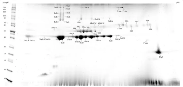
The sialome of G. m. submorsitans was investigated by 2D-gel electrophoresis separation of the WSE followed by colloidal blue staining (Fig. 1) and subsequent MALDI-TOF/MS identification. A total of 53 spots were observed with colloidal blue staining and could be picked for mass spectrometry analysis. Forty-seven spots were successfully identified by MALDI-TOF MS analysis providing a catalogue of seven salivary proteins (Table 1). For the spots of low molecular weight (< 25kD) no identi-fication could be obtained because the amount of material was too scanty. In contrast, low molecular weight spots are much more intense in 2-D electrophoresis gels ob-tained with G. m. morsitans saliva extracts obob-tained by centrifugation of salivary glands [41]. These differ-ences may be related to differdiffer-ences in the saliva composition of these two closely related subspecies but also to the saliva collection technique. Several spots with similar molecular weight but different iso-electric points led to the same identification suggest-ing the existence of isoforms for these proteins. All
spots were identified as related to G. m. morsitans salivary proteins [41]: the Tsetse salivary gland pro-tein 1 (Tsal1); the Tsetse salivary gland propro-tein 2 (Tsal2) and its two isoforms (Tsal2A and Tsal2B); the Tsetse Salivary Growth Factor 1 (TSGF-1); the Salivary Secreted Adenosine (SSA); the Adenosine Deaminase-related Growth Factor C (ADGF-C); the 5′Nucleotidase family salivary protein (5′-nuc) and the Tsetse Antigen 5 (TAg5).
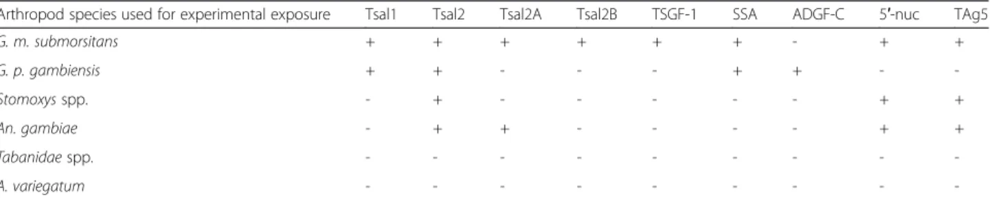
In order to identify G. m. submorsitans immunogenic salivary antigens that are specific of cattle exposure to tsetse flies, we established the 2D immunoblot profiles of serum samples from cows experimentally exposed to tsetse flies (G. m. submorsitans, G. p. gambiensis) or to other bloodsucking arthropods that are common in the study area (A. variegatum, An. gambiae, Tabanidae spp. or Stomoxys spp.). A number of spots were common be-tween the two tsetse species but differences could be ob-served (Fig. 2). Whereas ADFG-C appears to be immunogenic in the cow exposed to G. p. gambiensis, this salivary protein did not react with the G. m. submor-sitans serum. On the contrary, the G. p. gambiensis serum did not recognize any spot associated with the Tsal2 isoforms (Tsal2A and Tsal2B), TSGF-1, 5′-nuc and TAg5 families of salivary proteins. Importantly, cross re-actions (although weak) were also observed with the sera of cows experimentally exposed to Stomoxys spp. or An. gambiae which recognised Tsal2, 5′-nuc, TAg5 and Tsal2, Tsal2A, 5′-nuc, TAg5 respectively (Table 2). Based
Fig. 1 2D gel profile (SDS-PAGE) of Glossina morsitans submorsitans secreted salivary proteins. Whole saliva extracts were run on 2DE gels and stained with colloidal blue. Fifty-three spots were analysed by mass spectrometry and 47 lead to an identification. Molecular weight markers (MW) are indicated on the left. Abbreviations: Tsal1 (Tsetse salivary gland protein 1), Tsal2 (Tsetse salivary gland protein 2), Tsal2A (Tsetse salivary gland protein 2, isoform A), Tsal2B (Tsetse salivary gland protein 2, isoform B), TSGF-1 (Tsetse Salivary Growth Factor 1), SSA (Salivary Secreted Adenosine), ADGF-C (Adenosine deaminase-related growth factor C), 5′-nuc (5′nucleotidase family salivary protein) and TAg5 (Tsetse Antigen 5)
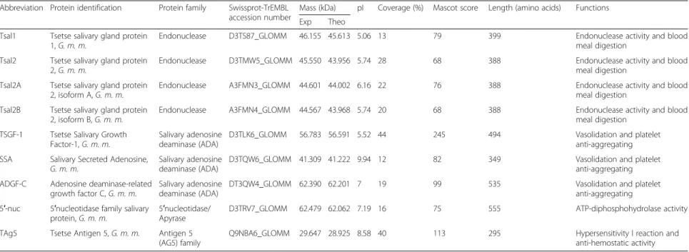
Table 1 Glossina morsitans submorsitans salivary secreted proteins identified by mass spectrometry. Database searches were performed against the Glossina entries of the SwissProt or TrEMBL databases with the MASCOT software. Molecular mass, pI and sequence coverage are shown. All the MASCOT scores are > 47 (p < 0.05)
Abbreviation Protein identification Protein family Swissprot-TrEMBL accession number
Mass (kDa) pI Coverage (%) Mascot score Length (amino acids) Functions Exp Theo
Tsal1 Tsetse salivary gland protein 1, G. m. m.
Endonuclease D3TS87_GLOMM 46.155 45.613 5.06 13 79 399 Endonuclease activity and blood meal digestion
Tsal2 Tsetse salivary gland protein 2, G. m. m.
Endonuclease D3TMW5_GLOMM 45.550 43.956 5.74 28 68 388 Endonuclease activity and blood meal digestion
Tsal2A Tsetse salivary gland protein 2, isoform A, G. m. m.
Endonuclease A3FMN3_GLOMM 44.601 44.002 6.16 22 76 388 Endonuclease activity and blood meal digestion
Tsal2B Tsetse salivary gland protein 2, isoform B, G. m. m.
Endonuclease A3FMN4_GLOMM 44.567 43.968 5.74 20 68 388 Endonuclease activity and blood meal digestion
TSGF-1 Tsetse Salivary Growth Factor-1, G. m. m.
Salivary adenosine deaminase (ADA)
D3TLK6_GLOMM 56.783 56.591 5.52 44 245 494 Vasolidation and platelet anti-aggregating SSA Salivary Secreted Adenosine,
G. m. m.
Salivary adenosine deaminase (ADA)
D3TQW6_GLOMM 41.309 41.222 9.94 12 82 349 Vasolidation and platelet anti-aggregating ADGF-C Adenosine deaminase-related
growth factor C, G. m. m.
Salivary adenosine deaminase (ADA)
DT3QW4_GLOMM 62.390 62.201 7 19 99 535 Vasolidation and platelet anti-aggregating 5′-nuc 5′nucleotidase family salivary
protein, G. m. m.
5′nucleotidase/ Apyrase
D3TRV7_GLOMM 62.479 62.062 7.19 16 75 555 ATP-diphosphohydrolase activity TAg5 Tsetse Antigen 5, G. m. m. Antigen 5
(AG5) family
Q9NBA6_GLOMM 29.647 28.925 8.58 40 113 295 Hypersensitivity I reaction and anti-hemostatic activity
G. m. m.: Glossina morsitans morsitans, Exp : experimental and Theo : Theoretical
Somda et al. Parasites & Vectors (2016) 9:149 Page 6 of 12
on these results we decided to focus the peptide de-sign on Tsal1 and SSA sequences as these salivary an-tigens were recognised only by the sera of animals individually exposed to both tsetse species and did not react with the sera of animals bitten by the other arthropod species.
Peptide design
Four and one sequences of 22 to 28 amino acid residues containing putative linear epitopes were identified from the G. m. morsitans Tsal1 and SSA sequences respectively (Additional file 1: Table S1). In order to avoid potential cross-reactivity with Abs against proteins from other bloodsucking arthropod species as well as from host
pathogens, these 5 peptides were submitted to the NCBI Blast T non redundant databases (Additional file 1: Table S1). The SSA65–92peptide showed some degree of
hom-ology with Culex quinquefasciatus and was therefore dropped out in further analyses. Similarly, among the 4 peptides identified to carry putative Tsal1 linear epi-topes, we selected Tsal152–65 and Tsal1145–166 as for
the other two peptides, closest matches were obtained for organisms to which cattle may be naturally ex-posed (Plasmodium yoelii yoelii and An. darlingi). After checking for Ab accessibility of these two pep-tides at the surface of the Tsal1 protein using 3D models (Fig. 3), Tsal152–75 and Tsal1145–166 were
syn-thesised and tested against cattle serum samples.
a
b
c
d
Fig. 2 Glossina morsitans submorsitans immunogenic salivary proteins in cattle. G. m. submorsitans whole salivary extracts were run on 2D gels and transferred to PVDF membranes. Membranes were then incubated with sera from cows experimentally bitten by (a) G. m. submorsitans, (b) G. p. gambiensis, (c) An. gambiae and (d) Stomoxys spp. Molecular weight markers (MW) are indicated on the left
Table 2 Glossina morsitans submorsitans salivary secreted proteins recognised by cows exposed to tsetse and other hematophagous arthropods
Arthropod species used for experimental exposure Tsal1 Tsal2 Tsal2A Tsal2B TSGF-1 SSA ADGF-C 5′-nuc TAg5
G. m. submorsitans + + + + + + - + + G. p. gambiensis + + - - - + + - -Stomoxys spp. - + - - - + + An. gambiae - + + - - - - + + Tabanidae spp. - - - -A. variegatum - - -
-‘+’ indicates Glossina morsitans submorsitans salivary proteins recognised on 2D gels by western blot with sera from animals exposed experimentally to several arthropod species
Ability of synthetic peptides to detect exposure to tsetse flies
The ability of the two synthetic peptide candidates to de-tect exposure of cattle to tsetse flies was first assessed on serum samples from cattle bred in different eco-climatic zones in Burkina Faso. Serum samples from the
cows experimentally exposed to the none-tsetse species were also included as negative controls (Fig. 4). Anti-Tsal152–75 IgG responses were significantly higher (P =
0.009) in cattle from tsetse infested area as compared to those from tsetse free area, and were the lowest in the control animals exposed to tabanids, stable flies, mos-quitoes or ticks. They also provided a better discrimin-ation between animals from tsetse infested or free areas as compared to the response directed against WSE (P = 0.116). In contrast no significant differences in the IgG response specific to Tsal1145–166 (P = 0.574) were
ob-served according to the exposure status, and this peptide was dropped out in further analyses. This may be due to the fact that as shown in Fig. 3 the Tsal1145–166sequence
is predicted to be involved in the formation of Tsal1 sec-ondary structures, which may have altered its immuno-genic properties.
Finally the performance of the Tsal152–75peptide as a
biomarker of exposure was further explored with experi-mental sera obtained from cows exposed to low or high regimen of tsetse bites (Fig. 5). In the low exposure group (10 flies/once a week), an early increase of the specific IgG response was observed after only three weeks of exposure and was maintained over the expos-ure period (11 weeks). Unexpectedly, very low IgG Ab levels were observed in the high exposure group (50 flies/twice a week), throughout the exposure period
Fig. 3 Tsal1 3D prediction model. The image was generated by the Pymol software (http://www.pymol.org) from the most probable structures published on the I-Tasser server [37]. N-ter is the first amino acid of the protein and C-ter, the last. Candidate biomarker peptides are colored in red for Tsal152–75and green for Tsal1145–166
Fig. 4 Cattle IgG responses against WSE and, Tsal152–75and Tsal1145–166peptides.The IgG responses directed against whole saliva extracts (WSE) and the two candidate synthetic peptides were investigated in 43 animals from a tsetse infested area, 17 animals from a tsetse free area and four animals exposed experimentally to A. variegatum, An. gambiae, Tabanidae spp. or Stomoxys spp. (other arthropods). IndividualΔOD values are represented by empty circles. In the scatter plot, the horizontal bars indicate the median value for each group. Statistical significance between the different groups is indicated (non-parametric Mann–Whitney U-test)
despite the fact that these animals mounted high IgG re-sponses to WSE.
Discussion
In this study, immuno-proteomics and bioinformatics tools were combined to design specific peptides as po-tential biomarker candidates for evaluating cattle expos-ure to tsetse flies. Indirect-ELISA tests using the identified peptides as antigens were then performed on sera from cattle exposed naturally or experimentally to tsetse bites. The Tsal152–75peptide appears as a
promis-ing candidate as anti-Tsal152–75IgG responses were
de-tected in both naturally exposed animals and in cows submitted to low tsetse exposure levels that where close to the tsetse challenge observed in the study area [42].
A number of tests have been developed to assess host exposure to tsetse bites. Early studies focused on the de-tection of host Abs raised against WSE from several tse-tse species: G. p. gambiensis to assess human exposure in West Africa [18]; G. m. submorsitans to assess cattle exposure in West Africa [19]; G. f. fuscipes to assess hu-man exposure in Central Africa [16, 17]; and G. m. mor-sitans to assess human exposure in East Africa [15]. These studies showed that proteins from the Tsal family are major constituents of tsetse saliva, and induce strong Ab responses in tsetse exposed hosts. These proteins were thus considered as interesting candidates to de-velop biomarkers of tsetse exposure. Consecutively, it was shown that a G. m. morsitans Tsal1 recombinant proteins could be used instead of WSE in mice and pigs experimentally exposed to tsetse flies [15, 26]. Because the production of Tsal recombinant proteins in large quantities is difficult, possibly due to the DNA binding/ endonuclease activity of Tsal proteins [43], these authors also developed a nanobody-based competitive immuno-assay to detect anti-Tsal Abs [44]. The advantage of this method is that the same test can be applied to a wide
range of hosts; nevertheless the test still requires the use of WSE, the production/storage of which can be a limi-tation in the context of laboratories from developing countries. In the present study, we evaluated the im-munogenic properties of G. m. submorsitans salivary an-tigens in cows exposed to two tsetse species and to other bloodsucking arthropods. Whereas G. m. submor-sitans Tsal proteins were shown to be highly immuno-genic, only anti-Tsal1 IgG Abs were specific to tsetse fly exposure as immune cross-reactions with Tsal2 proteins were observed in animals exposed to stable flies or An. gambiae. The results presented in the present work point out Tsal1 as the best salivary antigen candidate to de-velop a highly specific biomarker of cattle exposure. Never-theless, a study carried out on humans led to different results [18] as both Tsal proteins were recognised by sera from unexposed individuals. Instead this work led to the identification of a specific epitope within the TSGF-1 saliv-ary protein [29], which has now been validated to monitor human exposure to G. p. gambiensis during a vector con-trol campaign in Guinea [30]. It is noteworthy that in the present study, Ab response was detected against TSGF-1 only in the cow exposed to G. m. submorsitans. This illus-trates that results obtained in a given animal model cannot always be extrapolated to another. These differences are likely due (i) to the sequence diversity of salivary proteins between the different tsetse species; (ii) to host species specificities in immune recognition; but also (iii) to the range/level of biting insects or pathogens to which tsetse hosts are submitted to and that can vary greatly between mammals or eco-climatic contexts. Hence, available sero-logical tools, especially those relying on the recognition of a limited number of epitopes (such as it is the case for re-combinant proteins and synthetic peptides), should be care-fully evaluated prior to implementation as specificity and sensitivity of a given test may vary greatly according to the different contexts.
a
b
Fig. 5 Monitoring anti-Tsal152–75and anti-tsetse saliva antibody responses in cows experimentally exposed to low and high levels of tsetse bites. a Low exposure group (10 flies weekly) and (b) high exposure group (50 flies twice a week). Vertical bars above or below the curves are the standard errors of the group mean
Tsal152–75-based immunoassays, appear as promising
tools to assess cattle exposure in West Africa where G. morsitansand G. palpalis subspecies represent the main tsetse species. Because this peptide was designed from the G. m. morsitans Tsal1 sequence, it might also be ap-plied more widely as suggested by the results obtained experimentally on mice and pigs with the G. m. morsi-tansr-Tsal1 protein [26]. A surprising result was the fact that in our experimental conditions, no Ab response to Tsal152–75was observed in the group of cows submitted
to intensive tsetse fly bites despite the fact these cows exhibited strong Ab responses against WSE. This sug-gests that single epitopes behave differently in terms of immunogenicity according to the exposure conditions. Noteworthy, r-Tsal1 indirect-ELISA tests [26] or Tsal specific monoclonal nanobodies [44] were also less effi-cient than WSE to discriminate between mice or pigs exposed to different biting regimens. According to our results, the anti-Tsal152–75Ab response represents a
bio-marker of low exposure levels but is likely less useful to measure the intensity of cattle exposure. The mecha-nisms underlying this intriguing result are not yet under-stood but could be related to antigen specific B cell exhaustion or anergy induced by high antigenic stimula-tion levels. This is however, an interesting feature for a biomarker candidate as it suggests that the development of a qualitative Tsal152–75synthetic peptide-based
immu-nochromatographic rapid test to detect low tsetse expos-ure levels is a reachable goal.
Declaring tsetse free areas or detecting the possible re-emergence or reintroduction of tsetse flies after inter-ventions is an important aspect of tsetse eradication campaigns [45]. Using tsetse traps only is challenging because this entomological method is not sensitive, even less when tsetse densities are low [8]. Hence in such context, it underestimates the true tsetse density or in-correctly concludes to the absence of flies. Serological tests able to detect low exposure levels could thus repre-sent important alternative and complementary tools. Such tests could be used in the field on cattle herds or sentinel animals that are mobile baits naturally attractive for tsetse flies. Such sentinel animals are already com-monly used in the frame of tsetse vector control cam-paigns to monitor trypanosome infections, an indirect marker of tsetse exposure. In our experimental condi-tions, the bite by less than 30 flies over a period of three weeks was sufficient to induce anti-Tsal152–75 Ab
re-sponses. Further studies evaluating different biting regi-mens (number of flies, biting frequencies) as well as re-challenge experiments in previously exposed animals are required to determine more precisely the sensitivity of Tsal152–75-based immunoassays. Indeed anti-saliva Abs
were shown to be boosted by very low numbers of tsetse bites in re-challenged mice and pigs [26]. It will also be
useful to determinate the persistency of Tsal152–75 Ab
after an exposure to tsetse bites.
Conclusions
In conclusion, we identified a Tsal1 peptide whose Ab response is specific of cattle tsetse fly exposure. The IgG response directed to the Tsal152–75 synthetic peptide
could be a biomarker of low cattle exposure. These are promising results in the framework of developing simple Tsal152–75 based immunoassays (such as rapid
tests) to monitor the tsetse flies presence at low fly densities or to detect early reemergence in previously cleared areas.
Additional file
Additional file 1: Table S1. Synthetic peptides and putative linear epitopes from Glossina morsitans morsitans Tsal1 and SSA salivary proteins. (DOCX 31 kb)
Competing interests
The authors declare that they have no competing interests. Authors’ contributions
MBS: immunological analysis, peptides design, statistical analysis, data interpretation and drafting of the manuscript. SC: immunological analyses, peptides design, data interpretation and drafting of the manuscript. ZB: conception of the research idea, study design and its coordination. FMD: immunoblot assays and drafting of the manuscript. AP and FR: study design, implementation of immunological assays and reviewing the manuscript. ED: ELISA assays, statistical analysis and data interpretation. IS, JB and AS: reviewing the manuscript. EDV and MS: mass spectrometry analysis and interpretation. BB: designing of the study, co-coordination of the study and drafting of the manuscript. All authors read and approved the final version of the manuscript.
Acknowledgements
This work was supported by the French Ministry of Foreign Affairs (AIRES-Sud project), the“Institut de Recherche pour le Développment” (IRD), the International Atomic Energy Agency (IAEA) and the International Foundation for Science (IFS). Martin Bienvenu Somda received a PhD grant from the “Service de Coopération et d’Action Culturelle” from the French Embassy in Burkina Faso.
Author details
1Centre International de Recherche-Développement sur l’Elevage en zone
Subhumide (CIRDES), 01 BP 454 Bobo-Dioulasso 01, Burkina Faso.2Université
Polytechnique de Bobo-Dioulasso, 01 BP 1 091 Bobo-Dioulasso 01, Burkina Faso.3Institut de Recherche pour le Développement (IRD), Unité Mixte de Recherche 224, Maladies Infectieuses et Vecteurs: Ecologie, Génétique, Evolution et Contrôle (MIVEGEC), Montpellier 34394 Cedex 5, France.4CIRAD,
UMR CIRAD-INRA Contrôle des Maladies Animales, Campus International de Baillarguet, F34398 Montpellier, France.5Pan African Tsetse and
Trypanosomosis Eradication Campaign (PATTEC), Projet de Création de Zones Libérées Durablement de Tsé-tsé et de Trypanosomoses (PCZLD), Bobo-Dioulasso, Burkina Faso.6Institut de Génomique Fonctionnelle, CNRS
UMR 5203, INSERM U1191, UM1, UM2, Plate-forme de Protéomique Fonctionnelle CNRS UMS BioCampus 3426, 34094 Montpellier, France.
7Université de Ouagadougou, UFR/SVT, Laboratoire d’Entomologie
Fondamentale et Appliquée (LEFA), BP 9499 Ouagadougou 06, Burkina Faso.
8
Institut de Recherche pour le Développement, Unité Mixte de Recherche IRD-CIRAD 177, Interactions hôtes-vecteurs-parasites dans les maladies dues aux Trypanosomatidae, Campus International de Baillarguet, Montpellier 34398 Cedex 5, France.
Received: 18 November 2015 Accepted: 1 March 2016
References
1. Vreysen MJ, Seck MT, Sall B, Bouyer J. Tsetse flies: Their biology and control using area-wide integrated pest management approaches. J Invertebr Pathol. 2013;112:S15–25.
2. Cattand P. Food and Agriculture Organization, Programme Against African Trypanosomiasis (PAAT). Linking sustainable human and animal African trypanosomiasis control with rural development strategies. 2010. p. 76. PAAT technical and scientific series 10. Rome: Food and Agriculture Organization of the United Nations.
3. Kabayo JP. Aiming to eliminate tsetse from Africa. Trends Parasitol. 2002; 18(11):473–5.
4. FAO. Impacts of trypanosomosis on African agriculture, by B.M. Swallow. Rome: PAAT Technical and Scientific Series No. 2; 2000.
5. Feldmann U, Hendrichs J. The concept for integration of the sterile insect technique as a key component of future sub-regional, area-wide tsetse and trypanosomosis management operations. In: Feldmann U, Hendrichs J, editors. Animal Trypanosomosis: Vector and Disease Control Using Nuclear Techniques: Proceeding of the Second FAO/IAEA Seminar for Africa, November 27-December 1, 1995, Zanzibar, United Republic of Tanzania. Vienna: Backhuys Publishers; 1995. p. 193–214.
6. Maitima JM, Rodriguez LC, Kshatriya M, Mugatha S. Guidelines for assessing environmental and socio-economic impacts of tsetse and trypanosomiasis interventions. Nairobi: International Livestock Research Institute (ILRI). PO Box 30709–00100; 2007.
7. Schofield CJ, Kabayo JP. Trypanosomiasis vector control in Africa and Latin America. Parasit Vectors. 2008;1(1):24.
8. Dicko AH, Lancelot R, Seck MT, Guerrini L, Sall B, Lo M, et al. Using species distribution models to optimize vector control in the framework of the tsetse eradication campaign in Senegal. Proc Natl Acad Sci U S A. 2014; 111(28):10149–54.
9. Bouyer F, Seck MT, Dicko AH, Sall B, Lo M, Vreysen MJ, et al. Ex-ante benefit-cost analysis of the elimination of a Glossina palpalis gambiensis population in the Niayes of Senegal. PLoS Negl Trop Dis. 2014;8(8):e3112.
10. Gouteux JP, Artzrouni M, Jarry M. A density-dependent model with reinvasion for estimating tsetse fly populations (Diptera: Glossinidae) through trapping. Bull Entomol Res. 2001;91(3):177–84.
11. Barclay HJ, Vreysen MJB. The interaction of dispersal and control methods for the riverine tsetse fly Glossina palpalis gambiensis (Diptera: Glossinidae): a modelling study. Popul Ecol. 2013;55:53–68.
12. Ribeiro JM, Francischetti IM. Role of arthropod saliva in blood feeding: sialome and post-sialome perspectives. Annu Rev Entomol. 2003;48:73–88. 13. Oliveira F, Lawyer PG, Kamhawi S, Valenzuela JG. Immunity to distinct sand
fly salivary proteins primes the anti-Leishmania immune response towards protection or exacerbation of disease. PLoS Negl Trop Dis. 2008;2(4):e226. 14. Coutinho-Abreu IV, Guimaraes-Costa AB, Valenzuela JG. Impact of insect
salivary proteins in blood feeding, host immunity, disease, and in the development of biomarkers for vector exposure. Curr Opin Insect Sci. 2015; 10:98–103.
15. Caljon G, Van Den Abbeele J, Sternberg JM, Coosemans M, De Baetselier P, Magez S. Tsetse fly saliva biases the immune response to Th2 and induces anti-vector antibodies that are a useful tool for exposure assessment. Int J Parasitol. 2006;36(9):1025–35.
16. Poinsignon A, Remoue F, Rossignol M, Cornelie S, Courtin D, Grebaut P, et al. Human IgG antibody response to Glossina saliva: an epidemiologic marker of exposure to Glossina bites. Am J Trop Med Hyg. 2008;78(5):750–3. 17. Poinsignon A, Cornelie S, Remoue F, Grebaut P, Courtin D, Garcia A, et al.
Human/vector relationships during human african trypanosomiasis: initial screening of immunogenic salivary proteins of Glossina species. Am J Trop Med Hyg. 2007;76(2):327–33.
18. Dama E, Cornelie S, Somda MB, Camara M, Kambiré RFC, et al. Identification of Glossina palpalis gambiensis specific salivary antigens: towards the development of a serologic biomarker of human exposure to tsetse flies in West Africa. Microbes Infect. 2013;15(5):416–27.
19. Somda MB, Bengaly Z, Dama E, Poinsignon A, Dayo GK, Sidibe I, et al. First insights into the cattle serological response to tsetse salivary antigens: a promising direct biomarker of exposure to tsetse bites. Vet Parasitol. 2013;197:332–40.
20. Fontaine A, Diouf I, Bakkali N, Misse D, Pages F, Fusai T, et al. Implication of haematophagous arthropod salivary proteins in host-vector interactions. Parasit Vectors. 2011;4:187.
21. Drame PM, Poinsignon A, Besnard P, Cornelie S, Le Mire J, Toto JC, et al. Human antibody responses to the Anopheles salivary gSG6-P1 peptide: a novel tool for evaluating the efficacy of ITNs in malaria vector control. PLoS One. 2010;5(12):e15596.
22. Poinsignon A, Cornelie S, Mestres-Simon M, Lanfrancotti A, Rossignol M, Boulanger D, et al. Novel peptide marker corresponding to salivary protein gSG6 potentially identifies exposure to Anopheles bites. PLoS One. 2008;3(6):e2472.
23. Sanders ML, Glass GE, Scott AL, Schwartz BS. Kinetics and cross-species comparisons of host antibody responses to lone star ticks and American dog ticks (Acari: Ixodidae). J Med Entomol. 1998;35(5):849–56.
24. Drahota J, Martin-Martin I, Sumova P, Rohousova I, Jimenez M, Molina R, et al. Recombinant antigens from Phlebotomus perniciosus saliva as markers of canine exposure to visceral leishmaniases vector. PLoS Negl Trop Dis. 2014;8(1):e2597.
25. Schwarz A, Helling S, Collin N, Teixeira CR, Medrano-Mercado N, Hume JC, et al. Immunogenic salivary proteins of Triatoma infestans: development of a recombinant antigen for the detection of low-level infestation of triatomines. PLoS Negl Trop Dis. 2009;3(10):e532.
26. Caljon G, Duguma R, De Deken R, Schauvliege S, Gasthuys F, Duchateau L, et al. Serological responses and biomarker evaluation in mice and pigs exposed to tsetse fly bites. PLoS Negl Trop Dis. 2014;8(5):e2911.
27. Rizzo C, Ronca R, Fiorentino G, Verra F, Mangano V, Poinsignon A, et al. Humoral response to the Anopheles gambiae salivary protein gSG6: a serological indicator of exposure to Afrotropical malaria vectors. PLoS One. 2011;6(3):e17980.
28. Elanga Ndille E, Doucoure S, Damien G, Mouchet F, Drame PM, Cornelie S, et al. First Attempt To Validate Human IgG Antibody Response to Nterm-34 kDa Salivary Peptide as Biomarker for Evaluating Exposure to Aedes aegypti Bites. PLoS Negl Trop Dis. 2012;6(11):e1905.
29. Dama E, Cornelie S, Camara M, Somda MB, Poinsignon A, Ilboudo A, et al. In silico identification of a candidate synthetic peptide (Tsgf118-43) to monitor human exposure to tsetse flies in West Africa. PLoS Negl Trop Dis. 2013;7(9):e2455.
30. Courtin F, Camara M, Rayaisse JB, Kagbadouno M, Dama E, Camara O, et al. Reducing Human-Tsetse Contact Significantly Enhances the Efficacy of Sleeping Sickness Active Screening Campaigns: A Promising Result in the Context of Elimination. PLoS Negl Trop Dis. 2015;9(8):e0003727. 31. Shevchenko A, Wilm M, Vorm O, Mann M. Mass spectrometric sequencing
of proteins silver-stained polyacrylamide gels. Anal Chem. 1996;68(5):850–8. 32. Karas M, Hillenkamp F. Laser desorption ionization of proteins with molecular
masses exceeding 10,000 daltons. Anal Chem. 1988;60(20):2299–301. 33. Wilkins MR, Williams KL. Cross-species protein identification using amino
acid composition, peptide mass fingerprinting, isoelectric point and molecular mass: a theoretical evaluation. J Theor Biol. 1997;186(1):7–15. 34. Cornelie S, Remoue F, Doucoure S, Ndiaye T, Sauvage FX, Boulanger D, et al.
An insight into immunogenic salivary proteins of Anopheles gambiae in african children. Malar J. 2007;6:75.
35. Petersen TN, Brunak S, von Heijne G, Nielsen H. SignalP 4.0: discriminating signal peptides from transmembrane regions. Nat Methods. 2011;8(10):785–6. 36. Petersen B, Petersen TN, Andersen P, Nielsen M, Lundegaard C. A generic
method for assignment of reliability scores applied to solvent accessibility predictions. BMC Struct Biol. 2009;9:51.
37. Zhang Y. I-TASSER server for protein 3D structure prediction. BMC Bioinformatics. 2008;9:40.
38. Saha S, Raghava GPS. BcePred: Prediction of Continuous B-Cell Epitopes in Antigenic Sequences Using Physico-chemical Properties. In: Berlin S, editor. Artificial Immune Systems. Heidelberg. pp 197–204; 2004.
39. El-Manzalawy Y, Dobbs D, Honavar V. Predicting linear B-cell epitopes using string kernels. J Mol Recognit. 2008;21(4):243–55.
40. Hopp TP, Woods KR. Prediction of protein antigenic determinants from amino acid sequences. Proc Natl Acad Sci U S A. 1981;78(6):3824–8. 41. Alves-Silva J, Ribeiro JM, Van Den Abbeele J, Attardo G, Hao Z, Haines LR,
et al. An insight into the sialome of Glossina morsitans morsitans. BMC Genomics. 2010;11:213.
42. Dicko AH, Percoma L, Sow A, Adam Y, Mahama C, Sidibe I, et al. A Spatio-temporal Model of African Animal Trypanosomosis Risk. PLoS Negl Trop Dis. 2015;9(7):e0003921.
43. Caljon G, De Ridder K, Stijlemans B, Coosemans M, Magez S, De Baetselier P, et al. Tsetse salivary gland proteins 1 and 2 are high affinity nucleic acid binding proteins with residual nuclease activity. PLoS One. 2012;7(10):e47233. 44. Caljon G, Hussain S, Vermeiren L, Van Den Abbeele J. Description of a
nanobody-based competitive immunoassay to detect tsetse fly exposure. PLoS Negl Trop Dis. 2015;9(2):e0003456.
45. Barclay HJ, Hargrove JW. Probability models to facilitate a declaration of pest-free status, with special reference to tsetse (Diptera: Glossinidae). Bull Entomol Res. 2005;95(1):1–11.
• We accept pre-submission inquiries
• Our selector tool helps you to find the most relevant journal • We provide round the clock customer support
• Convenient online submission • Thorough peer review
• Inclusion in PubMed and all major indexing services • Maximum visibility for your research
Submit your manuscript at www.biomedcentral.com/submit