Analysis of neural circuits in vitro
by
Jennifer Lynn Wang
B.S., Physics, University of California, San Diego (2001)
B.A., Mathematics, University of California, San Diego (2001)
Submitted to the Department of Brain & Cognitive Sciences
in partial fulfillment of the requirements for the degree of
Doctor of Philosophy in Neuroscience
at the
MASSACHUSETTS INSTITUTE OF TECHNOLOGY
ARCHIVES
AASSACHUSrTI S 1'4Sb 1-1FUTE
SEP
08
2
.kBRARIES
June 2010
@
Massachusetts Institute of Technology 2010. All rights reserved.
A uthor
...
Department of Brain &
0ognitive
Sciences
May 12, 2010
Certified by ...
Accepted by...
H. S4ebastian Seung
Professor of Computational Neuroscience
Thesis Supervisor
...
. . . . . . . . . . . . . . . . . . . . . . . . . . . . . . . . . . . . . . .
Earl K. Miller
Picower Professor of Neuroscience
Chairman, Department Committee on Graduate Theses
Analysis of neural circuits in vitro
by
Jennifer Lynn Wang
Submitted to the Department of Brain & Cognitive Sciences
on May 12, 2010, in partial fulfillment of the
requirements for the degree of
Doctor of Philosophy in Neuroscience
Abstract
This thesis is a collection of manuscripts addressing connectivity of neural circuits in
cul-tured hippocampal neurons. These studies begin with an investigation of dopaminergic
modulation of excitatory synapses in small circuits of neurons grown on glial microislands.
We found that dopamine transiently depressed excitatory synaptic transmission. Scaling up
to larger circuits of neurons proved more challenging, since finding connected pairs became
combinatorially more improbable. The discovery and use of light-activatable ion channel
channelrhodopsin-2 (ChR2) promised to revolutionize the way in which we could map
con-nectivity in vitro. We successfully delivered the gene for ChR2 in hippocampal cultures using
recombinant adeno-associated virus and characterized the spatial resolution, as well as the
reliability of stimulating action potentials. However, there were limitations to this technique
that would render circuit maps ambiguous and incomplete. More recently, the engineering
of rabies virus (RV) as a neural circuit tracer has produced an exciting method whereby
viral infection can be targeted to a population of neurons and spread of the virus restricted
to monosynaptically connected neurons. We further investigated potential mechanisms for
previous observations which claim that RV spread is restricted to synaptically connected
neu-rons by manipulating neural activity and synaptic vesicle release. We found that RV spread
increased for blockade of synaptic vesicle exocytosis and for blockade of neural activity.
The underlying premise for pursuing these methods to elucidate connectivity is that the
computational power of the brain comes from changeable, malleable connectivity and that
to test network models of computation in a biological brain, we must map the connectivity
between individual neurons. This thesis builds a framework for experiments designed to
bridge the gap between computational learning theories and networks of live neurons.
Thesis Supervisor: H. Sebastian Seung
Contents
1 Introduction
5
2 Dopamine Transiently Depresses Excitatory Postsynaptic Currents in
Cul-tured Rat Hippocampal Neurons
9
3 Laser-evoked synaptic transmission in cultured hippocampal neurons
ex-pressing channelrhodopsin-2 delivered by adeno-associated virus
28
4 Changes in spread of rabies virus in response to perturbations of synaptic
vesicle release and neural activity in hippocampal culture
41
5 Concluding Remarks
59
Bibliography
60
Acknowledgements
I would first like to thank my advisor, Sebastian Seung, for being my scientific mentor, friend,
and an endless source of inspiration. I am honored to have been able to learn the ways of
science from him.
I am grateful to my committee members, Mark Bear, Carlos Lois, and Hongkun Park for
their guidance and insights into this thesis work.
For the dopamine work, Jung Choi heroically perfected the microisland culture protocol
we received from Yuki Goda.
For the ChR2 project, Maz Hasan graciously sent us the wonderful ChR2 virus and
provided infectious enthusiasm for the project. Jeannine Foley provided the culture, and
Seungeun Oh supplied optics wisdom for the laser stimulation system.
For the RV project, Ian Wickersham was the mastermind behind the constructs; Heather
Sullivan was the culture and virus making guru; Srinivas Turaga provided the convolutional
network (blank slate version); John Kaufhold supplied the crash course in bandpass filtering
images; Uygar Sumbul, Kannan UV, Ignacio Arganda, and Daniel Berger helped me look at
my images in different ways and taught me all about image analysis.
Seung lab members, past and present, who have contributed in many ways to my scientific
development: Russ Tedrake, Ben Pearre, Justin Werfel, Ila Fiete, Xiaohui Xie, Dezhe Jin,
Mark Goldman, Uri Rokni, Yonatan Loewenstein, Artem Starovoytov, Alan Chen, Naveen
Agnihotri, Brett Mensch, Sen Song, Viren Jain, and Matt Greene. All have been an amazing
source of ideas and discussion about science and life.
Many thanks and my eternal gratitude to my classmate, labmate, and officemate Neville
Sanjana, whose generous heart saw me through all my rough moments and believed in me,
both as a person and scientist.
I would also like to thank Amy Dunn, administrator extraordinaire, for processing the
countless orders and doing all the work that so often goes unnoticed but is so vital to getting
science done. Many thanks to Denise Heintze and Brandy Baker, graduate administrators
of Brain and Cognitive Sciences, for being so friendly and helping to keep me from falling
through the cracks.
I am indebted to my undergraduate advisor David Kleinfeld, who gave me my first
opportunity to do neuroscience by allowing me to work in his lab. Mrs. Packer, my high
school biology and anatomy and physiology teacher, encouraged me to pursue science, way
back in high school. Going back even further, I would like to thank Auntie Linda for taking
me a countless number of times to the Seattle Science Center to see what happens when
marshmallows get dunked in liquid nitrogen.
I've been incredibly lucky to have the following amazing climbing partners and friends,
who have provided support and encouragement over the years: Maria-Louisa Izamis, Jim
Wahl, John Cox, Dave Custer, Susan Ruff, Rachel Chapman, Laura Althoff, Nupur Lala,
Liz Yoder, Amy Sun, Pam Godde, Marieann Margossian, Brian Tran, Shahe Diermendjian,
and Kristin McLachlan (bluish green female sheep!).
I am grateful to Ming Wu and Jung Choi whose treatments and friendship keep me
balanced and sane.
Most importantly, I would like to thank all of the Wangs, Chengs, and Lockes who have
supported me all these years. I am especially grateful to my husband and best friend Simon
Peffers, who went without dinner for a month while I finished this thesis and to Sebastian
Peffers, whose 3 year old brain continues to fascinate me. Most of all, I thank my parents,
Evelyn and William Wang, who believed in me all these years and encouraged and supported
me with all their love, and my dear little brother Paul Wang, who makes me think about
the brain all the time.
Chapter 1
Introduction
The following collection of papers comprising this thesis represents a progression of thought
and technique addressing the study of monosynaptic connections in circuits of cultured
hip-pocampal neurons. The major contributions of this thesis are as follows:
" Application of dopamine transiently depresses excitatory synapses in cultured
hip-pocampal neurons grown on glial microislands
-
Developed microisland cultures which had small numbers of neurons isolated on
glial islands. This increased the probability that neurons were connected to each
other, and for isolated pairs of neurons, ruled out effects due to polysynaptic
interaction.
-
Used the patch clamp technique to record simultaneously from monosynaptically
connected pairs of neurons. Excitatory synapses were identified by reversal
po-tential.
-
Bath application of dopamine transiently depressed excitatory synapses.
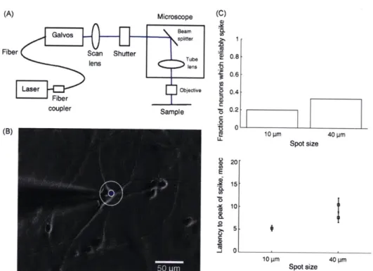
" Investigation of the spatial resolution and reliability of laser stimulation of neurons
expressing ChR2 and assessment of ChR2 stimulation to map connectivity of neurons
in culture
- Delivered ChR2 gene to cultured hippocampal neurons using rAAV.
-
Optical stimulation of action potentials was more reliable in neurons older than
DIV14.
-
A 40 pm diameter laser spot stimulated a larger fraction of neurons to reliably
fire action potentials than a 10 pm diameter spot.
-
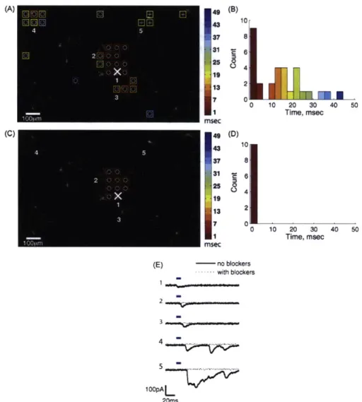
Scanned laser spot over culture to stimulate potential presynaptic partners of a
patch clamped neuron.
-
Proposed criteria distinguishing between monosynaptic and polysynaptic responses
recorded in a neuron postsynaptic to an optically-stimulated, ChR2-expressing
presynaptic neuron.
* Blockade of activity and synaptic vesicle release increased RV spread in hippocampal
culture
-
Validated the use of monosynaptically restricted RV in culture
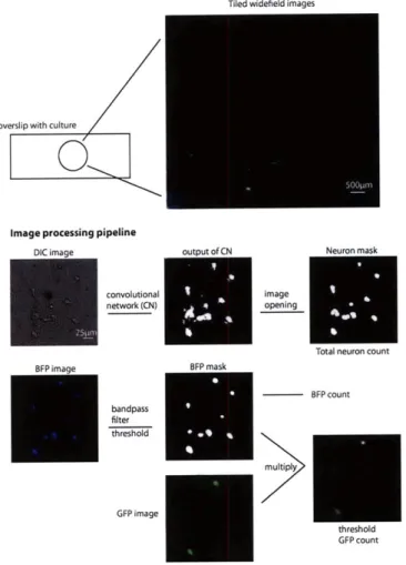
-
Developed an image analysis pipeline to automate analysis of large numbers of
images to assess the effects of drug manipulations on RV spread
-
Blocking synaptic vesicle release with botulinum toxin
-
A and tetanus toxin and
blocking neural activity with action potential blocker TTX and synaptic blockers
APV, CNQX, and bicuculline resulted in increased spread of RV. Application of
partial block of dynamin-mediated endocytosis with dynasore resulted in a slight
decrease of RV spread.
The study of neural circuits is the product of convergence in thought between the anatomical
studies of Ramon y Cajal [18] and the electrophysiological studies of Hubel and Weisel
[6]. There are many types of neurons in the brain, and how their connectivity could give
rise to the many functional properties neuroscientists have observed is both an inscrutable
and compelling problem
[4].
That synapses are the intermediary unit between these two
descriptions, places a large emphasis on understanding them, in particular, the modifications
by which synapses increase or decrease in strength. This importance is underscored by the
sheer immensity of literature on the experimental observations of synaptic modification and
the structures and mechanisms involved[7].
The experiments presented here were motivated by the fact that reward is a powerful
re-inforcer of behavior [8] and the subsequent finding that activity of dopamine neurons signaled
reward
[10].
Given the observation that stimulation of the ventral tegmental area modulated
cortical maps [1] and that dopamine could induce, as well as modulate certain forms of LTP
[5, 9], there was the possibility that dopamine could effect these changes by synapses being
the reward seeking agents themselves [11]. The hypothesis was that synaptic release,
fol-lowed by reward would result in an increased probability of release. Conversely, rewarding
synaptic failure would decrease the probability of release. Experimentally, this was a difficult
question to address, requiring measurements from neurons with small, stochastic synapses.
Because this was a hypothesis about vesicle release and failure, synaptically connected pairs
of neurons were required.
We chose to pursue this hypothesis using cultures of hippocampal neurons. The use of
culture to study connectivity is controversial because the endogenous 3 dimensional
struc-ture of the hippocampus is completely dissolved and networks regrown in a 2-dimensional
sheet. Arguably, it is unnatural and questionable whether the resulting connectivity has any
relationship to that of an intact brain. On the other hand, major phenomena observed in
culture, such as LTP and homeostatic plasticity, have also been observed in brain slice and
in vivo[2, 14], justifying the use of culture as a model system for investigating synapses. The
benefits to using culture are that it provides easy access for imaging, electrophysiological
recording, and genetic and pharmacological manipulation [13]. Our goal was not to claim
that the connectivity we observed in culture was a stand-in for an intact brain. Instead,
the goal was to verify a computational principle governing connectivity which might have
In chapter 2, we engineered connectivity in hippocampal culture by restricting growth
of neurons to glial microislands. Plating neurons at a low density on these microislands
resulted in circuits of neurons that had small numbers of neurons. Recording from islands
which contained only 2 synaptically connected neurons ensured that we were recording from a
monosynaptically connected pair, connections uncontaminated by polysynaptic input. From
those pairs we tested the effect of dopamine on excitatory synapses and found that dopamine
transiently depressed these synapses.
While this system held great promise for pursuing the hedonistic synapse hypothesis, we
found that microisland cultures were difficult to maintain, and the large synapses that were
characteristic of two neuron islands were not ideal for experiments which required small,
stochastic synapses. Using mass cultures provided a viable alternative. We found that
synapses in mass cultures were smaller than those in the microisland cultures, but finding
connected pairs of neurons was difficult. From pilot experiments, we observed a large range
of synaptic changes in response to contigent application of dopamine, resulting in the need
for many experiments to ensure reproducibility.
The search for a high-throughput method to identify small synapses begins in Chapter 3.
The report of successful transfection of neurons with ChR2 and the subsequent demonstration
that action potentials could be optically stimulated in these neurons with high temporal
precision [3] provided a promising way to screen for connected neurons in mass cultures.
We delivered the gene for ChR2 using recombinant adeno-associated virus and characterized
the reliability and spatial resolution of laser stimulation for producing action potentials in
neurons expressing ChR2. However, finding a criterion distinguishing between mono- and
poly-synaptic responses in a patch clamped postsynaptic neuron was elusive. Additionally,
there was a tradeoff between transfecting many neurons and preserving the spatial resolution
of the optical stimulation. The more densely packed ChR2 expressing neurons were, the
less likely sufficient optical stimulation would be guaranteed to stimulate only the targeted
neuron. Expressing ChR2 in fewer neurons meant having fewer potential presynaptic targets
to probe.
The engineering of monosynaptically restricted RV [17] appeared to be the ideal method
for elucidating connectivity in culture. By defining a population of host neurons and limiting
viral spread to neurons which were one synapse away, we could map all of the connections
onto a given neuron. Chapter 4 explores the use of monosynaptically restricted RV in
cul-ture, as well as the activity dependence of viral spread to putatively connected neurons. The
claim that RV spread is restricted to synaptically connected neurons comes from evidence
that systems with known connectivity can be traced with high precision using RV.
Addi-tionally, the lack of local spread outside these well-defined systems is a strong indication
of synaptic restriction [15, 12, 16]. Since synaptic transmission is a major determinant of
connectivity, we hypothesized that synaptic vesicle release should be required for the virus to
spread. Alternatively, neural activity might also be required. We investigated the effects of
activity blockade and synaptic transmission blockade on spread of RV and found that both
manipulations increased viral spread.
In closing, the hedonistic synapse hypothesis remains untested, but the framework and
methodology for finding monosynaptically connected pairs of neurons in hippocampal
cul-ture might be of use to researchers interested in probing such connections. With the notion
of cell type coming to the forefront of neural circuit analyses, there may be interest in
inves-tigating activity dependent plasticity mechanisms for different cell types. Additionally, the
interplay between activity dependent plasticity and homeostatic plasticity could be further
explored at the level of monosynaptically connected neurons. Recording from
monosynapti-cally connected pairs is a laborious task, and we hope that our contributions toward making
it more of a high-throughput endeavor will make it possible to try many more experimental
conditions, obtain larger samples and thus higher reproducibility of findings to enhance our
understanding of connectivity in the brain.
Chapter 2
Dopamine Transiently Depresses
Excitatory Postsynaptic Currents in
Cultured Rat Hippocampal Neurons
Dopamine Transiently Depresses Excitatory Postsynaptic
Currents in Cultured Rat Hippocampal Neurons
Jennifer Wangl'*, Jung H. Choi2, H. Sebastian Seung3
1 Department of Brain and Cognitive Sciences, Massachusetts Institute of Technology, Cambridge, MA, USA
2 Department of Brain and Cognitive Sciences, Massachusetts Institute of Technology, Cambridge, MA, USA
3 Department of Brain and Cognitive Sciences, Department of Physics, Massachusetts Institute of Technology, Howard Hughes Medical Institute, Cambridge, MA, USA
* E-mail: jenwangOmit.edu
Abstract
Background: Dopamine is a leading candidate for a reward signal in the brain and has also been implicated in many brain disorders. For these reasons, researchers have been interested in characterizing the effect of dopamine on synaptic transmission and plasticity. The reported effects of dopamine in the hippocampus have been diverse: different effects have been reported for different methodologies employed.
Methodology/Principal Findings: We have investigated the modulatory effects of dopamine on exci-tatory synaptic transmission using dual perforated patch clamp recordings of dissociated hippocampal neurons cultured from neonatal rats. Groups of a few neurons isolated on glial microislands were used for our experiments. Immunostaining revealed both D1
/D
5 and D2 subtypes of the dopamine receptor inour cultured neurons. Bath application of 10 pM dopamine (DA) caused a rapid depression of excitatory postsynaptic currents. The effect was transient, in that synaptic currents recovered to baseline during a 15 minute washout period. While the Di agonist SKF 38393 produced a weaker transient depression that was on the edge of statistical significance, it did not fully replicate the effect of dopamine. The D2
agonist quinpirole did not produce a significantly different result from control, nor did the combination of D1 and D2 agonists.
Conclusions/ Significance: Our results are similar to previous reports of immediate and transient depression in brain slices of hippocampal CA1, as well as neocortical areas of the hippocampal formation.
Introduction
Dopamine has become a leading candidate for a reward signal in the brain [1, 2], and plays an important role in current theories of reinforcement learning [3]. Dopamine has also been implicated in a number of brain disorders, such as Parkinson's disease [4, 5], schizophrenia [6], and ADHD [7]. For these reasons, researchers have been interested in characterizing the effects of dopamine on basic properties of neurons, such as intrinsic excitability [8-10] and synaptic transmission.
Electrophysiological studies have found that dopamine and its agonists have diverse effects on glu-tamatergic synaptic transmission. The effects depend on concentration, glutamate receptor subtype, dopamine receptor subtype, and brain region studied, which makes comparing results in the literature a confusing task. For brain slices of hippocampal CA1, researchers disagree over whether the effects of dopamine and its agonists are transient or persistent. Some find that synaptic transmission changes immediately upon bath application, and recovers to baseline within 10-15 minutes after washout. Others report that changes begin up to tens of minutes after bath application starts, and persist for up to hours after washout. As a rule, persistent changes have delayed onset, while transient changes develop rapidly (though one exception is noted below [11]). Those finding persistent changes do not report transient changes, and vice versa.
We now review the studies finding transient changes in more detail. Using intracellular recordings, Hsu showed that dopamine transiently depressed synaptic responses to Schaffer collateral stimulation [12]. Agonist and antagonist experiments suggested that this effect was mediated by the D2 receptor. Using
field potential recordings, Otmakhova and Lisman found that dopamine had little effect on synaptic responses to Schaffer collateral stimulation, except that the NMDA component was slightly depressed [13]. Instead, they found that dopamine transiently depresses synaptic responses to stimulation of the perforant path, including both the AMPA and NMDA components. Antagonist experiments suggested that both D1 and D2 receptors were involved.
In contrast to these findings of transient depression, Huang and Kandel found that D1/D5 agonists
induce persistent potentiation of field potential responses to Schaffer collateral stimulation [14]. Later on, Yang confirmed this result using intracellular recording via whole cell patch clamp [15], but Mockett et al. could not replicate it using field potential recordings [16]. Using whole cell patch clamp recording, Varela et al. found that a D1
/D
5 agonist could produce either persistent potentiation or depression [11].They found evidence that the sign of the effect depended on whether the NMDA receptors included NR2B subunits (potentiation) or NR2A subunits (depression). Since the NR2B subunit is predominant in the Schaffer collateral pathway, the Varela et al. result is consistent with the Huang and Kandel report of persistent potentiation. Since the NR2A subunit is predominant in the perforant pathway, the Varela et al. result is partially consistent with the Otmakhova and Lisman report of transient depression, except that their depression appears to be persistent and rapidly developing [13].
Finally, it should be mentioned that there is a subclass of the persistent studies in which dopamine is regarded as a modulator of long-term potentiation, defined as persistent potentiation induced by tetanic stimulation. Activation of D1
/D
5 receptors enhances the amplitude of early LTP [17], or is required forthe maintenance of late LTP [18]. According to these studies, dopamine by itself is not sufficient to induce persistent potentiation; tetanic stimulation is required.
The diversity of these results may not be surprising, in light of the complexity of molecular mechanisms
by which dopamine can affect glutamate receptors. Dopamine receptor activation can exert indirect
effects through the second messenger cAMP [19]. Another mechanism is direct, involving protein-protein interactions between dopamine receptors and glutamate receptors [20]. Further complicating the picture is the fact that dopamine can also directly block the NMDA receptor pore without mediation by dopamine receptors [21-23].
Given the inconsistencies between physiological studies in the literature, we thought it worth using a different method to reexamine the effects of dopamine on synaptic transmission in hippocampal neurons. We applied the perforated patch clamp technique to record from pairs of hippocampal neurons cultured on glial microislands. Dissociated cultures can be criticized as more unnatural than acute brain slices, but our preparation also has a number of advantages. By recording from monosynaptically connected pairs of neurons, we avoid potential confounds due to polysynaptic pathways in the brain slice preparation. Compared to whole cell patch clamp, perforated patch clamp may have less adverse effects on intracellular signaling pathways. The brain slice preparation contains endogenous stores of dopamine that can be released by electrical stimulation [24], while our cultures presumably do not. Finally, in and wash-out of dopamine is presumably faster in our cultures than in brain slices.
We have found that bath application of dopamine causes a transient depression of excitatory transmis-sion, with no sign of persistent effects. Furthermore, the D1 agonist SKF 38393 caused a weaker transient
D1 and D2 agonists produced no statistically significant changes. We suggest that D1 receptors could
mediate the transient depression produced by dopamine, with the caveat that the effect of D1 agonist
was significantly different from that of dopamine. Our results are similar to those of Otmakhova and Lisman for perforant path synapses [13], although it is unclear whether the synapses in our cultured hippocampal neurons are homologous to those of the perforant path or Schaffer collaterals. Our work is also related to the recent experiments of Zhang et al., who like us used dual intracellular patch recordings of hippocampal neurons grown in dissociated cultures. They found that dopamine transiently depresses the NMDA receptor-mediated component of glutamatergic currents [25].
Materials and Methods
Microisland cultures
Ethics Statement
P1 Sprague-Dawley rat pups were used. All animal procedures were approved by the MIT Committee on Animal Care, in compliance with standards for the ethical use of laboratory animals set by the state of Massachusetts, the city of Cambridge, and the United States Animal Welfare Act.
P1 rat pups were anesthetized by chilling on ice, followed by decapitation. Hippocampi were extracted and solutions were prepared in a similar fashion to [26]. Hippocampi were collected with dentate gyrus removed and cut into small (~ 1 mm) pieces in a dissection solution containing 25 mM HEPES in Hank's Balanced Salt Solution (HBSS), pH 7.3. Some pieces were immediately dissociated for culture by incubating the tissue for 30-40 min at 37C in HBSS containing 1 mM L-cysteine, 0.5 mM EDTA, 1.5 mM CaCl2, 20 U/ml Papain (Worthington), and 0.1 pg/ml DNAase. The remaining pieces were stored
for later use at 4C in Hibernate E medium (Brain Bits), supplemented with 2% B27 (Invitrogen). The enzymatically dissociated tissue was rinsed 3 times in culture medium containing 6 mg/ml glucose, 1 mM Na-Pyruvate (Invitrogen), 10% fetal bovine serum (Hyclone), 0.1% Mito serum extender (Invitrogen), 2%
B27 (Invitrogen), and 1 mM HEPES in Basal Medium Eagle (Invitrogen), pH 7.3 and then mechanically
triturated with a fire polished plastic pipette in culture medium conditioned by incubating overnight at
37C with a monolayer of glial cells in a culture flask.
on glass coverslips coated with rat tail collagen in a 24-well plate. After 1 day in the incubator, 100 pl of 24 pM cytosine beta-D-arabinofuranoside (Ara-C) in culture medium was added to dampen proliferation. After 2 to 3 weeks, several microislands of glial cells were usually visible. The islands were generally
50-100 pm in diameter. If neurons remained on the islands, the cultures were kept outside the incubator at
4C for a few hours to eliminate them.
Neurons were added to the glial islands 3-4 weeks later. These neurons were obtained by dissociating hippocampi that were either freshly dissected or stored for 3 days as described above. The cells were plated by aspirating culture medium from the well and adding 0.5 ml of cell suspension at 20,000 cells/ml on the glial microislands. A day or two later, 100 pl of culture medium was added (100pl of 24 pM Ara-C was added if needed to prevent glial proliferation). Neurons were used 8-14 days after plating on the glial microislands. We used microislands containing a few neurons, often just two.
Immunostaining
The cell culture was assayed for D1/D5 and D2 dopamine receptor subtypes [27] using rabbit anti-human dopamine Di (1:100) and goat anti-anti-human dopamine D2 (1:50) primary antibodies (Santa Cruz
Biotechnology, Inc.). The respective binding of these antibodies to their substrates was detected using secondary antibodies Alexa Fluor 488 donkey rabbit IgG (1:400) and Alexa Fluor 546 donkey anti-goat IgG (1:400) (Molecular Probes).
The cells were fixed in formalin for 20 min at room temperature, rinsed 3 times with phosphate buffered saline (PBS), and permeabilized in 0.25% Triton in PBS for 10 min at room temperature. Random binding of the primary was blocked with 4% donkey serum in PBS. The cells were then incubated at 37C for 1 hr with both primary antibodies. They were rinsed 3 times with PBS, incubated at 37C for 45 min with the secondary antibodies, and rinsed 3 more times with PBS. To control for random binding of the secondary antibodies, cells were fixed and permeabilized as above, and then incubated with the secondary antibodies only.
The cells were visualized with an inverted, phase contrast microscope (Olympus IX70). Excitation and emission of the secondary antibodies was produced using FITC and TRITC cubes (Chroma) with a mercury arc lamp source. Images were acquired using a Sensicam QE high speed CCD camera (Cooke), controlled by CamWare (Cooke) software.
Electrophysiology
Dual whole-cell perforated patch recordings were performed using the Multiclamp 700A patch clamp amplifier (Axon Instruments). Signals were filtered at 6 kHz and sampled at 10 kHz using a PCI-6052E
A/D board (National Instruments). Micropipettes were pulled from glass capillaries (Warner) and had
a 2-3 MQ resistance. The pipettes were back-filled with an internal solution containing 300 pg/ml amphotericin B (Sigma) in 1% DMSO for membrane perforation. The internal solution of the pipette contained (in mM) potassium gluconate 136.5, KCl 17.5, NaCl 9, MgCl2 2, pH 7.30. [28] We determined
that perforation had occurred when the access resistance stabilized and was no longer decreasing. This generally happened when the access resistance was between 20 MQ and 40 MQ with fluctuations less than 15%. The neurons were bathed in a HEPES-buffered saline (HBS) containing (in mM) NaCl 145, KCl 3, HEPES 10, CaCl2 3, glucose 8, MgCl2, pH 7.30. [28] Recordings were done at room temperature. To determine synaptic connectivity, the postsynaptic cell was stepped in voltage clamp, from -70 to -10 mV in 10 mV increments, while the presynaptic cell was stimulated in voltage clamp to fire an action potential in voltage clamp with a +120 mV pulse applied for 1.5 msec. In this way, the synaptic reversal potential could be determined. Typically, IPSCs reversed between -50 and -30 mV and had a
~100 ms time constant, while EPSCs (glutamatergic currents) showed no reversal for the given voltage
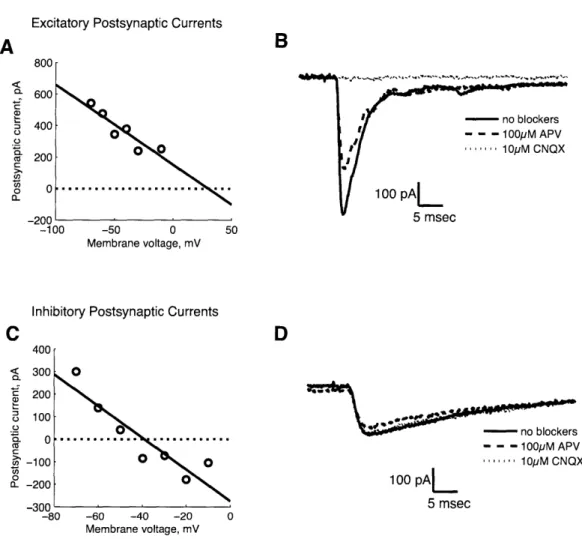
steps and had a shorter time constant of ~10 ms. The use of reversal potential to determine transmitter type was verified for a few examples by blocking glutamatergic currents with 10 AM CNQX and 100 pM APV (Sigma). In the representative example shown in Figure 1A, the estimated reversal potential was greater than -10 mV, so we classified the synaptic current as an EPSC. Application of CNQX was sufficient to block synaptic transmission (Figure 1B), confirming glutamatergic transmission. Conversely, in Figure 1C, the estimated reversal potential was -40 mV, so we classified the synaptic current as an
IPSC. Application of CNQX and APV did not alter synaptic transmission (Figure ID), which showed
that IPSCs were not mediated by glutamatergic transmission. We did not use synaptic blockers for all experiments; we measured the reversal potential instead, since it seemed a reliable indicator of transmitter type. For our experiments, we used pairs which had reversal potential greater than -10 mV.
To measure evoked EPSCs, the presynaptic cell was stimulated in voltage clamp by stepping from
-70 mV to 50 mV for 1 ms once every 30 sec while holding the postsynaptic cell at -70 mV. The PSC
amplitude was measured from a smoothed (0.5 ms boxcar) version of the current trace by taking the difference between the current just before onset of the PSC and the trough. PSC amplitudes ranged from
50-700 pA and had simple shapes, indicating monosynaptic responses. Pairs selected for analysis had
access resistance between 15 and 40 MQ and leak current less than -150 pA. Additionally, recordings in which postsynaptic currents exhibited correlated changes with access resistance were discarded. Custom software written in Matlab was used to perform the experiments and analyze the data.
Dopamine and agonists
The dopamine (DA) solution was prepared immediately before bath application, as dopamine is known to degrade rapidly through oxidation. If the solution was not freshly made, the effects reported in this paper became weaker (data not shown). We avoided using ascorbic acid as an anti-oxidant, because of its effects on neural excitability [29].
The D1 agonist R(+)SKF-38393 (Sigma) and D2 agonist (-)Quinpirole (Sigma) were kept at -20*C
as 10 mM stock solutions in Millipore filtered water. They were diluted to 10 pM in HBS immediately before bath application.
Bath application of drugs
All experiments consisted of three periods: (1) a baseline lasting 5 minutes, (2) drug application for 5
minutes, and (3) washout for 15 minutes. The bath solution was continually renewed using a peristaltic
pump (Rainin) at a base flow rate of 40 ml/hr. At the beginning of the drug application period, the
intake of the peristaltic pump was exchanged to bath solution containing the drug. The flow rate was increased to maximum for 30 seconds to rapidly exchange the new solution. For the rest of the drug application period, the flow rate was returned to the base value of 40 ml/hr. At the beginning of the washout period, the intake of the peristaltic pump was exchanged to bath solution with no drug, and a similar procedure was followed.
In control experiments, the flow rate of the peristaltic pump was manipulated with the same time course, but with no change of solutions.
Results
Using the procedures described in the Methods, we produced cultures containing glial microislands and hippocampal neurons at very low density (see Figure 2A1 for representative phase contrast image). In
vivo, the hippocampus receives dopaminergic innervation from the ventral tegmental area (VTA) [30]
and contains dopamine receptors [31,32]. Since our cultures presumably did not contain dopaminergic neurons, it was unclear whether the hippocampal neurons would continue to express dopamine receptors. This was checked by double immunostaining for D1 and D2 receptors, which revealed that expression is
present in vitro (Figure 2B and C).
Synaptic transmission between pairs of neurons was measured using dual patch clamp recordings (Figure 3A). For each recording, we located a glial microisland containing two to four neurons, and voltage clamped two of them. The presynaptic neuron was stimulated to produce an action potential while the postsynaptic neuron was recorded to measure synaptic transmission. If synaptic currents were observed, the reversal potential was found by varying the voltage of the postsynaptic neuron (see Methods). If the reversal potential was consistent with glutamatergic transmission, then an experiment was performed.
Every experiment used the same protocol. After the access resistance of the recording stabilized (see Methods), the presynaptic neuron was stimulated to generate a single action potential once every
30 seconds. The postsynaptic current was measured during three periods: (1) baseline for 10 minutes
(2) drug application for 5 minutes (3) washout for 15 minutes. For each recorded pair of neurons, the time series of EPSC amplitudes was normalized by dividing by the average EPSC amplitude during the baseline period. The normalized time series were averaged over neuron pairs to produce the graphs in Figure 3. Three drugs were each applied singly: dopamine (DA), the D1 agonist SKF 38393, and the D2
agonist quinpirole. In a fourth manipulation, both D1 and D2 agonists were applied in combination. All
drugs were used at 10 pM concentrations.
Figure 3B shows the average time course of EPSC amplitude during the DA experiments. The amplitude depresses immediately after the start of bath application of DA. By the end of the 5 minute DA period, the EPSC amplitude is less than 40% of its baseline value. During the 15 minute washout period, the EPSC recovers steadily. Shown for comparison is the average time series from control experiments with no drug.
In Figure 3C, application of D1 agonist causes depression of the EPSC amplitude. The effect is
strongest at the end of the 5 minute period of agonist application. The average depression during the agonist application is not as strong as that observed with DA. In Figure 3D, application of D2 agonist
the 5 minute period of agonist application. Overall, the depression is even weaker than that observed with D1 agonist.
It is not clear why DA produced a stronger depression than the agonists. We conjectured that DA produces a stronger effect by stimulating both D1 and D2 receptors. To test this hypothesis, we applied
a combination of D1 and D2 agonists, but this produced little or no effect (Figure 3E).
To test for the significance of these effects, we performed two types of statistical analysis. The first type of analysis is based on the various averages shown in Figure 3. In Figure 4A, each experiment is summarized by a single number quantifying the amount of depression, calculated by averaging the normalized EPSC amplitude during the 5 minute bath application period.
To compare the five conditions of (1) dopamine (n = 6), (2) D1 agonist (n = 8), (3) D2 agonist
(n = 8), (4) D1+D2 agonist (n = 7), and (5) control (n = 5), we performed an analysis of variance
(ANOVA), followed by the Tukey-Kramer test to identify pairs of conditions that were statistically
significant (MATLAB Statistics Toolbox). The ANOVA rejected the null hypothesis that all conditions are the same. By the Tukey-Kramer test, dopamine is significantly different from the other conditions
2-5, which include both the control and the agonists. Conditions 2-5 are not significantly different from
each other.
As a second test of statistical significance, we did not time average the data during the bath application period. Figure 4B shows these data for the different drugs. In this analysis, an experiment is not represented by a single average number, but by several data points taken at times during the bath application period. These data points are assumed to be statistically independent measurements. This assumption would be true if the fluctuations were due only to measurement noise. However, there also appear to be correlated fluctuations; the data points from a single experiment might tend to be all above-average or all below-above-average. Therefore we apply the second test of statistical significance with the caveat that its results are probably not conservative enough. We considered only data points from the second half of the bath application period, because it can take some time for the effects of the drug to set in.
Again the ANOVA rejected the null hypothesis that all conditions are the same. By the Tukey-Kramer test, the dopamine and D1 agonist experiments are significantly different from each other, and from all other conditions. The other conditions (D2 agonist, D1+D2 agonist, and control) are not significantly different from each other.
Discussion
We have found that bath application of dopamine causes a rapidly developing depression of excitatory synaptic transmission in hippocampal neurons. During the 15 minute washout period, synaptic currents recover to their baseline values, so that the depression is transient. Our finding of an immediate and transient depression is similar to previous reports in brain slices of hippocampal CA1 [12,13].
The D1 agonist SKF 38393 also induced a weak transient depression. However, the effect was not
significantly different from control by the Tukey-Kramer test based on the time averages of experiments. The effect was significant if the data points from an experiment were not averaged, but this statistical analysis cannot be completely trusted because the fluctuations at different times are likely to be correlated. We conclude that the effect of D1 agonist is on the borderline of significance. However, by both types of
statistical analysis, the effect of the D1 agonist is significantly different from that of dopamine. A weaker effect is consistent with the fact that SKF 38393 is regarded as a partial rather than full agonist of the D1 receptor [33]. The D2 agonist quinpirole caused an even weaker transient depression. The effect was
not significant by either statistical analysis.
We suggest that Di receptors could mediate the effects of dopamine on synaptic transmission. This is similar to the conclusion that Otmakhova and Lisman reached for perforant path synapses using antagonist experiments [13]. It does not match Hsu's finding that transient depression in the Schaffer collateral pathway is mediated solely by D2 receptors [12].
Finally, we found that a combination of the D1 and D2 agonists was not significantly different from
control. In other words, the effect produced by the combination of agonists was less than the effect produced by the D1 agonist. We speculate that this is related to the fact that the D1 and D2 receptors
have antagonistic effects on levels of the second messenger cAMP, as well as on phosphorylation of DARPP-32 [19].
Based on the results with agonists, we cannot completely exclude the possibility that dopamine is acting directly on the NMDA receptor pore, rather than indirectly through dopamine receptors [21-23].
Recently Zhang et al. published another study about the effects of dopamine on long-term plasticity of glutamatergic synapses between hippocampal neurons grown in dissociated cultures [25]. As an aside, they also mentioned that dopamine transient depresses synaptic transmission. They observed a significant effect when AMPA receptor currents were blocked. They also observed a weaker effect without synaptic
blockers, but the effect was not statistically significant. They did not report whether these effects could be replicated with dopamine agonists. To speculate about the slight differences between their results and ours, we note there are two differences in methods. They used ascorbic acid to retard the oxidation of dopamine in solution, and they cultured embryonic neurons rather than postnatal neurons.
In conclusion, we join the ranks of researchers who have reported transient depression of excitatory hippocampal synapses by dopamine [12, 13], as opposed to persistent potentiation [14, 15]. Transient depression is also a common finding in neocortical areas of the hippocampal formation. Behr et al. showed that dopamine and a D1 agonist transiently depress excitatory synaptic currents in subicular
neurons, while a D2 agonist did not [34]. Pralong and Jones found that dopamine transiently depressed
excitatory synaptic currents in entorhinal cortex neurons [35]. Based on antagonist experiments, they tentatively concluded that Di receptors were more likely to be involved. Similar results have also been reported in brain slices of prefrontal cortex [36], nucleus accumbens [37, 38], and basal forebrain [39].
Acknowledgments
References
1. Schultz W (1998) Predictive reward signal of dopamine neurons. J Neurophysiol 80: 1-27.
2. Schultz W (2002) Getting formal with dopamine and reward. Neuron 36: 241-263.
3. Montague P, Dayan P, Sejnowski T (1996) A framework for mesencephalic dopamine systems based
on predictive hebbian learning. Journal of Neuroscience 16: 1936-1947.
4. Dauer W, Przedborski S (2003) Parkinson's disease mechanisms and models. Neuron 39: 889-909.
5. Olanow C, Tatton W (1999) Etiology and pathogenesis of parkinson's disease. Annual review of
neuroscience 22: 123-144.
6. Davis K, Kahn R, Ko G, Davidson M (1991) Dopamine in schizophrenia: a review and
reconcep-tualization. American Journal of Psychiatry 148: 1474-1486.
7. LaHoste GJ, Swanson JM, Wigal SB, Glabe C, Wigal T, et al. (1996) Dopamine d4 receptor gene
8. Calabresi P, Mercuri NB, Sancesario G, Bernardi G (1993) Electrophysiology of dopamine-denervated striatal neurons. implications for parkinson's disease. Brain 116 ( Pt 2): 433-52.
9. Centonze D, Gubellini P, Pisani A, Bernardi G, Calabresi P (2003) Dopamine, acetylcholine and
nitric oxide systems interact to induce corticostriatal synaptic plasticity. Reviews in the neuro-sciences 14: 207-16.
10. Chen L, Bohanick JD, Nishihara M, Seamans JK, Yang CR (2007) Dopamine d1/5
receptor-mediated long-term potentiation of intrinsic excitability in rat prefrontal cortical neurons: Ca2+-dependent intracellular signaling. Journal of neurophysiology 97: 2448-64.
11. Varela J, Hirsch S, Chapman D, Leverich L (2009) D1/d5 modulation of synaptic nmda receptor
currents. Journal of Neuroscience 29: 3109-3119.
12. Hsu K (1996) Characterization of dopamine receptors mediating inhibition of excitatory synaptic transmission in the rat hippocampal slice. J Neurophysiol 76: 1887-1895.
13. Otmakhova N, Lisman J (1999) Dopamine selectively inhibits the direct cortical pathway to the
cal hippocampal region. J Neurosci 19: 1437-1445.
14. Huang Y, Kandel E (1995) D1/d5 receptor agonists induce a protein synthesis-dependent late potentiation in the cal region of the hippocampus. Proc Natl Acad Sci U S A 92: 2446-50.
15. Yang S (2000) Sustained enhancement of ampa receptor- and nmda receptor-mediated currents
in-duced by dopamine dl/d5 receptor activation in the hippocampus: an essential role of postsynaptic ca2+. Hippocampus 10: 57-63.
16. Mockett B, Brooks W, Tate W, Abraham W (2004) Dopamine d1/d5 receptor activation fails to
initiate an activity-independent late-phase ltp in rat hippocampus. Brain Res 1021: 92-100.
17. Otmakhova N, Lisman J (1996) D1/d5 dopamine receptor activation increases the magnitude of
early long-term potentiation at cal hippocampal synapses. J Neurosci 16: 7478-86.
18. O'Carroll CM, Morris RGM (2004) Heterosynaptic co-activation of glutamatergic and dopaminergic
afferents is required to induce persistent long-term potentiation. Neuropharmacology 47: 324-32.
20. Lee FJS, Xue S, Pei L, Vukusic B, Ch6ry N, et al. (2002) Dual regulation of nmda receptor functions
by direct protein-protein interactions with the dopamine dl receptor. Cell 111: 219-30.
21. Castro NG, de Mello MC, de Mello FG, Aracava Y (1999) Direct inhibition of the n-methyl-d-aspartate receptor channel by dopamine and (+)-skf38393. Br J Pharmacol 126: 1847-55. 22. Masuko T, Suzuki I, Kizawa Y, Kusama-Eguchi K, Watanabe K, et al. (2004) Monoamines
di-rectly inhibit n-methyl-d-aspartate receptors expressed in xenopus oocytes in a voltage-dependent manner. Neurosci Lett 371: 30-3.
23. Cui C, Xu M, Atzori M (2006) Voltage-dependent block of n-methyl-d-aspartate receptors by
dopamine d1 receptor ligands. Molecular Pharmacology 70: 1761-1770.
24. Frey U, Schroeder H, Matthies H (1990) Dopaminergic antagonists prevent long-term maintenance of posttetanic ltp in the cal region of rat hippocampal slices. Brain Res 522: 69-75.
25. Zhang JC, Lau PM, Bi GQ (2009) Gain in sensitivity and loss in temporal contrast of stdp by
dopaminergic modulation at hippocampal synapses. Proc Natl Acad Sci USA 106: 13028-33.
26. Hagler J, Goda Y (2001) Properties of synchronous and asynchronous release during pulse train
depression in cultured hippocampal neurons. J Neurophysiol 85: 2324-34.
27. Aizman 0, Brismar H, Uhl6n P, Zettergren E, Levey AI, et al. (2000) Anatomical and physiological
evidence for dl and d2 dopamine receptor colocalization in neostriatal neurons. Nat Neurosci 3:
226-30.
28. Bi G, Poo M (1998) Synaptic modifications in cultured hippocampal neurons: dependence on spike
timing, synaptic strength, and postsynaptic cell type. J Neurosci 18: 10464-72.
29. Sutor B, Bruggencate GT (1990) Ascorbic acid: a useful reductant to avoid oxidation of
cate-cholamines in electrophysiological experiments in vitro? Neurosci Lett 116: 287-92.
30. Gasbarri A, Verney C, Innocenzi R, Campana E, Pacitti C (1994) Mesolimbic dopaminergic neurons
innervating the hippocampal formation in the rat: a combined retrograde tracing and immunohis-tochemical study. Brain Res 668: 71-9.
31. Huang
Q,
Zhou D, Chase K, Gusella J, Aronin N, et al. (1992) Immunohistochemical localization of the dl dopamine receptor in rat brain reveals its axonal transport, pre- and postsynaptic local-ization, and prevalence in the basal ganglia, limbic system, and thalamic reticular nucleus. Proc Natl Acad Sci U S A 89: 11988-92.32. Levey A, Hersch S, Rye D, Sunahara R, Niznik H, et al. (1993) Localization of dl and d2 dopamine
receptors in brain with subtype-specific antibodies. Proc Natl Acad Sci U S A 90: 8861-5.
33. Setler P, Sarau H, Zirkle C, Saunders H (1978) The central effects of a novel dopamine agonist.
European journal of pharmacology 50: 419-430.
34. Behr J, Gloveli T, Schmitz D, Heinemann U (2000) Dopamine depresses excitatory synaptic trans-mission onto rat subicular neurons via presynaptic dl-like dopamine receptors. J Neurophysiol 84:
112-9.
35. Pralong E, Jones R (1993) Interactions of dopamine with glutamate- and gaba-mediated synaptic
transmission in the rat entorhinal cortex in vitro. Eur J Neurosci 5: 760-7.
36. Gao W, Krimer L, Goldman-Rakic P (2001) Presynaptic regulation of recurrent excitation by dl
receptors in prefrontal circuits. Proc Natl Acad Sci U S A 98: 295-300.
37. Harvey J, Lacey M (1996) Endogenous and exogenous dopamine depress epscs in rat nucleus
accumbens in vitro via dl receptors activation. J Physiol 492 ( Pt 1): 143-54.
38. Nicola S, Kombian S, Malenka R (1996) Psychostimulants depress excitatory synaptic transmission
in the nucleus accumbens via presynaptic d1-like dopamine receptors. J Neurosci 16: 1591-604.
39. Momiyama T, Sim J, Brown D (1996) Dopamine dl-like receptor-mediated presynaptic inhibition
of excitatory transmission onto rat magnocellular basal forebrain neurones. J Physiol 495 ( Pt 1):
97-106.
Excitatory Postsynaptic Currents
800 r 400 200 0 -200 -10te
- no blockers - --00M APV ''''''' 10pyM CNQX 100 pAL 5 msec -50 0 Membrane voltage, mVInhibitory Postsynaptic Currents
300-200 100 0--100 -200 -300 --80 ---- no blockers -- - 100pM APV 10pM CNQX
100 pA[
5 msec
-60 -40 -20 Membrane voltage, mVFigure 1. Validating reversal potential as a method to select glutamatergic synapses. In
panels (A) and (C), we show current-voltage (I-V) plots for two synapses, which serve as representative examples. The open circles are EPSC amplitudes (y-axis) measured for varying postsynaptic holding potentials (x-axis). The solid line is a least squares fit to indicate the estimated reversal potential (intersection with dotted line). In (A), the estimated reversal potential is greater than -10 mV, so we classified the synaptic current as an EPSC. Panel (B) shows that application of 10 1M CNQX is sufficient to block transmission, confirming that excitatory synaptic transmission is glutamatergic. An example of an inhibitory synapse is shown in (C) and (D). The estimated reversal potential was -40 mV (C), so we classified the synaptic current as an IPSC. Neither CNQX or APV had an effect on
synaptic transmission (D), consistent with the notion that IPSCs were not mediated by glutamatergic transmission.
O
0 .. .. . .. . .. ..
O. O..
Phase Contrast
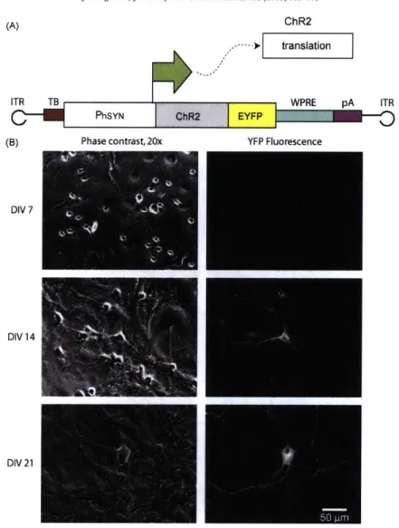
Figure 2. Hippocampal culture expresses D1 and D2 subtypes of the dopamine receptor.
Phase contrast image at 40x magnification of culture grown on glial microisland (A1). A magnified version is shown in A2. Pseudocolored fluorescent images showing Di (1:100) expression (B) and D2
(1:50) expression (C) using the same field of view as in A2.
.... .. . . .... .. ... ... .... .... ... .... .. .... ... ... ... ---- . ... . ... ... .. ...
10m. 100pA 1.5 2m-s E EE
~0.
Z 0 control 0 0. N control -10 -5 0 5 10 15 20 Time, min a) 1.5-05 C,13 N Z onro l E 0 (-)Quinpirole 0 O control 1.5- 20ms Z0
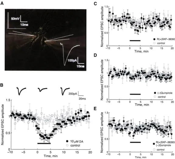
' -10 -5 0 5 10 15 20 Tm mTime, min a) 1.5 -E (-QunirlFigure 3. Bath application of dopamine and D1, D2 agonists results in a transient
depression of EPSC amplitude. Bath application of dopamine and D1/D2 agonists results in a transient depression of EPSC amplitude. A schematic (white) of the dual patch clamp experiment is shown in (A), overlaid on a 40x phase contrast image of two neurons isolated on a glial island. The presynaptic neuron (left) was stimulated in voltage clamp to fire an action potential by stepping the membrane potential of the cell from -70 mV to 50 mV for 1 ins. An EPSC was recorded in the voltage clamped postsynaptic neuron (right). The time series of averaged, normalized EPSC amplitudes are shown in (B-E, closed black circles) for the 3 phases of the experiment - baseline, drug application, and washout. The control condition, where no drug was applied, is shown for comparison (open gray circles, n = 5). The error bars represent the standard error. In (B), we applied 10 piM DA during the bath perfusion period (n = 6). Above the time series are three representative EPSCs selected from the corresponding phase of the experiment. In (C), we applied 10 piM D1 agonist R(+)SKF-38393 (n = 8)
during the drug application period, and in (D), we applied 10 piM D2 agonist (-)Quinpirole (nu 8). In
(E), we applied a combination of 10 piM R(+)SKF-38393 and 10 ptM (-)Quinpirole (n= 7).
1- x
0.8-
x
f
LU X 0.6- x E0.4 -0.2 - x 01-0
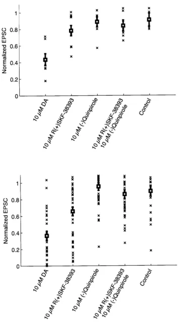
0.8 -Nx LU 0.6 - N .N E0.4 -0z 0.2 -0 Cp) 4k''Figure 4. Summary of data from the bath application experiments. (A) Distribution of
average normalized EPSC amplitude during the drug application period for each pair. Drug is indicated on the x-axis, and each point (x) represents a pair. The bold squares with error bars represent the
ANOVA estimate for the mean and standard error, respectively, for each group. The DA condition was
significantly different from control and all other conditions, using the Tukey-Kramer comparison test. (B) Distribution of EPSC amplitudes taken from the second half of the perfusion period. Each point (x) represents an individual, non-averaged value for EPSC amplitude. Drug is indicated on the x-axis. The
ANOVA estimate for the mean and standard error are also shown (bold squares and error bars,
respectively). The DA and Di conditions were significantly different from each other and all other conditions, including control, by the Tukey-Kramer comparison test.