Basolateral amygdala regulation of adult hippocampal
neurogenesis and fear-related activation of newborn neurons
The MIT Faculty has made this article openly available.
Please share
how this access benefits you. Your story matters.
Citation
Kirby, E D, A R Friedman, D Covarrubias, C Ying, W G Sun, K A
Goosens, R M Sapolsky, and D Kaufer. “Basolateral Amygdala
Regulation of Adult Hippocampal Neurogenesis and Fear-Related
Activation of Newborn Neurons.” Mol Psychiatry 17, no. 5 (June 14,
2011): 527–536.
As Published
http://dx.doi.org/10.1038/mp.2011.71
Publisher
Nature Publishing Group
Version
Author's final manuscript
Citable link
http://hdl.handle.net/1721.1/102418
Terms of Use
Creative Commons Attribution-Noncommercial-Share Alike
Basolateral amygdala regulation of adult hippocampal
neurogenesis and fear-related activation of newborn neurons
Elizabeth D. Kirby, B.S.1, Aaron R. Friedman, B.A.2, David Covarrubias, B.S.3, Carl Ying, B.A.2, Wayne G. Sun2, Ki A. Goosens, PhD4, Robert M. Sapolsky, PhD5, and Daniela Kaufer,
PhD*,1,2
1Helen Wills Neuroscience Institute, UC Berkeley 2Integrative Biology, UC Berkeley
3Molecular and Cell Biology, UC Berkeley
4McGovern Institute for Brain Research, Massachusetts Institute of Technology
5Department of Biological Sciences, Stanford University, Stanford, CA and Department of Neurology and Neurological Sciences, Stanford University, Stanford, CA
Abstract
Impaired regulation of emotional memory is a feature of several affective disorders, including depression, anxiety and post-traumatic stress disorder. Such regulation occurs, in part, by interactions between the hippocampus and the basolateral amygdala (BLA). Recent studies have indicated that within the adult hippocampus, newborn neurons may contribute to support of emotional memory, and that regulation of hippocampal neurogenesis is implicated in depressive disorders. How emotional information impacts newborn neurons in adults is not clear. Given the role of the BLA in hippocampus-dependent emotional memory, we investigated whether hippocampal neurogenesis was sensitive to emotional stimuli from the BLA. We show that BLA lesions suppress adult neurogenesis, while lesions of the central nucleus of the amygdala do not. Similarly, we show that reducing BLA activity through viral vector-mediated overexpression of an outwardly rectifying potassium channel suppresses neurogenesis. We also show that BLA lesions prevent selective activation of immature newborn neurons in response to a fear conditioning task. These results demonstrate that BLA activity regulates adult hippocampal neurogenesis and the fear context-specific activation of newborn neurons. Together, these findings denote functional implications for proliferation and recruitment of new neurons into emotional memory circuits.
Keywords
neurogenesis; hippocampus; fear conditioning; basolateral amygdala; emotion; stem cell
Users may view, print, copy, and download text and data-mine the content in such documents, for the purposes of academic research, subject always to the full Conditions of use:http://www.nature.com/authors/editorial_policies/license.html#terms
*Corresponding author: Daniela Kaufer, 3060 Valley Life Sciences Bldg. #3140, Berkeley, CA 94720, Phone: (510) 642-9346, Fax: (510) 643-6264, danielak@berkeley.edu.
HHS Public Access
Author manuscript
Mol Psychiatry. Author manuscript; available in PMC 2015 January 29.
Published in final edited form as:
Mol Psychiatry. 2012 May ; 17(5): 527–536. doi:10.1038/mp.2011.71.
Author Manuscript
Author Manuscript
Author Manuscript
Emotion strongly modulates memory function in adult mammals, altering the strength and longevity of memories and sometimes leading to memory dysfunction. Notably, disordered emotional memory contributes to several affective disorders, including depression, anxiety and post-traumatic stress disorder. Emotion-associated modulation of memory appears to rely largely on interaction between the memory processes of the hippocampus and emotional input provided by the basolateral complex of the amygdala (BLA)1-4. Behaviorally, BLA activity supports memory for emotionally salient stimuli in rodents and humans1, 5, 6 while at the neurophysiological level, BLA activity supports hippocampal long-term potentiation (LTP)7-10 and enhances hippocampal output to other brain areas11. BLA activity also modulates activation of hippocampal immediate early genes, another marker of hippocampal plasticity12, as well as hippocampal responses to stress13, 14, further suggesting a
modulatory role for the BLA in hippocampal function.
Recent studies suggest a role for adult hippocampal neurogenesis in emotional memory function15-19. In adult mammals, new hippocampal neurons arise from a resident population of neural stem cells located in the dentate gyrus20-23 and form a population of immature neurons that incorporate into existing networks within weeks of birth24. Ablation of adult neurogenesis by genetic knockout or irradiation impairs contextual fear memory15, 16 and may also modulate the transfer of fear-related memories from the hippocampus to other neural structures for long-term storage17. The heightened plasticity of newly born neurons appears to be key, as acceleration of their maturation impairs fear learning25. The
requirement of new neurons for fear memory suggests a clinical role for adult neurogenesis in several affective disorders3, 26, 27. However, the mechanisms by which adult neurogenesis responds to emotional stimuli to influence memory formation are not yet clear.
We explored the adult neurogenic response to emotional input from the BLA and its potential role in fear memory. Specifically, we investigated how BLA activity would affect adult neurogenesis and modulate activation of immature neurons in response to fear-associated context.
Materials and Methods
Animals
Adult male Sprague-Dawley rats (Charles River) were pair-housed on a 12h light/dark cycle. All animal procedures were approved by the UC Berkeley and MIT Animal Care and Use Committees.
Stereotaxic surgery
Excitotoxic lesions of the BLA or CeA or sham surgeries were performed as per28. Coordinates for BLA infusion were: -2.8 mm anterior/posterior (A/P), +/-5.1 mm medial/ lateral (M/L) relative to bregma; -6.8 mm (2 min) and -6.5 mm (1 min) relative to dura. Coordinates for CeA infusion were: -2.2 mm A/P and +/-4.4 mm M/L relative to bregma; -7.0 mm from dura (1 min). Six to 8 hrs after surgery all rats received an additional injection of buprenorphine (0.05 mg/kg, s.c.). For viral vector infusions, virus was infused 0.2 μl/min
Author Manuscript
Author Manuscript
Author Manuscript
for 10 min (2 μl total) at the same BLA coordinates. Viral vectors were prepared as per29 and titers were 106-108 infectious particles/ml.
Bromodeoxyuridine injections
5-Bromo-2’-deoxyuridine (BrdU, Sigma) was dissolved in physiological saline and injected intraperitoneally for all experiments.
Immunohistochemical staining
Rats were anesthetized with Euthasol euthanasia solution and transcardially perfused with ice cold 4% paraformaldehyde in 0.1M PBS. Brains were post-fixed for 24 hrs at 4°C, equilibrated in 30% sucrose in 0.1M PBS and then stored at -80°C. Immunostaining was performed on a 1 in 6 series of free-floating 30 μm cryostat sections.
For PCNA and BrdU staining, sections were rinsed in 0.1M Tris-buffered saline (TBS) and pretreated for 10min with 0.3% H2O2 in TBS. For BrdU only, sections were incubated in 2N
HCl at 37°C for 30 min. All sections were incubated in blocking solution (1% normal donkey solution, 0.3% triton-X 100 in TBS) for 30 min followed by overnight incubation at 4°C on rotation in primary antibody (mouse anti-PCNA, 1:500 in blocking, Abcam, Cambridge, MA; mouse anti-BrdU, 1:500, BD Biosciences, San Jose, CA). Sections were rinsed and transferred to secondary antibody (biotin anti-mouse, 1:500, Jackson
ImmunoResearch, West Grove, PA) for 2 hrs at room temperature. Following rinsing, sections were incubated in ABC reagent (Vector, Burlingame, CA) and then developed with DAB (Vector). Sections were mounted on gelatin-coated slides, dehydrated in alcohol and coverslipped with permount mounting medium.
Triple immunohistochemistry was performed similarly. Primary antibodies were: goat anti-DCX (1:200, Santa Cruz Biotechnology), mouse anti-S100β (1:200, Abcam), rat anti-MBP (1:100, Abcam), rabbit anti-cfos (1:100; Santa Cruz Biotechnology, Santa Cruz, CA). Secondary antibodies were: Cy5 anti-goat, Cy3 anti-mouse, Cy3 anti-rat, biotin anti-rabbit (1:500; Jackson ImmunoResearch). For the cFos/BrdU/DCX triple stain, a tertiary
incubation for 1 hr at room temperature was included with streptavidin-Alexa488 (1:1000 in TBS, Invitrogen, Carlsbad, CA) after secondary incubation. All sections were then incubated in 4% paraformaldehyde for 10 min, rinsed and incubated in primary antibody against BrdU as above (1:500, rat anti-BrdU, Abcam; mouse anti-BrdU, BD Biosciences). The next day, sections were rinsed and incubated in secondary antibody: FITC anti-rat; Cy3 anti-rat; biotin anti-rat (1:500, Jackson ImmunoResearch). For MBP/BrdU staining, a tertiary incubation for 1 hr at room temperature was added with streptavidin-Alexa488 (1:1000 in TBS,
Invitrogen). Sections were mounted on gelatin-coated slides and coverslipped with DABCO anti-fading medium.
BrdU, PCNA and cFos quantification
BrdU, PCNA or cFos positive cells were counted in the dentate gyrus and subgranular zone using a 40x air objective (Zeiss). The area sampled was calculated using StereoInvestigator software (MicroBrightfield, Williston, VT) and used to calculate the number of positive cells per m2.
Author Manuscript
Author Manuscript
Author Manuscript
Confocal analysis
25-50 BrdU or cFos positive cells were located in the dentate gyrus for each animal (or each hemisphere for unilateral lesion) and assessed in z-series of <1.0 um slices to determine if other markers (DCX, S100β, MBP, cFos, BrdU) were co-expressed. Confocal images were captured on a Zeiss 510 META/NLO confocal microscope with a 40x oil objective and adjusted for brightness and contrast using LSM Image Browser software.
Lesion and viral vector assessment
One series of sections was mounted and counterstained with cresyl violet for lesion assessment. If a lesion did not cover at least 75% of the area of interest without affecting surrounding areas, that rat was removed from analysis (Figure 1). For virus expression, if GFP expression was not found in the BLA or if expression was focused outside the BLA, the rat was removed from analysis.
Fear conditioning
Two weeks after unilateral lesion of the BLA or sham surgery, rats received 4 daily
injections of BrdU (100 mg/kg). Two weeks after the last BrdU injection, rats were exposed to fear conditioning. Fear conditioning chambers were 12 w × 10 l × 12 h inch boxes with an electrified grid floor inside a sound attenuating chamber (Coulbourn Instruments, Whitehall, PA). Chambers had two house lights and a house fan that were on at all times during testing and training. Rats were placed in the box and allowed 5 min to acclimate. The animals then received 10 unsignaled, 1mA, 1s duration shocks with an intertrial interval ranging from 10 to 120 s. Rats were left in the chamber for 3 min after the last shock and then returned to their home cage. Chambers were cleaned with 70% ethanol between trials. The next day, rats were placed back in the fear context or a novel context for 15 min without any shock delivery. The novel context was the fear conditioning chamber with the grid floor removed and laminated cork boarding covering three of the walls. Freezing behavior was tracked and analyzed with FreezeFrame software (Coulbourn). After context exposure, rats were returned to their home cages for 45 min and then perfused as in other studies. This time delay was chosen because it coincides with the elevation in IEG expression following context memory activation30.
Statistical analysis
Unpaired t-tests were used to assess the effects of bilateral lesion on proliferation and cell differentiation, context exposure on freezing behavior, and context exposure on BrdU co-labeling of sham operated rats (unequal variances assumed). Paired t-tests were used to assess proliferation differences in the unilateral lesion study, with an unpaired t-test to compare control PCNA/BrdU levels to that contralateral to BLA lesion. Repeated measures analysis of variance (ANOVA) was used to compare the effect of unilateral BLA versus CeA lesion on cell proliferation as well as the effect of novel versus fear context exposure on BrdU co-labeling in unilaterally BLA lesioned rats. Bonferroni post-hoc tests were used to assess the effect of hemisphere within each lesion group in both cases. p<0.05 was considered significant for all studies.
Author Manuscript
Author Manuscript
Author Manuscript
Results
Bilateral BLA lesions suppressed hippocampal neurogenesis
Adult hippocampal neurogenesis is a multi-phase process regulated by the proliferation, differentiation, migration, and survival of new cells23. To assess the effect of loss of BLA input on the neurogenic process, we excitotoxically lesioned the BLA of adult male rats bilaterally28, 31 (Figure 1) and investigated cell proliferation as well as differentiation (Figure 2a). BLA lesion reduced the number of proliferation cell nuclear antigen (PCNA) positive cells by 55.9% compared to rats who received bilateral sham surgery (Figure 2b). BLA-lesion also led to a 45.3% reduction in the number of BrdU positive dentate gyrus cells 5-10 days after proliferative cells were labeled by BrdU injection (Figure 2c). These results indicate suppression of cell proliferation following BLA lesion, resulting in a persistent reduction in immature cells. BLA lesion did not affect cell fate, with approximately 85-90% of new cells expressing the neural marker doublecortin (DCX) and less than 5% expressing the astrocytic marker S100β or the oligodendrocyte marker myelin basic protein (MBP) regardless of lesion (Figure 2d-e). These data indicate that BLA lesions cause a reduction in the pool of immature neurons and glia three weeks after lesion.
Unilateral BLA lesions suppressed hippocampal neurogenesis
We next investigated whether the suppression of adult hippocampal neurogenesis following BLA lesion is mediated by ipsilateral neural connections or by possible systemic changes (such as a change in circulating hormone levels). Because ipsilateral connections mediate the influence of BLA activity on hippocampal LTP10, 32, we predicted that BLA lesion-induced suppression of adult hippocampal neurogenesis would similarly rely on ipsilateral neural connections and be hemisphere-specific. To investigate this hypothesis, BrdU and PCNA positive cells were quantified three weeks following unilateral BLA lesion (Figure 3a).
Unilateral BLA lesion suppressed the number of PCNA-labeled proliferative hippocampal cells by 32.2% ipsilateral to the lesion compared to contralateral (Figure 3b). Similar results were obtained with BrdU-labeled cells (11.29±1.40 cells ipsilateral, 22.35±4.09 cells contralateral, 49.6% suppression, n = 7). The number of proliferative cells ipsilateral to BLA lesion was both similar to that in bilaterally lesioned rats and significantly lower than that in bilateral sham-operated rats (Figure 3b). Together, these results imply that BLA-associated regulation of neurogenesis is hemisphere-specific, most likely suppressing cell proliferation through ipsilateral neural connections.
CeA lesions did not suppress hippocampal neurogenesis
We next investigated the anatomical specificity of BLA lesion-induced suppression of hippocampal neurogenesis. Previous studies show that while BLA lesions reduce hippocampal LTP, lesions of the central nucleus of the amygdala (CeA) do not32. We therefore predicted that CeA lesions would also have no effect adult hippocampal neurogenesis.
Author Manuscript
Author Manuscript
Author Manuscript
Three weeks after surgery to lesion either the BLA or the CeA (Figure 1; Figure 3a), we found that while there were 26.8% fewer BrdU positive cells ipsilateral to BLA lesion compared to contralateral, there was no difference in the number of proliferative BrdU positive cells ipsilateral versus contralateral to CeA lesion (Figure 3c). We found a similar suppression in the number of proliferative PCNA positive cells in the dentate gyrus ipsilateral to BLA, but not CeA, lesion (Figure 3d). Over all the experiments of figure 3, both BrdU and PCNA cell number ipsilateral to BLA lesion was suppressed relative to control while cell number contralateral to lesion was not different from that in controls. Hippocampal proliferation levels in CeA lesioned rats, however, did not differ from controls either ipsilateral or contralateral to lesion. These results indicate that BLA lesions
ipsilaterally suppress hippocampal neurogenesis while CeA lesions do not, suggesting that the BLA influence over hippocampal neurogenesis is specific to loss of BLA input, and does not result non-specifically from ipsilateral excitotoxic cell death or surgical damage per se.
Viral vector-mediated reduction of BLA activity suppressed adult hippocampal neurogenesis
We next determined whether suppression of neural activity in the BLA without excitotoxic lesion is sufficient to modulate neurogenesis. To reduce BLA neural activity, we ectopically expressed the outwardly rectifying potassium channel Kv1.1 or a GFP-only control from a herpes viral vector in BLA neurons. This transgene construct has been shown to reduce basal neural firing29, 33. GFP-Kv1.1 overexpression reduced proliferative BrdU positive cell
number by 36.5% and 30.5% (Figure 4) as compared to GFP-only viral vector infusion and sham-operated controls, respectively. These results indicate that reduction of BLA activity via Kv1.1 overexpression is sufficient to suppress hippocampal neurogenesis ipsilaterally.
Fear memory activates newly born neurons
To investigate the functional importance of BLA regulation of adult hippocampal
neurogenesis, we examined how new neurons participate in BLA-dependent fear memory. Previous studies show that ablation of adult hippocampal neurogenesis causes impairments in BLA-dependent fear conditioning15-17, suggesting a functional role for new neurons in
fear-associated memory networks. Previous work also shows that new neurons may be particularly prone to integration into memory networks as they are preferentially activated by exposure to previously experienced contexts such as enriched environment, water maze and fear conditioning34-36.
To test the activation of newborn neurons by fear memory, we exposed sham-operated rats to a contextual fear conditioning task, which is dependent on both BLA and hippocampal activity3, 5, 6, 28, and then assessed expression of the immediate early gene (IEG) cFos in
newly born neurons. cFos expression reflects neuronal activation and incorporation into hippocampal memory circuits25, 34, 35. It is also critical to hippocampal mediation of
contextual fear conditioning specifically37, 38.
Two weeks after labeling proliferative cells with BrdU, rats were exposed to a series of 10 unpredictable shocks in a conditioning chamber (Figure 5a). Re-exposure to the shock-associated context (the fear context) the next day led to greater freezing compared to
Author Manuscript
Author Manuscript
Author Manuscript
exposure to a novel context (Figure 5b), indicating robust memory for the fear-associated environment. Forty-five minutes after re-exposure, rats were perfused and assessed for BrdU co-labeling with cFos. BrdU positive cells at this point were 15-19 days old, an age
characterized by preferential recruitment into memory networks and possibly enhanced importance for hippocampal memory function18, 34. Using DCX as a marker of immature neurons, we found that exposure to the fear context increased the proportion of BrdU cells co-expressing DCX and cFos compared to rats exposed to the novel context (Figure 5c and d). Exposure to fear context did not alter the total number of cFos-positive cells in the dentate gyrus (Figure 5e). These data suggest that new neurons are activated by fear-associated memory but not simply by exposure to a novel environment.
BLA lesions prevented activation of new neurons by fear memory
We next investigated whether BLA lesion could influence the activation of new neurons by fear memory. Unilaterally BLA lesioned rats showed greater freezing in the fear context than in the novel context, indicating robust fear memory despite the presence of lesion, as expected in the presence of one functional BLA (Figure 5f). We found that BLA lesion blocked the fear-context associated increase in BrdU/cFos/DCX co-expression ipsilaterally (Figure 5g). In fear context exposed rats, a lower proportion of BrdU cells expressed cFos/DCX+ ipsilateral versus contralateral to lesion. The proportion of triple labeled cells contralateral to lesion did not differ from that in sham-operated rats. In rats exposed to the novel context, by contrast, BLA lesion did not affect the proportion of cFos/DCX-labeled BrdU cells. These results suggest that BLA lesion blocks the selective activation of two week old immature neurons by a context associated with an aversive stimulus.
We also found that there were significantly fewer BrdU/DCX positive cFos cells ipsilateral to the BLA lesion compared to contralateral in rats exposed to the fear but not the novel context (Supplementary Figure 1). These results suggest that, of cells activated by exposure to a fear context (i.e. expressing cFos), the percentage representing immature neurons is reduced by ipsilateral BLA lesion. The overall percentage of BrdU cells expressing DCX was not affected by lesion (Supplementary Figure 2).
Discussion
We demonstrate for the first time that BLA activity regulates adult neurogenesis and potentially modifies the recruitment of new neurons into networks underlying emotional memory. Bilateral and unilateral BLA lesions suppressed hippocampal neurogenesis, suggesting that ipsilateral BLA input supports basal hippocampal neurogenesis. These results extend several previous lines of work showing that BLA activity can affect hippocampal plasticity and function. Previous work demonstrates, for example, that lesion or inactivation of the BLA suppresses dentate gyrus LTP ipsilaterally32. Activation of β-adrenoceptors in the BLA can also enhance hippocampal memory function in an inhibitory avoidance task while increasing expression of Arc, an immediate early gene and marker of hippocampal plasticity12. In addition, the BLA plays an integral role in how stress affects the hippocampus; the work of McGaugh et al. has shown the BLA is required for
stress-Author Manuscript
Author Manuscript
Author Manuscript
induced impairments of memory retrieval as well as stress-induced enhancements in memory consolidation4, 13, 14.
CeA lesions did not suppress hippocampal neurogenesis, suggesting that reduction in hippocampal neurogenesis is not attributable to generalized ipsilateral amygdala damage, and is a specific response to loss of BLA activity. Previous work on dentate gyrus-perforant path LTP similarly shows no effect of CeA lesions on LTP32. The dependence of
hippocampal plasticity on BLA but not CeA activity is consistent with the behavioral function of the BLA, which is required for hippocampus-dependent contextual fear conditioning. The CeA, by contrast, participates in hippocampus-independent behaviors such as cued fear conditioning39. Together with previous studies, our results suggest a general model wherein BLA input promotes multiple forms of hippocampal plasticity, thereby supporting hippocampal memory.
We further demonstrated regulation of hippocampal neurogenesis by BLA activity using a viral vector-mediated overexpression of the voltage gated outwardly-rectifying potassium channel, Kv1.1. Outwardly rectifying potassium channels such as Kv1.1 regulate neuronal excitability, with voltage gated subtypes aiding in repolarization after an action potential and thereby decreasing glutamate release probability40. Kv1.1 overexpression in neurons via HSV vector specifically causes reduced neural resting potential, decreases basal firing rates and is neuroprotective against excitotoxic insult (i.e. kainic acid and glutamate)29, 41. In the BLA, overexpression of outwardly rectifying potassium channels in neurons also reduces anxiety and stress responses in adult rats, suggesting that suppressing BLA activity by manipulating potassium channel expression has consequences for emotionality33. In the present study, we found that overexpression of Kv1.1 in the BLA suppressed hippocampal neurogenesis ipsilaterally, suggesting that neural activity in the BLA supports hippocampal neurogenesis.
The BLA sends the majority of its ipsilateral input to the hippocampus through two indirect pathways, relayed through either the medial septum or the entorhinal cortex10, 42. Both of these pathways have been implicated in BLA support of adult hippocampal LTP and provide excitatory input to the dentate gyrus10, 43. While the medial septum provides input through cholinergic projections to the dentate gyrus granule neurons, the entorhinal cortex primarily sends glutamatergic projections10, 43. Importantly, both acetylcholine and glutamate increase proliferation of adult neural precursor cells44-46, suggesting that input from either pathway could mediate BLA support of adult neurogenesis in the dentate gyrus. In addition, induction of dentate gyrus LTP itself stimulates hippocampal neurogenesis 47, 48, raising the
possibility that suppressed LTP following BLA lesion could in part underlie suppression of neurogenesis. Future studies will address the extent to which these two ipsilateral pathways contribute to the support of hippocampal neurogenesis.
New neurons, particularly those around two weeks old, influence emotional memory function15, 18. New neurons are also context-sensitive, integrating strongly into memory networks for contexts initially experienced around two weeks of age34. This preferential integration may rely on the heightened plasticity of immature neurons compared to their older counterparts in the dentate gyrus34. Indeed, acceleration of maturation beyond this
Author Manuscript
Author Manuscript
Author Manuscript
plastic phase can interfere with both integration of new neurons into non-fear related hippocampal memory networks as well as behavioral performance in hippocampus-dependent emotional memory tasks25.
We investigated whether new neurons in this highly plastic phase of their development were integrated into fear memory circuits using a contextual fear conditioning task two weeks after labeling newborn cells. Contextual fear conditioning relies on both the BLA and the hippocampus3, 5, 6, 28 and appears to be partially mediated by adult hippocampal
neurogenesis15, 49. We chose cFos expression as a marker of neuronal activation due to its role in memory for fear-associated context37, 38. We found that exposure to a fear-associated context activated two week old neurons more than exposure to a novel context, an effect that was blocked by ipsilateral BLA lesion. These results suggest that immature neurons are integrated into neural networks for fear-associated memory, but that this integration is dependent on BLA input. However, it is also possible that the new neuron activation could reflect participation in fear extinction. Future studies will further explore this possibility. One recent study using mice suggests that while fear conditioning activates newly born neurons36, this activation occurs in older neurons than those examined in our study (six weeks versus 2 weeks) and in a smaller proportion of new cells. A separate study further suggested that there was no cFos co-expression in two-week old neurons following exposure to a water maze task in mice25. Rather, IEG expression occurred only in new neurons 4-6 weeks old. Given that new neurons mature faster and potentially play a more influential role in hippocampal function in rats than mice50, these differences in activation and integration of new neurons likely relate to species differences in how new neurons function and mature. Our results demonstrate a novel mechanism for the influence of emotion over hippocampal memory function. By modifying adult hippocampal neurogenesis and altering the
incorporation of new neurons into emotional memory networks, BLA input could shape how emotional stimuli influence memory. Whether these two impairments are the result of two independent processes or contingent one upon the other (such as through selective
suppression of proliferation of cells that would later respond BLA input) is not clear and will require more research. To our knowledge, our results represent the first report of regulation of adult hippocampal neurogenesis and new neuron activation by BLA activity and fear-associated network activation. Future studies will further address the behavioral relevance of this phenomenon for emotional memory function as well as the underlying molecular mechanisms for emotion regulation of hippocampal proliferative capacity.
Supplementary Material
Refer to Web version on PubMed Central for supplementary material.
Acknowledgments
We would like to thanks Chloe LaLonde, Abhiram Gande, Anna Geraghty and Brandon Thai for technical assistance. EDK was supported by a California Institute for Regenerative Medicine pre-doctoral fellowship and a National Defense Science and Engineering Graduate Research fellowship from the Department of Defense. KG was supported by a NARSAD Young Investigator Award and the NIH (R01MH849662). DK was supported by a NARSAD Young Investigator Award and the NIMH BRAINS award (R01MH087495).
Author Manuscript
Author Manuscript
Author Manuscript
References
1. Richardson MP, Strange BA, Dolan RJ. Encoding of emotional memories depends on amygdala and hippocampus and their interactions. Nat Neurosci. 2004; 7:278–285. [PubMed: 14758364]
2. Tsoory MM, Vouimba RM, Akirav I, Kavushansky A, Avital A, Richter-Levin G. Amygdala modulation of memory-related processes in the hippocampus: potential relevance to PTSD. Prog Brain Res. 2008; 167:35–51. [PubMed: 18037005]
3. Roozendaal B, McEwen BS, Chattarji S. Stress, memory and the amygdala. Nat Rev Neurosci. 2009; 10:423–433. [PubMed: 19469026]
4. McGaugh JL. The amygdala modulates the consolidation of memories of emotionally arousing experiences. Ann Rev Neurosci. 2004; 27:1–28. [PubMed: 15217324]
5. Maren S. Neurotoxic basolateral amygdala lesions impair learning and memory but not the performance of conditional fear in rats. J Neurosci. 1999; 19:8696–8703. [PubMed: 10493770] 6. Maren S, Aharonov G, Fanselow MS. Retrograde abolition of conditional fear after excitotoxic
lesions in the basolateral amygdala of rats: absence of a temporal gradient. Behavioral Neuroscience. 1996; 110:718–726. [PubMed: 8864263]
7. Frey S, Bergado-Rosado J, Seidenbecher T, Pape H-C, Frey JU. Reinforcement of early long-term potentiation (early-LTP) in dentate gyrus by stimulation of the basolateral amgdala: heterosynaptic induction mechansims of late-LTP. J Neurosci. 2001; 21:3697–3703. [PubMed: 11331399] 8. Jas J, Almaguer W, Frey JU, Bergado J. Lesioning the fimbria-fornix impairs basolateral amygdala
induced reinforcement of LTP in the dentate gyrus. Brain Res. 2000; 861:186–189. [PubMed: 10751582]
9. Ikegaya Y, Saito H, Abe K. The basomedial and basolateral amygdaloid nuclei contribute to the induction of long-term potentiation in the dentate gyrus in vivo. Eur J Neurosci. 1996; 8:1833– 1839. [PubMed: 8921274]
10. Bergado JA, Frey S, Lopez J, Almaguer-Melian W, Frey JU. Cholinergic afferents to the locus coeruleus and noradrenergic afferents to the medial septum mediate LTP-reinforcement in the dentate gyrus by stimulation of the amygdala. Neurobiol Learn Mem. 2007; 88:331–341. [PubMed: 17596976]
11. Paz R, Pelletier JG, Bauer EP, Paré D. Emotional enhancement of memory via amygdala-driven facilitation of rhinal interactions. Nat Neurosci. 2006; 9:1321–1329. [PubMed: 16964249] 12. McIntyre CK, Miyashita T, Setlow B, Marjon KD, Steward O, Guzowski JF, et al.
Memory-influencing intra-basolateral amygdala drug infusions modulate expression of Arc protein in the hippocampus. PNAS. 2005; 102:10718–10723. [PubMed: 16020527]
13. Roozendaal B, Griffith QK, Buranday J, de Quervain DJ-F, McGaugh JL. The hippocampus mediates glucocorticoid-induced impairment of spatial memory retrieval: dependence on the basolateral amygdala. PNAS. 2003; 100:1328–1333. [PubMed: 12538851]
14. Roozendaal B, Nguyen BT, Power AE, McGaugh JL. Basolateral amygdala noradrenergic influence enables enhancement of memory consolidation induced by hippocampal glucocorticoid receptor activation. PNAS. 1999; 96:11642–11647. [PubMed: 10500230]
15. Saxe MD, Battaglia F, Wang JW, Malleret G, David DJ, Monckton JE, et al. Ablation of hippocampal neurogenesis impairs contextual fear conditioning and synaptic plasticity in the dentate gyrus. PNAS. 2006; 103:17501–17506. [PubMed: 17088541]
16. Hernández-Rabaza V, Llorens-Martín M, Velázquez-Sánchez C, Ferragud A, Arcusa A, Gumus HG, et al. Inhibition of adult hippocampal neurogenesis disrupts contextual learning but spares spatial working memory, long-term conditional rule retention and spatial reversal. Neurosci. 2009; 159:59–68.
17. Kitamura T, Y S, Takashima N, Murayama A, Niibori Y, Ageta H, et al. Adult neurogenesis modulates the hippocampus-dependent period of associative fear memory. Cell. 2009; 139:814– 827. [PubMed: 19914173]
18. Deng W, Saxe MD, Gallina IS, Gage FH. Adult-born hippocampal dentate granule cells undergoing maturation modulate learning and memory in the brain. J Neurosci. 2009; 29:13532– 13542. [PubMed: 19864566]
Author Manuscript
Author Manuscript
Author Manuscript
19. Dupret D, Fabre A, Döbrössy MD, Panatier A, Rodríguez JJ, Lamarque S, et al. Spatial learning depends on both the addition and removal of new hippocampal neurons. PLoS Biol. 2007; 5:1683– 1694.
20. Walker TL, White A, Black DM, Wallace RH, Sah P, Bartlett PF. Latent stem and progenitor cells in the hippocampus are activated by neural excitation. J Neurosci. 2008; 28:5240–5247. [PubMed: 18480280]
21. Eriksson PS. Neurogenesis in the adult human hippocampus. Nat Med. 1998; 4:1313–1317. [PubMed: 9809557]
22. Gould E. How widespread is adult neurogenesis in mammals? Nat Rev Neurosci. 2007; 8:481–488. [PubMed: 17514200]
23. Kirby, ED.; Kaufer, D. Stress and adult neurogenesis in the mammalian central nervous system. In: Soreq, H.; Friedman, A.; Kaufer, D., editors. STRESS: from molecules to behavior. Wiley-Blackwell; Weinheim, Germany: 2009. p. 71-91.
24. van Praag H, Schinder AF, Christie BR, Toni N, Palmer TD, Gage FH. Functional neurogenesis in the adult hippocampus. Nature. 2002; 415:1030–1034. [PubMed: 11875571]
25. Farioli-Vecchioli S, Saraulli D, Costanzi M, Pacioni S, Cinà I, Aceti M, et al. The timing of differentiation of adult hippocampal neurons is crucial for spatial memory. PLoS Biol. 2008; 6:2188–2204.
26. Revest J-M, Dupret D, Koehl M, Funk-Reiter C, Grosjean N, Piazza P-V, et al. Adult hippocampal neurogenesis is involved in anxiety-related behaviors. Mol Psychiatry. 2009; 14:959–967. [PubMed: 19255582]
27. Dere E, Pause BM, Pietrowsky R. Emotion and episodic memory in neuropsychiatric disorders. Behav Brain Res. 2010; 215:162–171. [PubMed: 20227444]
28. Goosens KA, Maren S. Contextual and auditory fear conditioning are mediated by the lateral, basal, and central amygdaloid nuclei in rats. Learn Mem. 2001; 8:148–155. [PubMed: 11390634] 29. Lee AL, Dumas TC, Tarapore PE, Webster BR, Ho DY, Kaufer D, et al. Potassium channel gene
therapy can prevent neuron death resulting from necrotic and apoptotic insults. J Neurochem. 2003; 86:1079–1088. [PubMed: 12911616]
30. Kee N, Teixeira CM, Wang AH, Frankland PW. Imaging activation of adult-generated granule cells in spatial memory. Nat Protocol. 2007; 2:3033–3044.
31. Paxinos, G.; Watson, C. The rat brain: in stereotaxic coordinates-the new coronal set. Elsevier Academic Press; Burlington, MA: 2004.
32. Ikegaya Y, Saito H, Abe K. Attenuated hippocampal long-term potentiation in basolateral amygdala-lesioned rats. Brain Res. 1994; 656:157–164. [PubMed: 7804830]
33. Mitra R, Ferguson D, Sapolsky RM. SK2 potassium channel overexpression in basolateral amygdala reduces anxiety, stress-induced corticosterone secretion and dendritic arborization. Mol Psychiatry. 2009; 14:847–855. [PubMed: 19204724]
34. Tashiro A, Makino H, Gage FH. Experience-specific functional modification of the dentate gyrus through adult neurogenesis: a critical period during an immature stage. J Neurosci. 2007; 27:3252– 3259. [PubMed: 17376985]
35. Kee N, Teixeira CM, Wang AH, Frankland PW. Preferential incorporation of adult-generated granule cells into spatial memory networks in the dentate gyrus. Nat Neurosci. 2007; 10:355–362. [PubMed: 17277773]
36. Stone SS, Teixeira CM, Zaslavsky K, Wheeler AL, Martinez-Canabal A, Wang AH, et al. Functional convergence of developmentally and adult-generated granule cells in dentate gyrus circuits supporting hippocampus-dependent memory. Hippocampus. 2010 e-pub ahead of print 7 September 2010. 10.1002/hipo.20845
37. Radulovic J, Kammermeier J, Spiess J. Relationship between fos production and classical fear conditioning: effects of novelty, latent inhibition, and unconditioned stimulus preexposure. J Neurosci. 1998; 18:7452–7461. [PubMed: 9736664]
38. Gao C, Gill MB, Tronson NC, Guedea AL, Guzmán YF, Huh KH, et al. Hippocampal NMDA receptor subunits differentially regulate fear memory formation and neuronal signal propagation. Hippocampus. 2010; 20:1072–1082. [PubMed: 19806658]
Author Manuscript
Author Manuscript
Author Manuscript
39. LeDoux JE. Emotion circuits in the brain. Ann Rev Neurosci. 2000; 23:155–184. [PubMed: 10845062]
40. Meir A, Ginsburg S, Butkevich A, Kachalsky SG, Kaiserman I, Ahdut R, et al. Ion channels in presynaptic nerve terminals and control of transmitter release. Physiological Reviews. 1999; 79:1019–1088. [PubMed: 10390521]
41. Wenzel HJ, Vacher H, Clark E, Trimmer JS, Lee AL, Sapolsky RM, et al. Structural consequences of Kcna1 gene deletion and transfer in the mouse hippocampus. Epilepsia. 2007; 48:2023–2046. [PubMed: 17651419]
42. Pikkarainen M, Ronkko S, Savander V, Insausti R, Pitkanen A. Projections from the lateral, basal and accessory basal nuclei of the amygdala to the hipoocampal formation in the rat. J Comp Neurol. 1999; 403:229–260. [PubMed: 9886046]
43. Wheal HV, Millera JJ. Pharmacological identification of acetylcholine and glutamate excitatory systems in the dentate gyrus of the rat. Brain Res. 1980; 182:145–155. [PubMed: 6243231] 44. Itou Y, Nochi R, Kuribayashi H, Saito Y, Hisatsune T. Cholinergic activation of hippocampal
neural stem cells in aged dentate gyrus. Hippocampus. 2010 e-pub ahead of print 6 January 2010. 10.1002/hipo.20761
45. Ge S, Sailor KA, Ming G-l, Song H. Synaptic integration and plasticity of new neurons in the adult hippocampus. J Physiol. 2008; 586:3759–3765. [PubMed: 18499723]
46. Joo J-Y, Kim B-W, Lee J-S, Park J-Y, Kim S, Yun Y-J, et al. Activation of NMDA receptors increases proliferation and differentiation of hippocampal neural progenitor cells. J Cell Sci. 2007; 120:1358–1370. [PubMed: 17389682]
47. Bruel-Jungerman E, Davis S, Rampon C, Laroche S. Long-term potentiation enhances
neurogenesis in the adult dentate gyrus. J Neurosci. 2006; 26:5888–5893. [PubMed: 16738230] 48. Chun SK, Sun W, Park JJ, Jung MW. Enhanced proliferation of progenitor cells following long-term potentiation induction in the rat dentate gyrus. Neurobiol Learn Mem. 2006; 86:322–329. [PubMed: 16824772]
49. Shors TJ, Townsend DA, Zhao M, Kozorovitskiy Y, Gould E. Neurogenesis may relate to some but not all types of hippocampal-dependent learning. Hippocampus. 2002; 12:578–584. [PubMed: 12440573]
50. Snyder JS, Choe JS, Clifford MA, Jeurling SI, Hurley P, Brown A, et al. Adult-born hippocampal neurons are more numerous, faster maturing, and more involved in behavior in rats than in mice. J Neurosci. 2009; 29:14484–14495. [PubMed: 19923282]
Author Manuscript
Author Manuscript
Author Manuscript
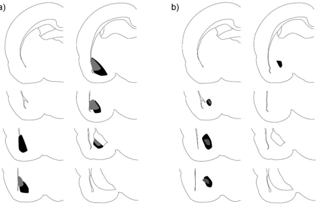
Figure 1. Excitotoxic lesions
Minimum (grey) and maximum (black) extent of excitotoxic BLA (a) and CeA (b) lesions.
Author Manuscript
Author Manuscript
Author Manuscript
Figure 2. Bilateral BLA lesions suppressed hippocampal neurogenesis
(a) Experimental timeline. (b) Bilaterally lesioned rats (n = 5) had significantly fewer PCNA positive cells than sham operated rats (n =6). *p<0.05. (c) Bilaterally lesioned rats also had significantly fewer BrdU positive cells than sham operated rats, representing a reduction in the number of 5-10 day old cells. *p<0.05. (d) BLA lesion did not affect the percent of BrdU positive cells expressing one of three cell fate markers: doublecortin (DCX), S100β or myelin basic protein (MBP). (e) Representative confocal images showing colocalization of BrdU with DCX, S100β or MBP. Scale bar = 10 μm.
Author Manuscript
Author Manuscript
Author Manuscript
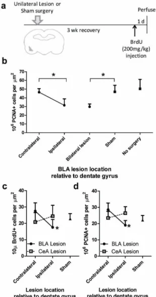
Figure 3. Unilateral lesions of the BLA, but not the CeA, ipsilaterally suppressed neurogenesis
(a) Experimental timeline. (b) There were significantly fewer dentate PCNA positive cells ipsilateral versus contralateral to unilateral BLA lesion (n = 5). Similar suppression of PCNA positive cells was found with bilateral BLA lesion (n = 6) as ipsilateral to unilateral lesion. The number of PCNA positive cells contralateral to lesion was similar to that found in the dentate gyrus of bilateral sham-operated animals (n = 3) and no surgery rats (n = 4). *p<0.05. (c) In BLA (n = 5), but not CeA (n = 6) lesioned rats, there were significantly fewer BrdU positive dentate cells ipsilateral versus contralateral to the lesion. Sham rats, n = 6. *p<0.01. (d) In BLA (n = 5), but not CeA (n = 6) lesioned rats, there were significantly fewer PCNA positive cells ipsilateral versus contralateral to the lesion. Sham rat, n = 6. *p<0.01.
Author Manuscript
Author Manuscript
Author Manuscript
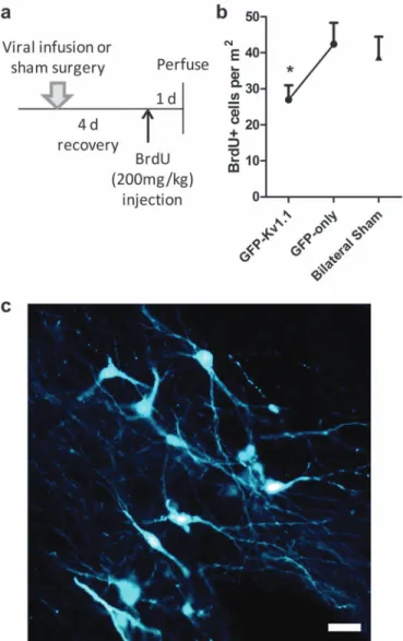
Figure 4. BLA silencing via potassium channel overexpression suppressed neurogenesis
(a) Experimental timeline. (b) In rats infused with GFP-Kv1.1 and GFP-only viral vectors to reduce BLA activity (n=8), there were significantly fewer BrdU positive dentate cells ipsilateral to GFP-Kv1.1 infusion. Sham rats, n = 15. *p<0.05. (c) Representative images of GFP expression in virus-infected BLA neurons. Scale bar = 10 μm
Author Manuscript
Author Manuscript
Author Manuscript
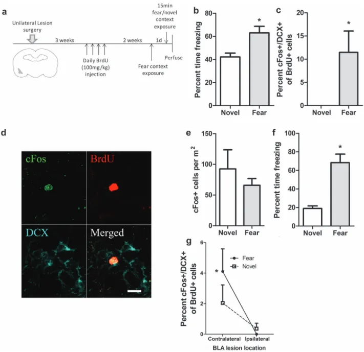
Figure 5. Unilateral BLA lesions blocked activation of immature neurons by exposure to a fear-associated context
(a) Experimental timeline. (b) Exposure to the fear context (n = 7) resulted in significantly more freezing than exposure to the novel context in sham operated rats (n = 5). (c) Sham-operated rats exposed to the fear context had a greater proportion of BrdU positive cells that co-expressed DCX and cFos. (d) Representative confocal image of a BrdU cell (red) expressing both DCX (blue) and cFos (green). Scale bar = 10 μm. (e) Sham-operated rats exposed to the fear context had similar numbers of cFos+ cells in the dentate gyrus as rats exposed to the novel context. (f) In unilaterally BLA lesioned rats, exposure to the fear context (n = 5) resulted in significantly more freezing than exposure to the novel context (n = 6). (g) In rats exposed to the fear but not the novel context, there was a lower percentage
Author Manuscript
Author Manuscript
Author Manuscript
of DCX/cFos labeled BrdU positive cells ipsilateral versus contralateral to the lesion. *p<0.05