HAL Id: tel-01918182
https://tel.archives-ouvertes.fr/tel-01918182
Submitted on 10 Nov 2018
HAL is a multi-disciplinary open access archive for the deposit and dissemination of sci-entific research documents, whether they are pub-lished or not. The documents may come from teaching and research institutions in France or abroad, or from public or private research centers.
L’archive ouverte pluridisciplinaire HAL, est destinée au dépôt et à la diffusion de documents scientifiques de niveau recherche, publiés ou non, émanant des établissements d’enseignement et de recherche français ou étrangers, des laboratoires publics ou privés.
Roles of ERK1/2 signaling in LMNA-cardiomyopathy
Maria Chatzifrangkeskou
To cite this version:
Maria Chatzifrangkeskou. Roles of ERK1/2 signaling in LMNA-cardiomyopathy. Cardiology and cardiovascular system. Université Pierre et Marie Curie - Paris VI; Freie Universität (Berlin), 2016. English. �NNT : 2016PA066380�. �tel-01918182�
Université Pierre et Marie Curie
Freie Universität
Ecole doctorale
Centre de recherche en Myologie, UPMC-Inserm UMRS974 CNRS FRE3617, - Institut de Myologie Institute of Chemistry and Biochemistry, Freie Universität Berlin
Roles of ERK1/2 signaling in LMNA-cardiomyopathy
Par Maria Chatzifrangkeskou
Thèse de doctorat de Biologie
Dirigée par Gisèle Bonne, Antoine Muchir et Petra Knaus
Présentée et soutenue publiquement le 08/11/2016
Devant un jury composé de :
Pr. Onnik Agbulut Pr, Université Pierre et Marie-Curie, Paris Pr. Sigmar Stricker Pr, Freie Universität Berlin
Pr. Roland Foisner Pr, Medical University Vienna, Austria Dr. Athanassia Sotiropoulos DR2, HDR, Institut Cochin, Paris Dr. Christian Hiepen Post-doc, Freie Universität Berlin
Table of Contents
Chapter 1 Introduction ... 13
1.1 Nucleus, nuclear lamina and laminopathies ... 13
1.1.1 Nuclear lamins ... 14
1.1.2 Structure of lamins ... 17
1.1.3 Posttranslational Processing and Modifications of the Nuclear Lamins ... 18
1.1.4 Lamin filament assembly and disassembly ... 18
1.1.5 Connections of A-type lamins with other nuclear envelope proteins ... 19
1.1.6 Connections between nucleus and cytoskeleton ... 21
1.1.7 Laminopathies ... 26
1.1.8 Pathophysiology ... 32
1.1.9 Animal models ... 33
1.1.10 Current therapeutic options for striated muscle laminopathies ... 36
1.1.11 Potential new treamtents... 36
1.1.12 Other therapeutic options ... 39
1.2 Actin... 41
1.2.1 Nucleator proteins ... 42
1.2.2 Profilin... 45
1.2.3 Capping protein (CP) ... 46
1.2.4 Severing proteins ... 46
1.2.5 Actin filaments in muscle ... 52
1.2.6 Regulation of sarcomeric actin filaments ... 54
1.3 TGF-β (Transforming growth factor-β) superfamily signaling ... 58
1.3.1 Non-Smad signaling pathways ... 62
1.3.2 Role of TGF-β in diseases ... 63
1.3.3 TGF-β in fibrosis ... 64
1.3.4 TGF-β-induction of connective tissue growth factor ... 67
Chapter 2 Manuscripts ... 70
3 | P a g e Chapter 4 Bibliography ... 150 Chapter 5 Appendices ... 174 5.1 Appendix I ... 175 5.2 Appendix II ... 180 Chapter 6 Acknowledgments ... 187
4 | P a g e
Table of figures
Figure 1: The eukaryotic nucleus ……….…………13
Figure 2: Schematic structure of lamin family members……….………17
Figure 3: Schematic illustration of the LEM-domain proteins……….………20
Figure 4: Schematic diagram of the nucleocytoskeleton interactions ………....22
Figure 5: Domain structure of the four main nesprin isoforms……….…….23
Figure 6: Schematic representation of the two perinuclear actin structures: actin cap and TAN lines….26 Figure 7: Laminopathies affecting striated muscles………...28
Figure 8: Morphological changes οf the heart in cardiomyopathy………..29
Figure 9: A simplified overview of MAPK pathways in mammals………..…...38
Figure 10: Schematic representation of the actin-binding proteins (ABPs) that influence the actin treadmilling………42
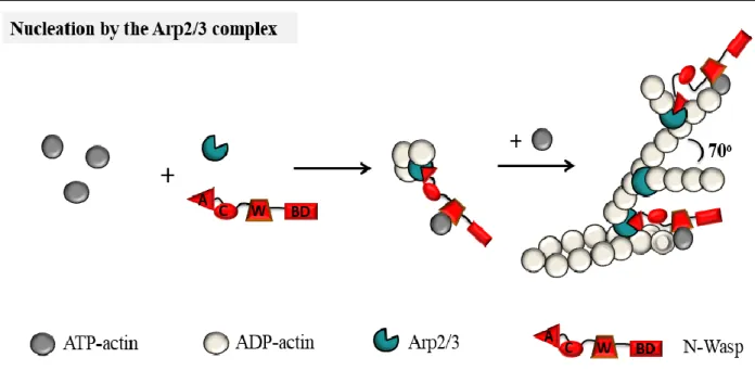
Figure 11: Arp2/3-mediated actin nucleation………..…….…43
Figure 12: Actin nucleation and elongation by the formin family proteins……….….…44
Figure 13: Actin nucleation by tandem monomer-binding nucleators……….……..45
Figure 14: Regulation of actin filament dynamics by ADF/cofilin, profiling, CAP, twinfilin and AIP1.…..….48
Figure 15: Schematic overview of the ADF/cofilin kinases and phosphatases and their upstream regulators………...….51
Figure 16: Schematic drawing of sarcomere structure and sarcomere-binding proteins……….….…53
Figure 17: Domains of tropomodulin (Tmod) and leiomodin (Lmod) proteins………..……56
Figure 18: Functional domains of the three Smads subfamilies ………..……59
Figure 19: Schematic illustration of myofibroblast transdifferentiation...60
Figure 20: Schematic representation of the TGF-β signaling pathway ………..……… ...65
Figure 21: The TGF–TAK1 pathway……….……….…….…67
Figure 22: Hypothetical model of increased TGF-β signaling LmnaH222P/H222P mice……….………….…….137
Figure 23: MRTF-SRF axis……….………143
Figure 24: Models of how abnormalities of A-type lamins may lead to hyperactivation of ERK1/2…... ..145
5 | P a g e
Abbreviations
ACE, angiotensin II converting enzyme ADF, actin-depolymerizing factor ALK, activin receptor-like kinases ALS, amyotrophic lateral sclerosis
AMH/MIS, anti-Müllerian hormone/Müllerian inhibiting substance
AP-1 activator protein-1
ATF-2, activated transcription factor-2 aWRN, atypical Werner syndrome BAF, Barrier to Autointegration Factor BMP, bone-morphogenetic protein
CaMKII, Ca2+/calmodulin-dependent protein kinase II,
CBD, chromatin binding domain Cdc42, cell division cycle 42, CDK, cyclin dependent kinases Cdk1, cyclin dependent kinase-1 CH, Calponin-homology domain CIN, chronophin
CMD, Congenital muscular dystrophy CMT2B1, Charcot-Marie Tooth type 2B1 Cobl, cordon-bleu
Co-Smads, common Smads, CP, capping protein
CTGF, Connective Tissue Growth Factor DAG, diaglycerol
DCM, dilated cardiomyopathy
DCM-CD, dilated cardiomyopathy with conduction system disease
ECG, electrocardiogram ECM, extracellular matrix
EDMD, Emery Dreifuss muscular dystrophy EF, ejection fraction
ERK, extracellular sign-regulated kinases
ERK1/2 extracellular signal-regulated kinase 1/2 ESCs, embryonic stem cells
F-actin, filamentous actin FAK, focal adhesion kinase FH1, formin-homology 1; FH2, formin-homology 2
FPLD, familial lipodystrophy of Dunnigan type FR, fractional shortening
FRAP, fluorescence-recovery after photobleaching FS, Fractional shortening
G-actin, globular actin
GDF, growth and differentiation factors family GPCR, G-protein-coupled receptors
GSK, Glycogen synthase kinase 3
HGPS, Hutchinson-Gilford progeria syndrome HP1, heterochromatin-associated protein-1 ICD, intracardiac cardioverter defibrillator ICMT, isoprenylcysteine carboxyl
methyltransferase
INM, inner nuclear membrane iPSCs, induced pluripotent stem cells I-Smads, inhibitory Smads,
JNK, c-Jun N terminal kinase JNK, c-JUN N-terminal kinase
KASH, Klarsicht/ANC-1/Syne-1 homologue LAD, lamina-associated domains
LAP, lamina-associated polypeptide LAP, latency-associated peptide
6 | P a g e
LBR, lamin B receptor
LEM, Lamina-associated polypeptide, Emerin and-MAN1
LGMD1B, limb-girdle muscular dystrophy type 1B
LIMK, Lin-11/Isl-1/Mec-3 kinase LINC, LInker of Nucleoskeleton and Cytoskeleton
Lmod, leiomodin LV, left ventricule
MAPK, mitogen-activated protein kinase MCLK, myosin light chain kinase MEK, MAP/ERK kinase
MLP, LIM protein
MRTF-A, myocardin-related transcription factor A NE, nuclear envelope
NEBD, nuclear envelope breakdown Nesprin, nuclear envelope spectrin repeat NF-Κb, nuclear factor-κB
NLS, nuclear localization signal NPC, nuclear pore complexes NPFs, Nucleation Promoting Factors
NRK, NCK-interacting kinase (NIK)-related kinase
N-WASP, neural WASP ONM, outer nuclear membrane PAK, p21-activated kinase
PI(4,5)P2, phosphatidylinositol 4,5-bisphosphate PKC, protein kinase C
PP1, protein phosphatase 1 PP2A, protein phosphatase 2A Rce1, Ras-converting enzyme 1
ROCK, RHO-associated coiled-coil-containing protein kinase
R-Smads, regulatory Smads RTK, receptor tyrosine kinase
RTK, receptors with intrinsic tyrosine kinase activity
SARA, Smad anchor for receptor activation Smurf2, Smad ubiquitination-related factor SRF, serum-response factor
SSH, slingshot
SUN, Sad1, UNC-84 homology TAK, TGF-β-activated kinase
TAN, transmembrane actin-associated nuclear TAZ, transcriptional coactivator with PDZ-binding motif
TESK, testis-specicific kinase TGF-β, transforming-growth factor Tmod, tropomodulin
TβR, TGF-β receptor
WH2, Wiskott-Aldrich Syndrome protein (WASp) homology 2
YAP, yes-associated protein (YAP)
Zmpst24, Zinc metalloprotease related to Ste24p α-SMA, α-smooth muscle
7 | P a g e
Abstract
Dilated cardiomyopathy is one of the leading causes of heart failure in Europe. Despite of the conventional medical care, there is no definitive and satisfactory treatment for the progressive cardiac dilatation and loss of contractility in LMNA cardiomyopathy often leading to sudden death or heart transplantation. LMNA gene encodes nuclear A-type lamins, which are the main constituents of the nuclear lamina. To explain how mutations in proteins of the nuclear envelope can cause a disease of the heart, it has been proposed that nuclear envelope abnormalities bring about cellular fragility and a decrease in the mechanical resistance to stress, which could partially explain the cardiac muscle disease, considering that the heart muscle is constantly subjected to mechanical force. In previous work, it has been showed that there is an abnormal activation of stress-activated ERK1/2 signaling in hearts that carry LMNA mutations. Administration of drugs inhibiting ERK1/2 signaling improves cardiac ejection fraction in mice, and blunts further increase in left ventricular dilatation. These studies clearly show that the abnormal ERK1/2 activation is involved in the pathophysiology of LMNA dilated cardiomyopathy. However, its role in the development of cardiac dysfunction remains unclear.
Inhibition of ERK1/2 signaling also slows progression of myocardial fibrosis, which is prominent in humans with dilated cardiomyopathy. I suggested that aberrant TGF-β signaling activity could participate to the abnormal ERK1/2 activation and be involved is the pathophysiology of left-ventricular contractile dysfunction in LMNA cardiomyopathy. My work led us to describe the TGF-β/ERK1/2/CTGF axis as a key player for the onset of myocardial fibrosis, which impairs left ventricular function, a major symptom of LMNA cardiomyopathy.
Given that the understanding of molecular and cellular mechanisms underlying the modulation of ERK1/2 signaling in the heart caused by LMNA mutation remains totally unclear, I tested the hypothesis that ERK1/2 abnormal modulation leads to alteration of cytosolic targets and alter cardiac cytoskeleton network. This may lead to LMNA cardiomyopathy. My work highlighted a novel partner of activated (phosphorylated) ERK1/2, ADF/cofilin-1. Cofilin promotes debranching of actin filaments. I showed that disrupted actin dynamics leads to abnormal destructuration of sarcomere and ADF/cofilin accumulation in the heart from a mouse model of LMNA cardiomyopathy, suggesting a defect in actin depolymerization. This project unraveled an unexpected role played by ERK1/2 signaling in actin dynamics and in the development of left-ventricular dysfunction in LMNA cardiomyopathy.
8 | P a g e
Rèsumè
La cardiomyopathie dilatée est l'une des principales causes d'insuffisance cardiaque en Europe. Dans le cadre de la cardiomyopathie liée aux mutations du gène LMNA, en dépit des soins médicaux conventionnels et de la transplantation cardiaque, aucun traitement satisfaisant ne permet de pallier à la dilatation cardiaque progressive et à la perte de la contractilité. Le gène LMNA code pour les lamines nucléaires de type A, qui sont les principaux constituants de la lamina nucléaire. L’impact des mutations dans un gène codant pour des protéines de l'enveloppe nucléaire sur le développement de maladies cardiaques demeure non élucidé. Afin d’expliquer cette corrélation, il a été proposé que des anomalies de l'enveloppe nucléaire pourrait engendrer une fragilité cellulaire et une diminution de la résistance mécanique aux contraintes. Ce phénomène pourrait- en partie - expliquer la pathologie musculaire cardiaque, en considérant le cœur comme constamment soumis à des forces mécaniques. De précédents travaux ont montré qu'il existe une activation anormale de la signalisation de réponse au stress ERK1/2 dans le cœur des patients porteurs de mutations du gène LMNA. L'administration médicamenteuse d’inhibiteurs de la voie de signalisation ERK 1/2 améliore la fraction d'éjection cardiaque chez la souris et atténue la progression de la dilatation ventriculaire gauche. Ces études démontrent que l'activation anormale de ERK 1/2 est impliquée dans la pathophysiologie de la cardiomyopathie dilatée liée aux mutations du gène LMNA. Cependant, son rôle dans le développement de la dysfonction cardiaque reste incertain.
L'inhibition de la voie ERK 1/2 ralentit également la progression de la fibrose myocardique, relativement développée chez l'homme, en cas de cardiomyopathie dilatée. Dans le cadre de ma thèse, j’ai suggéré que l'activité aberrante de la voie TGF-β pourrait participer à l’activation anormale de ERK 1/2 et être impliquée dans la physiopathologie de dysfonction contractile ventriculaire gauche dans la cardiomyopathie liée aux mutations du gène LMNA. Mon travail nous a conduits à décrire l’axe TGF-β/ERK1/2/CTGF comme un acteur clé de l'apparition de la fibrose myocardique, altérant la fonction ventriculaire gauche, symptôme majeur de la cardiomyopathie liée aux mutations du gène LMNA.
Les mécanismes moléculaires et cellulaires sous-jacents à la modulation de la signalisation ERK1/2, causée dans le cœur, par les mutations du gène LMNA, restent totalement incertains. De ce fait, j'ai testé l'hypothèse selon laquelle la modulation anormale de ERK1/2 induirait l'altération des cibles cytosoliques et modifierait le réseau du cytosquelette cardiaque. Cela conduirait ainsi à
9 | P a g e
la cardiomyopathie. Mon travail a mis en évidence un nouveau partenaire de la forme active (phosphorylée) d’ERK1/2 : cofiline-1. La cofiline induit la déramification des filaments d'actine. J’ai montré qu’une perturbation de la dynamique du réseau d'actine induisait une déstructuration anormale des sarcomères et une accumulation de la protéine ADF/cofiline dans le coeur d'un modèle murin de cardiomyopathie liée aux mutations du gène LMNA ; suggèrant ainsi un défaut de dépolymerisation de l’actine. Ce projet met en lumière un rôle inattendu joué par la signalisation ERK1/2 dans la dynamique de l'actine et dans le développement de la dysfonction ventriculaire gauche de la cardiomyopathie liée aux mutations du gène LMNA.
10 | P a g e
Zusammenfassung
Dilatative Kardiomyopathie ist eine der häufigsten Ursachen der Herzinsuffizienz in Europa. Neben einer konventionellen medizinischen Versorgung gibt es, außer der Herztransplantation, keine kurative Behandlung gegen die progressive Herzdilatation und den Verlust der Kontraktilität, die in der LMNA Kardiomyopathie auftreten. Das LMNA Gen kodiert die nuklearen A-typ Lamine, die den Hauptbestandteil der nuklearen Lamina bilden. Um zu erklären wie Mutationen in Proteinen der nuklearen Lamina zu Krankheiten des Herzens führen können, wird vermutet, dass die Anomalien der Kernhülle zu Fragilität und einer Abnahme der mechanischen Widerstandsfähigkeit der Zelle gegen mechanischen Stress führt. In der Erwägung, dass der Herzmuskel ständig mechanischen Kräften ausgesetzt ist, könnte dies zur Entstehung der Herzmuskelerkrankung beitragen. In früheren Arbeiten wurde bereits eine abnormale Aktivierung des stress-aktivierten ERK1/2 Signalwegs im Herzmuskelzellen, die eine LMNA Mutationen tragen, gezeigt. Die Verabreichung von Arzneimitteln, die die ERK1/2 Aktivierung hemmen, verbessert die Herzauswurffraktion und verhindert das weitere Fortschreiten der Dilatation des linken Ventrikels im Mausexperiment. Diese Studien zeigen deutlich, dass die abnormale ERK1/2 Aktivierung in der Pathophysiologie der dilatativen Kardiomyopathie beteiligt ist. Die genaue Rolle von ERK1/2 bei der Entstehung der Herzdysfunktion bleibt jedoch unklar.
Die Hemmung der ERK1/2 Aktivität verlangsamt außerdem das Fortschreiten der myokardialen Fibrose, die häufig bei Menschen mit dilatativer Kardiomyopathie beobachtet wird. Ich habe vermutet, dass eine anomale TGF-Beta Signalaktivität zu der abnormalen ERK1/2 Aktivität beiträgt und daher in die Pathophysiologie der linksventrikulären kontraktilen Dysfunktion der LMNA Kardiomyopathie involviert ist. Meine Arbeit zeigt den TGF-Beta/ERK1/2/CTGF Signalweg als einen wichtigen Akteur in der Entstehung der myokardialen Fibrose, welche die linksventrikuläre Funktion beeinträchtigt und ein Hauptsymptom der Kardiomyopathie darstellt. Da der molekulare und zelluläre Mechanismus der ERK1/2 Signaltransduktion in Herzen, versucht durch die LMNA mutation, völlig ungeklärt ist, habe ich die Hypothese getestet, dass die abnormale ERK1/2 Modulation zu Veränderungen der zytosolischen Interaktionspartner sowie zu einer Veränderung des Herz-Zytoskelettes führt. Diese Veränderungen wiederum könnten zur Kardiomyopathie führen.
11 | P a g e
Meine Arbeit zeigt einen neuen Interaktionspartner von aktiviertem ERK1/2, Cofilin1. Cofilin1 reguliert das Entzweigen von Aktin-filamenten. Ich konnte zeigen, dass eine gestörte Aktindynamik zu einer abnormalen Desorganisation des Sarkomers und einer Akkumulation von ADF/Cofilin im Herzen des Mausmodells der LMNA Kardiomyopathie führt. Dies weist auf einen Defekt in der Aktinpolymerisierung hin. Dieses Projekt deckt eine unerwartete Rolle der ERK1/2 Signaltransduktion in der Regulierung der Aktindynamik und in der Entstehung der linksventrikulären Dysfunktion in der LMNA Kardiomyopathie auf.
12 | P a g e
Preamble
Mutations in LMNA, encoding A-type lamins, intermediate-filament proteins of the inner nuclear membrane, cause a variety of diseases (i.e. ‘laminopathies’) that primarily affect striated muscles, and more particularly the heart. Despite our gaps in understanding many of their fundamental functions, much of the current research on the A-type lamins is focused on how mutations leading to alterations in these proteins cause striated muscle diseases. We previously demonstrated that the mechanically activated extracellular signal-regulated kinase (ERK) 1/2 is hyper-activated in striated muscle diseases caused by LMNA mutations (Muchir et al. 2007a). However, insights in the molecular mechanisms bridging ERK1/2 modulation and depressed muscle function are lacking. The main goal of the present work was to study the role of ERK1/2 signaling pathway in the pathogenesis and development of LMNA cardiomyopathy.
This doctoral thesis comprises three parts:
i/ a general introduction to the scientific topics addressed in the dissertation;
ii/ four manuscripts: two reviews and two peer-reviewed articles, which document and discuss the scientific work that has been conducted. One of the peer-reviewed manuscripts investigates the role of ERK1/2 and TGF-β signaling pathway in the development of fibrosis while the second manuscript focused on the role of ERK1/2 signaling on actin dynamics in the context of LMNA cardiomyopathy
13 | P a g e
Chapter 1
Introduction
1.1 Nucleus, nuclear lamina and laminopathies
The cell nucleus can be structurally and functionally divided into at least 2 separate regions, the nuclear envelope and the nuclear interior. The nuclear envelope separates the cytoplasm from the nucleus in eukaryotic cells. It is composed of the nuclear membrane, the nuclear lamina and the nuclear pore complexes (NPCs) (Figure 1). The nuclear membrane consists of 2 phospholipid bilayer membranes, the outer nuclear membrane (ONM), which is continuous with the endoplasmic reticulum, and the inner nuclear membrane (INM). The NPCs are large protein complexes made up of multiple copies of approximately 30 different proteins, called nucleoporins, that span through both the INM and ONM as well as the lamina and mediate the exchange of macromolecules such as nucleic acids and proteins with molecular weights exceeding ∼40 kDa between the nucleus and the cytoplasm (Schermelleh et al. 2008; Wente & Rout 2010). Nuclear lamina is a dense fibrous three-dimensional meshwork underlining the nucleoplasmic face of the INM.
The main function of the nuclear envelope is to provide protection to the genetic information as well as compartmentalization to concentrate enzymes and substrates for biochemical processes in the nucleus such as DNA replication, transcription, RNA processing and ribosome synthesis.
Figure 1: The eukaryotic nucleus. The nucleus of a cell eukaryotic cell contains the DNA, the nuclear envelope, the nucleolus and ribosomes. The nuclear envelope surrounds the nucleus with a double membrane (outer and inner) with nuclear pores. The ONM is continued with the rough ER. The nucleolus is a dense region of the nucleus where the ribosomal RNA is made.
14 | P a g e
1.1.1 Nuclear lamins
Nuclear lamins are the major structural components of the nuclear envelope that are implicated in a plethora of essential cellular processes, such as chromatin organization, nuclear positioning, spacing of the nuclear pore complexes, nuclear disassembly and reassembly during mitosis, DNA replication, RNA polymerase II-dependent gene expression, cell proliferation and differentiation, cell signaling, and cell division (Burke & Stewart 2012; Choi & Worman 2014; Collas et al. 2014; Kennedy & Pennypacker 2014; Shimi & Goldman 2014). Lamins are type V intermediate filament proteins that polymerize to form the nuclear lamina (Fisher et al. 1986; McKeon et al. 1986; Aebi et al. 1986) and divided into A-type lamins (lamins A and C) and B-type lamins (lamin B1 and lamin B2) (Figure 2). In addition to the major lamins A and B, mammalian cells express the minor isoforms lamin AΔ10 (Machiels et al. 1996) and germ cell-specific lamins C2 (Furukawa et al. 1994) and B3 (Furukawa & Hotta 1993). All metazoans examined to date, ranging from hydra to human, express at least one B-type lamin, but lamins are absent from unicellular organisms and plants (Cohen et al. 2001; Höger et al. 1990; Vorburger et al. 1989). Although plant cells have lamina, other proteins substitute for lamins (Ciska & Moreno DÃ-az de la Espina 2014).
A-type lamins
Lamins A and C, the main A-type lamins, arise by alternative splicing of a single gene, LMNA (Daniel Z Fisher et al. 1986; McKeon et al. 1986). The LMNA gene (Online inheritance in man (OMIM): #150330) is located on chromosome 1q21.2-q21.3 (Lin & Worman 1993; Wydner et al. 1996) and contains 12 exons encompassing around 58 kb. Lamin A is produced by alternative splicing of exon 10. It is 664 amino acids in length and the longest isoform of A-type lamins. The lamin C protein ends at exon 10 which makes it identical to lamin A up to codon 566 (Fisher et al. 1986; McKeon et al. 1986). The last six amino acids (567-572) are unique to lamin C (Lin & Worman 1993). Lamin A∆10 is identical to lamin A but lacks exon 10, shortening the tail domain by 30 amino acids (Machiels et al. 1996). In mouse, the LMNA gene has been shown to give rise to a fourth splice variant termed lamin C2 (Furukawa et al. 1994). Lamin C2 is expressed exclusively in germ cells and is identical to lamin C, except an alternative exon, 1C2, located in intron 1 of LMNA, codes for the N-terminal head domain (Jahn et al. 2010; Alsheimer & Benavente 1996). A-type lamins play a pivotal role in the maintenance of nuclear shape (Scaffidi 2006; Dahl et al. 2008; Lammerding et al. 2006), structure (Broers et al. 2005; Lammerding et al. 2006;
15 | P a g e
Stewart et al. 2007) and stability (Lammerding et al. 2004; Dahl 2004). In contrast to B-type lamins, A-type lamins are also found in a dynamic pool (Moir et al. 2000) in the nucleoplasm (Dechat et al. 2010), where interacts with chromatin. Numerous lines of evidence showed that lamins bind not only to heterochromatin as it was thought for a long time but also to euchromatin (Amendola & van Steensel 2014; Gruenbaum & Foisner 2015; Gesson et al. 2016).
A-type lamins are expressed at specific developmental stages or differentiated cells with high levels in skeletal and cardiac muscle (Krohne & Benavente 1986; Takamori et al. 2007; Lin & Worman 1997). Recent findings showed that small amounts of A-type lamins are also present in mouse ESCs (embryonic stem cells) and in preimplantation embryos (Eckersley-Maslin et al. 2013; Kim et al. 2013).In most tissues, lamin A and C are expressed at equal levels except in the brain where lamin C is the predominant form of A-type lamin (Yang et al. 2011).
In human, mutations in LMNA gene results in a range of phenotypic alterations, collectively termed laminopathies (Wilson & Foisner 2010; Zastrow et al. 2004). The pathogenic mechanism for the majority of LMNA mutations is still unclear. However, proposed disease mechanisms for laminopathies are closely linked to the function of lamins and lamin-binding proteins (Broers et al. 2006; Worman & Bonne 2007). Lamin-binding proteins include structural proteins: nesprin-1α (Mislow et al. 2002), nesprin-2 (Zhang et al. 2005), SUN1/2 (Haque et al. 2006; Haque et al. 2010), emerin (Lee et al. 2001; Sakaki et al. 2001), actin (Sasseville & Langelier 1998; Simon et al. 2010) and Nup88 (Lussi et al. 2011); proteins involved in gene transcription and cell signalling: c-Fos, Erk1/2 (González et al. 2008), MOK2 (Dreuillet 2002), PKCα (Martelli et al. 2002), SREBP1 (Lloyd 2002) and MAN1 (Mansharamani & Wilson 2005) and proteins involved in cell cycle regulation: pRb (Ozaki et al. 1994), cyclin D3 (Mariappan et al. 2007), PCNA (Shumaker et al. 2008) and Lap2α (Dechat et al. 2000).
16 | P a g e
B-type lamins
B-type lamins are encoded by either the LMNB1 gene on chromosome 5q23.3-q31.1 which encode lamin B1 or LMNB2 gene on chromosome 19p13.3, which encode lamin B2 and lamin B3 (Furukawa & Hotta 1993; Peter et al. 1989; Vorburger et al. 1989). Lamins B are constitutively expressed in essentially all somatic types including undifferentiated cells in early stages of embryonic development (Liu et al. 2011; Stewart & Burke 1987).
Mice deficient in lamin B1, lamin B2, or both are smaller than wild-type littermates at birth, have major defects in the lungs and brain, and die within a few minutes after birth (Vergnes et al. 2004; Coffinier et al. 2010; Kim et al. 2011). Fibroblasts from lamin B1 mutant mice display severely dysmorphic nuclei, impaired differentiation and premature senescence (Vergnes et al. 2004). B-type lamins have been associated with the assembly of the mitotic spindle matrix as well as DNA replication, genome organisation and apoptosis and are therefore thought to be essential for proliferation and cell survival (Guelen et al. 2008; Liu et al. 2000; Tsai et al. 2006). Another study showed that forebrain-specific removal of lamin B1 and B2 results in severe atrophy of the cortex, demonstrating an essential role in brain development (Coffinier et al. 2011; Yang et al. 2011). In contrast to A-type lamins, only a few disease-causing mutations have been identified in LMNB1 or LMNB2 such as leukodystrophy demyelinating autosomal dominant adult-onset (ADLD) for LMNB1 defects and partial acquired lipodystrophy associated with susceptibility variants of LMNB2 (Padiath et al. 2006; Zuela et al. 2012; Schreiber & Kennedy 2013; Brussino et al. 2010; Molloy et al. 2012; Schuster et al. 2011).
17 | P a g e
1.1.2 Structure of lamins
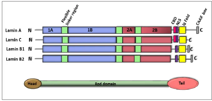
Similar to cytoplasmic intermediate filaments, lamins have a tripartite structure consisting of a helical rod domain flanked by globular N-terminal head and C-terminal tail domains (Aebi et al. 1986). Lamin A resembles lamin B in the head and rod domains, but has an expanded tail domain that comprises an extra 90 amino acid segment (Dittmer & Misteli 2011). The central rod domain of the lamins consists mainly of heptad repeats that are characteristic of α-helical proteins (McKeon et al. 1986; Stuurman et al. 1998). The tail domain contains a chromatin binding domain (CBD), a nuclear localization signal (NLS), an immunoglobulin (Ig Fold) sequence and a conserved CAAX box (Loewinger & McKeon 1988; Dhe-Paganon et al. 2002; Krimm et al. 2002). The NLS is located between the central rod and the Ig-fold and is required for the transport of lamins into the nucleus (Loewinger & McKeon 1988). The Ig fold domain is common to many proteins and mediates various protein interactions, and the CAAX box, a motif of serves as a substrate for extensive post-translational processing (Rusiñol & Sinensky 2006). Lamin C does not have a CaaX box, so it does not undergo modifications (Dechat et al. 2010) (Figure 2).
Figure 2: Schematic structure of lamin family members. Main characteristics are four central rod domains (1A, 1B, 2A, 2B), flanked by a globular head and a globular tail domain. In the globular tail domain, a nuclear localization signal (NLS; purple) can be identified as well as a chromatin-binding domain (CBD; pink) and a Caax motif, which is absent in lamin C.
18 | P a g e
1.1.3 Posttranslational Processing and Modifications of the Nuclear Lamins
Posttranslational modifications of the head and tail domains of lamins are required to control lamin assembly. In the first step, the C-terminal CaaX motif of prelamin A, lamin B1 and B2 undergoes farnesylation, and subsequently the terminal –AAX amino acids are cleaved by an endopeptidase, most likely Rce1 (Ras-converting enzyme 1) or FACE1. In the third step the cysteine residue undergoes a methylation step at the ER, a process catalyzed by the enzyme isoprenylcysteine carboxyl methyltransferase (ICMT) (Nigg 1992; Maske et al. 2003; Varela et al. 2005). Once incorporated into the nuclear lamina, only prelamin A is further cleaved 15 amino acids away (in human lamin A after Tyr646) from its farnesylated/carboxymethylated cysteine of the C terminus by the protease Zmpst24 (Zinc metalloprotease related to Ste24p)/FACE1 (Pendas et al. 2002; Corrigan et al. 2005). This cleavage event occurs 30–60 minutes post-synthesis—after assembly into the nuclear lamina (Gerace et al. 1984; Pendas et al. 2002; Bergo et al. 2002). This final modification step completes the post-translational modification of prelamin A to mature lamin A and it is thought to aid localization of lamin A to the nuclear envelope.
1.1.4 Lamin filament assembly and disassembly
Similarly to other intermediate filament proteins, both A-type and B-type lamins can self-associate to form filaments in vitro and in vivo (Foeger et al. 2006). The first step in this assembly involves a polar head-to-tail polymerization of the lamin dimers in parallel association of two α-helical rod domains into a left handed superhelix (Heitlinger et al. 1991; Stuurman et al. 1996) and these polarized arrays associate laterally in an antiparallel fashion to build tetrameric protofilaments. The interaction between four protofilaments form the characteristic ∼10-nm-diameter lamin filament (Heitlinger et al. 1991; Stuurman et al. 1996; Ben-Harush et al. 2009). In contrast, the lamin filaments assembly in vitro produces large paracrystalline arrays which do not exist in vivo under normal conditions (Stuurman et al. 1998). The apparent difference between lamin assembly in vivo (10-nm filaments) and in vitro (paracrystals) demonstrates that lamin organization in vivo is regulated by interactions with other molecules.
The nuclear lamins are rapidly disassembled during the prophase/metaphase transition in vertebrate cells, in a process called nuclear envelope breakdown (NEBD) (Fields & Thompson
19 | P a g e
1995). The disassembly of A-type lamins is triggered in a phosphorylation-dependent manner by cyclin dependent kinase-1 (Cdk1), protein kinase C (PKC) and other mitotic kinases leading to their depolymerization and solubilization (Heald & McKeon 1990). In contrast,B-type lamins maintain proximity with the nuclear membrane. After mitosis, the A-type lamins are dephosphorylated and reassembled into the nuclear envelope along to B-type lamins.
1.1.5 Connections of A-type lamins with other nuclear envelope proteins
At the nuclear membrane, several proteins form connections with the underlying lamina structure on the one hand or the cytoskeleton on the other. In particular, the lamin proteins interact with INM proteins, like emerin, LBR (Lamin B receptor) and LAP (lamina-associated polypeptide), and chromatin-binding proteins, like BAF (Barrier to Autointegration Factor), at the nuclear periphery to form a stable network that supports the membrane and links the INM to the chromatin (Ellenberg & Siggia 1997; Moir et al. 2000; Wilson & Foisner 2010).
Emerin is a serine-rich transmembrane protein encoded by the EMD gene and is located at the inner nuclear membrane (Bione, Maestrini, Rivella, et al. 1994; Nagano et al. 1996). The N-terminal of emerin contains a ∼45-residue structural motif known as LEM (for Lamina-associated polypeptide, Emerin and-MAN1) domain which binds BAF, a small chromatin-binding molecule (Lee et al. 2001) (Figure 3). Emerin interacts with several proteins such as lamin A/C (Clements et al. 2000; Lee et al. 2001; Sakaki et al. 2001; Dreger et al. 2001), lamin B receptor (LBR) (Martins et al. 2000), actin (Fairley et al. 1999) and nesprins (Warren et al. 2005; J. M. K. Mislow et al. 2002; J. M. . Mislow et al. 2002). The localization of emerin to the nuclear envelope is lamin A-dependent (Sullivan et al. 1999; Vaughan et al. 2001). Mutations on EMD are associated with X-linked Emery-Dreifuss muscular dystrophy (X-EDMD) and are mostly nonsense mutations resulting in a loss of emerin (Bione, Maestrini, Rivella, et al. 1994; Nagano et al. 1996).
Lamin-associated polypeptide (LAP) family is encoded by two genes LAP1 and LAP2 (Martin et al. 1995; Harris et al. 1995; Furukawa et al. 1995; Berger et al. 1996). The mammalian LAP2 gene encodes six alternative spliced isoforms (LAP2α, β, γ, δ, ε, ζ) that play an important role in the regulation of nuclear architecture by binding lamin B1 and chromosomes in a manner regulated by phosphorylation during mitosis (Berger et al. 1996; Dechat et al. 2000; Furukawa et al. 1994).
20 | P a g e
LAP2α is linked to a laminopathy-type dilated cardiomyopathy and Lap2α knock out mice showed delayed satellite cells differentiation, although delayed muscle regeneration was not observed (Taylor et al. 2005; Gotic et al. 2010; Gotic et al. 2010).
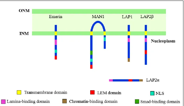
LAP1A and LAP1B have been shown to bind to A and B-type lamins, whereas LAP2C only binds lamin B (Foisner & Gerace 1993). Mutations on the gene encoding for LAP1B protein has been found in patients with dystonia associated with dilated cardiomyopathy as well as in individuals with muscular dystrophy and cardiomyopathy (Kayman-Kurekci et al. 2014; Dorboz et al. 2014). MAN1 is an integral protein of the INM (Bengtsson 2007; Paulin-Levasseur et al. 1996). The N-terminal domain of MAN1 contains a LEM domain which binds Lamin A/C and chromatin via BAF and C-terminal region has an RNA-recognition motif (RR-motif) which is a binding site for regulatory Smads (R-Smads) (Bengtsson 2007). MAN1 binds R-Smads but not inhibitory Smads, and therefore acts as an inhibitor of BMP (bone-morphogenetic protein) and TGF-β
(transforming-Figure 3: Schematic illustration of the LEM-domain proteins. LAP2α, LAP2β, emerin and MAN1 contain the LEM domain, which interacts with the DNA-binding protein barrier-to-autointegration factor (BAF). LAP, lamina-associated polypeptide; NLS, nuclear localization signal; INM, inner nuclear membrane; ONM, outer nuclear membrane
21 | P a g e
growth factor) signaling (Lin et al. 2005). Overexpression of MAN1 has been shown to block transcription of BMP and TGF-β targets upon cytokine stimulation and loss of MAN1 at the INM results in increased TGF-β signaling (Lin et al. 2005). Loss of-function mutations in MAN1 result in bone-related diseases such as osteopoikilosis and Buschke-Ollendorff syndrome (Mumm et al. 2007), phenotypes characterised by increased bone density and associated with increased TGF-β signaling.
Lamin B receptor (LBR) is an inner nuclear membrane protein and belongs to the ERG4/ERG24 family. LBR is targeted to the INM via 8 putative transmembrane domains (Worman et al. 1990). The LBR anchors B-type lamins to the INM and has been shown to interact with emerin (Martins et al. 2000), lamin associated protein-2β (LAP2β) (Ye & Worman 1994), double strand DNA, histones and the heterochromatin-associated protein-1 (HP1) (Ye & Worman 1996). Mutations in the LBR gene cause Pelger-Huet anomaly, an hematology particularity and HEM/Greenberg skeletal dysplasia, a bone disease (Hoffmann et al. 2002; Waterham et al. 2003).
1.1.6 Connections between nucleus and cytoskeleton
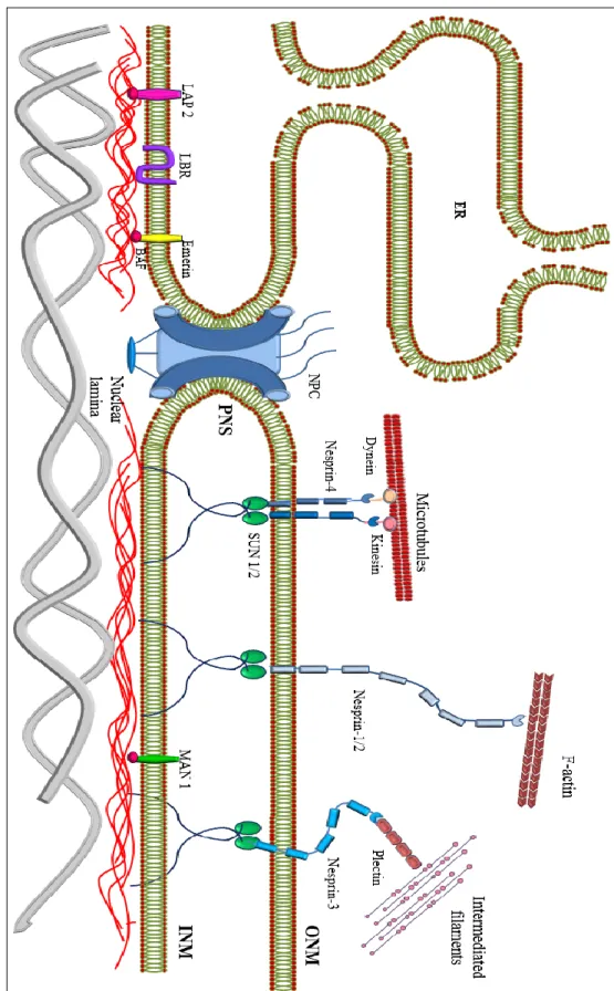
Besides the connection between the inner nuclear membrane and the nuclear lamina, the nucleus is also connected to the cytoskeletal filaments through linker proteins (Dittmer & Misteli 2011) (Figure 4). This link between nucleus and cytoskeleton has been implicated in a variety of functions important for nuclear positioning, migration, morphology and mechanics (Starr & Han 2003).
Nesprins (Nuclear envelope spectrin repeat proteins) comprise a large protein family of spectrin-repeat-containing proteins localized to the ONM and INM depending on their size and interaction partners (Razafsky & Hodzic 2009). Nesprins are encoded by four genes (SYNE1-4) (Mellad et al. 2011), that give rise to multiple isoforms. Their structure contains a central rod region with several spectrin repeats, variable in length, and a C-terminal KASH (Klarsicht/ANC-1/Syne-1 homologue) transmembrane domain that mediates nuclear membrane localization and
22 | P a g e Figure 4: Sche m at ic diagr am of the nu cl eocyt os kelta l intera ct ion s. The nuc le ar en velo pe c on sist s t he nucl ear m em br anes, nucl ear la m ina, an d pore com plexes . T he nucl ear la m in a li es on the in ner surface of the inn er nucl ea r m em br ane (INM), wh ere it serv es to m ai ntain nu cl ea r sta bili ty , orga nize chrom at in and bind nu cl ea r pore com plexes (NPCs) N uclear en velo pe protei ns that ar e bound to the lam ina include nes pr in, em erin, la m ina -associat ed p ro tei ns 1 and 2 (L AP1 a nd L AP 2), t he la m in B r ecepto r (LBR ) a nd MA N1.
23 | P a g e
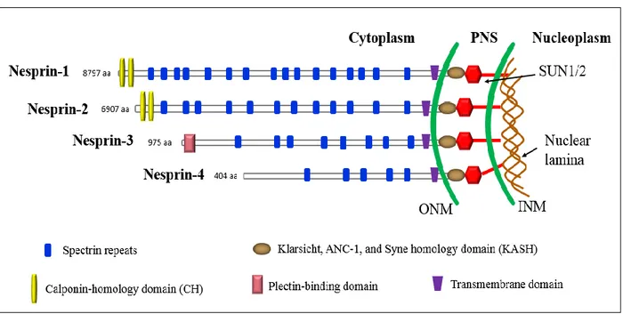
interacts with the SUN-domain in the perinuclear space (Crisp et al. 2006; Zhang et al. 2001; J. M. K. Mislow et al. 2002) (Figure 4). Nesprin-1 and -2 (giant isoforms) harbor an N-terminal actin-binding Calponin-homology domain (CH) (Zhen et al. 2002; Padmakumar et al. 2004) (Figure 5). Nesprin-3 contains an N-terminal binding domain for plectin which interacts with intermediate filament proteins (Wilhelmsen et al. 2005; Ketema et al. 2007) and nesprin-4 binds to kinesin-1, a microtubule motor protein participating in the nuclear positioning process (Roux et al. 2009) (Figure 4). Missense mutations in SYNE1 and SYNE2 (Zhang et al. 2007) genes in humans as well as mice harbouring KASH-domain deletions (Puckelwartz et al. 2009; Zhang et al. 2007), are associated with a skeletal muscle phenotype resembling EDMD, whereas other mutations in SYNE1 are associated with non-muscle pathologies, including arthrogryposis and cerebellar ataxia (Gros-Louis et al. 2007; Voit et al. 2002).
Figure 5: Domain structure of the four main nesprin isoforms. The nesprins-1 and -2 contain two N-terminal actin-binding Calponin homology (CH) domains, a central region comprised of multiple Spectrin repeats (the number depends on the isoform) and a C-terminal KASH domain that is required for their nuclear envelope localization. Nesprin-3 and nesprin-4 are smaller isoforms which anchor to plectin and microtubules, respectively. The length of each protein in amino acids is indicated.
24 | P a g e
Table 1: Functions of LINC complex
SUN (Sad1, UNC-84 homology) domain proteins SUN1 and SUN2 are located to the INM. SUN proteins have a central helical coil domain that mediates dimerization (Tzur et al. 2006), an N-terminal domain which interacts directly with A-type lamins (Haque et al. 2006), emerin and short nesprin-2 isoforms (Haque et al. 2010) and a C-terminal domain. The latter interacts with the KASH domain found in giant nesprins 1 and 2 in the luminal space between the INM and the ONM (Padmakumar et al. 2005; Crisp et al. 2006; Ketema et al. 2007), forming a mechanical link, called LINC (LInker of Nucleoskeleton and Cytoskeleton) complex (Stewart et al. 2007; Crisp et al. 2006; Padmakumar et al. 2005). SUN proteins, bind to A-type lamins (Padmakumar et al. 2005; Worman & Gundersen 2006; Hodzic et al. 2004; Crisp et al. 2006; Haque et al. 2006) as well as to nuclear pore complexes (NPCs).
LINC complex is essential for a multitude of cellular functions including nucleocytoskeletal connection and organization, nuclear morphology, nuclear positioning, cell polarity, cell differentiation, organelle positioning and mechanotransduction (Lombardi et al. 2011; Zhang et al. 2010). Table 1 summarizes the cellular functions in which LINC plays role. The first experimental clues for the role of LINC complex in mechanotransduction come from a seminal study in which magnetic tweezers were used to pull on nesprin-1 antibody-coated beads attached to isolated nuclei
Functions References
Association with actin and microtubules networks (Starr & Han 2003; Starr & Fridolfsson 2010; Münter et al. 2006)
Formation of TAN lines in migrating cells; cell migration; cell polarization
(Luxton et al. 2011; Chancellor et al. 2010)
Force propagation from FA and nucleus (Lombardi et al. 2011; Crisp et al. 2006)
Centrosome association (Dawe et al. 2009)
Chromatin separation from the nuclear envelope and organization of the mitotic spindle
(Turgay et al. 2014) Nuclear positioning at NMJ (Grady et al. 2005) Cell division via interaction with telomeres (Ding et al. 2007) Formation of actin cap (Khatau et al. 2009) Stretch-induced nuclear rotation and
stretch-induced inhibition of differentiation
25 | P a g e
(Guilluy et al. 2014). Pulling on nesprin-1 resulted in local nuclear stiffening accompanied by an enhanced recruitment of A-type lamins to the LINC complex and Src-dependent phosphorylation of emerin (Tyr74 and Tyr95). This phosphorylation reinforces the connection between lamin A/C and the LINC complex. In support of their role in the direct force transmission to the nucleus, the nuclear deformation response to mechanical strain was found abrogated upon LINC disruption using dominant-negative nesprin and SUN constructs (Lombardi et al. 2011). Disrupting the LINC complex and the nucleo-cytoskeletal linkage can perturb normal gene expression in both myoblasts and cardiomyocytes (Nikolova-Krstevski et al. 2011; Brosig et al. 2010).
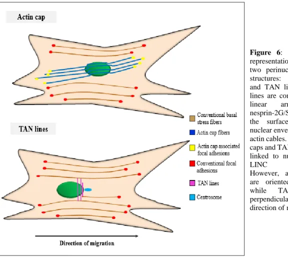
A remarkable finding showed that, LINC complexes also participate in the formation of the transmembrane actin-associated nuclear (TAN) lines at specialized apical regions of the nucleus (Luxton et al. 2010) (Figure 6). In a series of papers, Gundersen and coworkers showed that Nesprin-2 giant, Sun-2 and myosin II are essential components of the TAN lines. TAN lines play a major role in nuclear movement and polarity in migrating fibroblasts and myoblasts (Luxton et al. 2010; Chang et al. 2015). The INM proteins Samp and emerin were also found in these complexes (Borrego-Pinto et al. 2012; Chang et al. 2013).
A subset of cytoskeletal actin filaments are directly attached to the nucleus, forming the perinuclear actin cap (Khatau et al. 2009). These specialized actin fibers cover the top of the nucleus, as opposed to conventional basal stress fibers lying at the basal surface of the cell. Although both TAN lines and actin cap are connected to the surface of the nuclear envelope through the lamin A/C and LINC complex, actin cap stress fibers are oriented parallel and TAN lines perpendicularly with the direction of migration (Figure 6). Apart from the role of the actin cap as nuclear shape regulator, it has been also proposed to serve rapid transduction of information about the mechanical properties of the cellular environment into the nucleus (Chambliss et al. 2013; Khatau et al. 2009). The absence of LINC complexes in cellular models carrying LMNA mutations results in the disappearance of actin caps (Hotulainen & Lappalainen 2006).
26 | P a g e
1.1.7 Laminopathies
Lamin A/C alterations/substitutions result in a vast range of tissue-specific disorders broadly termed laminopathies (Worman 2012; Butin-Israeli et al. 2012) and can be separated into primary and secondary laminopathies. To date, more than 450 mutations (see http://www.umd.be/LMNA/) in the LMNA gene have been identified in so called primary laminopathies; the majority of these mutations are linked to muscular dystrophies, but some mutations have little or no effect on muscle tissue. These include point mutations, frameshift mutations, deletions, and two nonsense mutations.
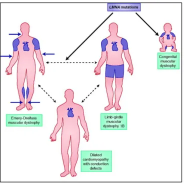
The spectrum of ∼14 distinct primary LMNA laminopathies can be broadly classified into 1) diseases that affecting the striated muscles include LMNA-related congenital muscular dystrophy (L-CMD), Emery-Dreifuss muscular dystrophy (EDMD), limb-girdle muscular dystrophy type 1B (LGMD1B) and dilated cardiomyopathy with conduction system disease(DCM-CD) (Figure 7),
Figure 6: Schematic representation of the two perinuclear actin structures: actin cap and TAN lines. TAN lines are composed of linear arrays of nesprin-2G/SUN2, on the surface of the nuclear envelope along actin cables. Both actin caps and TAN lines are linked to nucleus via LINC complex. However, actin caps are oriented parallel while TAN lines perpendicularly to the direction of migration.
27 | P a g e
Table 2: Diseases caused by mutations in LMNA gene
2) diseases affecting the adipose tissue include familial lipodystrophy of Dunnigan type (FPLD), 3) peripheral neuropathy associated with demyelination of motor neurons such as axonal neuropathy Charcot-Marie Tooth type 2B1 (CMT2B1), and 4) premature aging syndromes which include Hutchinson-Gilford progeria syndrome (HGPS) and atypical Werner syndrome (aWRN) (Table 2) (De Sandre-Giovannoli et al. 2002; Shackleton et al. 2000; Muchir et al. 2000; Fatkin et al. 1999; Eriksson et al. 2003; Chen et al. 2003). The mechanism whereby different mutations in a single gene that is widely expressed cause such a broad spectrum of diseases remains a puzzle. The clinical profile of the same LMNA mutation often varies in severity among members of the same family (Fatkin et al. 1999; Cao & Hegele 2000; Brown et al. 2011).
Secondary laminopathies are caused by mutations in ZMPSTE24 gene, leading to defects in posttranslational modification of A-type lamins and include restrictive dermopathy (Navarro et al. 2004) and Mandibuloacral dysplasia (Agarwal et al. 2003). Description of secondary laminopathies is beyond the scope of this thesis.
Laminopathies Reference
Affecting striated muscles
AD-Emery Dreifuss Muscular Dystrophy (AD-EDMD) (Bonne et al. 1999)
AR-Emery Dreifuss Muscular Dystrophy (AR-EDMD) (Raffaele Di Barletta et al. 2000) Limb-girdle muscular dystrophy type 1B (LGMD1B) (Muchir et al. 2000)
LMNA-associated congenital muscular dystrophy (L-CMD) (Quijano-Roy et al. 2008) Dilated-cardiomyopathy with conduction defects (DCM-CD) (Fatkin et al. 1999) Lipodystrophy syndromes
Dunnigan-type familial partial lipodystrophy (FPLD2) (Shackleton et al. 2000) Peripheral neuropathy
Charcot Marie Tooth Disease type 2B1 (CMT-2B1) (De Sandre-Giovannoli et al. 2002)
Accelerating aging syndromes
Hutchinson-Gilford Progeria Syndrome (HGPS) (Eriksson et al. 2003)
Atypical Werner Syndrome (aWRN) (Chen et al. 2003)
28 | P a g e
Emery-Dreifuss Muscular Dystrophy (EDMD)
One of the tissues most frequently affected by changes in genes encoding lamin A/C is striated muscle (Figure 7). Emery-Dreifuss muscular dystrophy (EDMD) is the most prevalent disorder of nuclear envelopathies, a group of diseases caused by mutations of genes that encode proteins of the nuclear envelope. EDMD is a genetically heterogenous disorder with X-linked (XL-EDMD: OMIM #310300), autosomal dominant (AD-EDMD: OMIM #181350) and autosomal recessive (AR-EDMD: OMIM #604929) forms. The genes responsible for the three genetic forms were identified. In 1994, Bione et al. identified emerin (EMD) as the gene on chromosome Xq28 that is mutated in X-linked EDMD (Bione, Maestrini, Rivella, et al. 1994; Raffaele Di Barletta et al. 2000). Most mutations in the X-EDMD gene are point mutations or small deletions/insertions of emerin that cause interruption of transcription or translation (Bione, Maestrini & Rivella 1994; Klauck et al. 1995; Nigro et al. 1995; Wulff et al. 1997). Later, Bonne et al. demonstrated that mutations in LMNA gene cause AD-EDMD and AR-EDMD (Bonne et al. 1999). Missense mutations introducing amino acid changes in the coding sequence appear to be the most common mutations in the LMNA gene (Bonne et al. 1999; Raffaele Di Barletta et al. 2000).
The three forms of EDMD are characterized clinically by slowly progressive muscle weakness and wasting in a scapulo-humeroperoneal distribution, early contractures of the elbows, ankles,
Figure 7: Laminopathies affecting striated muscles present in childhood or adulthood (Emery-Dreifuss muscular dystrophy, limb-girdle muscular dystrophy 1B and dilated cardiomyopathy with conduction defects) are shown with affected muscle groups illustrated in blue. Adapted from Lu et al. 2011
29 | P a g e
Achilles tendons and posterior neck. Cardiac conduction defecits and arrythmias may occur, resulting in life-threatening dilated cardiomyopathy. Cardiac disease, in most cases, begins at the end of the second decade with no direct relationship to the severity of the skeletal muscle involvement.
Dilated cardiomyopathy with conduction system disease (DCM-CD)
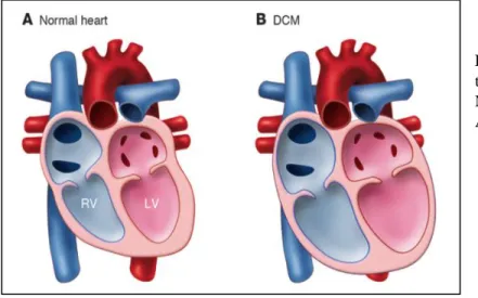
Soon after the identification of the first LMNA mutations in EDMD, Fatkin et al. reported that LMNA mutations can cause dilated cardiomyopathy associated with conduction system disease (DCM-CD: OMIM #115200) with minimal to no skeletal muscle involvement (Fatkin et al. 1999). Dilated cardiomyopathy is characterized by progressive weakening of the heart muscles, thinning of the left ventricle wall and insufficient pumping that typically leads to heart failure. Similar to other dilated cardiomyopathies, LMNA cardiomyopathy is characterized by chamber enlargement and systolic dysfunction of one or both ventricles (Figure 8). Several reports emphasized that different lamin A/C mutations cause very similar cardiac phenotypes, characterized by atrial fibrillation, conduction system disease, sudden death and heart failure (Fatkin et al. 1999; Bécane et al. 2000; Taylor et al. 2003; Kärkkäinen et al. 2004; van Berlo et al. 2004). In addition, both atrial and ventricular arrhythmias are common among LMNA mutation carriers. Some studies have pointed out that sudden death may be the first disease manifestation, which could be due to ventricular arrhythmias and therefore occur despite the implantation of pacemaker (Bécane et al. 2000; Sanna et al. 2003; van Berlo et al. 2004; Van Berlo et al. 2005; Kärkkäinen et al. 2004).
Figure 8: Morphological changes of the heart in cardiomyopathy. (A) Normal heart. (B) DCM heart. Adapted from (Mcnally et al. 2013)
30 | P a g e
Thus, implantation of cardioverter-defibrillator (ICD) is required for primary prevention of ventricular arrhythmias and sudden cardiac death (Meune et al. 2006; Charron et al. 2012). The course of LMNA cardiomyopathy is aggressive due to a high rate of malignant ventricular arrhythmias and end-stage heart failure, often leading to premature death or cardiac transplantation (Taylor et al. 2003; Van Berlo et al. 2005). By the age of 60 years, 55% of LMNA mutation carriers die of cardiovascular death or receive a heart transplant, compared to 11% of patients with idiopathic cardiomyopathy without LMNA mutation (Taylor et al. 2003). Diagnosis of LMNA-related DCM relies on sequence analysis. Conduction system disease and arrhythmias are detected by electrocardiogram (ECG) and left ventricular dilatation and reduced systolic function are most commonly identified with two-dimensional echocardiography.
The recurrence of common DCM-causing mutations is low and the level of pleiotropy of some mutations is unusual. For instance, the mutation R644C displays extreme pleiotropic diversity. As such, the mechanism through which mutated lamins cause cardiac dysfunction remains obscure. DCM can also be caused by mutations in emerin, nesprin-1 and -2, LUMA, an inner nuclear
Clinical terms
Cardiomyopathy: disease or dysfunction of heart muscle.
Conduction system disease: reduction of electrical signal through the heart conduction system Arrhythmia: abnormal electrical impulses leading to defects in heart rhythm; can be too fast (tachyarrhythmia), too slow (bradyarrhythmia) or irregular.
Systolic function: estimated by a measure of the LV ejection fraction or fractional shortening. Reduced systolic function is defined as ejection fraction of less than 50% or a fractional shortening of less than 25%-30%.
Ejection fraction (EF): percentage of blood that heart pumps with each contraction.
Fractional shortening (FS): the change in the diameter of the left ventricle between the contracted and relaxed states. It is calculated as (FS = [LVEDd − LVEDs]/LVEDd) where LVEDd is LV dimension in end diastole and LVEDs is LV end dimension in systole
Electrocardiogram: electrical activity of the heart recorded by electrodes attached to the patient’s chest, arm or leg.
Echocardiogram: ultrasound of the heart, allowing visualization of chamber size, pumping function and velocity of blood flow of the heart.
Implantable pacemaker: device that uses electrical impulses to regulate the heart rhythm
Intracardiac cardioverter defibrillator (ICD): device placed under the skin that keeps track of the heart rate.
31 | P a g e
membrane protein that binds emerin, and LAP2α (Bengtsson & Otto 2008; Schreiber & Kennedy 2013). Many of the disease mechanism may mirror those for muscular dystrophy described above and it remains unclear why some mutations specifically affect cardiac but not skeletal muscle.
Limb-Girdle muscular dystrophy type 1B
Limb-girdle muscular dystrophy (LGMD) is a heterogeneous group of disorders characterized by a limb girdle distribution of weakness. Patients with limb-girdle muscular dystrophy can also develop cardiomyopathy, although at a lesser frequency than those with EDMD. LGMD1B (OMIM #159001) is a form of LGMD inherited as an autosomal dominant trait. It is slowly progressive muscle dystrophy displaying a classic limb-girdle pattern of muscle atrophy with age related atrioventricular cardiac conduction defect and dilated cardiomyopathy (van der Kooi et al, 1996). Mutations in the LMNA gene were identified in LGMD1B (Muchir et al. 2000).
LMNA-associated congenital muscular dystrophy (L-CMC)
Congenital muscular dystrophies (CMDs) are rare genetic myopathies characterized by dystrophic features on muscle biopsy as well as severe hypotonia, diffuse limb and axial muscle weakness and atrophy or delayed motor development from the first few months of life. CMDs are genetically and phenotypically heterogeneous disorders caused by several different genes; among them is LMNA (L-CMD: OMIM #613205) (Finsterer et al. 2010). All children have a progressive course with an initial rapid decline in cervical/axial strength followed by a period of slower progression (Quijano-Roy et al. 2008). Cardiac involvement was reported in some cases (i.e., severe ventricular arrhythmias associated with sudden cardiac death).
32 | P a g e
1.1.8 Pathophysiology
Many hypotheses have been proposed attempting to link the pathophysiology of laminopathies to known or emerging functions of A-type lamins. Prominent among these hypotheses include those based on functions that A-type likely have in maintaining the mechanical integrity of cells subject to stress (i.e. the ‘mechanical stress hypothesis’) or in regulating tissue-selective transcription or signal transduction (i.e. the ‘gene expression hypothesis’). Recent studies have also led some investigators to hypothesize that abnormalities in cell proliferation or differentiation underlie the pathogenesis of laminopathies (Favreau et al. 2004).
1.1.8.1 Mechanical hypothesis
The ‘mechanical stress hypothesis’ posits that striated muscles are constantly subjected to mechanical forces. Alteration of A-type lamins expression disrupts the nuclear integrity leading to decreased ability of the nucleus to resist stress. This is supported by the findings that muscle cells lacking A-type lamins or expressing A-type lamins with amino acid substitutions found in individuals with striated muscle diseases, have visible abnormalities in nuclear architecture and abnormalities in the peripheral chromatin (Favreau et al. 2004; Ostlund et al. 2001; Fidziańska & Hausmanowa-Petrusewicz 2003; Sylvius et al. 2005). Additionally, impaired mechano-transduction and reduced resistance under repetitive mechanical strain as indicated by the weakened activity of the mechanical stress-responsive transcription factor NF-κB (Nuclear factor-κB), yes-associated protein (YAP)/transcriptional coactivator with PDZ-binding motif (TAZ) signaling pathway and mechanosensitive transcription factor MRTF-A (myocardin-related transcription factor A) signaling, have also been reported in cells lacking or carrying mutations on Lmna gene (Broers et al. 2004; Lammerding et al. 2004; Lammerding et al. 2006; Bertrand et al. 2014; Ho et al. 2013). We can, thus, speculate that some features of the striated muscle diseases caused by LMNA mutations arise from defective nuclear mechanics.
33 | P a g e
1.1.8.2 Gene expression hypothesis
The ‘gene expression hypothesis’ is based on the notion that A-type lamins expression is crucial for tissue-specific gene expression, as evidenced by the findings that A-type lamins bind to chromatin (Glass et al. 1993), transcription factors such as SREBP1 and HOK2 (Dreuillet 2002; Mancini et al. 1994; Lloyd 2002) and members of cell signaling cascades including ERK/12 and TGF-β (Emerson et al. 2009; González et al. 2008; Muchir et al. 2007a; Muchir et al. 2007b). However, there are few direct data showing that abnormalities in A-type lamins lead to altered gene expression and regulation. A recent interesting study claimed that mutation in LMNA interfere with the formation of heterochromatic lamina-associated domains (LADs) that disrupt developmental epigenetic programming (Perovanovic et al. 2016).
More studies support the notion that A-type lamins interact with proteins involved in cell cycle progression and control. Nucleoplasmic lamins A/C directly interact with the cell cycle regulator pRb in complex with LAP2α, tethering pRb to the nucleoskeleton where the cyclin D/cdk4 and 6 complex hyperphosphorylates pRb resulting in the release of E2F and the activation of genes involved in G1/S transition (Markiewicz 2002; Markiewicz et al. 2005). Other studies showed that A-type lamins interact with cyclin D3 which is involved in G1/S transition and the DNA elongation factor PCNA (proliferating cell nuclear antigen)(Mariappan et al. 2007; Shumaker et al. 2008).
1.1.9 Animal models
Several mouse models of the striated muscle laminopathies have been extremely helpful in studying the pathogenic mechanisms as well as potential innovative pharmacological therapies. These include mice that are null for either Lmna or Emd and mice carrying Lmna missense mutations such as p.H222P (Arimura et al. 2005), p.N195K (Mounkes et al. 2005) and p.M371K (Wang et al. 2006).
Lmna-/- mouse
The first lamin A mouse model was lamin A and C knockout (Lmna-/-) (Sullivan et al. 1999). However, there is no report of human patient completely lacking lamins A/C apart from an individual who has haploinsufficiency of LMNA and a fetus homozygous for a premature stop codon in LMNA that died in gestation (Bonne et al. 1999; van Engelen et al. 2005; Muchir et al.
34 | P a g e
2003). While this model was considered as ‘null’ for many years, it has been recently shown that the mice express a very low levels of the truncated (deletion of exons 8-11) Lmna gene product at mRNA and protein level. The resulting truncated lamin D8-11 could act as hypoactive protein (loss-of-function) or a toxic molecule (gain-of-function) to explain the phenotype of the Lmna-/-
mice. These mice showed post-natal growth defects by 2 weeks of age and die at 6-7 weeks. Lmna -/- mice have a reduced lifespan and exhibit regional skeletal myopathy that resemble human EDMD
associated with the development of DCM-CD with hallmark characteristics such as ventricular dilation, decreased fractional shortening as measured by echocardiogram, and decreased heart conductivity as measured by electrocardiogram (Cohen et al. 2013; Nikolova et al. 2004; Sullivan et al. 1999). Heterozygous mice are normal at early age but develop atrioventricular conduction defects with atrial and ventricular arrhythmias (Wolf et al. 2008).
LmnaH222P/H222P mouse
A knock-in mouse line carrying a missense mutation (histidine-to-proline substitution at amino acid 222 (H222P) which causes AD-EDMD in humans, was established in 2005 (Arimura et al. 2005). The LmnaH222P/H222P mice develop normally without growth defects and live significantly longer than Lmna-/- mice, dying at 9 months of age. Adult male homozygous mice develop dilated cardiomyopathy, showing decreased fractional shortening, ventricular dilation, and increased fibrosis in the myocardium. These mice show decreased locomotion and an abnormal stiff walking posture, which in combination with the wide variation in muscle fiber size and centrally located nuclei, are classical signs of muscular dystrophy. This was the first Lmna mouse model mimicking human laminopathy from the gene mutation to the clinical characteristics.
LmnaN195K/N195K mouse
A second knock-in mouse model was created, that carries the missense mutation N195K (asparagine-to-lysine substitution at amino acid 195) (Mounkes et al. 2005). The introduction of Lmna N195K missense mutation resulted in the expression of a mutant protein, which caused death in homozygous mutant mice at 3 months of age due to a heart-specific pathology reminiscent of dilated cardiomyopathy type 1A. Phenotypes observed in the LmnaN195K/N195K mice consistent with dilated cardiomyopathy included dilation of heart chambers, increased heart weight, increased interstitial fibrosis, upregulation of a fetal gene expression profile and conduction defects. Through the implantation of continuous electrocardiographic monitoring transmitters, they demonstrated
35 | P a g e
that the LmnaN195K/N195K mice have severe arrhythmic events, which became progressively worse
and eventually resulted in death. These mice showed minimal or no indication of muscular dystrophy.
LmnaΔΚ32
An Lmna mutant mouse model deleted for lysine 32 of lamin A/C (LmnaΔK32 mouse) was engineered in 2012 (Bertrand et al. 2012). Homozygous LmnaΔK32/ΔK32 mice exhibit maturation defects of skeletal and cardiac muscles, and severe metabolic disorders responsible for premature death at 2 weeks of age. Interestingly, at this age, heterozygous LmnaΔK32/+ mice did not have an obvious pathological phenotype but later develop a cardiac specific phenotype caused by the lamin A/C haploinsufficiency, which is in part due to delK32 lamin A/C degradation through the ubiquitin-proteasome system (Cattin et al. 2013). A-type lamins are absent from the NE and found exclusively in the nucleoplasm and this fact is thought to affect the cell proliferation and differentiation via the regulation of Rb function. Heterozygous LmnaΔΚ32/+ mice die between 10 and 20 months of age from heart failure.
Lmna M317K mouse
A model of overexpression of mutant A-type lamin in the heart has also been created and studied. In an alternative approach, cardiac-specific expression in transgenic mice (with two endogenous wildtype Lmna alleles) of human lamin-A M371K (methionine-to-lysine substitution at amino acid 371), which is encoded by an LMNA mutant that causes AD-EDMD in humans, leads to acute or subacute heart damage without fibrosis or severe inflammation, whereas similar overexpression of wildtype human lamin A does not cause significant abnormalities (Wang et al. 2006). Heterozygous mice expressing the mutant M371K lamin A showed a low birth rate and died by 2-7 weeks of age.
36 | P a g e
1.1.10 Current therapeutic options for striated muscle laminopathies
To date, no specific cure exists for human patients with striated muscle laminopathies. However, cardiac defects remain the most life-threatening clinical manifestation of striated muscle laminopathies. The management of LMNA-related DCM is focused on treatment of conduction system disease, arrhythmia, and heart failure. The conduction system abnormalities often necessitate the implantation of a permanent pacemaker.Current treatments for heart failure include conventional medications such as angiotensin II converting enzyme (ACE) inhibitors, angiotensin receptor blockers, beta blockers and aldosterone antagonists. Even though these approaches are able to reduce the risk of heart failure, patients still experience left ventricular dysfunction. In most cases of cardiomyopathy caused by LMNA mutations, implantation of an ICD is required to prevent arrhythmias and sudden cardiac death (Meune et al. 2006). However, this invasive procedure carries a risk of infection, can be traumatic to patients, and is technically more demanding and liable to complication in young children and infants. Therefore, cardiac transplantation is frequently the definitive treatment for DCM.
1.1.11 Potential new treamtents
Importantly, findings in animal models of the disease have yielded promising results that might be translated into novel pharmacological therapy. The mechanism through which mutated A-type lamins cause cardiac disease remains still obscure. To explore the pathogenesis of LMNA cardiomyopathy, a genome-wide expression analysis on the heart tissue from emerin null and LmnaH222P knockin mice, has been carried out. This analysis showed that increased extracellular signal-regulated kinase 1/2 (ERK1/2) branch of the mitogen-activated protein kinase (MAPK) signaling cascade and activation of downstream target genes (Muchir et al. 2007a; Muchir, et al. 2007b) and that this activation of MAPK pathway plays role in the pathogenesis of cardiac disease in EDMD.
Inhibition of MAPK pathways
MAPK pathways belong to a family of serine/threonine protein kinases and are major ubiquitously expressed signaling cascades, which transduce signal from extracellular mitogens, growth factors and cytokines at the cell surface to the nucleus. MAPK pathways include MAP kinases, MAP


![[PDF] Formation Matlab : éléments de programmation et graphisme | Cours informatique](data:image/gif;base64,R0lGODlhAQABAIAAAP///wAAACH5BAEAAAAALAAAAAABAAEAAAICRAEAOw==)