Effect of molecular ordering on
spin and charge injection in rubrene
The MIT Faculty has made this article openly available.
Please share
how this access benefits you. Your story matters.
Citation
Raman, K. V. et al. “Effect of molecular ordering on spin and charge
injection in rubrene.” Physical Review B 80.19 (2009): 195212. ©
2009 The American Physical Society
As Published
http://dx.doi.org/10.1103/PhysRevB.80.195212
Publisher
American Physical Society
Version
Final published version
Citable link
http://hdl.handle.net/1721.1/52587
Terms of Use
Article is made available in accordance with the publisher's
policy and may be subject to US copyright law. Please refer to the
publisher's site for terms of use.
Effect of molecular ordering on spin and charge injection in rubrene
K. V. Raman,1,
*
S. M. Watson,2J. H. Shim,1 J. A. Borchers,2 J. Chang,1,3and J. S. Moodera11Francis Bitter Magnet Laboratory, MIT, Cambridge, Massachusetts 02139, USA 2National Institute of Standards and Technology, Gaithersburg, Maryland 20899, USA 3Center for Spintronics Research, Korea Institute of Science and Technology, Seoul, Korea
共Received 30 July 2009; revised manuscript received 29 October 2009; published 30 November 2009兲
Studies have shown that interfaces play a crucial role for efficient spin injection and transport. Here, we address the complex role of interface in spin and charge injection into organic materials by various interface-sensitive characterization tools. Inelastic tunneling spectroscopy and polarized neutron reflectometry were mainly adopted to explore the interfaces of high mobility organic semiconductor rubrene sandwiched by two ferromagnetic electrodes. The dramatic difference in the reported magnetotransport properties and charge injection characteristics in trilayer magnetic junctions has been attributed to the different growth morphology of rubrene molecules at the interface dictated by the presence or absence of a 0.6 nm alumina seed layer. The magnetic contribution of the top ferromagnetic electrode is also influenced by the morphology of the rubrene layer underneath, directly affecting the spin injection efficiency. This work highlights the importance of inter-face engineering in the development of organic-based spintronics devices.
DOI:10.1103/PhysRevB.80.195212 PACS number共s兲: 72.25.Dc, 72.25.Mk, 72.80.Le, 75.47.⫺m
Spin transport in -conjugated organic semiconductors 共OSs兲 is necessarily a vast and complex area that is just beginning to be explored.1 Recent demonstrations of spin transport in organic materials2–6have been promising, giving impetus to explore the potential of OSs in spintronics appli-cations. Although their study can be extremely challenging, it is expected to be rich in knowledge. For instance, unlike inorganic semiconductors, OSs have shown significantly complex charge injection and transport mechanisms. To date, several theories have dealt with the transport phenomena: modeling the strong electron-phonon coupling 共polarons兲 and the structural disorder of the molecules to account for the observed transport characteristics in different organic systems.7–9 Growth-related structural changes in OS films have also been known to influence the transport properties10 and, consequently, the spin injection and transport, as ob-served in this work. Due to the complexity associated with organic systems, appropriate characterization methods need to be implemented to understand the spin transport.
In our recent work, we demonstrated a large tunneling magnetoresistance共TMR兲 of 6% at room temperature, show-ing a spin-diffusion length of 13 nm in thin amorphous ru-brene films共C42H28兲.11The study was performed in the
tun-nel junction structure for hybrid 共rubrene film grown on alumina as tunnel barrier兲 and rubrene 共rubrene film grown without alumina seed layer兲 barriers. In this work, we ad-dress the role of interfaces, their influence on the growth morphology of the OS, and hence on the transport properties by performing interface-sensitive characterization by inelas-tic tunneling spectroscopy 共IETS兲 and polarized neutron re-flectometry共PNR兲 measurements. Charge and spin transport measurements were extended to thick rubrene barriers 共⬎20 nm兲 to obtain information on the evolution of the film morphology with thickness in both types of junctions.
Samples for IETS measurements were prepared as a metal-insulator-metal structure with a thin rubrene barrier film to form hybrid junctions 共HJs兲 and rubrene junctions 共RJs兲. The layers were all grown in situ using a shadow mask
technique3,11in a deposition chamber with a base pressure of 6⫻10−8 Torr. In the case of HJs, the bottom electrode and a
thin Al layer共0.5 nm兲 were grown at low temperature 共80 K兲. This was followed by short共6–8 s兲 oxygen plasma exposure 共500 W at 60 mTorr兲 to form a thin alumina seed layer. The rubrene barrier and the top electrode were then deposited at room temperature to form the junction共200⫻200 m2兲.
IETS is a powerful tool to analyze the active vibrational modes of molecules within the barrier, in probing their orientation, structural, electronic, and chemical modifications.12,13 The strong electron-phonon coupling in organic materials makes IETS a highly interface-sensitive characterization tool compared to other spectroscopic meth-ods such as Raman and infrared共IR兲. Unlike Raman and IR spectroscopy, the wavelength of the probing tool 共electrons兲 in IETS is much smaller. As a result, the selection rules, governing the symmetry of the vibrating dipoles with respect to the probing wavelength, are different in IETS and depend only on the orientation of the molecules as predicted and observed experimentally.13
IETS was carried out at 4.2 K using a lock-in technique, with an ac modulation of 5 mV at 495 Hz to improve the signal detection. Figure1shows the IETS results 共dVd2I2 vs V兲 for the HJ and the RJ. All the major peaks in the IETS plot were carefully examined for reproducibility. The computed Raman and IR spectroscopy peaks with relative intensities for single-crystal rubrene by Weinberg-Wolf et al.14are also shown in Fig. 1. The IETS data were also obtained for con-trol junctions with only Al2O3barrier共no rubrene兲 to identify
the peaks due to rubrene molecules in the barrier. The Al-phonon mode at⬃33 mV 共Ref.15兲 and the Al-O stretching
modes at⬃107 mV 共Ref.16兲 are identified. For the HJ, the
peak position and the relative intensity of the different active phonon modes are found to match well with the Raman and IR peaks obtained for the free standing rubrene single crys-tals, showing that the rubrene molecules are not structurally altered in the barrier of our junctions. However, for the RJ, strong low-energy modes with additional trap-assisted
reso-nant states at 100 and 121 mV are observed, suggesting a disordered growth of rubrene films. Based on a previous study on trap-assisted tunneling,17 the energy level of the trap 共Vt兲 above the electrode Fermi level and the physical
position of the trap 共dt兲 from the bottom electrode are
ex-tracted using Vt= VrVf/共Vr+ Vf兲 and dt= doVf/共Vr+ Vf兲, where
Vf and Vr are the position of the trap peak in forward and
reverse bias, respectively, of the IETS data共reverse bias not shown in the IETS plot for clarity兲 and do is the effective
electrical thickness of the barrier. A dielectric constant of 3.0 for rubrene was used in estimating do.18The calculations for
the trap states at 100 mV and 121 mV gave a value of Vt
= 50 mV, dt= 44 Å and Vt= 60 mV, dt= 47 Å, respectively.
However, the number of trap states and their corresponding location within the barrier were not the same from sample to
sample, suggesting extrinsic origin, due to growth-related structural defects.
In order to understand the difference in the active vibra-tion modes and their relative intensities observed in the two types of junctions, the IETS data were compared with the molecular vibration simulations reported by Weinberg-Wolf et al.14,19 It is known that in IETS, the tunneling electrons interact preferably with the vibrational modes that involve oscillating bond dipoles parallel to the direction of electron flow.20–22 In HJ, the peak observed at 81 mV, corresponding to an IR mode 共650 cm−1兲, involves oscillations along the
tetracene backbone axis of the rubrene molecule. Similarly, the intense active modes at 150 mV共1201 cm−1 IR mode兲
and 165 mV共1331 cm−1Raman mode兲 also show vibrations
along the tetracene backbone axis 关see Fig. 1共b兲 共i and ii兲兴, strongly suggesting azimuthal 共vertically up兲 growth of ru-brene molecules on the alumina seed layer. In RJ, however, these modes are absent, whereas other modes at 53 mV 共432 cm−1Raman mode兲, 76 mV 共616 cm−1IR mode兲, and
163 mV共1307 cm−1Raman mode兲 are observed, which cor-respond to oscillations transverse to the tetracene backbone axis involving the side rings关see Fig.1共b兲共iii and iv兲兴. This indicates that in the latter case, the rubrene molecule prefers to grow flat on the electrode resulting in disordered growth of the film. Similar observations of different growth mor-phologies of OSs are reported by other techniques as well10,23 and may be attributed to the different electronic coupling of the organic molecules with the growth surface controlled by the surface local density of states.24
Higher-energy modes at 177.5 and 193.5 mV were also seen in both the HJ and RJ, corresponding to the vibrational modes in the side phenyl rings of the rubrene molecule. Few low intensity modes with transverse oscillations are also noted in the HJ at 47 mV共377 cm−1IR mode兲 and 122 mV 共978 cm−1 IR or Raman mode兲, which we interpret as
aris-ing from the top rubrene-metal interface. In Fig.1, the IETS data are shown for junctions with Al electrodes since it was found to give less background noise and help comprehend the information contained in these plots better. With ferro-magnetic共FM兲 electrodes 关Co and Py 共or Fe兲 as the bottom and top electrodes, respectively兴, a large nonlinear back-ground was present, which smeared out the peaks at lower bias共⬍⬃70 mV兲. However, a systematic and careful study on multiple samples, both with FM and Al electrodes, showed similar activity of the molecular vibrations leading to the same conclusions.
We also investigated the evolution of the growth morphol-ogy of rubrene with increasing thickness. For thin films 共⬍10 nm兲, the IETS results concur with the conclusions from the cross-sectional transmission electron microscope images taken on magnetic tunnel junction structures.11It re-producibly showed a considerable dependence of the rubrene film thickness on the growth surface: nearly double the bar-rier thickness in HJ compared to RJ, for the same nominal rubrene thickness共read from the in situ quartz monitor兲. For thicker films共⬎10 nm兲, IETS loses the sensitivity to distin-guish between the interface and bulk molecular layers. This is experimentally observed with the presence of additional second-order peaks in the IETS plot. In such cases, we tried to study the evolution of the rubrene morphology using
FIG. 1. 共Color online兲 共a兲 IETS plots of RJ 关Al共8兲/Rub共10兲/ Al共10兲兴 共circle兲 and HJ 关Al共8兲/Al2O3共0.6兲/Rub共6兲/Al共10兲兴
共square兲 with IETS plot of control junction 共CJ兲 关Al共8兲/Al2O3共1兲/Al共8兲兴 共triangles兲, and computed Raman 共solid
bar兲 and IR 共hollow bar兲 peaks for single-crystal rubrene included for reference共all thicknesses are in nm兲. The IETS plots are shifted vertically for clarity. The arrows on the x-axis correspond to the position of Al phonon mode共⬃33 mV兲 and Al-O stretching mode 共⬃107 mV兲, 共b兲 Active molecular vibrational modes observed in hybrid junctions at共i兲 1201 cm−1and共ii兲 650.1 cm−1共arrows rep-resents vibrations in plane along the tetracene axis兲 and in rubrene junctions at 共iii兲 432 cm−1and 共iv兲 616.4 cm−1共arrows represent
vibrations out of plane perpendicular to the tetracene axis兲. Sche-matics of the molecular vibration shown here is taken from Ref.19.
RAMAN et al. PHYSICAL REVIEW B 80, 195212共2009兲
thickness measurement and electrical transport measure-ments in an attempt to separately probe the bulk and the interface properties. Thickness measurements were per-formed on thicker rubrene films grown simultaneously on Al and Al2O3surfaces with the same nominal thickness 共of 15
and 20 nm兲, using a profilometer, and is shown in Fig.2共a兲. A notable difference in the real thickness of rubrene film for the two types of samples is observed. However, the differ-ence is much less than when the films are thinner.11 These results imply that molecular ordering is an interface effect driven by the underlying seed layer. With increasing thick-ness, the bulk disorder smears the preferential azimuthal growth in HJs. Nevertheless, as we will show next, we do observe some remanence of the interface ordering, which affects the transport even at 20-nm-film thickness. Figure
2共b兲 schematically shows a model for the rubrene layer growth and its effects on the transport properties.
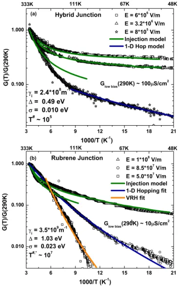
Conductance versus temperature measurements 关G共T兲兴 were performed for thicker rubrene films and fitted well with existing transport models,9,25 providing information about the electronic coupling共and hence molecular ordering兲, both at the injection interface and within the bulk. Figure3shows
G共T兲 for the HJ and the RJ at different applied electric field 共F兲 for the nominal rubrene film thickness of 20 nm. It should be noted that the rubrene films were grown simulta-neously for HJ and RJ to avoid any growth-related variation for the two cases. The charge injection model developed by Arkhipov et al.9 was used to analyze the results. Here, the injected current is found by considering injection into the Gaussian distribution of states 共with variance 兲 in the OS followed by either the return of the carrier to the electrode or its diffusive escape into the bulk. The injected current Iinjis therefore found as a product of the tunneling probability exp共−2␥ixo兲 共i.e., the probability of the carrier reaching the
position x0 in the first jump兲 and the escape probability
wesc共x0兲 共Ref.9兲 Iinj⬁
冕
a ⬁ dxoexp共− 2␥ixo兲wesc共xo兲 ⫻冕
−⬁ ⬁dE Bol共E兲g关U共xo兲 − E兴,
where x = a共⬃0.6 nm兲 is chosen as the surface of organic film,␥iis the interface related inverse localization radius and
Bol共E兲 is the Boltzmann factor
Bol共E兲 =
再
exp共− E/kBT兲, E ⬎ 01, E⬎ 0,
冎
kB is the Boltzmann constant
and U共x兲 is the energy barrier for injection given by U共x兲 =−16q2
orx− qFx, whereis the energy difference between
the Fermi level of the electrode and the highest occupied molecular-orbital level of rubrene, q is the electronic charge, or is the dielectric constant arising due to image forces,
and F is the applied electric field. It should be noted that the inverse localization length共␥兲 contains information about the electronic coupling that can provide structure-related infor-mation. Furthermore, they can have different values at the interfaces 共␥i兲 and within the bulk 共␥b兲.
The injection model fits our experimental curves in the two junctions with the extracted parameters共,,␥i兲 shown
in Fig. 3. For the optimized fitting parameters, the model reproducibly matches the experimental curve for different F, supporting the validity of the fits. A largefor RJ共1.03 eV兲 is inferred compared to HJ 共0.49 eV兲, in agreement with previous studies of thin rubrene barrier tunnel junctions.11In addition ␥i
r/␥ i
h⬃10 共r and h denote RJs and HJs,
respec-tively兲 suggests the anisotropic tunneling injection efficien-cies in the two junctions: better tunneling injection in HJs compared to that in RJs. This may be due to the presence of the alumina tunnel barrier and also due to the azimuthal growth of the rubrene molecules at the interface leading to a better electronic overlap of the rubrene-electron cloud with the metal. Such effects can be expected to influence spin injection and transport. In our earlier work, related differ-ences were observed in terms of the spin-polarized tunneling and TMR signals.11
The injection model begins to deviate at low T and low F. Bulk transport,25 viz., one-dimensional 共1D兲 hopping
FIG. 2. 共Color online兲 共a兲 Shows the rubrene film thickness measured using a profilometer for Al共6兲/Rub共x兲/Al共3兲 and Al共6兲/Al2O3共0.7兲/Rub共x兲/Al共3兲 with two different values of the
nominal thickness: x = 15 and 20 nm. 共b兲 Schematics: 共Left兲 azi-muthal growh of rubrene molecules on the alumina seed layer in HJ leading to stronger inter-molecular electronic coupling;共Right兲 dis-ordered nonazimuthal growth of rubrene on metal electrode in RJ leading to a strong hopping dominant transport.
兵G共T兲␣exp关−共T#/T兲−1/2兴其 and variable range hopping 共VRH兲
兵G共T兲␣exp关−共Tⴱ/T兲−1/4兴其, where T#,ⴱ␣␥
b 共bulk-related
in-verse localization radius兲, were instead fitted under these conditions, showing a gradual change from charge injection to the bulk transport-limited regime. At such low bias and temperature, a constant density of states near the Fermi level in the organic film is assumed to contribute to bulk conduc-tion. A change from 1D hopping to VRH hopping was ob-served in RJ at lower F. In contrast, no sign of such strong temperature dependence of G was noted in HJ. Only at low electric fields 共F⬍ ⬃1⫻106 V/m兲 and low temperatures was the 1D hopping model found to fit the curve for HJ. The fit is shown for the lowest measurable value of F 共8⫻105 V/m兲. The relatively stronger temperature
de-pendence of G observed in RJ compared to HJ suggests the highly disordered growth of rubrene molecules 共or weaker electronic coupling兲 in our RJs. This is supported by the inequality ␥br⬎␥bh共T#,ⴱ␣␥b兲 obtained from the parameters
T#,ⴱ, extracted using bulk models, signifying stronger
inter-molecular coupling in HJs. These results clearly show that the bottom seed layer共Al2O3in our case兲 plays an important
role for efficient spin injection by influencing the growth of rubrene molecules at the interface. Further, the results sug-gest some remanence of this ordering into the bulk共up to 20 nm in this case兲, providing efficient spin transport.
The interface magnetization was probed by PNR mea-surements using the NG1 reflectometer at NIST. Large area samples共⬃1.1 cm2兲 with a structure similar to the junctions
used for transport studies, with Co and Fe/CoO as the bottom and top layers, were prepared on unetched Si substrates. The nominal thickness of the rubrene film was 10 nm and 20 nm for the hybrid and the rubrene samples, respectively. The samples were cooled down in a magnetic field of 0.7 T to achieve exchange pinning of the Fe layer at the Fe/CoO in-terface. Using a supermirror polarizer and analyzer, the neu-tron beam was polarized parallel to the magnetic field, which was applied in the sample plane, as described in Ref.26. The reflectivity data were corrected for beam footprint, instru-ment background, and efficiencies of the polarizing eleinstru-ments 共typically⬎97%兲. Four reflectivity cross sections were mea-sured: R+ + and R−− labeled nonspin flip共NSF兲 as the
neu-tron retains its original polarization after scattering from the sample, and R+ −and R− +, labeled spin flip共SF兲, where the neutron spin flips it polarization, from the up 共+兲 to down state 共−兲 and vice versa, upon scattering. The nuclear scat-tering length density 共SLD兲 of a material or chemical SLD 共chem兲 can be determined from fits to the NSF reflectivity
data26–28in order to extract a profile of the chemical compo-sition of the film as a function of depth. In addition, the vector magnetization of the film as a function of depth can be ascertained from simultaneous fits to the NSF and SF cross sections. Specifically, the splitting between the NSF reflec-tivity cross sections is sensitive to the component of the magnetization parallel to the applied field. The SF reflectiv-ity is entirely of magnetic origin and is sensitive only to the component of the magnetization perpendicular to the applied field. The NSF PNR data were fit29 with the REFLPAK共Ref.
30兲 and GAREFL 共Ref. 31兲 software suites to obtain the
chemical and magnetic depth profiles. While spin-flip scat-tering was measured at all fields considered in this study, the scattering was observed to be negligible 共data not shown兲, indicating no significant moment perpendicular to the ap-plied field.
The reflectivity data collected at 5 K confirmed expecta-tions from SQUID magnetometry that the two FM layer magnetizations are aligned parallel 共P兲 in high fields 共Ha = 0.7 T兲 and antiparallel 共AP兲 in low fields 共Ha= 10 mT兲.
Figure4shows the NSF reflectivity measurement共R++ and R − −兲 for both the rubrene and the hybrid sample at Ha
= 0.7 T, along with the corresponding depth profile of the nuclear SLD and the layer magnetic moment for the two samples obtained from the fit to the reflectivity data. The most significant difference between the samples includes a lower value of the rubrene nuclear SLD and a larger value of the rubrene layer thickness relative to its nominal value共i.e., ⬃1.5 times the nominal thickness, compared to 1.1 times兲 obtained for the hybrid sample relative to the rubrene sample 共Fig.4兲. This result is in line with the different morphology
and packing density of the rubrene molecules共Fig.2兲 grown
FIG. 3. 共Color online兲 Normalized conductance 共log scale兲 vs inverse temperature plotted in 共a兲 HJ 关Co共8兲/Al2O3共0.6兲/ Rub共40兲/Fe共8兲兴 and 共b兲 RJ 关Co共8兲/Rub共40兲/Fe共8兲兴. The fit of ex-perimental curves to charge injection and bulk transport models with the extracted parameters are shown.
RAMAN et al. PHYSICAL REVIEW B 80, 195212共2009兲
on different seed layers 共i.e., Co or Al2O3兲, as described earlier. The structural characteristics of the underlying Co layer in both samples, as determined from PNR, are similar. Specifically, the structural roughness 共i.e., corresponding to the width of the interface between the Co layer and the layer above兲 at the Co/rubrene and Co/Al2O3interfaces in rubrene and hybrid samples, respectively, is comparable in both samples 共Fig. 4兲. Similar magnetic moments 共⬃1100 emu/cc兲 for the free bottom Co layer were also observed in the P and AP states of the hybrid and the rubrene sample. We thus conclude that there is minimal in-plane magnetic domain formation in the bottom FM electrode in the P and AP states because the structural disorder within the Co layer is limited.
In general, the moments for all the FM layers in both samples were found to be lower than the bulk values 共Co-1422 emu/cc and Fe-1700 emu/cc兲, which is consistent with expectations for fine-grained polycrystalline thin FM layers and more so for the top FM layers with additional interfacial disorder. The average magnetic moment of the top Fe layer in the hybrid sample was considerably lower, by⬃20% for both AP and P states, compared to the average Fe moment in the rubrene sample 共Fig. 4兲. This difference although
puz-zling is an interesting result and requires careful
interpreta-tion within the context of the corresponding structural mor-phology of the upper layers in the two samples. First, it is unlikely that oxidation of the Fe is the source for the reduced moment in the hybrid structure because the samples were well protected with a thick bilayer of Al共7 nm兲/ Al2O3共5 nm兲 over it 共Fig. 4兲. The nuclear SLD of the CoO
layer adjacent to the Fe was found to be lower than the bulk value共⬃4.27⫻10−6 Å
⬘
−2兲 in the hybrid sample, suggesting areduced structural density of the upper layers rather than en-hanced oxidation. Another possibility is that the reduced Fe moment is a direct consequence of higher roughness of the rubrene layer in hybrid structure. However, the PNR fitting analysis showed that the roughness of the rubrene/Fe layer is similar 共⬃3 nm兲 in the two samples though there exists a higher roughness at the top interface of the Fe layer in the hybrid sample 共⬃1.3 nm兲 compared to in the rubrene sample 共⬃0.7 nm兲.
A more significant finding is a lower nuclear SLD for Fe in the hybrid sample 共Fig.4兲 presumably indicating that the
structural density is reduced from that of bulk Fe. A possible origin of this reduction emerges from an understanding of the growth of Fe at the first few layers, which can be directly influenced by the structural properties of the underlying ru-brene layer. Given the roughness of the ruru-brene/Fe interface,
FIG. 4. 共Color online兲 共a兲 Measured NSF reflectivity data 共R++ and R−−兲 and the corresponding fits for both the hybrid Si/SiO共1兲/Co共8兲/Al2O3共0.6兲/Rub共15兲/Fe共8兲/CoO共1.5兲 and rubrene: Si/SiO共1兲/Co共8兲/Rub共20兲/Fe共8兲/CoO共1.5兲 samples at Ha= 0.7 T.
共b兲 Depth profile of the nuclear SLD 共top兲 and the layer magnetic moment 共bottom兲 obtained from the PNR fits for the hybrid and rubrene samples.
a significant disorder of Fe at this interface共continuing into the bulk兲 is probable in both samples. In hybrid structures, due to the lower packing density and the azimuthal growth of rubrene molecules, one can expect such disorder to be more pronounced in Fe leading to the reduced SLD. The structural properties of the top Fe electrode in hybrid samples therefore affect its magnetic response showing the reduced moment. Also, they lead to a higher interface roughness of the top Fe layer in the hybrid structure. This is expected to have a direct influence on the exchange coupling and tend to weaken the pinning of the Fe layer. Correspondingly, the magnetic re-sponse of Fe at the Fe/rubrene interface can differ from the rest of the layer. This will require a careful and systematic study that will be followed in the future.
The weak exchange pinning due to the rubrene/Fe inter-face behavior in HJ may lead to random spin fluctuations that are detrimental for spin injection and transport. A way to mitigate this influence was to modify the junction stack. We decided to exchange bias the bottom smooth Co layer and make the top Fe layer free. MR measurements were per-formed for the HJs with a rubrene thickness of 25 nm共shown in Fig. 5兲. A MR of 7% was observed at 4.2 K, reducing to
⬃4.5% at 77 K. No MR was noticed at room temperature. A sharp switching between the P and the AP states are noted, suggesting the advantage of having a strongly exchange pinned Co layer and a free top Fe layer. The gradual decrease in MR with bias at 77 K共see Fig.5inset兲 indicated the good
quality of the junctions. Further, we were able to detect spin transport signals through thicker rubrene films 共25 nm兲, a significant improvement in our device performance. In com-parison, small共⬃0.4%兲 or no MR was measured even in the most stable thin rubrene barrier junctions共5 nm兲, confirming the strong influence of interface ordering on spin injection.
In conclusion, we have observed the influence of the seed layer on the growth morphology of the rubrene molecules at the interface. Different growth mechanisms lead to a highly anisotropic conduction mechanism that affects both charge
and spin injection and transport properties. Although this study reveals better spin transport in molecularly ordered films, PNR result shows a corresponding complex nature of interface magnetic behavior, highlighting the open chal-lenges that have to be carefully addressed and tailored if organic materials are to reach their theoretical expectations in spintronics application.
This work was supported by the KIST-MIT program funds, the ONR Grant No. N00014-09-1-0177, and the NSF Grant No. DMR-0504158.
*vkarthik@mit.edu
1W. J. M. Naber, S. Faez, and W. G. van der Wiel, J. Phys. D 40,
R205共2007兲.
2Z. H. Xiong, Z. Di Wu, Valy Vardeny, and Jing Shi, Nature
共London兲 427, 821 共2004兲.
3T. S. Santos, J. S. Lee, P. Migdal, I. C. Lekshmi, B. Satpati, and
J. S. Moodera, Phys. Rev. Lett. 98, 016601共2007兲.
4V. Dediu, L. E. Hueso, I. Bergenti, A. Riminucci, F. Borgatti, P.
Graziosi, C. Newby, F. Casoli, M. P. De Jong, C. Taliani, and Y. Zhan, Phys. Rev. B 78, 115203共2008兲.
5V. N. Prigodin, N. P. Raju, K. I. Pokhodnya, J. S. Miller, and A.
J. Epstein, Adv. Mater. 14, 1230共2002兲.
6Ö. Mermer, G. Veeraraghavan, T. L. Francis, Y. Sheng, D. T.
Nguyen, M. Wohlgenannt, A. Köhler, M. K. Al-Suti, and M. S. Khan, Phys. Rev. B 72, 205202共2005兲.
7V. Coropceanu, J. Cornil, D. A. da Silvo Filho, Y. Olivier, R.
Silbey, and J.-L. Bre’das, Chem. Rev.共Washington, D.C.兲 107, 926共2007兲.
8M. A. Baldo and S. R. Forrest, Phys. Rev. B 64, 085201共2001兲. 9V. I. Arkhipov, E. V. Emelianova, Y. H. Tak, and H. Bässler, J.
Appl. Phys. 84, 848共1998兲.
10S. C. B. Mannsfeld, A. Virkar, C. Reese, M. F. Toney, and Z.
Bao, Adv. Mater. 21, 2294共2009兲.
11J. H. Shim, K. V. Raman, Y. J. Park, T. S. Santos, G. X. Miao, B.
Satpati, and J. S. Moodera, Phys. Rev. Lett. 100, 226603共2008兲.
12R. C. Jaklevic and J. Lambe, Phys. Rev. Lett. 17, 1139共1966兲; J.
Lambe and R. C. Jaklevic, Phys. Rev. 165, 821共1968兲.
13P. K. Hansma, Tunneling Spectroscopy: Capabilities, Application
and New Techniques共Plenum Press, New York, 1982兲.
14J. R. Weinberg-Wolf, L. E. McNeil, S. Liu, and C. Kloc, J. Phys.:
Condens. Matter 19, 276204共2007兲.
15A. L. Geiger, B. S. Chandrashekhar, and J. G. Adler, Phys. Rev.
188, 1130共1969兲.
16D. C. Tsui, R. E. Dietz, and L. R. Walker, Phys. Rev. Lett. 27,
1729共1971兲.
17M. Wang, W. He, and T. P. Ma, Appl. Phys. Lett. 86, 192113
FIG. 5. 共Color online兲 MR measurement 共minor loop兲 per-formed on HJ: CoO共1兲/Co共8兲/Al2O3共0.6兲/Rub共25兲/Fe共12兲 at 77
and 4.2 K. The bottom Co electrode is pinned to the CoO layer by cooling the sample in negative field. Insets: top shows the increase in junction resistance with cool down and the bottom shows the gradual decrease in MR with applied bias at 77 K. The shift in the curve 共toward left兲 is due to a residual current from the power supply, flowing through the magnet.
RAMAN et al. PHYSICAL REVIEW B 80, 195212共2009兲
共2005兲.
18S. Fratini, H. Xie, I. N. Hulea, S. Ciuchi, and A. F. Morpurgo,
New J. Phys. 10, 033031共2008兲.
19http://www.physics.unc.edu/project/mcneil/MolecularAnimations/
anim.php
20J. Kirtley, D. J. Scalapino, and P. K. Hansma, Phys. Rev. B 14,
3177共1976兲.
21N. M. D. Brown, R. B. Floyd, and D. G. Walmsley, J. Chem.
Soc., Faraday Trans. 2 75, 17 共1979兲; N. M. D. Brown, W. J. Nelson, and D. G. Walmsley, ibid. 75, 32共1979兲.
22J. T. Hall and P. K. Hansma, Surf. Sci. 77, 61共1978兲.
23R. J. Kline, M. D. Mcgehee, and M. F. Toney, Nature Mater. 5,
222共2006兲.
24G. E. Thayer, J. T. Sadowski, F. Meyer zu Heringdorf, T.
Saku-rai, and R. M. Tromp, Phys. Rev. Lett. 95, 256106共2005兲.
25N. F. Mott, Philos. Mag. 19, 835共1969兲. 26G. P. Felcher, Phys. Rev. B 24, 1595共1981兲.
27M. R. Fitzsimmons and C. F. Majrkzak, Modern Techniques for
Characterizing Magnetic Materials 共Kluwer, Norwell, MA,
2005兲.
28K. V. O. Donovan and N. F. Berk, Neutron Scattering from
Mag-netic Materials共Elsevier, Amsterdam, The Netherlands兲.
29C. F. Majkrzak, Physica B 221, 342共1996兲.
30P. A. Kienzle, K. V. O’Donovan, J. F. Ankner, N. F. Berk, and C.
F. Majkrzak, http://www.ncnr.nist.gov/reflpak共2000–2006兲.
31P. A. Kienzle, M. Doucet, D. J. McGillivray, K. V. O’Donovan,
N. F. Berk, and C. F. Majkrzak, http://www.ncnr.nist.gov/reflpak 共2000–2006兲.