Dynamic Regulation and Functions of Locus-specific DNA Methylation By
Yuelin Song B.S. Biological Sciences Zhejiang University, 2014
Submitted to the Department of Biology
In Partial Fulfillment of the Requirements for the Degree of Doctor of Philosophy
At the
MASSACHUSETTS INSTITUTE OF TECHNOLOGY May 2020
© 2020 Massachusetts Institute of Technology. All rights reserved
Signature of Author ………... Department of Biology May, 2020 Certified by………... Rudolf Jaenisch Professor of Biology Thesis Supervisor Accepted by.………... Mary Gehring Associate Professor of Biology; Member, Whitehead Institute Co-Chair, Biology Graduate Committee
Dynamic Regulation and Functions of Locus-specific DNA Methylation By
Yuelin Song
Abstract
The role and regulation of DNA methylation at various genetic elements have gathered tremendous interest over decades. The methylomes of many cell types have been described, revealing a dynamic and tissue-specific pattern of DNA methylation (tissue-specific differentially methylated regions, T-DMRs) in the distal regulatory elements, such as enhancers. The formation of T-DMRs still remain mysterious, however, one of their interesting features observed in mouse ES cells (mESCs) is the low-to-intermediate levels of average DNA methylation resulted from inter-cellular epigenetic heterogeneity. Given the transcriptional repressive role of DNA methylation at promoters, such non-zero levels of enhancer methylation is interesting to characterize. Prior to this thesis, a reporter for genomic DNA methylation (RGM) has been developed in the Jaenisch lab, when targeted into T-DMRs of interest, the surrounding locus-specific DNA methylation will be reported as on-and-off of fluorescent signals in single cells. We further modified RGM to investigate the regulation of DNA methylation at pluripotency super-enhancers Sox2 and MiR290 at single allele level in mESCs. We found that enhancer DNA methylation is surprisingly dynamic with two alleles independently being demethylated and methylated within days. Such dynamics is the basis of epigenetic and transcriptional heterogeneity and is coupled with changes in histone modifications and transcription factor binding. Furthermore, epigenetic heterogeneity was also observed in the developing pre-implantation embryos. Our work provided a paradigm to functionally investigate locus-specific DNA methylation in heterogenous tissues in diseases and development.
The regulation of locus-specific DNA methylation is highly context dependent and sensitive to the environment. Our understanding of how locus-specific DNA methylation is regulated in vivo is still restricted to a few genomic elements. The appendix of this thesis attempts to generate an animal model to expand the scope of research on DNA methylation to retroelement-associated metastable epialleles.
Thesis Supervisor: Rudolf Jaenisch
Dedication
I dedicate this thesis to my parents who have always been unconditionally supporting my pursuit of knowledge and happiness.
Acknowledgements
The completion of this thesis wouldn’t have been possible without the tremendous support and help I received over years from my mentors and colleagues. First foremost, I would like to thank my thesis advisor Dr. Rudolf Jaenisch, whose work I’ve always admired since college and I almost knew that I would join his lab when applying to graduate school. What I appreciate the most for being a graduate student in the Jaenisch lab is the great scientific freedom. Rudolf has always encouraged me when I took challenges and believed in me when things didn’t work as expected. I thank Rudolf for supporting me to propose projects, independently design experiments and collaborate outside of the lab, and providing me the opportunities to attend and speak at scientific conferences. I enjoyed writing my first scientific paper with Rudolf. He was very patient and edited all my smallest typos rounds after rounds. He taught me how to convey messages clearly and effectively. The journey of publishing was never a smooth sail, but thanks to Rudolf’s strong support, I was able to get through the most frustrated moments and continue to grow as a scientist. Rudolf’s acuity to science and the ability of immediately pointing at critical things always amaze me, and I am constantly inspired by his genuine interest and excitement for science. I especially want to thank Rudolf for his generous support when I wanted to explore alternative careers while in graduate school, without which I wouldn’t be so sure about my continuing pursuit for academia. Apart from science, Rudolf has a passion for life that is so infectious and I love listening about his hardcore hiking adventures and exchanging with my amateur camping experiences, all of which little things made my PhD years very enjoyable. Rudolf, thank you for being a great mentor.
I would like to thank another two postdoc colleagues who mentored me: Dr. Yonatan Stelzer and Dr. Frank Soldner. Yonatan taught me all the baby steps in DNA methylation research and mouse genetics by hand. Frank is so knowledgeable and experienced, without whose help I wouldn’t be able to get through many of my experimental crises and drainages of ideas. The daily coffee chats with Yonatan and Frank on science, politics, life or literally everything were so much fun and inspiring. The unforgettable intense scientific arguments among us were probably one of the biggest drivers for me to stay in science, as they made me realize how much we love and care about what we do. Both of them are extremely smart and insightful scientists, whom I deeply admire and learnt greatly from.
I also would like to thank formal graduate student Dr. Chikdu Shivalila, who taught me cloning, gene targeting, and importantly, how to enjoy graduate school. Many thanks to Dr. Malkiel Cohen for his help throughout my PhD and the free drives to the WI retreat every year. I would like to thank Ruthie Flannery for her “mouse-mommy-like” attentive care for our animal facility and her help in my never-ending genotyping requests. I would like to thank Dr. Styliani Markoulaki, Jesse Drotar, Nick Rosenau, and Dina Rooney for their support in mouse experiments, Raaji Alagappan for her strong sense of responsibility to care for the whole lab, Dongdong Fu for her fast and reliable work on tissue-processing, and Gerry Kemske, Robert Burger and Carrie Garrett-Engele for making our lab running smoothly. I thank all current and past Jaenisch lab members for their help and the supportive environment they contribute to create. I would also like to thank Dr. Richard Young, Dr. Stefan Semrau and their students and postdocs for the great collaboration experience.
I would like to thank my thesis committee, Dr. Phil Sharp and Dr. Tyler Jacks, for their feedbacks during my thesis committee meetings, preliminary exams, and their strong support in my academic career.
I am forever grateful to my undergraduate mentors, Dr. Yingjie Wang and Dr. Binghui Shen. Both of them care deeply for nurturing the next generation of Chinese scientists. When they started their joint lab at Zhejiang University after returning from the US, they gave me my own bench and interesting projects to explore despite the fact that I was only a sophomore. They both helped me to connect with US research labs through their network after they learnt that I would like to pursue my PhD in the US. With the help from Dr. Binghui Shen, I had the opportunity to conduct summer research at Dr. Kun-Liang Guan’s lab at USCD working with a PhD student on different isoforms of PKC; with the help from Dr. Yingjie Wang, I was able to have the research experience in Dr. Robert G. Roeder’s lab at the Rockefeller University under the guidance of Dr. Zhanyun Tang and Dr. Weiyi Chen on chromatin biology. Both experiences were so valuable and rare for international students at that time, and they shaped my scientific interest in epigenetics. I thank Dr. Guan and Dr. Roeder labs for the training and their generous support in my graduate school application.
Finally, to my parents, thank you for giving me unconditional supports for whichever path I choose in life. Thank you for being my biggest champions. You are the reason why I can enjoy all the freedom and privileges, and be who I am today.
Table of Contents
Abstract ... 3
Dedication ... 4
Acknowledgements ... 5
Chapter 1. Introduction. ... 10
1.1 The Spatial and Temporal Landscape of Genomic DNA CpG Methylation in Mouse Embryonic Stem Cells ... 10
1.2 Enzymatic Regulations of DNA Methylation Dynamics in ESCs ... 13
1.3 DNA Methylation in Transcriptional Regulation at Different Genetic Elements ... 16
1.4 Cross-talk Between DNA Methylation and Histone Modification ... 19
1.5 Cross-talk Between DNA Methylation and Transcription Factor Binding ... 21
1.6 Super-Enhancers and the Mediator Complex ... 25
1.7 Enhancer DNA Methylation Heterogeneity ... 27
1.8 Genomic DNA Methylation Reporter for Locus-specific Studies in Single Cells ... 28
1.9 Why Is Enhancer DNA Methylation Heterogenous and What Does It Mean Functionally? ... 30
References ... 31
Chapter 2. Dynamic Enhancer DNA Methylation as Basis for Transcriptional and Cellular Heterogeneity of ESCs. ... 43
Summary ... 45 Introduction ... 45 Results ... 47 Discussion ... 60 Methods ... 63 Acknowledgement ... 72 References ... 88 Supplementary Information ... 94
Chapter 3. Future Directions ... 114
3.1 Developmental Impacts of DNA Methylation Heterogeneity by Epigenetic Lineage Tracing .... 114
3.3 Metabolic Regulation of DNA Methylation in Early Embryonic Development ... 117
3.4 DNA Methylation and Transcriptional Condensates ... 118
Concluding Remarks ... 119
Reference ... 119
APPENDIX I. Metastable Epiallele Reporter Mouse Model Development for Studying Environmental Regulation of DNA Methylation ... 121
Background ... 121
Result ... 124
CHAPTER 1 FIGURE 1 ... 10 FIGURE 2 ... 11 FIGURE 3 ... 15 FIGURE 4 ... 16 FIGURE 5 ... 30 CHAPTER 2 FIGURE 1 ... 75 FIGURE 2 ... 77 FIGURE 3 ... 79 FIGURE 4 ... 81 FIGURE 5 ... 83 FIGURE 6 ... 85 FIGURE 7 ... 87 CHAPTER 3 FIGURE 1 ... 115 APPENDIX FIGURE 1 ... 122 FIGURE 2 ... 123 FIGURE 3 ... 124 FIGURE 4 ... 125 FIGURE 5 ... 125 FIGURE 6 ... 126 FIGURE 7 ... 127 FIGURE 8 ... 128 FIGURE 9 ... 129 FIGURE 10 ... 131 FIGURE 11 ... 132 FIGURE 12 ... 133 FIGURE 13 ... 135
Chapter 1. Introduction.
1.1 The Spatial and Temporal Landscape of Genomic DNA CpG Methylation in Mouse Embryonic Stem Cells
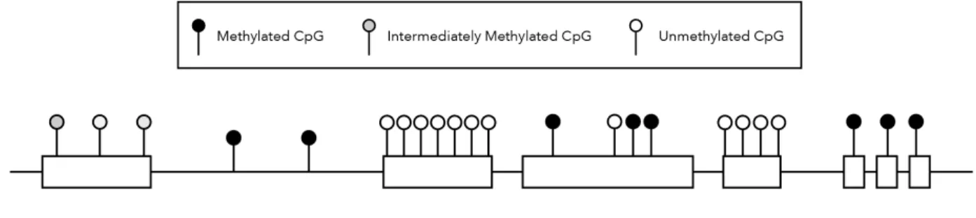
5’methyl-cytosine in the context of DNA CpG dinucleotides (referred as DNA methylation below) has been proposed as an inheritable epigenetic mark regulating gene expression and cell fate since 19751. However, the specific roles of genomic DNA methylation across different genomic elements still remain to be elucidated. In the vertebrate genome most CpGs exist in CpG-islands (CGI, CpG rich regions with approx. 1kb), which are protected from DNA methylation in somatic cells2,3. CpG-islands residing in more than half of the genome tend to be associated with transcriptional start sites (TSSs) of house-keeping and developmental genes2. The rest of the genome is depleted of CpGs but the remaining non-CGI CpGs are heavily methylated (>70%)4, especially at gene bodies, transposable elements and gene deserts3. Non-coding regulatory elements, such as enhancer and insulators have also low CpG densities. These regions tend to have more variable levels of DNA methylation in a tissue-specific manner5-8 (Figure 1).
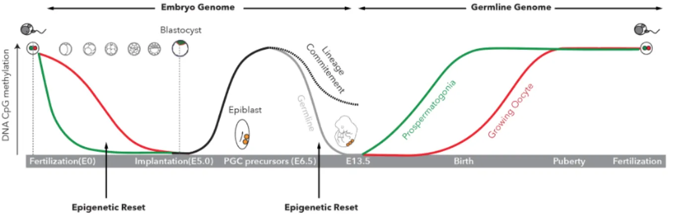
DNA methylation is essential to mammalian development and most dynamic at early developmental stages. There are two waves of DNA methylation reset throughout mammalian development (Figure 2). The first one is a global erasure of DNA methylation except for genomic imprinted regions in the paternal and maternal genomes post-fertilization. This wave of reset brings the embryonic genome to a hypomethylated state at the blastocyst stage (E3.5-4.5 in mouse and E5-6 in human)9,10. Upon implantation, global de novo methylation occurs in the epiblast, derived from the inner cell mass of the blastocyst, which gives rise to the embryo proper10-12. Tissue-specific regulatory regions undergo demethylation during embryonic development or adult stem cell differentiation6,13,14. The second reset occurs in the developing primordial germ cells (PGCs), where parent-of-origin DNA methylation is removed after which germ-line- and sex-specific methylation patterns are established at different times in male and female embryos12,15-17.
Figure 2. DNA Methylation Reprogramming (Resets) During Development
Mouse embryonic stem cells (mESCs) are isolated from the inner cell mass at the blastocyst stage and can be maintained at the pluripotent state in different media conditions in-vitro18-20. mESCs possess pluripotent signatures of genomic DNA
cell fate decisions21. The total level of DNA methylation of mESCs is similar to that of liver and kidney cells in vivo22. Different from many other precursor cells as well as
conventionally grown human ESCs, mESCs can tolerate global loss of methylation when depleted of all methyl-transferases and still remain pluripotent gene signatures 23-27. These DNA methylation deficient mESCs are capable to proliferate however fail to differentiate, indicating that DNA methylation is dispensable for maintaining the pluripotent state but essential to cells assuming a specific lineage in mouse28. CGI-promoters of pluripotency genes are usually hypomethylated in mESCs, such as those of Oct4 and Nanog29,30. Immunoprecipitation of methylated DNA combined with DNA
microarrays (mDIP) revealed that promoter methylation contributes to silencing of some developmental genes in ESCs31. Nevertheless, a majority of CGI-associated promoters of developmental genes in mESCs remain unmethylated as shown using reduced representation bisulfite sequencing (RRBS) that specifically enriches for CpG dense regions32. Whole-genome bisulfite sequencing (WGBS) sufficiently covering comparatively less-studied and hypermethylated CpG-poor regions showed that, in certain tissues or cell types, short stretches of DNA are hypomethylated, which constitutes differentially methylated regions (DMRs)5,33,34. mESC-specific DMRs identified in WGBS studies by comparing to other somatic tissues are predominantly found at distal regulatory regions of pluripotency genes5,13,17,35,36. Interestingly, unlike CGI-associated proximal promoters that are usually protected from DNA methylation, tissue-specific DMR (T-DMR)-associated enhancers show low-to-intermediate levels of DNA methylation5. Methylation of these regions is more dynamically regulated during development than CGI promoters, which is consistent with the role of enhancers in instructing tissue-specific gene expression6,13,37,38. However, the identities and biological functions of tissue-specific DMRs, especially causal links to gene regulations and cell states, still remain to be elucidated.
1.2 Enzymatic Regulations of DNA Methylation Dynamics in ESCs
The processes of methylation and demethylation concertedly regulate methylation levels in ESCs (Figure 3). The mouse and human genomes both encode 5 DNMTs: DNMT1, DNMT2, DNMT3A and DNMT3B, and DNMT3L, although DNMT2 mainly has tRNA methyl-transferase activity39. DNMT3L has no enzymatic activity but is essential in facilitating DNMT3A-mediated de novo methylation in germ-cells and also protects DNMT3A2 from degradation in ESCs40. De novo methylation is enzymatically carried out by DNMT3A and DNMT3B, which is essential for establishing the methylation pattern in the developing embryo41-43. In ESCs, the predominantly active isoforms are DNMT3A, DNMT3A2, and DNMT3B1, the deletion of which result in progressive loss of methylation42. DNMT3A and DNMT3B have overlapping and distinct genomic targets in ESCs and both are excluded from active enhancers and promoters44. DNMT3B targets actively transcribed gene bodies, consistent with intragenic methylation’s role in promoting transcriptional efficiency44. DNMT3A appears to target distal promoters and de novo methylates pluripotency enhancers upon differentiation42,45,46. Maintenance methylation is mainly mediated by DNMT1 in partnership with UHRF1, although it has been shown that DNMT3s also contributes to DNA methylation maintenance47,48. DNMT1 is targeted to the replication fork and interacts with PCNA, preferentially methylating hemi-methylated DNA49. Passive demethylation is caused by the absence of maintenance methylation activity and it critically contributes to the methylation dynamics throughout development. Genetic ablation of DNMT1 and UHRF1 leads to rapid demethylation of ESCs23,50-52. The post-fertilization maternal genome undergoes demethylation by excluding oocyte-specific DNMT1o from the nucleus53. In PGC epigenetic reprogramming, demethylation was shown to be achieved by nuclear extrusion or transcriptional repression of UHRF1 while
PGCs undergo several rounds of division54. Primed mESCs cultured in LIF/serum transition into the naïve state resembling the ICM when switched to “2i” (GSKβi/MEKi) media with the global demethylation during this process being mainly achieved by passive demethylation as a consequence of DNMT1/UHRF1 downregulation55,56. Passive demethylation also contributes to lineage-specific demethylation, however, how it is regulated in a locus-specific manner and to what degree it matters in comparison to active demethylation has not been fully elucidated57.
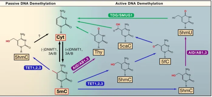
Active or active-passive demethylation is initiated by TET enzymes converting 5’ methyl-cytosine (5mC) into 5’ hydroxymethyl-cytosine (5hmC). 5hmC can be further oxidized to 5’ formalcytosine (5fC) and 5’ carboxylcytosine (5caC) by TET enzymes, or deaminated by AID/APOBEC family members into 5’ hydroxymethyluracil (5mU). 5mC can also be directly deaminated by AID/APOBEC into thymidine, which together with 5caC and 5mU are recognized by DNA base-excision repair pathway (BER) members such as TDG and subsequently replaced by newly synthesized cytosine58. Active demethylation has been shown to be present in many cell types and plays an important role during development58-60. Compared to the maternal genome where demethylation is mainly passive, the paternal genome undergoes TET3-mediated active demethylation post-fertilization with much faster kinetics61. Another example supporting the mechanism of active demethylation is the observation of cycles of methylation and demethylation within 100min periods in response to environmental stimuli in the absence of cell division62,63. Post-mitotic neurons also show high levels of 5hmC and activity-dependent active demethylation mediated by TET1 and APOBEC1 was observed in adult mouse brain64,65.
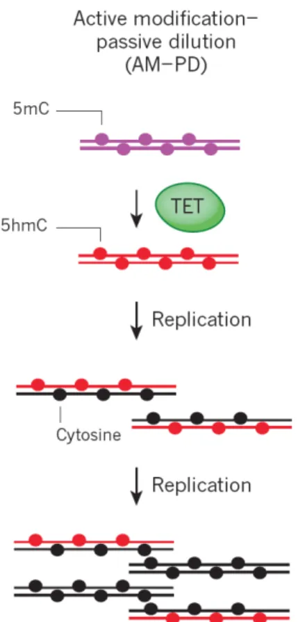
5hmC inhibits the maintenance methylation machinery and therefore causes passive demethylation in the subsequence cell division (active-passive demethylation, Figure 4). An example of active-passive demethylation can be found in PGC
reprogramming, where genomic imprints are erased. 5hmC accumulates after initial active demethylation and is then diluted in a replication-dependent manner60. In mESCs, both TET1 and 2 are highly expressed and 5hmC can be readily detected. TET1 is enriched at CGI promoters and TET2 mostly at actively transcribed gene bodies and enhancers66,67. TET3 is expressed at low level and contributes to ~2% of 5hmC68. Similar to DNMTs, loss of all TET enzymes does not affect ESC maintenance but impairs cell differentiation. TET triple-knockout mice show gastrulation defects causing embryonic lethalality69. Genetic ablation of the downstream BER component TDG revealed wide-spread accumulations of 5fC and 5caC at distal regulatory elements and poised promoters. Although the exact function of oxidized 5mC in transcriptional regulation is still unclear, they could be bound by other proteins and mediate local changes of chromatin59.
Figure 3. DNA Methylation and Demethylation Pathways (Active or Passive). Bhutani et al. Cell. 2011.
Figure 4. Active-passive Demethylation (AM-PD: active modification-passive demethylation). Kohli et al. Nature. 2013.
1.3 DNA Methylation in Transcriptional Regulation at Different Genetic Elements It is challenging to generalize a universal rule of DNA methylation in gene regulation at different genomic elements. The function of DNA methylation is most extensively characterized at proximal promoters, where it serves as a stable silencing mark3,70. Around 50% CGIs are associated with TSSs and remain hypomethylated at house-keeping and developmental genes, and many of the CGIs remain hypomethylated even after transcription becomes inactive70. However, during development there are small subsets of CGI promoters become de novo methylated, which usually leads to long-term gene silencing2. In ESCs, promoter DNA methylation
contributes to long-term silencing of germline-specific genes, such as Dazl, whereas promoters of highly expressed pluripotency genes such as Oct4, Nanog, Dppa4, Tdgf1 are unmethylated31,71-73. The mechanism of DNA methylation mediating promoter silencing involves concerted alterations of chromatin structure as well as histone modifications74,75. As ESCs differentiate, promoters of other lineage-specific genes become de novo methylated after gene silencing occurs, leading to long-term repression via heterochromatin formation, subsequently restricting pluripotency and promoting lineage commitment76. Similarly, in X chromosome inactivation, DNA methylation at gene promoters occurs several days post-inactivation and maintains repression of silenced genes on the inactivated X (Xi) whereas “escaper” genes have lower methylation at promoters77-79. Therefore, DNA methylation at promoters serves to stably maintain but likely not to initiate gene silencing.
In contrast, DNA methylation at gene bodies is not associated with repression in mammals. Gene-bodies are in general CpG poor and therefore are highly methylated37,70,80. Methylated gene bodies are associated with increased expression levels in cancer and somatic cells81-83, and perturbation of gene body methylation using demethylating agents such as 5’azacytidine leads to down-regulation of gene expression81. Some “orphan CGIs” are also found at gene-bodies and thought to possess potential intra-genic alternative promoter activities84. “Orphan CGIs” are more methylated than promoter CGIs (20~30% vs 3% methylation)2,84,85. Mechanistically, DNA methylation at gene bodies does not block elongation despite repressive histone modifications such as H3K9me3 in cancer cells86. In mESCs DNA methylation even facilitates transcriptional elongation via preferential binding by histone variant H2A.B at gene bodies not promoters87. On the other hand, due to its inhibitory role in transcriptional initiation, DNA methylation at gene bodies could also enhance
transcriptional efficiency by suppressing alternative promoter usage, retroelement activation and regulatory anti-sense ncRNA production88-90.
The regulation and function of DNA methylation at enhancers are less characterized than that of promoters3, potentially due to the fact that most enhancers locate at CpG poor regions where sufficient coverage at single-base resolutions is only achieved with WGBS and variable levels of methylation complicating the interpretation of correlations5,13,91. Nevertheless, locus-specific studies have shown that methylated enhancers tend to correlate with reduced transcription of cognate genes, although some enhancers still remain unmethylated even after becoming dormant13,92. The evidence of DNA methylation at enhancers being a negative regulator of transcription is variable. In breast cancer92,93 a strong anti-correlation of gene expression with promoter methylation is seen, whereas in blood and skin cells this relationship is only moderate and even varies between different sub-group cells types94. However, enhancer methylation is becoming increasingly important as growing evidence supports the notion that T-DMRs that are only hypomethylated in certain lineages are preferentially associated with tissue-specific enhancers5,13,95,96. Therefore, understanding the mechanism of how T-DMRs at enhancers regulate tissue-specific gene expression is a crucial issue. The MyoD enhancer was shown to be hypermethylated in non-muscle cells but unmethylated in embryonic myogenic cells in a human MyoD enhancer transgenic mouse model. Though essential for MyoD tissue-specific expression, MyoD enhancer demethylation does not immediately cause MyoD expression95, which is similar to the delayed chicken clys gene expression after enhancer demethylation97. Together these examples indicate that enhancer demethylation may be a necessary but not sufficient step for gene activation. Interestingly, mice carrying a CpG-mutant MyoD enhancer, which is unable to become methylated, have no aberrant ectopic MyoD expression, suggesting that enhancer
methylation is not required for gene silencing in this context95. This echoes the induced liver-specific demethylation of the Tat enhancer by glucocorticoids (GC). GC withdraw after initial exposure does not remethylate the enhancer, despite Tat expression being shut down in the absence of stimuli98. In other cases, aberrant enhancer hypomethylation correlating with cognate gene upregulation have been found in ER-positive tumors as well as in neurons from psychosis patients consistent with the importance of proper enhancer methylation in normal physiology92,99. In contrast to the delayed transcriptional activation after enhancer demethylation of MyoD and clys, demethylation of an enhancer-like Treg-specific demethylated region (TSDR) at the Foxp3 locus is sufficient to induce Foxp3 expression in Tregs100,101. However, this may not be generalizable as the TSDR is proximal to Foxp3 promoter and contains CpG-islands which is less typical for most distal regulatory elements5,13,102. In mESCs, enhancer demethylation by genetic ablation of DNA methyl-transferases led to both up- and down-regulation of target gene expression, with the transcriptionally suppressive roles of DNA methylation validated at only a few loci103.
Therefore, the answer as to the exact role and detailed steps of enhancer methylation dynamics in gene regulation is far from being conclusive and highly context-dependent37,104. Since enhancers are major transcription factor binding sites and are subjected to other complex epigenetic regulations to be discussed below, a more comprehensive view of all these components will facilitate a better understanding of how enhancer T-DMRs regulate gene expression.
1.4 Cross-talk Between DNA Methylation and Histone Modification
DNA methylation and histone modifications represent two parallel and complementary mechanisms in gene expression regulation. In many scenarios of promoter silencing, two processes are coordinated through direct interactions
between DNMTs and histone modifiers. For example, DNMTs can recruit the Polycomb repressive complex 2 by interacting with EZH2 to catalyze H3K27me3. H3K9 methyl-transferase G9a can recruit DNMT3s to de novo methylate pluripotency gene promoters. It was also shown that interactions between DNMT3s with SUV39H1 and SETDB1 mediate pericentric heterochromatinization74. On a different level, histone modifications can directly affect DNMT binding. For example, H3K36me3 at gene-bodies is recognized by the PWWP domain of DNMT3s and is required for intragenic DNA methylation105. H3K4me2 and H3K4me3 modified nucleosomes exclude DNMTs by disrupting binding from the ADD domain, thus protecting the CGI from being methylated106. Reciprocally, DNA methylation status can influence histone modifications. Unmethylated CGIs are bound by the CXXC domains of some H3K4 methyl-transferases as well as of CXXC finger protein 1 (CFP1), which recruits H3K4me3 methyltransferases that do not have the CXXC domain to activate CGI promoters107,108. In contrast, DNA methylation at a repressor domain upstream of H19 can recruit DNA methylation binding protein MeCP2, which facilitates H3K9 methylation and gene repression109.
H3K27ac is a shared marker between active enhancers and promoters. In general, H3K27ac is inversely-correlated with DNA methylation as acetylated histones promote chromatin accessibility and transcriptionally permissivity110,111. Methyl-binding MeCP2, beside facilitating H3K9 methylation, is also in complex with histone deacetylase (HDAC)112. Histone deacetylation condenses chromatin inhibiting transcription factor access and facilitating gene silencing on methylated DNA. Treatment of live cells with HDAC inhibitor can induce global hypomethylation by altering DNMT1 nuclear dynamics and protein level113,114. On the other hand, knocking out DNMT1 leads to extensive changes of the H3K27ac landscape in mESCs, with both gains and losses at different enhancers, indicating a more complicated and
locus-dependent relationship between DNA methylation and H3K27ac103. H3K4me1 is distinguishably enriched at enhancers compared to promoters. In contrast to H3K27ac, H3K4me1-marked enhancers are usually primed to be active. The presence of H3K4me1 at enhancers often precedes nucleosome depletion and H3K27 acetylation, leaving an opportunity for enhancer activation104. A meta-analysis of high-throughput profiles of DNA methylation and histone modifications observed a pattern of H3K4me1 enrichment at enhancers with intermediate DNA methylation level, such as at the enhancer of c-Myc and Sox2. In ESCs, regions with low DNA methylation level has elevated H3K4me3 level and diminished H3K4me1, which seems to mark an enhancer-promoter transition. The molecular details of this correlation cannot yet be explained by any intermediate mechanisms such as binding of known MBD-containing proteins or 5hmC patterns115. The inconclusive understanding of how DNA methylation and enhancer histone modifications are connected is partially due to current sequencing-based technologies that use bulk-cell populations at a snapshot time-point and thus have limited temporal and locus-specific resolution. Observations yielded from the genome-wide high-throughput approaches are valuable yet mostly correlative. Therefore, it is important to utilize new systems that allow functional explanation of the various (anti-)correlations between DNA methylation and histone modifications at different genomic loci.
1.5 Cross-talk Between DNA Methylation and Transcription Factor Binding
Tissue-specific enhancers are bound by transcription factors (TFs). On one hand, the binding activity of a TF can be influenced both positively and negatively by DNA methylation. On the other hand, TF binding can recruit DNA (de)methylation machineries inducing local changes of DNA methylation. It is still challenging to
distinguish the hierarchical cross-talk between DNA methylation changes and TF binding events in transcriptional regulation.
Due to the transcriptionally suppressive role of DNA methylation at promoters and some enhancers, TF binding is traditionally thought to be inhibited by methylated DNA either directly or indirectly through methyl-binding proteins116. Crystal structure studies have shown that 5mC resides in the major groove of DNA, and, because the bulky methyl-group narrows the minor groove, the altered structure of the DNA can directly affect TF binding117. MBD-containing proteins can bind methylated DNA directly at CpGs, such as MeCP2, or to any methylated DNA independent of the sequence, such as MBD1,2,4118. These methyl-binding proteins are associated with protein partners that mediate heterochromatin formation and transcriptional repression. They also exclude the demethylase TET1 from methylated DNA118,119,120 and thus indirectly inhibit TF binding to methylated DNA. However, evidence indicates that DNA methylation can both promote and inhibit TF binding. A systematic SELEX (systematic evolution of ligands by exponential enrichment) assay applied a collection of 542 full-length human TFs and DNA binding domains to CpG-methylated DNA and showed that the methylated DNA inhibited binding of many major classes of TFs including bHLH, bZIP, and ETS. In contrast, TFs containing homeodomains such as POU and NEAT were shown to preferentially bind to methylated DNA121. However, the forcefully expressed POU-domain containing TF Oct4 in colon carcinoma cells can only bind to DNA when it is unmethylated122, raising the caution that the knowledge about TF-binding and DNA methylation gained through in vitro TF-binding motifs binding assays still requires in vivo validation, as nucleosomal chromatin presents a very different target for TF binding as compared to naked DNA118. Nevertheless, a few in
vivo studies have yielded valuable insights on the cross-talk between DNA methylation and TF-binding in a regulated chromatin environment. By mapping DNaseI
hypersensitivity sites (DHS) in the absence of DNA methylation in DNMT triple knockout (TKO) cell , the study showed that NRF1 gained additional thousands binding sites especially in the CpG-poor distal regulatory elements, which were originally methylated in WT mESCs. The appearance of new NRF1 binding sites in TKO cells was concomitant with enrichment of H3K27ac and initiation of aberrant transcription, indicating that DNA methylation in wild-type cells was safeguarding against aberrant transcription by blocking NRF1 binding123. In contrast, KLF4 ChIP-bisulfite sequencing experiments have shown that KLF4 binds to two motifs differently in hESCs. KLF4 binding to one motif is preferred when the sequence is methylated whereas binding to the other motif is inhibited by methylation, providing an example of context-dependent methylation sensitivity of TF-binding124.
The above-mentioned examples show that the DNA methylation status could act upstream excluding or attracting certain TFs. As the genomic DNA methylation pattern correlates with cell-type, locus-specific DNA methylation needs to be inherited mitotically to maintain a certain cell state. To maintain a given differentiation state DNA methylation at promoters and enhancers may serve to prevent TFs from aberrantly activating genes which are not supposed to be expressed in the given cell type, and meanwhile attracting methyl-binding repressors to secured the silenced state. Similarly, the pre-existing unmethylated promoters and enhancers need to be actively protected from de novo methylation for maintaining cell-type specific gene expression.
However, the chick-and-egg question is whether TF binding induces DNA methylation changes or whether the pre-existing DNA methylation state regulates TF binding affinity. The above-mentioned KLF4 is a pioneer transcription factor in iPSC programming and initiates remodeling of the chromatin, opening up pluripotency enhancers and TSSs125. In fact, most pioneer TFs instruct cell-fate transitions and a common view of tissue-specific enhancer activation is that pioneer TF binding instructs
local changes of the heterochromatin by interactions with various epigenetic modifiers and chromatin remodelers, which further leads to TF-dependent coactivator assembly and transcription. Therefore, the ability to bind methylated enhancers is an important attribute of pioneer TF function. In addition to KLF4, other pioneer TFs such as HOXA5, HOXA9, GATA3, GATA4, FOXC1, FOXK1, FOXK2, and FOXA1 have been shown in vitro118 to have methyl-CpG binding activity. However, in vivo data for these pioneer TFs binding being sufficient to change DNA methylation is either lacking or supports the opposite view, i.e. that the pre-existing DNA methylation status determines whether the pioneer TF initiates transcription. For example, FOXA1 binds to target enhancers during neural differentiation, which is inhibited by DNA methylation contrary to previous in vitro observations126,127. An in vivo example of TF driving methylation changes is MEF2 and SIX binding to the upstream of the Myogenin promoter. Myogenin starts muscle-specific expression at around E8.5 following a rostro-caudal gradient. Its weak-CpG island promoter is initially methylated and bound by ZBTB38 and MBD2 to maintain the silenced state, both of which are degraded upon the onset of differentiation with the concomitant demethylation of Myogenin promoter leading to activation of the gene. Mutations of either MEF2 or SIX binding sites abolishes DHS formation illustrating the requirement for binding of two TFs in initiating demethylation, which precedes the activation of Myogenin128,129. Perhaps the most robust evidence of TF binding driving DNA methylation changes comes from the insulator CTCF and the transcriptional repressor REST. Stadler et al. showed CTCF binds regulatory elements those overlap with low-methylated regions (LMR). A reporter construct with or without CTCF binding motif was inserted into the same genomic locus of mESCs. The constructs carrying a mutated CTCF binding motif became methylated in mESCs whereas reduced methylation was observed in cells containing the functional CTCF binding motif construct. A construct containing
pre-methylated CTCF binding site was then further inserted into the same locus causing CTCF binding-induced local reduction of methylation in vivo, confirming the driver role of CTCF binding in creating LMRs5. Similar to CTCF, REST also occupies LMRs in ESCs and genetic deletion of REST led to increased methylation at its target LMRs. Reintroduction of REST restored the LMR, showing the necessity and sufficiency of REST binding in creating and maintaining DNA methylation patterns5.
High-throughput in vitro biochemical studies as well as genome-wide epigenetic and TF binding profiling yielded valuable correlative patterns between TF binding and DNA methylation. However, the relationship between genomic DNA methylation and binding activities of many TFs in a physiologically relevant setting still awaits further mechanistic dissection, especially at enhancers, the activation of which is tissue-specific. With the development of genomic and epigenomic editing technologies, manipulations of TF binding sites or of locus-specific methylation status endogenously should bring a clearer picture of this mechanistic hierarchy.
1.6 Super-Enhancers and the Mediator Complex
Classical enhancers are defined as DNA elements bound by transcription factors (TF), which activate transcription of a gene independently of distance or orientation with respect to the gene130. Super-enhancers are a large cluster of enhancers that often have unusually high levels of coactivator binding, such as the Mediator complex, and display active histone modifications, such as H3K27ac131. In mESCs, super-enhancers are defined by either of the following criteria: (1) the binding of all three master TFs Oct4, Sox2 and Nanog, (2) the proximity of stitched enhancer within 12.5kb, (3) a ranking of H3K27ac or MED1 ChIP-seq signal where the intensity of the enhancer is above the slope of 1130-132. Compared to normal enhancers, super-enhancers are large and usually locate at genes controlling cell identity with their activity being more
sensitive to the knockdown of the Mediator complex subunits. Therefore, super-enhancers tend to be associated with T-DMRs. Most super-super-enhancers in mESCs are associated with pluripotency genes such as Sox2, Oct4, Nanog, Prdm14, Esrrb, Klf4 and microRNA clusters such as Mir290-295. Individual enhancers within a super-enhancer can cooperate in activating transcription consistent with that super-enhancers usually drive high level of target gene expression. Strong enrichment of the Mediator complex plays a central role in coordinating transcription and was initially used to identify super-enhancers in mESCs132,133. The Mediator complex consists of approximately 30 subunits, including MED1 to MED31, and the cyclin-dependent kinase (CDK) 8-cyclin C pair as well as several paralogs. The subunits of the Mediator complex are not fixed and can vary between different complex isoforms. The earliest discovered function of the Mediator complex was a “bridge” to bring TF-bound enhancers to PolII and general transcription factor (GTF) machineries to promoters by a “looping” mechanism and to form the pre-initiation complex (PIC)134. Besides initiation, the Mediator complex has also been shown to interact with histone modifiers, lncRNAs, transcriptional elongation, termination and RNA splicing factors133.
Recently, an alternative view of how transcription factors interacting with DNA regulatory elements has evolved when the Mediator complex together with other chromatin associated coactivators was found to form phase-separated condensates in vitro and in vivo135,136. Transcriptional factor and coactivator condensates form when proteins reach a critical concentration and therefore are dependent on the density and affinity of TF-binding sites at enhancers137. As TF and coactivators may recruit or exclude DNA methylation machinery from binding to enhancers in a locus-dependent manner, it is unresolved how DNA methylation affects transcriptional condensates. Direct or indirect interactions between the Mediator Complex and the DNA (de) methylation machineries or methyl-binding proteins may be involved in coordinating
transcriptional activity. It was shown that Mediator CDKs have repressive role in transcription of C/EBPβ target genes, and this repressive effect is achieved in part through recruiting histone arginine methyl-transferase PRMD5. The product of H4R3me2 correlated with DNMT3A recruitment and methylation at target gene promoters138. The Mediator complex subunit MED14 in Arabidopsis thaliana was shown to promote transcription at highly methylated transposable elements with med14-3 mutation inducing decreased non-CG DNA methylation at chromosomal pericentromeric regions139. In summary, it remains to be elucidated whether the Mediator complex can directly cross-talk with DNMTs, TETs or methyl-binding proteins at enhancers.
1.7 Enhancer DNA Methylation Heterogeneity
DNA methylation at a certain CpG in a single cell at a given time is a binary event with values of 0 (biallelically unmethylated), 0.5 (mono-allelically methylated), or 1 (biallelically methylated). However, many active enhancers show a range of low-level value of methylation between 0-0.5, which could be a result of averaging methylated and unmethylated alleles from bulk-cell sequencing. In a cultured cell population, for example ESCs, such continuous values of low-level methylation (0-0.5) of a specific loci indicates cell-to-cell DNA methylation heterogeneity. Recent development of single cell RRBS (scRRBS) and WGBS (scWGBS) in ESCs indeed revealed widespread locus-specific methylation heterogeneity, especially at LMR active enhancers140-142. Interestingly, transcription levels of ESC genes have also been shown to be heterogeneous143 suggesting the possibility that the transcriptional level heterogeneity has an epigenetic basis. However, most sequencing-based observations are correlative lacking functional validation. As cells differentiate, decommissioning enhancers gain DNA methylation to a critical level that shuts down target gene
expression. ESCs represent a transient pluripotent state in vivo, and this leads to a hypothesis that the heterogenous DNA methylation level may be a specific feature of active pluripotency enhancers, which is regulated in the way to permit both demethylation and de novo methylation upon environmental and developmental signaling, similar to the bivalent H3K4me3 and H3K27me3 modifications at “poised” developmental enhancers104.
The methylation heterogeneity of rapidly dividing ESCs allows a dynamic view where enhancers of any cell can adopt the methylated or unmethylated state at a given time, and the heterogeneous levels of methylation collectively constituting the low-to-intermediate level of bulk-cell methylation. Unfortunately, bisulfite sequencing even at single base resolution does not allow validation of such hypothesis, because each profile represents a snapshot with little means to trace individual cells longitudinally. In addition, many of the CpG poor enhancers are not sufficiently covered by WGBS or scWGBS and allelic information is even harder to retrieve if SNP information is not available. Therefore, a different approach such as using reporters that allows real-time resolution is needed to validate the aforementioned hypothesis as well as to further investigate the causal link between epigenetic and transcriptional level of heterogeneity.
1.8 Genomic DNA Methylation Reporter for Locus-specific Studies in Single Cells To date, many technologies have been adopted to study DNA methylation. Those measuring genomic average methylation level include WGBS and RRBS, methylated DNA immunoprecipitation (MeDIP), mass spectrometry and HPLC related methods. Single-cell bisulfite sequencing can reveal intercellular differences of genome-wide methylation. Combined-bisulfite restriction analysis (COBRA) and other site-specific methylation-sensitive restriction digestion analysis, and pyro-sequencing,
on the other hand, can generate sensitive quantification of DNA methylation at a locus-specific level144. However, neither of these methods can provide single-cell, locus-specific and real-time resolution of DNA methylation at the same time, especially in vivo.
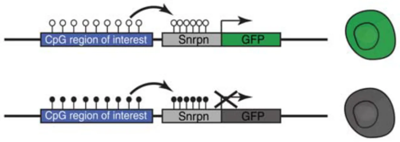
Previously, Stelzer et al. in the Jaenisch lab generated a genomic reporter of DNA methylation (RGM) with the aim to trace dynamic changes of methylation locus-specifically in single cells. The RGM consists of a methylation sensitive Snrpn minimal promoter driving the expression of a fluorescent protein. The reporter construct can be readily inserted into T-DMR of interest and the methylation status of the Snrpn promoter follows that of the surrounding genomic loci. If unmethylated, the RGM drives fluorescent protein expression in a single cell (Figure 5). The faithfulness of the RGM in reporting endogenous genomic DNA methylation has been validated in different genetic elements such as CGI promoters145, imprinting control regions (ICR)146, retro-transposable elements (unpublished), and enhancers145. The fluorescent signal allows flow cytometry assisted sorting (FACS) to purify cells with a defined DNA methylation state at a particular locus, as well as real-time monitoring of the changes of DNA methylation both in vitro and in vivo. Especially for rare populations of cells that exhibit different methylation patterns different from the majority, the resolution given by RGM is superior to current existing sequencing-based technologies. For example, parent-of-origin imprinting is thought to be faithfully maintained in adult somatic tissues147. Using RGM, loss of imprinting pattern at the Dlk1-Dio IG DMR was found in multiple adult somatic tissues, which also leads to corresponding cell-type specific allelic transcription of genes controlled by this ICR146. Compared to other methylation analysis methods, RGM provides continuous tracing of locus-specific methylation changes in live cells on the allelic level. The basis, regulation and functional impacts of DNA methylation at tissue-specific enhancers and its intricate relationship with histone
modification, TF binding and transcription still remain to be elucidated at the mechanistic level, and the RGM system provides an attractive approach to address this question in single cells and on individual alleles.
Figure 5. RGM Reporter Translates Genomic DNA Methylation Information into Fluorescent Signal in Single Cells. Stelzer et al. CSH Symposia on Quantitative Biology, 2015.
1.9 Why Is Enhancer DNA Methylation Heterogenous and What Does It Mean Functionally?
Analysis of bulk-WGBS and scWGBS data all pointed to the fact that enhancer methylation is heterogeneous and that the LMRs created by such heterogeneity being an enhancer-specific feature has functional impact on transcription. To test this hypothesis and to further investigate its functional implication requires separation of cells with heterogenous methylation status at enhancers, which is a task the RGM can fulfill. Mouse ESCs are developmentally equivalent cells which have shown DNA methylation heterogeneity at enhancer-associated T-DMRs. The high homologous recombination rate of mESCs allows efficient gene targeting and the self-renewal and differentiation potentials allow examination of enhancer DNA methylation during
dynamic cell-fate changes. As two alleles of enhancers could have non-synchronized activity and epigenetic states, I created allele-specific RGM targeted mESCs and transgenic animals at pluripotency super-enhancers Sox2 and Mir290. The aim of my thesis is to answer the following questions: (1) Is DNA methylation heterogeneity the molecular basis of the low-to-intermediate level of enhancer methylation in mESCs? (2) If so, what creates DNA methylation heterogeneity at super-enhancers. (3) What is the functional impact of enhancer methylation heterogeneity and does it exist in the developing mouse embryos?
References
1 Holliday, R. & Pugh, J. E. DNA modification mechanisms and gene activity during development. Science 187, 226-232 (1975).
2 Deaton, A. M. & Bird, A. CpG islands and the regulation of transcription. Genes Dev 25, 1010-1022, doi:10.1101/gad.2037511 (2011).
3 Jones, P. A. Functions of DNA methylation: islands, start sites, gene bodies and beyond. Nat Rev Genet 13, 484-492, doi:10.1038/nrg3230 (2012).
4 Feng, S. H. et al. Conservation and divergence of methylation patterning in plants and animals. P Natl Acad Sci USA 107, 8689-8694, doi:10.1073/pnas.1002720107 (2010).
5 Stadler, M. B. et al. DNA-binding factors shape the mouse methylome at distal regulatory regions. Nature 480, 490-495, doi:10.1038/nature10716 (2011). 6 Ziller, M. J. et al. Charting a dynamic DNA methylation landscape of the human
genome. Nature 500, 477-481, doi:10.1038/nature12433 (2013).
7 Elliott, G. et al. Intermediate DNA methylation is a conserved signature of genome regulation. Nat Commun 6, doi:Artn 636310.1038/Ncomms7363 (2015).
8 West, A. G., Gaszner, M. & Felsenfeld, G. Insulators: many functions, many mechanisms. Genes Dev 16, 271-288, doi:10.1101/gad.954702 (2002).
9 Cockburn, K. & Rossant, J. Making the blastocyst: lessons from the mouse. J Clin Invest 120, 995-1003, doi:10.1172/JCI41229 (2010).
10 Messerschmidt, D. M., Knowles, B. B. & Solter, D. DNA methylation dynamics during epigenetic reprogramming in the germline and preimplantation embryos. Genes Dev 28, 812-828, doi:10.1101/gad.234294.113 (2014).
11 Zernicka-Goetz, M., Morris, S. A. & Bruce, A. W. Making a firm decision: multifaceted regulation of cell fate in the early mouse embryo. Nat Rev Genet 10, 467-477, doi:10.1038/nrg2564 (2009).
12 Smallwood, S. A. & Kelsey, G. De novo DNA methylation: a germ cell perspective. Trends Genet 28, 33-42, doi:10.1016/j.tig.2011.09.004 (2012).
13 Hon, G. C. et al. Epigenetic memory at embryonic enhancers identified in DNA methylation maps from adult mouse tissues. Nat Genet 45, 1198-U1340, doi:10.1038/ng.2746 (2013).
14 Reizel, Y. et al. Postnatal DNA demethylation and its role in tissue maturation. Nat Commun 9, 2040, doi:10.1038/s41467-018-04456-6 (2018).
15 Saitou, M., Kagiwada, S. & Kurimoto, K. Epigenetic reprogramming in mouse pre-implantation development and primordial germ cells. Development 139, 15-31, doi:10.1242/dev.050849 (2012).
16 Smith, Z. D. et al. DNA methylation dynamics of the human preimplantation embryo. Nature 511, 611-615, doi:10.1038/nature13581 (2014).
17 Seisenberger, S. et al. The Dynamics of Genome-wide DNA Methylation Reprogramming in Mouse Primordial Germ Cells. Mol Cell 48, 849-862, doi:10.1016/j.molcel.2012.11.001 (2012).
18 Keller, G. Embryonic stem cell differentiation: emergence of a new era in biology and medicine. Genes Dev 19, 1129-1155, doi:10.1101/gad.1303605 (2005). 19 Evans, M. J. & Kaufman, M. H. Establishment in culture of pluripotential cells from
mouse embryos. Nature 292, 154-156, doi:10.1038/292154a0 (1981).
20 Ying, Q. L. et al. The ground state of embryonic stem cell self-renewal. Nature 453, 519-523, doi:10.1038/nature06968 (2008).
21 Smith, Z. D. & Meissner, A. DNA methylation: roles in mammalian development. Nat Rev Genet 14, 204-220, doi:10.1038/nrg3354 (2013).
22 Biniszkiewicz, D. et al. Dnmt1 overexpression causes genomic hypermethylation, loss of imprinting, and embryonic lethality. Mol Cell Biol 22, 2124-2135, doi:10.1128/mcb.22.7.2124-2135.2002 (2002).
23 Tsumura, A. et al. Maintenance of self-renewal ability of mouse embryonic stem cells in the absence of DNA methyltransferases Dnmt1, Dnmt3a and Dnmt3b. Genes Cells 11, 805-814, doi:10.1111/j.1365-2443.2006.00984.x (2006).
24 Liao, J. et al. Targeted disruption of DNMT1, DNMT3A and DNMT3B in human embryonic stem cells. Nat Genet 47, 469-478, doi:10.1038/ng.3258 (2015). 25 Trowbridge, J. J., Snow, J. W., Kim, J. & Orkin, S. H. DNA Methyltransferase 1 Is
Essential for and Uniquely Regulates Hematopoietic Stem and Progenitor Cells. Blood 114, 163-163 (2009).
26 Fan, G. P. et al. DNA hypomethylation perturbs the function and survival of CNS neurons in postnatal animals. J Neurosci 21, 788-797 (2001).
27 Jackson-Grusby, L. et al. Loss of genomic methylation causes p53-dependent apoptosis and epigenetic deregulation. Nat Genet 27, 31-39 (2001).
28 Panning, B. & Jaenisch, R. DNA hypomethylation can activate Xist expression and silence X-linked genes. Gene Dev 10, 1991-2002, doi:DOI 10.1101/gad.10.16.1991 (1996).
29 Hattori, N. et al. Epigenetic control of mouse Oct-4 gene expression in embryonic stem cells and trophoblast stem cells. J Biol Chem 279, 17063-17069, doi:10.1074/jbc.M309002200 (2004).
30 Imamura, M. et al. Transcriptional repression and DNA hypermethylation of a small set of ES cell marker genes in male germline stem cells. Bmc Dev Biol 6, doi:Artn 3410.1186/1471-213x-6-34 (2006).
31 Fouse, S. D. et al. Promoter CpG methylation contributes to ES cell gene regulation in parallel with Oct4/Nanog, PcG complex, and histone H3K4/K27 trimethylation. Cell Stem Cell 2, 160-169, doi:10.1016/j.stem.2007.12.011 (2008).
32 Meissner, A. et al. Genome-scale DNA methylation maps of pluripotent and differentiated cells. Nature 454, 766-U791, doi:10.1038/nature07107 (2008). 33 Slieker, R. C. et al. Identification and systematic annotation of tissue-specific
differentially methylated regions using the Illumina 450k array. Epigenet Chromatin 6, doi:Artn 2610.1186/1756-8935-6-26 (2013).
34 Song, F. et al. Tissue specific differentially methylated regions (TDMR): Changes in DNA methylation during development. Genomics 93, 130-139, doi:10.1016/j.ygeno.2008.09.003 (2009).
35 Leung, D. et al. Regulation of DNA methylation turnover at LTR retrotransposons and imprinted loci by the histone methyltransferase Setdb1. P Natl Acad Sci USA 111, 6690-6695, doi:10.1073/pnas.1322273111 (2014).
36 Kobayashi, H. et al. Contribution of Intragenic DNA Methylation in Mouse Gametic DNA Methylomes to Establish Oocyte-Specific Heritable Marks. Plos Genet 8, doi:ARTN e100244010.1371/journal.pgen.1002440 (2012).
37 Jones, P. A. Functions of DNA methylation: islands, start sites, gene bodies and beyond. Nature Reviews Genetics 13, 484-492, doi:10.1038/nrg3230 (2012). 38 Ong, C. T. & Corces, V. G. Enhancer function: new insights into the regulation of
tissue-specific gene expression. Nature Reviews Genetics 12, 283-293, doi:10.1038/nrg2957 (2011).
39 Lyko, F. The DNA methyltransferase family: a versatile toolkit for epigenetic regulation. Nature Reviews Genetics 19, 81-92, doi:10.1038/nrg.2017.80 (2018). 40 Veland, N. et al. DNMT3L facilitates DNA methylation partly by maintaining
DNMT3A stability in mouse embryonic stem cells. Nucleic Acids Res 47, 152-167, doi:10.1093/nar/gky947 (2019).
41 Kaneda, M. et al. Essential role for de novo DNA methyltransferase Dnmt3a in paternal and maternal imprinting. Nature 429, 900-903, doi:10.1038/nature02633 (2004).
42 Chen, T. P., Ueda, Y., Dodge, J. E., Wang, Z. J. & Li, E. Establishment and maintenance of genomic methylation patterns in mouse embryonic stem cells by Dnmt3a and Dnmt3b. Mol Cell Biol 23, 5594-5605, doi:10.1128/Mcb.23.16.5594-5605.2003 (2003).
43 Okano, M., Bell, D. W., Haber, D. A. & Li, E. DNA methyltransferases Dnmt3a and Dnmt3b are essential for de novo methylation and mammalian development. Cell 99, 247-257, doi:Doi 10.1016/S0092-8674(00)81656-6 (1999).
44 Baubec, T. et al. Genomic profiling of DNA methyltransferases reveals a role for DNMT3B in genic methylation. Nature 520, 243-U278, doi:10.1038/nature14176 (2015).
45 Gu, T. P. et al. DNMT3A and TET1 cooperate to regulate promoter epigenetic landscapes in mouse embryonic stem cells. Genome biology 19, doi:ARTN 8810.1186/s13059-018-1464-7 (2018).
46 Petell, C. J. et al. An epigenetic switch regulates de novo DNA methylation at a subset of pluripotency gene enhancers during embryonic stem cell differentiation. Nucleic Acids Res 44, 7605-7617, doi:10.1093/nar/gkw426 (2016).
47 Walton, E. L., Francastel, C. & Velasco, G. Maintenance of DNA methylation Dnmt3b joins the dance. Epigenetics-Us 6, 1373-1377, doi:10.4161/epi.6.11.17978 (2011).
48 Feng, J. et al. Dnmt1 and Dnmt3a maintain DNA methylation and regulate synaptic function in adult forebrain neurons. Nat Neurosci 13, 423-U437, doi:10.1038/nn.2514 (2010).
49 Vilkaitis, G., Suetake, I., Klimasauskas, S. & Tajima, S. Processive methylation of hemimethylated CpG sites by mouse Dnmt1 DNA methyltransferase. J Biol Chem 280, 64-72, doi:10.1074/jbc.M411126200 (2005).
50 Bostick, M. et al. UHRF1 plays a role in maintaining DNA methylation in mammalian cells. Science 317, 1760-1764, doi:10.1126/science.1147939 (2007).
51 Jeltsch, A. & Jurkowska, R. Z. New concepts in DNA methylation. Trends Biochem Sci 39, 310-318, doi:10.1016/j.tibs.2014.05.002 (2014).
52 Piccolo, F. M. & Fisher, A. G. Getting rid of DNA methylation. Trends Cell Biol 24, 136-143, doi:10.1016/j.tcb.2013.09.001 (2014).
53 Cardoso, M. C. & Leonhardt, H. DNA methyltransferase is actively retained in the cytoplasm during early development. J Cell Biol 147, 25-32, doi:DOI 10.1083/jcb.147.1.25 (1999).
54 Kagiwada, S., Kurimoto, K., Hirota, T., Yamaji, M. & Saitou, M. Replication-coupled passive DNA demethylation for the erasure of genome imprints in mice. Embo Journal 32, 340-353, doi:10.1038/emboj.2012.331 (2013).
55 Marks, H. et al. The Transcriptional and Epigenomic Foundations of Ground State Pluripotency. Cell 149, 590-604, doi:10.1016/j.cell.2012.03.026 (2012). 56 von Meyenn, F. et al. Impairment of DNA Methylation Maintenance Is the Main
Cause of Global Demethylation in Naive Embryonic Stem Cells (vol 62, pg 848, 2016). Mol Cell 62, 983-983, doi:10.1016/j.molcel.2016.06.005 (2016).
57 Oda, M., Oxley, D., Dean, W. & Reik, W. Regulation of lineage specific DNA hypomethylation in mouse trophectoderm. PloS one 8, e68846, doi:10.1371/journal.pone.0068846 (2013).
58 Bhutani, N., Burns, D. M. & Blau, H. M. DNA demethylation dynamics. Cell 146, 866-872, doi:10.1016/j.cell.2011.08.042 (2011).
59 Wu, X. & Zhang, Y. TET-mediated active DNA demethylation: mechanism, function and beyond. Nat Rev Genet 18, 517-534, doi:10.1038/nrg.2017.33 (2017).
60 Kohli, R. M. & Zhang, Y. TET enzymes, TDG and the dynamics of DNA demethylation. Nature 502, 472-479, doi:10.1038/nature12750 (2013).
61 Oswald, J. et al. Active demethylation of the paternal genome in the mouse zygote. Curr Biol 10, 475-478, doi:10.1016/s0960-9822(00)00448-6 (2000). 62 Kangaspeska, S. et al. Transient cyclical methylation of promoter DNA. Nature
452, 112-115, doi:10.1038/nature06640 (2008).
63 Metivier, R. et al. Cyclical DNA methylation of a transcriptionally active promoter. Nature 452, 45-50, doi:10.1038/nature06544 (2008).
64 Guo, J. U., Su, Y., Zhong, C., Ming, G. L. & Song, H. Hydroxylation of 5-methylcytosine by TET1 promotes active DNA demethylation in the adult brain. Cell 145, 423-434, doi:10.1016/j.cell.2011.03.022 (2011).
65 Mellen, M., Ayata, P. & Heintz, N. 5-hydroxymethylcytosine accumulation in postmitotic neurons results in functional demethylation of expressed genes. Proc Natl Acad Sci U S A 114, E7812-E7821, doi:10.1073/pnas.1708044114 (2017).
66 Hon, G. C. et al. 5mC Oxidation by Tet2 Modulates Enhancer Activity and Timing of Transcriptome Reprogramming during Differentiation. Mol Cell 56, 286-297, doi:10.1016/j.molcel.2014.08.026 (2014).
67 Huang, Y. et al. Distinct roles of the methylcytosine oxidases Tet1 and Tet2 in mouse embryonic stem cells. Proc Natl Acad Sci U S A 111, 1361-1366, doi:10.1073/pnas.1322921111 (2014).
68 Lu, F., Liu, Y., Jiang, L., Yamaguchi, S. & Zhang, Y. Role of Tet proteins in enhancer activity and telomere elongation. Genes Dev 28, 2103-2119, doi:10.1101/gad.248005.114 (2014).
69 Dawlaty, M. M. et al. Loss of Tet enzymes compromises proper differentiation of embryonic stem cells. Developmental cell 29, 102-111, doi:10.1016/j.devcel.2014.03.003 (2014).
70 Bird, A. P. CpG-rich islands and the function of DNA methylation. Nature 321, 209-213, doi:10.1038/321209a0 (1986).
71 Farthing, C. R. et al. Global mapping of DNA methylation in mouse promoters reveals epigenetic reprogramming of pluripotency genes. Plos Genet 4, e1000116, doi:10.1371/journal.pgen.1000116 (2008).
72 De Smet, C., Lurquin, C., Lethe, B., Martelange, V. & Boon, T. DNA methylation is the primary silencing mechanism for a set of germ line- and tumor-specific genes with a CpG-rich promoter. Mol Cell Biol 19, 7327-7335 (1999).
73 Kelly, T. K. et al. H2A.Z maintenance during mitosis reveals nucleosome shifting on mitotically silenced genes. Molecular cell 39, 901-911, doi:10.1016/j.molcel.2010.08.026 (2010).
74 Cedar, H. & Bergman, Y. Linking DNA methylation and histone modification: patterns and paradigms. Nature Reviews Genetics 10, 295-304, doi:10.1038/nrg2540 (2009).
75 Newell-Price, J., Clark, A. J. L. & King, P. DNA methylation and silencing of gene expression. Trends Endocrin Met 11, 142-148, doi:Doi 10.1016/S1043-2760(00)00248-4 (2000).
76 Mohn, F. et al. Lineage-specific polycomb targets and de novo DNA methylation define restriction and potential of neuronal progenitors. Molecular cell 30, 755-766, doi:10.1016/j.molcel.2008.05.007 (2008).
77 Lock, L. F., Takagi, N. & Martin, G. R. Methylation of the Hprt gene on the inactive X occurs after chromosome inactivation. Cell 48, 39-46, doi:10.1016/0092-8674(87)90353-9 (1987).
78 Sharp, A. J. et al. DNA methylation profiles of human active and inactive X chromosomes. Genome Res 21, 1592-1600, doi:10.1101/gr.112680.110 (2011). 79 Beard, C., Li, E. & Jaenisch, R. Loss of methylation activates Xist in somatic but
not in embryonic cells. Genes Dev 9, 2325-2334, doi:10.1101/gad.9.19.2325 (1995).
80 Ball, M. P. et al. Targeted and genome-scale strategies reveal gene-body methylation signatures in human cells (vol 27, pg 361, 2009). Nat Biotechnol 27, 485-485, doi:10.1038/nbt0509-485b (2009).
81 Yang, X. J. et al. Gene Body Methylation Can Alter Gene Expression and Is a Therapeutic Target in Cancer. Cancer Cell 26, 577-590, doi:10.1016/j.ccr.2014.07.028 (2014).
82 Rauch, T. A., Wu, X. W., Zhong, X., Riggs, A. D. & Pfeifer, G. P. A human B cell methylome at 100-base pair resolution. P Natl Acad Sci USA 106, 671-678, doi:10.1073/pnas.0812399106 (2009).
83 Arechederra, M. et al. Hypermethylation of gene body CpG islands predicts high dosage of functional oncogenes in liver cancer (vol 9, 3164, 2018). Nat Commun 9, doi:ARTN 397610.1038/s41467-018-06482-w (2018).
84 Maunakea, A. K. et al. Conserved role of intragenic DNA methylation in regulating alternative promoters. Nature 466, 253-257, doi:10.1038/nature09165 (2010).
85 Illingworth, R. S. et al. Orphan CpG islands identify numerous conserved promoters in the mammalian genome. Plos Genet 6, e1001134, doi:10.1371/journal.pgen.1001134 (2010).
86 Nguyen, C. T., Gonzales, F. A. & Jones, P. A. Altered chromatin structure associated with methylation-induced gene silencing in cancer cells: correlation of accessibility, methylation, MeCP2 binding and acetylation. Nucleic Acids Res 29, 4598-4606, doi:DOI 10.1093/nar/29.22.4598 (2001).
87 Chen, Y. B., Chen, Q., McEachin, R. C., Cavalcoli, J. D. & Yu, X. C. H2A.B facilitates transcription elongation at methylated CpG loci. Genome research 24, 570-579, doi:10.1101/gr.156877.113 (2014).
88 Neri, F. et al. Intragenic DNA methylation prevents spurious transcription initiation. Nature 543, 72-+, doi:10.1038/nature21373 (2017).
89 Aporntewan, C. et al. Hypomethylation of Intragenic LINE-1 Represses Transcription in Cancer Cells through AGO2. PloS one 6, doi:ARTN e1793410.1371/journal.pone.0017934 (2011).
90 Tufarelli, C. et al. Transcription of antisense RNA leading to gene silencing and methylation as a novel cause of human genetic disease. Nat Genet 34, 157-165, doi:DOI 10.1038/ng1157 (2003).
91 Lister, R. et al. Human DNA methylomes at base resolution show widespread epigenomic differences. Nature 462, 315-322, doi:10.1038/nature08514 (2009). 92 Fleischer, T. et al. DNA methylation at enhancers identifies distinct breast cancer
lineages. Nat Commun 8, doi:Artn 137910.1038/S41467-017-00510-X (2017). 93 Aran, D. & Hellman, A. DNA Methylation of Transcriptional Enhancers and
Cancer Predisposition. Cell 154, 11-13, doi:10.1016/j.cell.2013.06.018 (2013). 94 Bock, C. et al. DNA Methylation Dynamics during In Vivo Differentiation of Blood
and Skin Stem Cells. Mol Cell 47, 633-647, doi:10.1016/j.molcel.2012.06.019 (2012).