ORIGINAL PAPER
Aging the oldest turtles: the placodont affinities
of Priscochelys hegnabrunnensis
Torsten M. Scheyer
Received: 21 January 2008 / Revised: 1 April 2008 / Accepted: 3 April 2008 / Published online: 9 May 2008
# Springer-Verlag 2008
Abstract Priscochelys hegnabrunnensis, a fragmentary piece of armour shell from the Muschelkalk of Germany (Upper Triassic) with few diagnostic morphological fea-tures, was recently proposed to represent the oldest known stem turtle. As such, the specimen is of high importance because it shifts the date of the first appearance of turtles back about 20 Ma, which equals about 10% of the total stratigraphic range of the group. In this paper, I present new morphologic, histologic and neutron tomographic (NT) data that relate to the microstructure of the bone of the specimen itself. In opposition to the previous morphologic descrip-tions, P. hegnabrunnensis was found to share several distinctive features (i.e. bone sutures congruent with scute sulci, absence of a diploe structure with interior cancellous bone, thin vascular canals radiating outwards from distinct centres in each field and rugose ventral bone surface texture consisting of mineralised fibre bundles) with cyamodontoid placodonts (Diapsida: Sauropterygia) and fewer with stem turtles (i.e. depth of sulci). Two aspects that were previously thought to be relevant for the assignment to the turtle stem (conical scutes and presence of foramina) are argued to be of dubious value. P. hegnabrunnensis is proposed to represent a fragmentary piece of cyamodontoid armour consisting of fused conical plates herein. The specimen is not a part of the turtle stem and thus does not represent the oldest turtle. Accordingly, P. hegnabrunnensis does not shorten the ghost lineage to the potential sister group of turtles.
Keywords Placodontia . Testudinata .
Neutron tomography . Bone histology . Stratigraphic gap
Introduction
Fossils are of great importance to identify the stratigraphic age, range and dates of the first appearance of extant (crown) taxa. The dating of the Tree of Life increasingly relies on the usage of molecular clocks, but estimates remain of limited use if they are not well calibrated (Brochu 2004; Benton and Donoghue2007; Marjanović and Laurin 2007), which is usually done by the first appearance of members of a (crown) group in the fossil record (e.g. Müller and Reisz 2005). Lately, morphological and molec-ular analyses have been presented that hypothesise turtles as being diapsid reptiles, although it is still under debate if turtles have closer affinities to archosaurs or lepidosaurs (e.g. Kumazawa and Nishida1999; Müller2003; Rest et al. 2003; Iwabe et al.2004; Hill2005; Scheyer2007a). Others view turtles as parareptiles (Laurin and Reisz, 1995; Lee, 2001), and at least one recent molecular phylogeny is compatible with this position (Frost et al. 2006). This debate on the origin of turtles so far prevented turtles from being included as calibration points in analyses of more inclusive taxa. Once this controversy is settled, the role of turtles may increase in dating the Tree of Life.
The recovery of new fossil representatives of the turtle stem can potentially shorten the stratigraphic gap, expressed by a ghost lineage, to the potential sister group of turtles, independent of the discussion about the sister group relationships. Recently, a purported new species of basal turtle, Priscochelys hegnabrunnensis Karl,2005, was described based on a single specimen (holotype SMNS 80141) from the Upper Muschelkalk (Lower Ladinian, Communicated by G. Mayr
T. M. Scheyer (*)
Paläontologisches Institut und Museum, Universität Zürich, Karl Schmid-Strasse 4,
CH-8006 Zürich, Switzerland e-mail: [email protected]
Middle Triassic) of southern Germany. Previously, all other basal turtles were exclusively known from the Upper Triassic (e.g. Joyce2007). P. hegnabrunnensis thus would push back the stratigraphic age of turtles by about 20 million years. At that time, the coastal regions of the Muschelkalk sea were inhabited by placodont reptiles (Diapsida: Sauropterygia), i.e. cyamodontoid placodonts that superficially resemble turtles in encasing their body in extensive armour shells (e.g. Rieppel 2002), raising the possibility that SMNS 80141 represents a fragment of cyamodontoid placodont armour.
As stated in the re-description of the holotype (Joyce and Karl 2006) that significantly improved upon the original description by Karl (2005), the specimen consists of a highly fragmentary piece of bony armour with many glued and cast areas, which has a pattern of scute impressions (sulci) superimposed on the external bone surface. The presence and arrangement of these shield impressions, supposedly representing the sulci of supra-marginals, led to the hypothesis that the specimen is the oldest and probably most basal member of Testudinata (sensu Joyce et al.2004).
Whereas extant vertebrates can be studied with a great variety of methods to gather, for example, physiological, developmental and molecular data, the data gleaned from fossils are largely restricted to outer morphology. In case of SMNS 80141, (Joyce and Karl2006: 106) thus concluded that, although the“anatomical identification remains uncer-tain” and “it is impossible to unambiguously orient the specimen,” there is no morphological data that contradict an assignment to Testudinata.
Bone histology has established itself as a second, powerful method to analyse fossils, thus providing the possibility to verify morphology-based hypotheses. Espe-cially in poorly known material lacking diagnostic features, i.e. fragmentary osteoderms and shell armour like SMNS 80141, the bone microstructure provides valuable data that can be used to verify and resolve phylogenetic hypotheses (e.g. Hill2005; Scheyer et al.2007). This approach is also validated by the presence of a phylogenetic signal in bone histological and microanatomical characters (Laurin et al. 2004; Cubo et al. 2005).
The aim of this paper is to review the description of SMNS 80141 given by Joyce and Karl (2006) and to provide details on outer morphology and the inner bone microstructure that were previously not assessed. Addition-al non-invasive neutron tomography (NT) images were used to determine whether the previous categorisation of SMNS 80141 as the oldest member of the turtle stem is valid. As was recently argued by Schwarz et al. (2005), one major advantage of NT for visualisation in vertebrate fossils in contrast to the well-established X-ray computed tomog-raphy technique (e.g. Clarke et al. 2005; Balanoff and
Rowe 2007) is the excellent detection of fractured, glued and cast areas within fossils (i.e. the re-modelled areas in SMNS 80141).
Materials and methods
The holotype and single specimen of P. hegnabrunnensis, SMNS 80141, was studied with a special focus on the interior structure of the bone. As invasive analysis by thin sectioning, the standard method applied in studying bone histology, was not appropriate in this case, natural breaks and cuts of SMNS 80141 were analysed instead with a LEICA stereo-microscope MZ16 and were then compared with histological and anatomical features of turtle shell bones and placodont armour plates (species summarised in Scheyer2007a,b).
Additionally, NT scans were done at the Neutron Trans-mission Radiography Station NEUTRA, Paul-Scherrer-Institute (PSI) at Villingen, Switzerland. The specimen was mounted upright on a rotary table (position of analysis: 3, see Vontobel et al. 2003) between the neutron emitter and scintillator screen. Images were taken with a Peltier-cooled 1,024×1,024-pixel charge-coupled device camera (DV434; Andor Technology). The rotation of the specimen over 180° and processing of the data set with OCTOPUS 8.1 resulted in a total image stack of 673 slices (974×974 pixels; approximate slice distance 0.104 mm; isometric voxels). Detailed descriptions of the method of NT scanning and the NEUTRA facility at the PSI were published by Vontobel et al. (2003) and Schwarz et al. (2005). The 3D volumetric reconstruction of the image stack and segmentation of the volumetric reconstruction was done in AMIRA 4.1. NT scan images were further processed in Adobe Photoshop. Geological setting The geological setting and the palaeo-ecological significance of P. hegnabrunnensis have been discussed extensively in Joyce and Karl (2006). According to these authors, P. hegnabrunnensis did not necessarily live in a marine environment, even though the specimen was found in marine Muschelkalk sediments.
Terminology The microscopic terminology used herein is mainly based on Scheyer and Sander (2007) and Scheyer (2007a). Placodont-specific terminology is based on Rieppel (2002) and (Scheyer2007b).
Institutional abbreviations The following abbreviations for the institutions were used in this study: MAGNT, Museum and Art Gallery of the Northern Territory, Darwin, Australia; MB, Naturhistorisches Forschungsinstitut and Museum für Naturkunde, Humboldt-Universität zu Berlin, Germany; MHI, Muschelkalkmuseum Hagdorn Ingelfingen, Germany;
SMNS, Staatliches Museum für Naturkunde in Stuttgart, Germany.
Results
The specimen SMNS 80141 was thoroughly described and discussed by Joyce and Karl (2006), and if not stated otherwise, I agree with their interpretations. This paper focuses on the bone microstructure and on new interpreta-tions. I agree with Joyce and Karl (2006) that the sculptured surface represents the dorsal side and the flatter surface the ventral side of the specimen. Furthermore, for comparative reasons, I adopt their orientation of the specimen (Fig.1) and its division in six separate fields, marked by Roman numerals I–VI (Joyce and Karl2006: Fig. 2).
Interior bone structures NT reveals areas of SMNS 80141 that are filled with glue or plaster, especially the small fractures in field VI and the large plaster fillings between field VI and fields IV and V (Figs. 1a–c and 2a–c). All fields have a compact interior bone structure. The bone centre is devoid of larger or longer bone trabeculae; thus, a spongy diploe structure has not been found anywhere in the specimen. The bone has little vascularisation, with only few large vascular canals that are mainly restricted to the innermost regions of SMNS 80141. In addition to what was deduced from bone surface breaks, the interior vascular canals radiate outward from specific centres (Fig.1a–c) in each field. The distinct vascular centres are situated in the central interior areas of the smaller fields (I–V) and interior to the‘pronounced central tubercles’ of field I–IV (Joyce and Karl2006: 107). In field VI, the interior canals radiate from a centre adjacent to the median anterior margin towards the posterior margins where field VI contacts fields I–V (Fig. 1a–c). The interior centre of radiating vascular canals is most pronounced in the posterior part of field VI. Dorsal bone surface structure Next to the presumably postmortem pockmark structure mentioned by Joyce and Karl (2006), the dorsal bone surface is mostly smooth. In few small areas in field VI, deeper compact bone layers and their vascular patterns are visible (Fig. 3a). Few vascular canals extend dorsoventrally through the compact bone and may lead to small foramina on the dorsal bone surface. Far more frequently, canals are parallel to the surface. These canals converge slightly towards the anterior-most part of field VI. In fields III and IV, vascular canals are mostly small and dorsoventrally oriented.
Ventral surface structures The ventral surface of SMNS 80141 is heterogeneous in that field V shows a deeper
Fig. 1 Dorsal view of P. hegnabrunnensis (SMNS 80141). Fields of the specimen are marked with roman numerals I–VI according to Joyce and Karl (2006). a–c Three dorsoventrally arranged NT images that section horizontally through the specimen. In the NT images, the cast and glued areas in the specimen appear in light colours, whereas the bone tissue appears in grey scale. In a, the white arrow indicates the centre of vascularisation in the large field VI from which small vascular canals radiate outwards towards the margins of the field. In b and c, the radiating canals of fields VI and II are visible, respectively. d–f The positions of three representative sagittal NT images that section the specimen obliquely are indicated here (a–c in Fig.2)
level of bone tissue that is not observable in fields I–IV and VI. In the anterior part of field V, the bone tissue shows faint growth marks (Fig. 3b). Numerous sub-parallel thin vascular canals are perpendicular to the growth marks of the bone tissue thus creating a radiating pattern (Fig. 3b). The vascular canals extend towards the sulcus between fields V and VI. The ventral surface of fields I–IV and VI shows a rugose bone structure, based on the relief of a meshwork of randomly oriented mineralised fibre bundles (Fig. 3c). Between the meshwork, small foramina lead to canals that extend into the bone tissue (Fig.3d). No smooth compact bone layer is encountered in any of the fields. Delineation of the dorsal fields is faint at best in ventral view.
‘Marginal bone surface structures’ Dorsally, the fields I– IV and VI show narrow, flat marginal depressions inter-preted as scute sulci. Field V lacks sulci as its margins are either completely broken off or reconstructed. Sulci are most complete in fields II and III. In dorsal view, a light ‘striation pattern’ of the bone is observable in the sulci where fields contact each other (i.e., where fields I–III meet field VI) and at the free margins of the fields (I–IV). In lateral view, the free margins exhibit thin protruding vertical bone lamellae, especially in fields II–IV (Fig.3e).
At the anterior margin of field VI, the bone exhibits a low ridge, causing the anterior-most plate margin to curve
ventrally; the lateral margin of the field is thus visible in ventral view. The bone shows also some slight ‘striation’ here; however, this may result from erosive or preparatory artefacts because several evenly spaced holes posterior and directly adjacent to the curved margin are visible (Fig. 3f).
Discussion
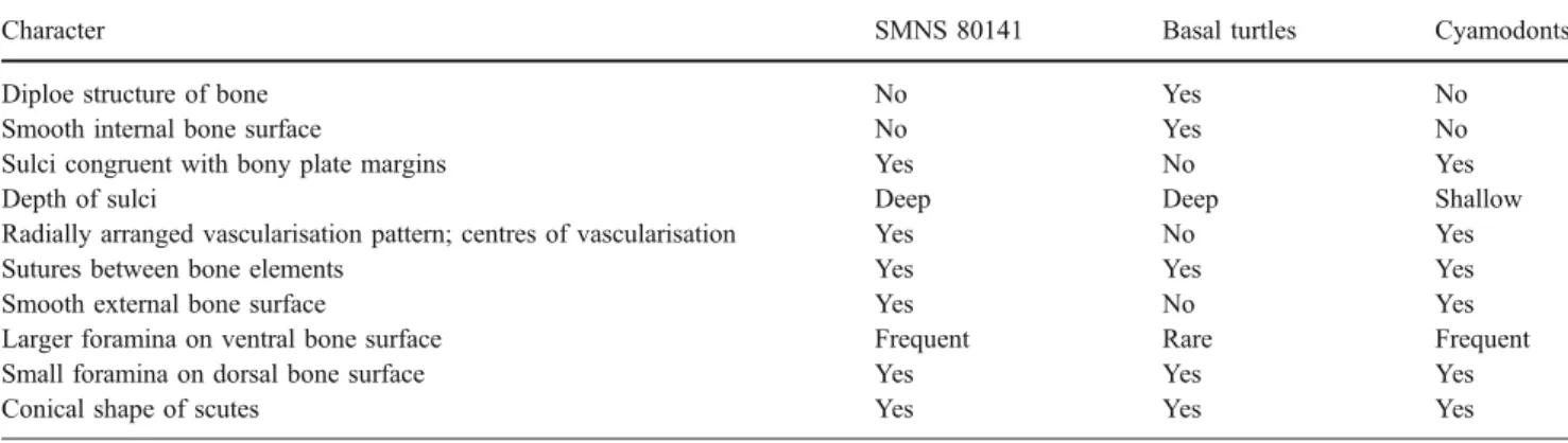
(Joyce and Karl2006: 108) summarised that“SMNS 80141 lacks bony sutures, exhibits irregularly sized, conical scutes, and highly distinct sulci,” leading to the conclusion that P. hegnabrunnensis, represented only by the holotype, is the oldest basal turtle (i.e. a stem turtle). In the following, I will list and discuss the points (marked by quotes) that lead to their original assignment to Testudinata in light of novel data on the bone microstructure of SMNS 80141. Note that some of the original points have been combined or simplified. Histological results are summarised in Table1.
‘Ventral surface rather smooth, lack of notable features’ I agree with Joyce and Karl (2006) that the ventral surface is rather smooth in that it lacks any sculpturing or the presumably postmortem pockmarks of the dorsal surface. On a finer scale, however, the ventral surface of the specimen is not smooth but rather rugose as seen in Fig. 3c,d, an obvious morphological feature that has not been taken into account before. There is also no indication that a smooth compact bony layer once covered the rugose pattern, as neither field carries a remnant of such a bone tissue. Moreover, the meshwork of fibre bundles is largely unaffected by erosion. This is in accordance with Joyce and Karl’s (2006: 107) assessment that the ventral surface of SMNS 80141 was somehow ‘protected from scavenging.’ It is therefore argued here that the rugose bone surface structure is a primary feature instead of an erosive or preparatory artefact.
The presence or absence of this feature is important because a (fine-scale) smooth bone surface usually indi-cates a compact layer of parallel fibered of lamellar bone tissue. Such a compact bone layer (Fig. 3g) suggests a diploe structure as is typically found in the turtle shell, including that of stem turtles, with the exception of species that show strong to extreme adaptations to the aquatic environment (Scheyer and Sander2007). In these taxa (e.g. Dermochelyidae), the internal compact bone layer of the shell can be partly or completely reduced (Zangerl 1969; Scheyer and Sander2007; Scheyer2007a).
A rugose bone surface structure (Fig. 3h) instead of a smooth bone layer, on the other hand, was found in several cyamodontoid placodont armour plates (Scheyer 2007b), and a cross-hatching pattern of mineralised fibres has been Fig. 2 Sagittal NT images (a–c) of P. hegnabrunnensis (SMNS
80141). As is visible on all three images (positions marked in Fig.1), the bone is rather compact and lacks a distinct diploe structure, where dorsal and ventral compact bone layers frame inner cancellous bone. The interior‘spaces’ seen within the bone in section b are identified as a network of fractured and glued bone parts and do not represent areas of highly vascular cancellous bone tissue. Note radiation artefacts around the specimen are most prominent in the vicinity of those parts that are largely re-modelled
described for the ventral surfaces of plates of Cyamodus hildegardis, Psephosaurus suevicus and Psephosauriscus sp. (e.g. Haas 1969; Westphal 1976; Rieppel 2002). The rugose texture in cyamodontoid armour plates is based on a meshwork of randomly arranged mineralised fibre bundles, as in SMNS 80141.
Neutron tomography data A diploe structure as is present in turtle shell bones is not seen in the NT images (Figs. 1 and 2). Instead, the NT data reveal the presence of interior vascularisation centres and radiating vascular canals in each field of SMNS 80141, features that are well known from placodont armour plates (Westphal 1976; Scheyer 2007b). Fig. 3 Microstructures of holotype of P. hegnabrunnensis (SMNS
80141) (a–f) and comparisons with g left hyoplastron of the extant turtle Carettochelys insculpta (MAGNT R12640) and h an armour plate of the placodont Psephosaurus suevicus (MHI 1426/3). a Area of damaged bone surface of SMNS 80141 reveals small vascular canals that extend sub-parallel to the centre of the anterior margin of field VI. Few canals are oriented dorsoventrally, appearing as small foramina. b Detail of the (damaged) ventral bone surface of field V that shows finely spaced sub-parallel vascular canals that extend oblique to weakly observable bone growth layers (marked by white arrows). c Ventral bone surface structure of the posterior part of SMNS 80141 (fields II-IV, posterior part of VI). The rugose texture is caused by protruding randomly oriented mineralised fibre bundles. d
Close-up of the rugose ventral surface structure. Numerous small foramina insert between the mineralised fibre bundles into the bone interior. e Focus on the posterior margins of fields II–IV in dorsolateral view. Short vertical bone lamellae (white arrows) are interpreted as remnants of sutures between fields II and IV and additional fields that were not preserved. f Close-up of the anterior margin of field VI in ventral view. Several scattered holes (marked by white arrows) that occur directly adjacent to the bony rim of the specimen are interpreted as preparation artefacts. g Close-up of smooth internal cortical bone layer of the turtle plastron bone in ventral view. White arrows mark the location of scattered larger and smaller foramina. h Close-up of rugose internal bone surface structure of the armour plate (in ventral view) caused by protruding mineralised fibre bundles
The NT scans clearly show that not only the missing parts of the specimen were cast but also that the interior parts of the bone were strongly damaged and re-fitted together as the glued parts clearly delineate the bone fragments in the NT images. Thus, the interior glued and cast areas do not mask a diploe structure.
‘All edges of element broken, no presence of sutures’ Joyce and Karl (2006) interpreted all margins of SMNS 80141 as showing ‘signs of mechanical breakage,’ and the lack of sutures is one of the main reasons why SMNS 80141 is interpreted as a basal turtle carrying an extra row of supramarginal scutes, with fields I–V tentatively assigned to represent supramarginals 9–13 (Joyce and Karl 2006: Fig. 3). The ‘striation’ patterns, which are not superficial structures but are encountered in all sulci of the dorsal bone surface and at the lateral margins of fields II–IV (see Fig. 3e,f), argue against the mechanical breakage of the margins.
Although the fields appear to be completely fused, the interior ‘striation’ extending perpendicular to the dorsal sulci probably represents former sutures between fields. Especially between fields I and IV, the‘striation’ is visible macroscopically as well as in tomographic images. Ac-cordingly, if sutures are hypothesised to be present between all fields, the dorsal scute sulci would coincide directly with the sutures. Such a coincidence of sutures and scute sulci is typically not found in turtle shells (e.g. Zangerl1969; Joyce 2007). On the other hand, the congruence of bony plate margins and scute boundaries has been described and figured for specimen SMNS 7113 of the placodont Psephosaurus suevicus (Rieppel2002: Fig. 21A1). ‘Fusion of elements’ The fusion of individual turtle bones, thought to indicate maturity in basal turtles (Joyce and Karl 2006), usually involves a complete re-modelling of interior bone structures; thus, the sutures become indistinguishable (e.g. Scheyer 2007a). Sutures between individual
cyamo-dontoid armour plates are usually preserved throughout ontogeny (see Rieppel 2002). However, diagenetic and taphonomic effects may obscure delineations of plates, which may resemble fused elements of turtles or cyamo-dont shells (personal observation). Again, as noted above, the ‘striation’ patterns found at the margins of the fields (see Fig. 3e) are taken as indicators for the presence of initial sutures between the fields of SMNS 80141.
Bone tissue visible on the ventral surface of field V The interior radial vascularisation pattern of field V occurs because small vascular canals radiate from a vascular centre, which is not preserved, towards the margins of field V. A similar bone structure was recently described in the cortical bone of specimen SMNS 91009 of the placodont Psephosaurus sp. (Scheyer 2007b: Fig. 3), whereas such a bone tissue and vascularisation pattern was not encountered in turtle shell bones (Scheyer 2007a).
‘Depth of sulci’ Based on the depth of the sulci of SMNS 80141, Joyce and Karl (2006) concluded that it is plausible to assume that the specimen represents a turtle instead of a placodont where sulci are usually shallower (Rieppel2002). The bone histology does not provide additional data, so the depth of sulci seen in SMNS 80141 could be interpreted as a contradictory point to the new hypothesis presented herein.
‘Scutes of all placodonts are rather flat, those of primitive turtles are somewhat conical’ In turtles, keratinous scute morphologies can roughly be inferred by the underlying bone surfaces, so many basal turtles do have somewhat conical scutes. The same applies to cyamodontoid armour plates, however, which include procumbent and recumbent spiked armour plate morphologies. The large majority of hexagonal plates may indeed have been covered by rather flat scutes; however, there is no indication why conical or spiked plates should be covered by flat instead of conical Table 1 Comparison of bone histological and morphological details in P. hegnabrunnensis (SMNS 80141), basal turtles (i.e. Proganochelys quenstedti, SMNS 17203 and MB.R. 3449.2; Proterochersis robusta, SMNS 16442) and derived cyamodontoid placodonts
Character SMNS 80141 Basal turtles Cyamodonts
Diploe structure of bone No Yes No
Smooth internal bone surface No Yes No
Sulci congruent with bony plate margins Yes No Yes
Depth of sulci Deep Deep Shallow
Radially arranged vascularisation pattern; centres of vascularisation Yes No Yes
Sutures between bone elements Yes Yes Yes
Smooth external bone surface Yes No Yes
Larger foramina on ventral bone surface Frequent Rare Frequent
Small foramina on dorsal bone surface Yes Yes Yes
scutes so this character remains ambiguous. In the placodont Placochelys placodonta, the conical shape of larger and smaller bony armour plates is well visible, indicating that this taxon also had conical scutes similar to basal turtles (Jaekel1907: plate 9, Figs. 1–3).
‘Absence of foramina’ Smaller (mostly on dorsal bone surface) and numerous larger foramina (ventrally) were found on the bone surface of SMNS 80141. Foramina are known from turtle shell bones (e.g. Scheyer2007a,b; Joyce and Bell2004: Figs. 108, 109) as well as from placodont armour plates (Rieppel 2002; Scheyer 2007b; personal observation). Due to the lack of quantitative data on foramina occurrence, no clear assignment to either turtles or placodonts is possible.
To summarise, apart from the greater depth of scute sulci, the outer morphology is only weakly supporting turtle affinities. On the other hand, there is overwhelming data, mainly based on new fine-scale morphological details and bone histological observations, which support a relationship of P. hegnabrunnensis with placodont reptiles.
Conclusions
Given the new evidence and interpretations presented herein and taking into account the fragmentary nature of SMNS 80141 as the oldest basal turtle, it is most plausible that the specimen represents a fragmentary piece of armour of a cyamodontoid placodont rather than the oldest known turtle. Fields I–V are hypothesised to represent fused, once separately sutured armour plates instead of supramarginal scutes of a basal turtle shell.
Based on the overall shape of SMNS 80141 and the presence of the peculiar striation patterns in the bone tissue caused by distinct centres of radial vascularisation, it is further proposed that P. hegnabrunnensis is related to the slightly younger cyamodontoid genus Psephosaurus Fraas, 1896; the remains of which (i.e. armour fragments) are known from the Upper Ladinian of southern Germany (see Rieppel2002).
The occurrence of the Lower Ladinian P. hegnabrun-nensis does not correspond with the minimum stratigraphic age of turtles but falls well within the stratigraphic range of Triassic placodont reptiles. Until older, unambiguous fossil turtles are recovered, Proterochersis robusta Fraas, 1913 from the Early Norian, Upper Triassic, remains the oldest turtle known.
Acknowledgement Rainer Schoch (Stuttgart) is thanked for the possibility to study the holotype of P. hegnabrunnensis, as well as basal turtles and placodonts. Further turtle and placodont material was kindly provided by Gavin Dally (Darwin), Hans Hagdorn (Ingelfingen),
Thomas Mörs (Stockholm) and Martin Sander (Bonn). My gratitude goes to Eberhard Lehmann and colleagues at the Paul-Scherrer-Institute, Villingen PSI, Switzerland, for the possibility to obtain the NT scans of the specimen. Jasmina Hugi (Zürich) helped with the preparation of the NT scans. Marcelo Sánchez-Villagra (Zürich) and Martin Sander and Nicole Klein (Bonn) are thanked for discussion or reading earlier drafts of the manuscript. Olaf Dülfer (Bonn) is acknowledged for preparing thin sections and Rosi Roth (Zürich) for taking photographs. I thank Michel Laurin (Paris) and Walter Joyce (New Haven) for their constructive comments in reviewing the manuscript. The project was funded by the‘Fonds zur Förderung des akademischen Nachwuchses’ of the Universität Zürich (FAN).
References
Balanoff AM, Rowe T (2007) Osteological description of an embryonic skeleton of the extinct elephant bird, Aepyornis (Palaeognathae: Ratitae). Soc Vertebr Paleontol Memoir 9:1–53 Benton MJ, Donoghue PCJ (2007) Paleontological evidence to date
the Tree of Life. Mol Biol Evol 24(1):26–53
Brochu CA (2004) Calibration age and quartet divergence date estimation. Evolution 58:1375–1382
Clarke JA, Tambussi CP, Noriega JI, Erickson GM, Ketcham RA (2005) Definitive fossil evidence for the extant avian radiation in the Cretaceous. Nature 433:305–308
Cubo J, Ponton F, Laurin M, de Margerie E, Castanet J (2005) Phylogenetic signal in bone microstructure of sauropsids. Syst Biol 54:562–574
Fraas E (1896) Die schwäbischen Trias-Saurier nach dem Material der Kgl. Naturalien-Sammlung in Stuttgart zusammengestellt. E Schweizerbart, Stuttgart
Fraas E (1913) Proterochersis, eine pleurodire Schildkröte aus dem Keuper. Jahresh Ver Vaterl Naturkd Württemb 80:1–30 Frost DR, Grant T, Faivovich J, Bain RH, Haas A, Haddad CFB,
de Sá RO, Channing A, Wilkinson M, Donnellan SC, Raxworthy CJ, Campbell JA, Blotto BL, Moler P, Drewes RC, Nussbaum RA, Lynch JD, Green DM, Wheeler WC (2006) The amphibian tree of life. Bull Am Mus Nat Hist 297:1–370
Haas G (1969) The armour of placodonts from the Muschelkalk of Wadi Ramon (Israel). Isr J Zool 18:135–147
Hill RV (2005) Integration of morphological data sets for phylogenetic analysis of Amniota: the importance of integumentary characters and increased taxonomic sampling. Syst Biol 54(4):530–547 Iwabe N, Hara Y, Kumazawa Y, Shibamoto K, Saito Y, Miyata T,
Katoh K (2004) Sister group relationship of turtles to the bird-crocodilian clade revealed by nuclear DNA-coded proteins. Mol Biol Evol 22(4):810–813
Jaekel O (1907) Placochelys placodonta aus der Obertrias des Bakony. In: von Loczy L et al (ed) Resultate der Wissenschaft-lichen Erforschung des Balatonsees, 1. Band. 1. Teil. Paläonto-logischer Anhang III. Band. vol. 8. Hoelzel, Wien, p 1–91 Joyce WG (2007) Phylogenetic relationships of Mesozoic turtles. Bull
Peabody Mus Nat Hist 48(1):3–102
Joyce WG, Bell CJ (2004) A review of the comparative morphology of extant testudinoid turtles (Reptilia: Testudines). Asiat Herpetol Res 10:53–109
Joyce WG, Karl H-V (2006) The world’s oldest fossil turtle: fact versus fiction. In: Danilov IG, Parham JF (eds) Fossil Turtle Research, Vol. 1, Russ J Herpetol, 13(Suppl):104–111
Joyce WG, Parham JF, Gauthier JA (2004) Developing a protocol for the conversion of rank-based taxon names to phylogenetically defined clade names, as exemplified by turtles. J Paleontol 78:989–1013
Karl H-V (2005) The homology of supramarginals in turtles (Reptilia: Chelonii). Stud Geol Salmant 41:63–75
Kumazawa Y, Nishida M (1999) Complete mitochondrial DNA sequences of the green turtle and blue-tailed mole skink: statistical evidence for archosaurian affinity of turtles. Mol Biol Evol 16(6):784–792 Laurin M, Reisz RR (1995) A reevaluation of early amniote
phylogeny. Zool J Linn Soc 113:165–223
Laurin M, Girondot M, Loth M-M (2004) The evolution of long bone microstructure and lifestyle in lissamphibians. Paleobiology 30:589–613
Lee MSY (2001) Molecules, morphology, and the monophyly of diapsid reptiles. Contrib Zool 70:1–22
Marjanović D, Laurin M (2007) Fossils, molecules, divergence times, and the origin of lissamphibians. Syst Biol 56:369–388 Müller J (2003) Early loss and multiple return of the lower temporal
arcade in diapsid reptiles. Naturwissenschaften 90:473–476 Müller J, Reisz RR (2005) Four well-constrained calibration points
from the vertebrate fossil record for molecular clock estimates. BioEssays 27:1069–1075
Rest JSR, Ast JC, Austin CC, Waddell PJ, Tibbetts EA, Hay JM, Mindell DP (2003) Molecular systematics of primary reptilian lineages and the tuatara mitochondrial genome. Mol Phylogenet Evol 29:289–297
Rieppel O (2002) The dermal armor of the cyamodontoid placodonts (Reptilia, Sauropterygia): morphology and systematic value. Fieldiana Geol N S 46(1517):1–41
Scheyer TM (2007a) Comparative bone histology of the turtle shell (carapace and plastron): implications for turtle systematics, functional morphology and turtle origins. Ph.D. thesis, pp. 343 [URN: http://nbn-resolving.de/urn:nbn:de:hbz:5N-1229; URL:
http://hss.ulb.uni-bonn.de/diss_online/math_nat_fak/2007/ scheyer_torsten], Mathematisch-Naturwissenschaftliche Fakultät, Institute of Palaeontology, University of Bonn
Scheyer TM (2007b) Bone histology of the dermal armor of the Placodontia: the occurrence of postcranial chondroid bone and its developmental implications. J Anat 211:737–753
Scheyer TM, Sander PM (2007) Terrestrial palaeoecology for basal turtles indicated by shell bone histology. Proc R Soc Lond B 274:1885–1893
Scheyer TM, Sander PM, Joyce WG, Böhme W, Witzel U (2007) A plywood structure in the shell of fossil and living soft-shelled turtles (Trionychidae) and its evolutionary implications. Org Divers Evol 7(2):136–144
Schwarz D, Vontobel P, Lehmann EH, Meyer CA, Bongartz G (2005) Neutron tomography of internal structures of vertebrate remains: a comparison with X-ray computed tomography. Palaeontol Electronica 8(2):30A, http://palaeo-electronica.org/paleo/ 2005_2/icht/issue2_05.htm
Vontobel P, Lehmann EH, Frei G (2003) Performance characteristics of the tomography setup at the PSI NEUTRA thermal neutron radiography facility. In: Proceedings of Computed Tomography and Image Processing for Industrial Radiology, June 23–25, Berlin, pp 37–44. Available at:http://people.web.psi.ch/vontobel/ images/berlin_ct-ip2003.pdf
Westphal F (1976) The dermal armour of some Triassic placodont reptiles. In: Bellairs AdA, Cox CB (eds) Morphology and biology of reptiles. Academic, London, p 31–41
Zangerl R (1969) The turtle shell. In: Gans C, Bellairs AdA, Parsons TS (eds) Biology of the reptilia, vol. 1. Morphology A, Academic, London, p 311–339